Next Article in Journal
Ligand-Free Silver Nanoparticles: An Innovative Strategy against Viruses and Bacteria
Previous Article in Journal
Non-O1/Non-O139 Vibrio cholerae—An Underestimated Foodborne Pathogen? An Overview of Its Virulence Genes and Regulatory Systems Involved in Pathogenesis
 
 
microorganisms-logo
Article Menu

Article Menu

Article

Epidemiological and Clinical Aspects of Cutaneous and Mucosal Leishmaniases in Portugal: Retrospective Analysis of Cases Diagnosed in Public Hospitals and Reported in the Literature between 2010 and 2020

by
Rafael Rocha
1,2,3,
Cláudia Conceição
1,2,
Luzia Gonçalves
1,2,4,5,
Ana Cláudia Carvalho
6,
André Maia
7,
André Martins
8,
António Carujo
9,
António Maio
10,
Catarina Forra
11,
Catarina Melita
12,
Daniela Couto
13,
Diana Fernandes
14,
Dulce Pereira
15,
Ema Leal
16,
Helena Sarmento
8,
Inês Sousa
17,
Jean-Pierre Gonçalves
18,
Joana Marinho
19,
Joana Vasconcelos
20,
João Cunha
21,
João Rodrigues
22,
José Miguel Silva
23,
Lídia Caley
24,
Luís Malheiro
25,
Luís Santos
16,
Margarida Garcia
16,
Maria Cunha
17,26,
Maria Lima
27,
Maria Margarida Andrade
28,
Marta Marques
15,
Miguel Alpalhão
17,
Mónica Silva
29,
Rita Ferraz
30,
Rui Soares
31,
Salomão Fernandes
32,
Samuel Llobet
17,
Sofia Cruz
33,
Teresa Guimarães
34,
Tiago Branco
35,
Tomás Robalo-Nunes
36,
Vasco Almeida
16 and
Carla Maia
1,2,*
add Show full author list remove Hide full author list
1
Instituto de Higiene e Medicina Tropical (IHMT), Universidade Nova de Lisboa (UNL), Rua da Junqueira N°100, 1349-008 Lisboa, Portugal
2
Global Health and Tropical Medicine (GHTM), Associate Laboratory in Translation and Innovation Towards Global Health (LA-REAL), Instituto de Higiene e Medicina Tropical (IHMT), Universidade Nova de Lisboa (UNL), Rua da Junqueira N°100, 1349-008 Lisboa, Portugal
3
Centro Hospitalar Universitário de São João, Alameda Prof. Hernâni Monteiro, 4200-319 Porto, Portugal
4
Centro de Estatística e Aplicações da Universidade de Lisboa, Faculdade de Ciências, Universidade de Lisboa, Campo Grande, 1749-016 Lisboa, Portugal
5
Z-Stat4life, Espaço Cowork Baldaya, Palácio Baldaya, Estrada de Benfica N° 701ª, 1549-011 Lisboa, Portugal
6
Hospital de Braga, Sete Fontes—São Victor, 4710-243 Braga, Portugal
7
Centro Hospitalar de Trás-os-Montes e Alto Douro, Avenida da Noruega, 5000-508 Vila Real, Portugal
8
Hospital da Senhora da Oliveira Guimarães, Rua dos Cutileiros, Creixomil, 4835-044 Guimarães, Portugal
9
Centro Hospitalar Universitário de Santo António, Rua Prof. Vicente José de Carvalho N° 37, 4050-366 Porto, Portugal
10
Centro Hospitalar do Baixo Vouga, Av. Artur Ravara, 3810-501 Aveiro, Portugal
11
Unidade Local de Saúde de Castelo Branco, Avenida Pedro Álvares Cabral, 6000-085 Castelo Branco, Portugal
12
Hospital Professor Doutor Fernando Fonseca, IC 19, 2720-276 Amadora, Portugal
13
Centro Hospitalar Universitário Cova da Beira, Alameda Pêro da Covilhã, 6200-251 Covilhã, Portugal
14
Centro Hospitalar de Leiria, Rua das Olhalvas, 2410-197 Leiria, Portugal
15
Centro Hospitalar Tondela-Viseu, Avenida Rei D. Duarte, 3504-509 Viseu, Portugal
16
Centro Hospitalar Universitário de Lisboa Central, Rua José António Serrano, 1150-199 Lisboa, Portugal
17
Centro Hospitalar Universitário Lisboa Norte, Avenida Professor Egas Moniz, 1649-035 Lisboa, Portugal
18
Instituto Português de Oncologia de Lisboa Francisco Gentil, Rua Professor Lima Basto, 1099-023 Lisboa, Portugal
19
Centro Hospitalar Universitário de Coimbra, Praceta Professor Mota Pinto, 3004-561 Coimbra, Portugal
20
Centro Hospitalar de Lisboa Ocidental, Rua da Junqueira N° 126, 1349-019 Lisboa, Portugal
21
Hospital Distrital de Santarém, Avenida Bernardo Santareno, 2005-177 Santarém, Portugal
22
Unidade Local de Saúde da Guarda, Avenida Rainha Dona Amélia, 6300-858 Guarda, Portugal
23
Unidade Local de Saúde do Norte Alentejano, Avenida de Santo António, Apartado 234, 7301-853 Portalegre, Portugal
24
Centro Hospitalar do Médio Tejo, Avenida Maria de Lourdes de Mello Castro, Ap. 118, 2304-909 Tomar, Portugal
25
Centro Hospitalar de Vila Nova de Gaia/Espinho, Rua Conceição Fernandes, 4434-502 Vila Nova de Gaia, Portugal
26
Faculdade de Medicina, Universidade de Lisboa, Avenida Professor Egas Moniz, 1649-028 Lisboa, Portugal
27
Centro Hospitalar de Setúbal, Rua Camilo Castelo Branco, Apartado 140, 2910-446 Setúbal, Portugal
28
Hospital de Cascais Dr. José de Almeida, Avenida Brigadeiro Victor Novais Gonçalves, 2755-009 Alcabideche, Portugal
29
Centro Hospitalar Barreiro Montijo, Avenida Movimento das Forças Armadas, 2834-003 Barreiro, Portugal
30
Centro Hospitalar do Tâmega e Sousa, Avenida do Hospital Padre Américo, N° 210, Guilhufe, 4560-136 Penafiel, Portugal
31
Instituto Português de Oncologia de Coimbra Francisco Gentil, Avenida Bissaya Barreto N° 98, 3000-075 Coimbra, Portugal
32
Hospital Beatriz Ângelo, Avenida Carlos Teixeira, N° 3, 2674-514 Loures, Portugal
33
Hospital de Vila Franca de Xira, Estrada Carlos Lima Costa N°2, 2600-009 Vila Franca de Xira, Portugal
34
Unidade Local de Saúde do Nordeste, Avenida Abade de Baçal, 5301-852 Bragança, Portugal
35
Centro Hospitalar Universitário do Algarve, Rua Leão Penedo, 8000-386 Faro, Portugal
36
Hospital Garcia de Orta, Avenida Torrado da Silva, 2805-267 Almada, Portugal
*
Author to whom correspondence should be addressed.
Microorganisms 2024, 12(4), 819; https://doi.org/10.3390/microorganisms12040819
Submission received: 28 March 2024 / Revised: 14 April 2024 / Accepted: 15 April 2024 / Published: 18 April 2024
(This article belongs to the Section Parasitology)

Abstract

:
Leishmania infantum, a zoonotic vector-born parasite, is endemic in the Mediterranean region, presenting mostly as visceral (VL), but also as cutaneous (CL) and mucosal leishmaniasis (ML). This study aimed to describe the epidemiological and clinical aspects of the CL and ML cases diagnosed in mainland Portugal between 2010 and 2020. Collaboration was requested from every hospital of the Portuguese National Health System. Cases were screened through a search of diagnostic discharge codes or positive laboratory results for Leishmania infection. Simultaneously, a comprehensive literature search was performed. Descriptive statistics and hypothesis testing were performed using IBM® SPSS® Statistics. A total of 43 CL and 7 ML cases were identified, with a predominance of autochthonous cases (86%). In CL, immunosuppressed individuals constituted a significant proportion of patients (48%), and in this group, disseminated CL (22%) and simultaneous VL (54%) were common. In autochthonous cases, lesions, mostly papules/nodules (62%), were frequently observed on the head (48%). The approach to treatment was very heterogeneous. ML cases were all autochthonous, were diagnosed primarily in older immunosuppressed individuals, and were generally treated with liposomal amphotericin B. The findings suggest a need for enhanced surveillance and reporting, clinical awareness, and diagnostic capacity of these forms of leishmaniasis to mitigate underdiagnosis and improve patient outcomes. A holistic One Health approach is advocated to address the multifaceted challenges posed by leishmaniases in Portugal and beyond.

1. Introduction

Leishmaniases are a group of diseases caused by protozoan parasites of the genus Leishmania. These parasites are transmitted by infected female phlebotomine sand flies, and the disease is zoonotic in most settings [1]. The clinical spectrum of symptomatic disease is usually grouped into two main syndromes, visceral leishmaniasis (VL) and cutaneous leishmaniasis (CL) [1], both of which are endemic and geographically widespread in the Mediterranean region. In this region, L. infantum, which belongs to the L. donovani complex, is the etiologic species of most autochthonous human leishmaniasis cases [2]. Infection with L. infantum, when symptomatic, usually presents as VL, although cases of simultaneous or independent CL and mucosal leishmaniasis (ML) caused by this species are increasingly recognized [3]. In the western Mediterranean regions where L. infantum is endemic, including in Portugal, Phlebotomus perniciosus is the main vector [4], and dogs are the main reservoir for human infection [5].
Cutaneous leishmaniasis is traditionally considered a rare disease in Portugal, with very few cases documented prior to 2002 [6]. However, since reporting of CL is not mandatory, information regarding cases of this clinical form is dispersed between databases of different hospitals and a few publications in national and international journals and master/doctoral theses. Most cases described after 2000 are locally acquired and likely caused by L. infantum (even though species identification was seldom reported [7,8]); few cases suggested imported disease by dermotropic species from the New World (including those from the L. Viannia subgenus [9]). ML cases caused by endemic L. infantum have also been described [10,11,12]. In contrast to other European endemic countries, such as France, where CL cases are mostly imported [13], in Portugal, most published cases seem to be autochthonous. In most of these countries, however, CL is not regularly monitored at a national level [2]; in Portugal, no reports or reviews addressed CL nationally, and only one did so at a regional level (in Cova da Beira and Beira Interior Norte, describing 13 patients [14]). Consequently, the epidemiology of CL in Portugal is largely unknown, and attention should be focused on understanding the contexts of endemic disease, as well as the trends in imported cases following recent waves of migration from and increased travel to dermotropic species endemic countries, especially those in South America [15].
Therefore, this study aimed to describe the cases of cutaneous or mucosal leishmaniases diagnosed in hospitals of the Portuguese National Health Service between 2010 and 2020, or reported in the scientific literature in an equivalent period.

2. Materials and Methods

2.1. Study Population

Mainland Portugal is located in southwest Europe, bordering Spain and the Atlantic Ocean, and is divided into seven NUTS2 regions and 24 NUTS3 regions [16]. According to the 2021 national census, the population of mainland Portugal was 9,857,593 inhabitants [17]. Between 2010 and 2020, hospital-based healthcare services were provided by the Portuguese National Health Service (NHS) in approximately 100 general and specialized hospitals in mainland Portugal, according to data from the Directorate-General for Health (DGS) of Portugal [18]. Some of these hospitals are grouped into hospital centers. By protocol, every episode of emergency or hospitalization in these hospitals is given a code on discharge for primary and secondary diagnoses, following the International Classification of Diseases (ICD).
In this multicenter retrospective study, individuals diagnosed with cutaneous and/or mucosal leishmaniases in one of the hospitals of the Portuguese NHS, located in mainland Portugal, between 2010 and 2020, inclusively, were included in this study. Only laboratory confirmed cases were included, which consisted of the presence of a compatible clinical picture and meeting at least one of the following criteria: (i) detection of Leishmania DNA in cutaneous and/or mucosal samples; (ii) visualization of intracellular organisms in macrophages, compatible with Leishmania amastigotes in biopsy material or cytological examination; (iii) growth of Leishmania from a clinical sample inoculated in a specific culture medium.

2.2. Data Collection

Every hospital or hospital center of the NHS in mainland Portugal was contacted, and collaboration in this study was requested. Cases in each included hospital were screened through a search of the following diagnostic discharge codes: 085, 085.1, 085.2, 085.3, 085.4, 085.5, 085.9 (ICD-9); B55, B55.1, B55.2, B55.9 (ICD-10). In hospitals where codification of diagnosis was incomplete or unavailable for the whole or parts of the period of analysis, the listing of cases was complemented by searching skin/mucosa samples in which Leishmania DNA was detected by PCR and cytology, as well as histopathology reports in which observation of Leishmania amastigotes was mentioned. Sociodemographic and clinical data for the cases identified was extracted from the medical records of each episode, codified, and inserted into a digital database. Data extraction was carried out by different professionals; a common database was used, and a protocol for filling in the required information was provided to every collaborator.
Additionally, a comprehensive literature search was performed on 3 March 2024 by sourcing National Library of Medicine (NLM) resources through PubMed (https://pubmed.ncbi.nlm.nih.gov/, accessed on 3 March 2024) using the following Boolean string: (“cutaneous” OR “mucosal” OR “mucocutaneous”) AND “leishmaniasis” AND “Portugal”. Search results were saved as a comma-separated value (CSV) file, and subsequently imported into Microsoft Excel® (Version Office 365, Microsoft Corp, Redmond, USA) Study eligibility was manually assessed. All records were screened according to the title and abstract, if available. Only studies published between 2011 and 2022 and in which at least one of the affiliations of the authors was a Portuguese hospital were included. This time frame was selected to match the cases diagnosed in the hospitals between 2010 and 2020, considering a 1–2 year delay between the diagnosis and publishing of the cases. Only case reports or series of confirmed cutaneous and/or mucosal leishmaniases were retained, including those published in English or Portuguese languages.
Cases of CL/ML obtained from the two sources (hospitals and publications) were matched, considering the following individual details, whenever available: age and sex of patient, comorbidities, region of residence at the time of diagnosis, year of admission to the hospital, diagnostic techniques, and treatment strategy. For duplicated cases, data from both sources were merged into a single entry in the final database. Categorical variables extracted from the clinical records or scientific articles were analyzed, mostly using the categories provided as options in the standardized database, but regrouping was performed in some cases. NUTS regions and municipalities were defined according to the latest organizational definition, implemented in 2024. The term “migrant” was used for people born abroad. Cases were defined as autochthonous if no species other than L. infantum was identified and if (a) there was no lifetime history of travel or residence abroad in CL endemic regions (any of the countries listed as endemic by the WHO for 2022 [19]); (b) there was a history of travel or residence in CL endemic region(s), but it occurred more than 12 months before the beginning of symptoms, and there was no change in immune status since the stay abroad; (c) or there was no information regarding travel history. Cases not meeting any of these criteria were considered as imported. Time to presentation represented the amount of time elapsed since the beginning of signs/symptoms related to leishmaniasis and the first visit to healthcare providers/institutions. Time to diagnosis represented the amount of time elapsed since presentation to healthcare providers and the confirmation of the diagnosis of leishmaniasis (according to the criteria above). Time to treatment was measured as the amount of time elapsed since the confirmation of diagnosis and the start of Leishmania directed therapy. For the purposes of this study, a patient was considered immunosuppressed if one or more of the following conditions were present: HIV infection with a CD4 cell count <500/µL; any primary immunodeficiency; active solid or hematologic malignancy; prior solid organ or bone marrow transplantation; current treatment with immunosuppressive/immunomodulatory drugs (as listed in [20]). The types of lesions were defined according to clinical records. Disseminated CL was defined by the presence of over 10 lesions in multiple non-contiguous sites; MCL (mostly associated with L. braziliensis complex) was defined as a condition in which, following (or simultaneously with) a non-adjacent primary cutaneous lesion, parasites disseminate towards the mucosa; ML was defined as a condition in which localized Leishmania lesions in the mucosa occur without primary skin involvement, or in which skin involvement presents concurrently only in contiguous areas [21]. Non-improvement was defined as persistence or worsening of signs/symptoms, despite appropriate therapy, and was assessed at seven and thirty days after starting treatment. These two timeframes were defined by the authors to allow homogeneous data collection regarding outcome in the different hospitals involved. European guidelines propose a definition of non-response for CL as no clinical improvement at four weeks after start of therapy [21]. Relapses were defined as recurrence of signs/symptoms and positive culture/PCR/microscopy in a skin/mucosa sample after completing primary treatment with clinical improvement at 30 days.

2.3. Statistical Analysis

Mean annual incidence of CL was estimated based on the following formula: Incidence = (New Cases)/(Population × Timeframe), considering a timeframe of 11 years and an at-risk population, for each region, consisting of the average value between the number of inhabitants estimated in the census of 2011 and the census of 2021, according to the National Institute of Statistics [17]. The corresponding 95% confidence intervals (CIs) for the incidence rate were obtained using a substitution method [22].
Descriptive statistics and hypothesis testing were performed using IBM® SPSS® Statistics Version 29.0. Bar charts were built using Microsoft® Excel®. Geographical representation and analysis of results were achieved using QGIS® Version 3.22.
For categorical variables, absolute frequencies and percentages were calculated. Symmetric continuous variables were summarized by means with standard deviations, and asymmetric continuous variables (e.g., age, lesion size) by medians with interquartile intervals (IQIs). Missing or unknown data were excluded from denominators, unless stated otherwise.
Comparisons between CL and ML were performed using the Pearson Chi-square test (CST) for categorical variables, or Fisher’s exact test (FET), in case of failure of the assumptions of the CST. For continuous variables, after checking the assumptions of normality and homogeneity of the variances, the Mann–Whitney U test (MWT) was used for comparing two independent groups.

3. Results

Data from 42 of the 45 hospitals or hospital centers in mainland Portugal were available for analysis. A total of 42 cases of CL and 7 cases of ML were diagnosed between 2010 and 2020 in the hospitals included. A total of 79 articles were obtained from the PubMed database search. Of these, six articles were selected, according to selection criteria, representing a total of three cases of CL [8,9,23] and three cases of ML [10,11,12]. Of these cases, five were matched with cases retrieved through the hospital searches. Consequently, combining the two sources of data, 43 cases of CL and 7 cases of ML were available for analysis.

3.1. Sociodemographic Characteristics and Comorbidities

Sociodemographic characteristics of CL/ML cases are represented in Table 1. The median age was 48 years old (IQI 33–61.2) and was significantly higher in ML patients compared to CL patients (66 vs. 47, p = 0.026, U = 71.0). Male sex was predominant globally and in both forms of leishmaniasis. Seven cases of CL (16.3%) were imported (from Brazil n = 3, Morocco n = 2, Mexico n = 1, Tunisia n = 1); all cases of ML were autochthonous. Migrants represented approximately 25% of the patients diagnosed.
Immunosuppressing conditions were present in 48.0% of patients, including HIV infection/AIDS, reported in nearly one-third of patients (78.6% had CD4 cell counts <200/µL). Additionally, chronic pharmacologic immunosuppression for inflammatory diseases was reported in 12.2% of patients (n = 6), most commonly consisting of regimens containing anti-TNFα (n = 2) and methotrexate (n = 3). Chronic organ dysfunction was present in 28.6% of patients, especially those with ML (p = 0.005, FET).
Of the 50 cases of CL/ML identified, 46 were considered primary (or incident) cases, and 4 were relapsing cases (first episode diagnosed before 2010). The estimated annual incidence of CL/ML in mainland Portugal between 2010 and 2020 was 0.036 cases/100,000 population/year. Table 2 and Figure 1 show the number of cases of CL/ML diagnosed between 2010 and 2020 and the estimated annual incidence in this period by NUTS2 and NUTS3 region.

3.2. Clinical Aspects of CL

Globally, the median time from onset of symptoms to presentation to healthcare services was 16 weeks (IQI 8–48), and the median time from presentation to diagnosis was 16 days (IQI 7–35). Hospital admission occurred in 60.5% of patients and was significantly more common in immunosuppressed patients (77.3% of cases; p = 0.021, χ2 = 5.324, df = 1).
The clinical aspects of autochthonous cases of CL (n = 36) are represented in Table 3. Approximately 40% of patients had multiple lesions; the median size of the largest lesion was 30 mm (IQI 10–40). The most common type of lesion was a papule/nodule (present in 62.1% of patients), followed by an ulcer (24.1%). The head was the most common anatomical location (48.3%), followed by the upper and lower limbs (each in 31.0%). Skin bacterial superinfection was reported in 11.1% of cases. The lesions were similar in immunosuppressed and non-immunosuppressed patients, except that they were more frequently localized in the trunk (p = 0.002, FET) and were painful (p = 0.037, FET) in the former group. Additionally, 54.5% of immunosuppressed patients had simultaneous VL, contrasting with 4.2% in the non-immunosuppressed group (p < 0.001, CST, χ2 = 14.369, df = 1). Disseminated CL was only seen in immunosuppressed patients, occurring in 22.2% of these cases (n = 4, all of them people living with HIV).
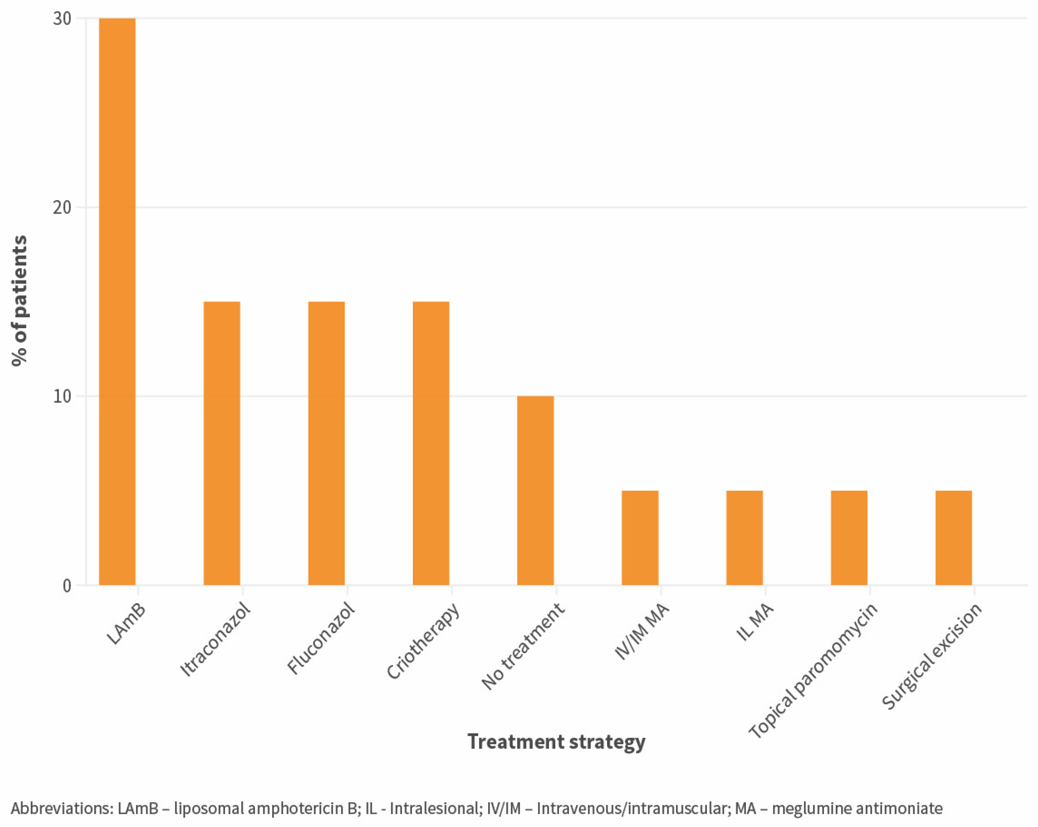
Skin samples of autochthonous CL cases were mostly obtained by biopsy, and by smear/scrapping in only in 2.4% of cases. Microscopy was used in all cases and was positive in 100% of these cases. PCR was used in 28.6% of cases and was positive in 85.7% of these. Leishmania species/complex was identified in 19.4% of cases (by molecular biology techniques); all belonged to the L. donovani complex (n = 6). Successful identification was available for 4/7 imported cases (L. donovani complex: n = 2, L. Viannia sp.: n = 2). Serology was used as part of the diagnostic workup in 34.6% of cases and was positive in 37.5% of these. Treatment for CL was introduced in 93.8% of patients, and the median time from diagnosis to treatment was four days (IQI 0–31). Most patients were treated with monotherapy (92.5%). Systemic treatment was used in 80.0% of patients and significantly more frequently in immunosuppressed patients (p = 0.033, FET). Detailed strategies used for primary treatment of patients who had exclusively CL (without simultaneous visceral involvement) (n = 20) are represented in Figure 2. Improvement by day 7 or 30 after starting therapy was mentioned in 60.0 and 76.2% of patients, respectively, and was not significantly different in immunosuppressed patients. Switching to a different regimen or retreatment due to non-improvement was implemented in 19.2% of patients. Relapse was reported in two patients, both immunosuppressed.
Time from presentation to diagnosis was the only variable that differed significantly between NUT2 regions (p = 0.037, KWT = 6.616, df = 2), being shorter in the Norte, followed by AML and Centro.

3.3. Clinical Aspects of ML

Leishmaniasis with mucosal involvement (n = 7) represented ML in all cases (no cases of MCL were identified). One patient was a person living with HIV, three were immunosuppressed for other reason, and two were non-immunosuppressed adults (one not defined). In five cases, only the nasal mucosa was involved (one with septal perforation), and in one case, both the oral and the nasal mucosa were involved. All cases were diagnosed by biopsy of the lesions and identification of amastigotes using microscopy, and PCR was used additionally in five cases. All cases were treated with liposomal amphotericin B except one, which was treated with IV meglumine antimoniate. One patient, who presented with simultaneously VL, died.

4. Discussion

The present study reinforces that the incidence of autochthonous CL cases seems to be lower than that of VL at a national level [24], although marked differences were noted between the NUTS2 and NUTS3 regions. Few data were previously available in the country and were mostly derived from case reports and case series, since no national reporting system is in place [25]. These data already suggested that the Beiras e Serra da Estrela region could be an important focus of CL [14], which was also pointed out in the present study. Recent cases revealed in this study in regions where CL has not been previously described in the literature, such as the Aveiro region, should be further investigated. Additionally, further studies could help elucidate whether or not this heterogeneity could be explained by an increased clinical awareness in certain regions or the presence of a distinct, particularly dermotropic L. infantum genotype. In support of the first hypothesis, it should be noted that in areas of neighboring Spain, the incidence of autochthonous CL was similar to that of VL [26]; however, CL notification is mandatory in Spain, as opposed to the protocol for Portugal [2] and in the present study, the underestimation of CL incidence could be due to inadequate coding at the hospital level and insufficient laboratory information.
Imported CL still represents a minority of cases in Portugal, as opposed to other European endemic countries, such as (metropolitan) France [27]; however, this could be expected to change in upcoming years due to increasing migration from CL endemic countries, such as Brazil [15]. Since no systematic clinical screening program is implemented in migrant populations in Portugal, leishmaniasis cases, especially (spontaneously resolving) CL, could go unnoticed and translate into an underestimation of imported cases.
In the autochthonous CL group, 48.0% were immunosuppressed, and in this group, 54.5% presented with simultaneously VL, which suggests that immunosuppressed people with CL benefit from a more intensive diagnostic workup in a setting where most CL is assumed to be caused by L. infantum [28]. Overall, time from onset of symptoms to presentation to healthcare and time from presentation to diagnosis were long, likely reflecting, on one hand, low concern of the patients for the lesions and, on the other hand, the low awareness of clinicians of the disease, or their unfamiliarity with the availability and performance of diagnostic techniques. In terms of lesion characteristics, the findings of the present study overlap those of other case series in the Mediterranean context, where L. infantum is endemic and includes frequent multiple lesions, a lower prevalence of ulcerated lesions compared to nodules/papules or plaques, and a predominance of head/neck lesions [3,29].
According to the present study, the diagnosis of CL in Portugal between 2010 and 2020 relied mostly on microscopy. As PCR was performed in only 42.1% of cases, Leishmania species was not identified in all imported cases, and therapy was commonly selected based on the probable geographical location of the infection. However, species identification in New World CL could have implications for individual management, especially in areas where multiple species co-circulate, since species in the Viannia subgenus, especially L. braziliensis, have been more frequently associated with MCL, requiring initial screening for this form and longer follow-up time [30]. On the other hand, species identification in Old World CL could be of public health interest, in the context of surveillance and assessment of risk of introduction of species such as dermotropic L. tropica. This anthroponotic species has been reported as the most imported species from refugees [31], and Phlebotomus sergenti, a specific vector of L. tropica, is widely distributed in Southern Europe, including in Portugal [32].
Treatment of CL cases was very heterogeneous but often relied on systemic therapy, especially IV/IM in immunosuppressed patients, which is in accordance with European recommendations [21]. Overall, rates of non-improvement (or failure) at 30 days after starting treatment (23.8%) were similar to those reported in previous studies in settings where L. infantum is endemic [3].
Although mucosal involvement is more commonly associated with L. (Viannia) sp., ML is increasingly recognized in the Old World in the context of L. infantum infection and especially in immunosuppression [33]. In the present study, seven cases were identified, all were autochthonous, and two were not immunosuppressed. Nose and throat physicians in endemic regions should be alert to the presentation, diagnostic approach, and management specificities of ML, particularly in regards to non-immunosuppressed patients and lesions located in the oral, pharyngeal, and laryngeal mucosa.
Finally, this study presents some limitations, beginning with the fact that in some hospitals, not all information was collected due to a lack of collaboration or the absence of patient consent. The coding of the diagnosis for inpatients was not uniformly performed and digitalized in every hospital for the whole duration of the study period, and coding for outpatients was irregularly performed in hospitals, so cases were screened via laboratory results, whenever feasible. Some hospitals required internal personnel to access information, so in some cases, interpretation of variables could be different, despite using the same database. Regarding the literature review of CL cases diagnosed in Portugal, a notable limitation may arise from the possibility that further cases might have been reported solely in national journals not indexed in PubMed or exclusively presented at conferences or congresses.

5. Conclusions

This study sheds light on the epidemiological and clinical landscape of CL and ML in Portugal between 2010 and 2020. While the incidence of autochthonous CL was low, CL was more common than previously reported, but still less common than in neighboring countries, possibly translating into significant underdiagnosis. Regional disparities highlight the importance of localized surveillance efforts. Programs to control leishmaniasis should focus not only on reducing underreporting, but also on raising awareness for the disease’s different clinical forms among healthcare practitioners and providing tools for earlier diagnosis. Clinical suspicion should be particularly heightened for immunosuppressed people, who are disproportionately affected.
The present findings also underscore the potential for the underestimation of imported CL cases, particularly in the context of increasing migration from endemic regions, in the absence of systematic clinical screening programs. Given the implications for individual management and public health surveillance, there is a need for greater emphasis on species identification in imported cases.
Systematically combining clinical and national surveillance data could allow for a more detailed assessment of the epidemiologic situation and an evaluation of the progress in clinical practice, uncovering gaps that need to be addressed in the near future. In order to improve the overall outcome for leishmaniasis patients, human data should also be integrated with data from vectors and mammal hosts to produce holistic strategies to control the disease in several stages of the life cycle, following a One Health approach.

Author Contributions

Conceptualization, R.R., C.C., L.G. and C.M. (Carla Maia); methodology, R.R., C.C., L.G., A.C.C., A.M. (André Maia), A.M. (André Martins), A.C., A.M. (António Maio), C.F., C.M. (Catarina Melita), D.C., D.F., D.P., E.L., H.S., I.S., J.-P.G., J.M., J.V., J.C., J.R., J.M.S., L.C., L.M., L.S., M.G., M.C., M.L., M.M.A., M.M., M.A., M.S., R.F., R.S., S.F., S.L., S.C., T.G., T.B., T.R.-N., V.A. and C.M. (Carla Maia); validation, R.R., C.C., L.G. and C.M. (Carla Maia); formal analysis, R.R.; investigation, R.R., A.C.C., A.M. (André Maia), A.M. (André Martins), A.C., A.M. (António Maio), C.F., C.M. (Catarina Melita), D.C., D.F., D.P., E.L., H.S., I.S., J.-P.G., J.M., J.V., J.C., J.R., J.M.S., L.C., L.M., L.S., M.G., M.C., M.L., M.M.A., M.M., M.A., M.S., R.F., R.S., S.F., S.L., S.C., T.G., T.B., T.R.-N. and V.A.; resources, A.C.C., A.M. (André Maia), A.M. (André Martins), A.C., A.M. (António Maio), C.F., C.M. (Catarina Melita), D.C., D.F., D.P., E.L., H.S., I.S., J.-P.G., J.M., J.V., J.C., J.R., J.M.S., L.C., L.M., L.S., M.G., M.C., M.L., M.M.A., M.M., M.A., M.S., R.F., R.S., S.F., S.L., S.C., T.G., T.B., T.R.-N. and V.A.; writing—original draft, R.R.; writing—review and editing, C.C., L.G., A.C.C., A.M. (André Maia), A.M. (André Martins), A.C., A.M. (António Maio), C.F., C.M. (Catarina Melita), D.C., D.F., D.P., E.L., H.S., I.S., J.-P.G., J.M., J.V., J.C., J.R., J.M.S., L.C., L.M., L.S., M.G., M.C., M.L., M.M.A., M.M., M.A., M.S., R.F., R.S., S.F., S.L., S.C., T.G., T.B., T.R.-N., V.A. and C.M. (Carla Maia); visualization, R.R.; supervision, C.C., L.G. and C.M. (Carla Maia). All authors have read and agreed to the published version of the manuscript.

Funding

R.R. was supported by the Portuguese Ministry of Education and Science (via Fundação para a Ciência e a Tecnologia, I.P.—FCT) through a PhD grant (UI/BD/151067/2021). The authors would also like to acknowledge FCT for funding through contracts with GHTM (UID/Multi/04413/2020), CEAUL (UIDB/00006/2020 and UIDP/00006/2020), and LA-REAL (LA/P/0117/2020). The work of C.M. was supported by the European Commission grant 101057690 and UKRI grants 10038150 and 10039289 and is catalogued by the CLIMOS Scientific Committee as CLIMOS number 006 (http://www.climos-project.eu, accessed on 17 April 2024). The contents of this publication are the sole responsibility of the authors and do not necessarily reflect the views of the European Commission, the Health and Digital Executive Agency, or UKRI. Neither the European Union, the granting authority, nor UKRI can be held responsible for this work. The funders had no role in study design; data collection and analysis; the decision to publish; or the preparation of the manuscript. For the purposes of open access, the authors have applied a CC BY public copyright license to any Author Accepted Manuscript version arising from this submission. The six Horizon Europe projects, BlueAdapt, CATALYSE, CLIMOS, HIGH Horizons, IDAlert, and TRIGGER, form the Climate Change and Health Cluster.

Institutional Review Board Statement

This study received a favorable opinion of the Ethics Committees of all the involved institutions, namely: Instituto de Higiene e Medicina Tropical, Universidade Nova de Lisboa (reference 1.22); Centro Hospitalar do Baixo Vouga (16-01-2022/CES); Centro Hospitalar Barreiro Montijo; Centro Hospitalar de Entre o Douro e Vouga (29/2022); Centro Hospitalar de Leiria (34/2023); Centro Hospitalar de Lisboa Ocidental (2293); Centro Hospitalar do Médio Tejo (080/2022); Centro Hospitalar do Oeste; Centro Hospitalar de Setúbal (018/2022F); Centro Hospitalar do Tâmega e Sousa (63/2021); Centro Hospitalar Tondela Viseu (08/19/11/2021); Centro Hospitalar de Trás-os-Montes e Alto Douro; Centro Hospitalar de Vila Nova de Gaia e Espinho (65/2022); Centro Hospitalar Universitário do Algarve (172/2021); Centro Hospitalar Universitário de Coimbra (OBS.SF.083-2022); Centro Hospitalar Universitário Cova da Beira (03/2022); Centro Hospitalar Universitário de Lisboa Central (1287/2022); Centro Hospitalar Universitário Lisboa Norte; Centro Hospitalar Universitário de Santo António (2022.060); Centro Hospitalar Universitário de São João (06/2022); Hospital Beatriz Ângelo (4018/2022); Hospital de Braga (29_2022); Hospital de Cascais Dr. José de Almeida (1/CE); Hospital Distrital de Santarém (31/2022); Hospital do Espírito Santo de Évora; Hospital Garcia de Orta; Hospital Professor Doutor Fernando Fonseca; Hospital da Senhora da Oliveira Guimarães (07/2022); Hospital de Vila Franca de Xira; Instituto Português de Oncologia de Coimbra Francisco Gentil; Instituto Português de Oncologia de Lisboa Francisco Gentil (73/2022); Unidade Local de Saúde do Alto Minho (30/2022); Unidade Local de Saúde do Baixo Alentejo (10/2021); Unidade Local de Saúde de Castelo Branco; Unidade Local de Saúde da Guarda (112/2022); Unidade Local de Saúde do Nordeste (59/2022); Unidade Local de Saúde do Norte Alentejano. Additionally, the study was authorized by the administration council of all the involved hospitals. In the three hospitals where informed consent was required, collection of data was only performed for patients who provided written consent.

Data Availability Statement

The datasets generated and analyzed during the current study are not publicly available due to a confidentiality commitment with the health institutions and the participants.

Acknowledgments

The authors would like to acknowledge the directors of the all the clinical and laboratory departments involved in this study and the administration councils of the hospitals that authorized the study.

Conflicts of Interest

The authors declare no conflicts of interest.

References

  1. Farrar, J.; Garcia, P.; Hotez, P.; Junghanss, T.; Kang, G.; Lalloo, D.; White, N. Manson’s Tropical Diseases, 24th ed.; Elsevier: Amsterdam, The Netherlands, 2024. [Google Scholar]
  2. ECDC. Surveillance, Prevention and Control of Leishmaniases in the European Union and Its Neighbouring Countries. Stockholm. 2022. Available online: https://www.ecdc.europa.eu/sites/default/files/documents/leishmaniasis-surveillance-eu.pdf (accessed on 3 March 2024).
  3. Gaspari, V.; Gritti, T.; Ortalli, M.; Santi, A.; Galletti, G.; Rossi, A.; Rugna, G.; Mattivi, A.; Matteo, G.; Belloli, G.L.; et al. Tegumentary Leishmaniasis in Northeastern Italy from 2017 to 2020: A Neglected Public Health Issue. Int. J. Environ. Res. Public Health 2022, 19, 16047. [Google Scholar] [CrossRef] [PubMed]
  4. Phlebotomine sandflies maps. European Centre for Disease Prevention and Control and European Food Safety Authority. Stockholm: ECDC. Available online: https://ecdc.europa.eu/en/disease-vectors/surveillance-and-disease-data/phlebotomine-maps (accessed on 3 March 2024).
  5. Maia, C.; Dantas-Torres, F.; Campino, L. Parasite Biology: The Reservoir Hosts. In The Leishmaniases: Old Neglected Tropical Diseases; Springer International Publishing: Cham, Germany, 2018; pp. 79–106. [Google Scholar]
  6. Campino, L.; Abranches, P. Leishmaniose cutânea: Uma doença rara em Portugal? Acta. Med. Port. 2002, 15, 387–390. [Google Scholar] [PubMed]
  7. Borges-Costa, J.; Pacheco, D.; Soares-Almeida, L.; Sacramento-Marques, M. Pápulas marrones y violáceas localizadas en la espalda de un paciente con infección por el virus de la inmunodeficiencia humana. Enferm. Infecc. Microbiol. Clin. 2013, 31, 410–411. [Google Scholar] [CrossRef] [PubMed]
  8. Costin, A.; Bonito, F.; Alves, J.; Barreiros, H. Tratamiento de la leishmaniasis localizada mediante el antimoniato de meglumina intralesional y la terapia fotodinámica. Actas Dermosifiliogr. 2020, 111, 897–899. [Google Scholar] [CrossRef] [PubMed]
  9. Vasconcelos, J.; Torres, J.; Granado, J.; Baptista, T.; Mansinho, K. Cutaneous leishmaniasis in non-endemic countries: An emerging yet neglected problem. IDCases 2019, 17, e00570. [Google Scholar] [CrossRef] [PubMed]
  10. Marques, N.; Bustorff, M.; Cordeiro Da Silva, A.; Pinto, A.I.; Santarém, N.; Ferreira, F.; Nunes, A.; Cerqueira, A.; Rocha, A.; Ferreira, I.; et al. Visceral Dissemination of Mucocutaneous Leishmaniasis in a Kidney Transplant Recipient. Pathogens 2020, 10, 18. [Google Scholar] [CrossRef] [PubMed]
  11. Lopes, L.; Vasconcelos, P.; Borges-Costa, J.; Soares-Almeida, L.; Campino, L.; Filipe, P. An atypical case of cutaneous leishmaniasis caused by Leishmania infantum in Portugal. Dermatol. Online J. 2013, 19, 20407. [Google Scholar] [CrossRef] [PubMed]
  12. Matos, A.L.; Figueiredo, C.; Alves, F.; Pereira, N.; Gonçalo, M. Mucocutaneous leishmaniasis complicating systemic lupus erythematosus and responding to high dose amphotericin B. J. Eur. Acad. Dermatol. Venereol. 2021, 35, pe756. [Google Scholar] [CrossRef] [PubMed]
  13. Van der Auwera, G.; Davidsson, L.; Buffet, P.; Ruf, M.T.; Gramiccia, M.; Varani, S.; Chicharro, C.; Bart, A.; Harms, G.; Chiodini, P.L.; et al. Surveillance of leishmaniasis cases from 15 European centres, 2014 to 2019: A retrospective analysis. Eurosurveillance 2022, 27, 2002028. [Google Scholar] [CrossRef] [PubMed]
  14. Azevedo, N. Leishmaniose cutânea na Beira Interior: Um Diagnóstico Diferencial Obrigatório. Master’s Thesis, Universidade da Beira Interior, Covilhã, Portugal, 2009. [Google Scholar]
  15. Fundação Francisco Manuel dos Santos. PORDATA Base de Dados de Portugal Contemporâneo. População Residente Estrangeira Segundo os Censos: Total e por País de Nacionalidade. 2023. Available online: https://www.pordata.pt/portugal/populacao+residente+estrangeira+segundo+os+censos+total+e+por+pais+de+nacionalidade-3786 (accessed on 3 March 2024).
  16. Fundação Francisco Manuel dos Santos. PORDATA. O que são NUTS? Available online: https://www.pordata.pt/o+que+sao+nuts (accessed on 3 March 2024).
  17. Instituto Nacional de Estatística. Censos 2021 Resultados Definitivos—Portugal, Lisboa; Instituto Nacional de Estatística: Lisbon, Portugal, 2022.
  18. Fundação Francisco Manuel dos Santos. PORDATA Base de Dados de Portugal Contemporâneo. SNS: Hospitais por Tipo—Continente. 2022. Available online: https://www.ine.pt/xportal/xmain?xpid=INE&xpgid=ine_publicacoes&PUBLICACOESpub_boui=66196836&PUBLICACOESmodo=2 (accessed on 3 March 2024).
  19. World Health Organisation. Status of Endemicity of Cutaneous Leishmaniasis: 2022. 2023. Available online: https://apps.who.int/neglected_diseases/ntddata/leishmaniasis/leishmaniasis.html (accessed on 8 October 2023).
  20. Nemhauser, J. Travelers with Additional Considerations. In CDC Yellow Book 2024; Oxford University Press: New York, NY, USA, 2023; pp. 139–180. [Google Scholar]
  21. Gradoni, L.; López-Vélez, R.; Mokni, M. Manual on Case Management and Surveillance of the Leishmaniasis in the WHO European Region; World Health Organization: Geneva, Switzerland, 2017. [Google Scholar]
  22. Daly, L.E. Confidence Limits Made Easy: Interval Estimation Using a Substitution Method. Am. J. Epidemiol. 1998, 147, 783–790. [Google Scholar] [CrossRef] [PubMed]
  23. Antunes, J.; da Costa, J.B.; Guimarães, M.; Vaz-Pinto, I.; Soares-Almeida, L.; Filipe, P. Post-Kala-azar dermal leishmaniasis due to Leishmania donovani in Europe—Case report. Int. J. Dermatol. 2013, 52, 1584–1586. [Google Scholar] [CrossRef] [PubMed]
  24. Maia, C.; Conceição, C.; Pereira, A.; Rocha, R.; Ortuño, M.; Muñoz, C.; Jumakanova, Z.; Pérez-Cutillas, P.; Özbel, Y.; Töz, S.; et al. The estimated distribution of autochthonous leishmaniasis by Leishmania infantum in Europe in 2005–2020. PLoS Negl. Trop. Dis. 2023, 17, e0011497. [Google Scholar] [CrossRef] [PubMed]
  25. Maia, C.; Campino, L. Leishmaniose em Portugal no início do século XXI. An. Inst. Hig. Med. Trop. 2014, 13, 25–28. [Google Scholar]
  26. Humanes-Navarro, A.M.; Herrador, Z.; Redondo, L.; Cruz, I.; Fernández-Martínez, B. Estimating human leishmaniasis burden in Spain using the capture-recapture method, 2016–2017. PLoS ONE 2021, 16, e0259225. [Google Scholar] [CrossRef] [PubMed]
  27. Pasquier, G.; Demar, M.; Lami, P.; Zribi, A.; Marty, P.; Buffet, P.; Desbois-Nogard, N.; Gangneux, J.P.; Simon, S.; Blaizot, R.; et al. Leishmaniasis epidemiology in endemic areas of metropolitan France and its overseas territories from 1998 to 2020. PLoS Negl. Trop. Dis. 2022, 16, e0010745. [Google Scholar] [CrossRef]
  28. Campino, L.; Pratlong, F.; Abranches, P.; Rioux, J.A.; Santos-Gomes, G.; Alves-Pires, C.; Cortes, S.; Ramada, J.; Cristovão, J.M.; Afonso, M.O.; et al. Leishmaniasis in Portugal: Enzyme polymorphism of Leishmania infantum based on the identification of 213 strains. Trop. Med. Int. Health 2006, 11, 1708–1714. [Google Scholar] [CrossRef] [PubMed]
  29. Giavedoni, P.; Iranzo, P.; Fuertes, I.; Estrach, T.; Alsina Gibert, M. Leishmaniasis cutánea. Experiencia de 20 años en un hospital español de tercer nivel. Actas Dermosifiliogr. 2015, 106, 310–316. [Google Scholar] [CrossRef]
  30. Pan American Health Organization. Guideline for the Treatment of Leishmaniasis in the Americas, 2nd ed.; Pan American Health Organization: Washington, DC, USA, 2022. [Google Scholar]
  31. Rocha, R.; Pereira, A.; Maia, C. Non-Endemic Leishmaniases Reported Globally in Humans between 2000 and 2021—A Comprehensive Review. Pathogens 2022, 11, 921. [Google Scholar] [CrossRef] [PubMed]
  32. European Centre for Disease Prevention and Control (ECDC). Phlebotomus sergenti-Current Known Distribution: August 2023. 2023. Available online: https://www.ecdc.europa.eu/en/publications-data/phlebotomus-sergenti-current-known-distribution-august-2023 (accessed on 13 October 2023).
  33. Faucher, B.; Pomares, C.; Fourcade, S.; Benyamine, A.; Marty, P.; Pratlong, L.; Faraut, F.; Mary, C.; Piarroux, R.; Dedet, J.P.; et al. Mucosal Leishmania infantum leishmaniasis: Specific pattern in a multicentre survey and historical cases. J. Infect. 2011, 63, 76–82. [Google Scholar] [CrossRef] [PubMed]
Figure 1. Estimated mean annual incidence, per 100,000 population, between 2010 and 2020 of cutaneous leishmaniasis by NUTS3 region.
Microorganisms 12 00819 g001
Figure 2. Strategies used for primary treatment of patients with autochthonous exclusively cutaneous leishmaniasis (no evidence of simultaneous visceral involvement) (n = 20).
Microorganisms 12 00819 g002
Table 1. Sociodemographic characteristics and comorbidities of leishmaniasis cases, globally and by form of disease.
GlobalCLMLp-Value
Number50437
Median age, years (IQI)4847660.026 *
[33–61.25][33–59][50–75](U = 71.0)
Male sex (%)68.069.857.10.666
(34/50)(30/43)(4/7)(FET)
Country of birth (%)
 Native75.672.51000.313
(FET)
(34/45)(29/40)(5/5)
 Migrant24.427.50
(11/45)(11/40) a(0/5)
Origin of infection (%)
 Autochthonous86.083.71000.573
(FET)
(43/50)(36/43)(7/7)
 Imported14.016.30
(7/50)(7/43) b(0/7)
Immunosuppression (%)
 Yes48.046.557.10.697
(24/50)(20/43)(4/7)(FET)
 Unknown/Not reported2.0014.3
(1/50)(0/43)(1/7)
HIV infection/AIDS
 Yes (%)32.734.916.70.649
(16/49)(15/43)(1/6)(FET)
 CD4 cell count <200/µL (%)78.684.600.214
(11/14)(11/13)(0/1)(FET)
Chronic pharmacologic immunosuppression (%)
 Inflammatory/autoimmune diseases c12.29.333.30.151
(6/49)(4/43) d(2/6) e(FET)
 Other4.12.316.7
(2/49)(1/43) f(1/6) g
Chronic dysfunction/condition (%)
 Yes28.619.483.30.005 *
(12/42)(7/36) h(5/6) i(FET)
a Brazil: n = 4; Cape Verde: n = 2; Guinea-Bissau: n = 1; Morocco: n = 1; São Tomé e Príncipe: n = 1; Senegal: n = 1; unknown: n = 1
b Brazil: n = 3; Morocco: n = 2; Mexico: n = 1; Tunisia: n = 1
c Systemic lupus erythematosus: n = 2; ankylosing spondylitis: n = 1; Crohn’s disease: n = 1; psoriasis: n = 1; rheumatoid arthritis: n = 1
d Adalimumab: n = 1; adalimumab + methotrexate: n = 1, methotrexate: n = 1; methotrexate + prednisolone: n = 1
e Methotrexate: n = 1; mycophenolate mofetil + prednisolone: n = 1
f Lymphoma: n = 1
g Kidney transplant: n = 1
h Chronic kidney disease n = 6; chronic heart failure n = 1; chronic obstructive respiratory disease n = 1; chronic hepatic disease n = 1; diabetes mellitus n =1
i Chronic kidney disease: n = 2; chronic heart failure: n = 1; chronic obstructive respiratory disease: n = 1; diabetes mellitus: n = 3
* Statistically significant
Abbreviations: CL—cutaneous leishmaniasis; ML—mucosal leishmaniasis; HIV—human immunodeficiency virus; AIDS—acquired immunodeficiency syndrome; IQI—interquartile interval; FET—Fisher’s exact test.
Table 2. Number of cases of cutaneous and mucosal leishmaniasis diagnosed between 2010 and 2020, inclusively, and mean annual incidence in this period, per 100,000 population, by NUTS2 and NUTS3 region.
RegionAverage Population in 2011–2021 *Number of CL/ML CasesMean Annual CL/ML Incidence **95% CI
Mainland Portugal9,951,765390.0360.024–0.047
 Norte3,638,13470.0170.006–0.033
  Alto Minho238,05100NA
  Cávado413,38700NA
  Ave421,93300NA
  Área Metropolitana do Porto1,747,87640.0210.003–0.046
  Alto Tâmega89,19500NA
  Tâmega e Sousa420,77610.0220.001–0.120
  Douro194,51610.0470.001–0.260
  Terras de Trás-os-Montes112,39900NA
 Centro1,695,204130.0700.037–0.119
  Região de Aveiro368,89830.0740.015–0.216
  Região de Coimbra448,50000NA
  Região de Leiria290,69200NA
  Viseu Dão Lafões260,20520.0700.008–0.252
  Beira Baixa103,59710.0880.002–0.489
  Beiras e Serra da Estrela223,31250.2040.066–0.475
 Oeste e Vale do Tejo823,94800NA
  Oeste363,02500NA
  Médio Tejo219,26600NA
  Lezíria do Tejo241,65700NA
 Grande Lisboa2,052,392130.0580.031–0.098
 Península de Setúbal793,65130.0340.007–0.100
 Alentejo489,25910.0190.000–0.069
  Alentejo Litoral97,18300NA
  Baixo Alentejo120,77700NA
  Alto Alentejo111,71410.0810.002–0.453
  Alentejo Central159,58500NA
 Algarve459,17420.0400.005–0.143
* Arithmetic mean between the population size estimated in the National Census of 2011 and 2021. ** Number of new cases per 100,000 population, per year, based on the following formula: Incidence = (New Cases)/(Population × Timeframe). Abbreviations: CL—cutaneous leishmaniasis; ML—mucosal leishmaniasis; NA—not applicable; CI—confidence interval.
Table 3. Clinical presentation and management of autochthonous cutaneous leishmaniasis cases between 2010 and 2020.
Type of Lesion (%)
 Papule/Nodule62.1
(18/29)
 Ulcer24.1
(7/29)
 Macule/Plaque20.7
(6/29)
Multiple lesions (%)41.9
(13/31)
Median size of largest lesion, mm (IQI)30
[10–40]
Location of lesions (%)
 Head48.3
(14/29)
 Upper limbs31.0
(9/29)
 Lower limbs31.0
(9/29)
 Trunk20.7
(6/29)
Disseminated cutaneous leishmaniasis (%)12.5
(4/32)
Local pain (%)25.0
(5/20)
Skin superinfection a (%)11.1
(3/27)
Simultaneous visceral leishmaniasis (%)38.9
(14/36)
Technique used in skin/mucosa sample (%)
 Microscopy100
(31/31)
  Positive result100
(30/30)
 Polymerase chain reaction28.6
(8/28)
  Positive result85.7
(6/7)
Identification of species (%)19.4
(6/31)
Serology (%)
  Yes b34.6
(9/26)
  % positive37.5
(3/8)
Treatment of primary episode (%)
 Yes93.8
(30/32)
  Median time from diagnosis to treatment, days (IQI)4
[0–31]
  Monotherapy93.3
(28/30)
  Systemic80.0
(24/30)
  Topical23.3
(7/30)
  Side effects15.4
(2/13)
Outcome of treatment (%)
Improvement at 7 days60.0
(9/15)
Improvement at 30 days76.2
(16/21)
 Switch of treatment/retreatment (non-improvement)19.2
(5/26)
 Relapse5.6
(2/36)
a methicillin-sensitive Staphylococcus aureus: n = 1; Pseudomonas aeruginosa: n = 1; non-identified n = 1
b immunofluorescent antibody test: n = 5; unknown: n = 4
Abbreviations: IQI—interquartile interval.
Disclaimer/Publisher’s Note: The statements, opinions and data contained in all publications are solely those of the individual author(s) and contributor(s) and not of MDPI and/or the editor(s). MDPI and/or the editor(s) disclaim responsibility for any injury to people or property resulting from any ideas, methods, instructions or products referred to in the content.