Article

The Identification of the Mitochondrial DNA Polymerase γ (Mip1) of the Entomopathogenic Fungus Metarhizium brunneum

by
Stylianos P. Varassas
1,
Sotiris Amillis
2,
Katherine M. Pappas
1 and
Vassili N. Kouvelis
1,*
1
Section of Genetics and Biotechnology, Department of Biology, National and Kapodistrian University of Athens, 15784 Athens, Greece
2
Section of Botany, Department of Biology, National and Kapodistrian University of Athens, 15784 Athens, Greece
*
Author to whom correspondence should be addressed.
Microorganisms 2024, 12(6), 1052; https://doi.org/10.3390/microorganisms12061052
Submission received: 27 April 2024 / Revised: 16 May 2024 / Accepted: 20 May 2024 / Published: 23 May 2024
(This article belongs to the Section Molecular Microbiology and Immunology)

Abstract

:
Replication of the mitochondrial (mt) genome in filamentous fungi is under-studied, and knowledge is based mainly on data from yeasts and higher eukaryotes. In this study, the mitochondrial DNA polymerase γ (Mip1) of the entomopathogenic fungus Metarhizium brunneum is characterized and analyzed with disruption experiments and its in silico interactions with key proteins implicated in mt gene transcription, i.e., mt RNA polymerase Rpo41 and mt transcription factor Mtf1. Disruption of mip1 gene and its partial expression influences cell growth, morphology, germination and stress tolerance. A putative in silico model of Mip1-Rpo41-Mtf1, which is known to be needed for the initiation of replication, was proposed and helped to identify potential amino acid residues of Mip1 that interact with the Rpo41-Mtf1 complex. Moreover, the reduced expression of mip1 indicates that Mip1 is not required for efficient transcription but only for replication. Functional differences between the M. brunneum Mip1 and its counterparts from Saccharomyces cerevisiae and higher eukaryotes are discussed.

1. Introduction

DNA polymerase γ, also designated as Mip1 in fungi and most specifically in yeasts [1,2], plays the most important role in mitochondrial (mt) replication in fungi and consists of a single subunit, which is homologous to the catalytic subunit PolγA (POLG1) in humans [3]. To initiate the molecular process of replication of mtDNA, the complex Rpo41-Mtf1 is also required [4,5,6], since Rpo41, the mt RNA polymerase, when combined with the Transcription Factor 1 Mtf1, has activity of mitochondrial primase [7]. Both Mip1 and Rpo41 have homology with the corresponding genes of the bacteriophages T5 and T3/T7, respectively [8,9,10], but especially in the cases of mitochondrial DNA polymerases, like the yeast Mip1 and human POLG1, N- and C-terminal extensions (of different sizes), which have not found in bacterial and phage polymerases and have been identified and studied [11,12].
While the majority of research on mt genome diversity and its functions has been focused on yeasts among the fungi [13,14,15], it is important to expand studies to other subphyla of Ascomycota, and specifically to Pezizomycotina, which include model species, like Neurospora crassa and Aspergillus nidulans; biotechnologically important species, like the entomopathogenic fungi used as Biological Control Agents (BCAs) for the protection of crops; pathogens to humans, animals and plants, like Aspergillus fumigatus, Beauveria bassiana and Fusarium oxysporum, respectively; and symbionts, like the ones in lichens and mycorrhiza. Therefore, there is a need to fully clarify the similarities of the mt gene expression mechanisms in these species that are not putatively anaerobes, as yeasts are. Entomopathogenic fungi like the hypocrealean species Metarhizium brunneum may act as model organisms for studying the DNA replication mechanism of mt genes for two reasons. Firstly, the mt genome is already known and has been comparatively analyses within the genus Metarhizium [16] in addition to its whole-genome analysis [17]. Secondly, species of the fungal genus Metarhizium have been used as BCAs, as they have proven to be benign alternatives for the protection of several different crops worldwide [18,19,20]. Gaining insights into the functional mechanisms of their mt genomes (e.g., mtDNA replication) may provide a starting point for future genetic modifications of these genomes, with a final aim of improving the efficacy of their entomopathogenic activity against pests like aphids and whiteflies which destroy crops like maize, potatoes and tomatoes [18]. In this work, the mip1 gene from M. brunneum was isolated and partially inactivated in order to investigate its impact on the phenotype of the fungus. Additionally, an in silico analysis was performed in order to find the putative amino acid residues of Mip1 which interact with the Rpo41-Mtf1 complex in order to act as primase during replication.

2. Materials and Methods

2.1. Media, Strains and Growth Conditions

Complete (CM) and Minimal media (MM) containing carbon and nitrogen sources, as well as salt and trace element solutions, were used in liquid or solid form complemented with 1.5% (w/v) agar. Potato dextrose agar (PDA, AppliChem, Darmstadt, Germany) was also used as complete medium. In all cases, the pH was set at 6.5. Unless otherwise stated, glucose was used as a carbon source at a concentration of 1% (w/v). Sodium nitrate (NaNO3) was used as a nitrogen source at a final concentration of 10 mM. The salt solution, consisting of KCl, MgSO4 and KH2PO4, and the trace element solution consisting of Na2B4O7, CuSO4, FeO4P, MnSO4, Na2MoO4 and ZnSO4, were used as described in similar experiments of Aspergillus nidulans [URL: https://www.aspergillus.org.uk/lab_protocols/care-and-feeding-of-aspergillus-fgsc/ (accessed on 19 May 2024)] in the Fungal Genetics Stock Center (FGSC). Radial growth on solid MM was assessed at 25 °C, pH 6.5 for a time period of 3–10 days, whereas the growth of conidia suspensions in liquid MM for downstream applications was assessed at 25 °C, pH 6.5, 150 rpm for a time period of 12–24 h. M. brunneum strain ARSEF 3297 was used in all experiments. The Escherichia coli DH5α strain was used for cloning purposes.

2.2. Nucleic Acid Manipulations

Genomic DNA from M. brunneum was isolated according to the protocol described in Apostolaki et al. [21]. Plasmid purification and DNA gel extraction were performed using the Nucleospin Plasmid and the Nucleospin Extract II kits (Macherey-Nagel, Düren, Germany). Restriction enzymes, T4-ligases and phosphatases were from Takara Bio (Kusatsu, Japan). DNA sequences were determined by CeMIA SA (Larissa, Greece). Routine and high-fidelity PCR amplifications were performed using KAPA Taq DNA and Kapa HiFi polymerases (Kapa Biosystems, Merck SA, Athens, Greece). Oligonucleotides 5UTR-MIP1[ApaI]-F, 5UTR-MIP1[SpeI]-R, MTS-MIP1[SpeI]-[5]F and MTS-MIP1[NotI]-[3]R carrying specific enzyme adaptor sequences are presented in Table 1 and were used to amplify the relevant fragments from genomic DNA. The gene for hygromycin B phosphotransferase from Klebsiella pneumoniae was amplified from plasmid pAG32 [22] using oligonucleotides HYGrB-pAG32-[XbaI]-F and HYGrB-pAG32-[SpeI]-R (Table 1). These fragments were subsequently joined and cloned into the pGEM-T vector (Promega, Madison, WI, USA) using single-step ligation reactions. For the construction of a cassette overexpressing mip1, the relevant sequence of the alcAp promoter of the putative alcohol dehydrogenase homologue in M. brunneum was amplified using oligonucleotides alc-prom-[SpeI]-F and alc-prom-[SpeI]-R (Table 1), and the resulting fragment was cloned in frame between hygB and mip1 ORF. Targeted integrations of gene fusions were achieved by the amplification of those linear cassettes, also carrying the marker hygB, resulting in resistance to hygromycin B.

2.3. Transformation

Transformation in M. brunneum was performed as described in Koukaki et al. [23] for Aspergillus nidulans with some modifications. In brief, a conidial suspension of approximately 108–109 conidia of M. brunneum isolated from five plates grown for 7 days on MM 25 °C, after being passed through a nylon filter (mesh size 75 μm) to separate conidia from other irrelevant mycelial structures, were cultured in MM at 25 °C/130 rpm for 12–14 h, until conidial germ tube emergence. Germlings were subsequently incubated for 5 h in 1.2 M MgSO4, 10 mM orthophosphate, pH 5.8 supplemented with 500 mg cell wall lysing enzymes (Sigma-Aldrich, St. Louis, MO, USA), followed by a brief incubation of the resulting protoplasts with 1–2 μg linear DNA cassette, in 1 M Sorbitol, 10 mM Tris-HCl pH 7.5 and 10 mM CaCl2 in the presence of 60% (w/v) PEG6000. Transformants were selected after incubation as an overlay on MM plates supplemented with 1 M Saccharose and 0.25–1 mg/mL Hygromycin B for 7 days at 25 °C. Transformants were verified by PCR analysis and sequencing.

2.4. Microscopy

Samples were incubated in 3 cm petri dishes on coverslips in liquid MM supplemented with glucose as carbon and NaNO3 as nitrogen sources at 23–30 °C for 0–24 h. Samples were observed on an Axioplan Zeiss microscope (Zeiss, Jena, Germany) with Nomarski interference contrast and the resulting images were acquired with a Zeiss-MRC5 digital camera, using the ZEN 2.0 software. Conidial length-width ratio was calculated using the ZEN 2.0 (graphics tab-adding annotations) and SigmaPlot 14.5 software [24].

2.5. Protein Molecular Modeling

Prediction of the Mip1 secondary structure was performed using the PSIPRED 4.0 Workbench (UCL-CS Bioinformatics, London, UK) [25]. For homology modeling of the Mip1 protein, the Hidden Markov Model-based tool HHPred [26] and MODELLER 9.25 [27] were used, based on the highly similar crystal structure of a bacteriophage T7 DNA replication complex (Protein Data Bank, PDB 1T7P) with 97.74% similarity and 1.8 × 10⁻²² E-value, as described previously [28]. EDock software (replica exchange MC simulation) was used to describe the folding of the above predicted Mip1 of M. brunneum ARSEF 3297 with a 10 base-pair DNA molecule (ligand) [29]. Using MergeStructs Plugin, Version 1.1 (URL: https://www.ks.uiuc.edu/Research/vmd/plugins/mergestructs/ accessed on 19 May 2024), a cryo-EM structure of yeast mitochondrial RNA polymerase transcription initiation complex (6YMW) [30] was combined with our predicted Mip1 structure. Protein structures were visualized and compared using PyMOL 2.4 (https://pymol.org). In all programs, the default parameters were used.

2.6. RNA Extraction, First-Strand cDNA Synthesis and Quantitative Real-Time PCR

Total cellular RNA was isolated from 50–100 mg grinded mycelium using the TRIzol™ Reagent (Invitrogen, Carlsbad, CA, USA), following the manufacturer’s instructions. The isolated total RNA (1 μg verified for its purity and quantity using Nanodrop, Thermo Fischer Scientific, Waltham, MA, USA) was reverse transcribed in a 20 μL reaction mixture to generate single-stranded complementary DNA (cDNA) using 100 U SuperScript™ II Reverse Transcriptase (Invitrogen, Carlsbad, CA, USA), 40U RNaseOUT Recombinant Ribonuclease Inhibitor (Invitrogen, Carlsbad, CA, USA) and 10 μM gene-specific primer (Table 1). Reverse transcription was performed at 42 °C for 50 min, followed by enzyme inactivation at 70 °C for 15 min. As a control for cDNA synthesis efficiency, the cDNAs from different target RNAs and a negative control (DNAse-treated RNA, no reverse transcription) were used as a template for amplification with specific primers by PCR (KAPA HiFi HotStart ReadyMix PCR Kit, Kapa Biosystems, Merck SA, Athens, Greece). In addition, DNA fragments generated with KAPA HiFi HotStart DNA Polymerase (β-actin: 194 bp, DNA polymerase γ: 174 bp, ATP synthase F0 subunit 9: 220 bp) were used directly for blunt-end cloning in vector pBluescript II SK (Stratagene, Agilent Technologies, Santa Clara, CA, USA) and sequencing in both directions using the M13 universal primers. Amplicon sequences were analysed using the “Sequence Scanner v2” software (Thermo Fisher Scientific Inc., Waltham, MA, USA). qPCR assays were performed on the StepOne™ (Applied Biosystems, Carlsbad, CA, USA) using SYBR Green I dye for the quantification of the genes of interest. A 10 μL reaction mixture, containing 5 μL Kapa SYBR Fast Universal 2× qPCR master mix (Kapa Biosystems, Roche Diagnostics, Wilmington, USA), 10 ng of cDNA template and 200 nM of each specific primer, was used in a PCR protocol (95 °C for 3 min, followed by 40 cycles of denaturation at 95 °C for 15 s, primer annealing and extension at 60 °C for 1 min), followed by typical melt curve analyses to distinguish specific amplicons from non-specific products and/or primer dimers. All qPCR reactions were performed using two technical replications for each tested sample and target, and the average Ct of each duplicate was used in quantification analyses. Transcripts were detected for the mitochondrial ATP synthase subunit 9 (atp9) and the mitochondrial DNA polymerase γ (mip1). The housekeeping gene β-actin was used as a reference control for normalization. In detail, the Ct values obtained from our WT and mutant RNA samples were directly normalized to the housekeeping gene of β-actin and then compared. We assumed that the amplification efficiencies of the mip1 and the atp9 and β-actin genes were close to 100 percent (as we had a calibration curve slope of −3.32). Firstly, the difference between the Ct values (ΔCt) of the mip1, the atp9 and the β-actin genes were calculated for each experimental sample. Then, the difference in the ΔCt values between the experimental and control samples ΔΔCt was calculated. The fold-change in expression of the mip1 and atp9 between the two samples was then equal to 2−ΔΔCt.

2.7. Statistical Analysis

Statistical significance of differences between compared datasets was assessed using the analysis of variance (ANOVA) test, following the evaluation of homogeneity of variance across samples (F-test p ≤ 0.05). Datasets were then subjected to means separation using Tukey’s honest significant difference (HSD) test. Scale bars were added using SigmaPlot14.5 software.

3. Results

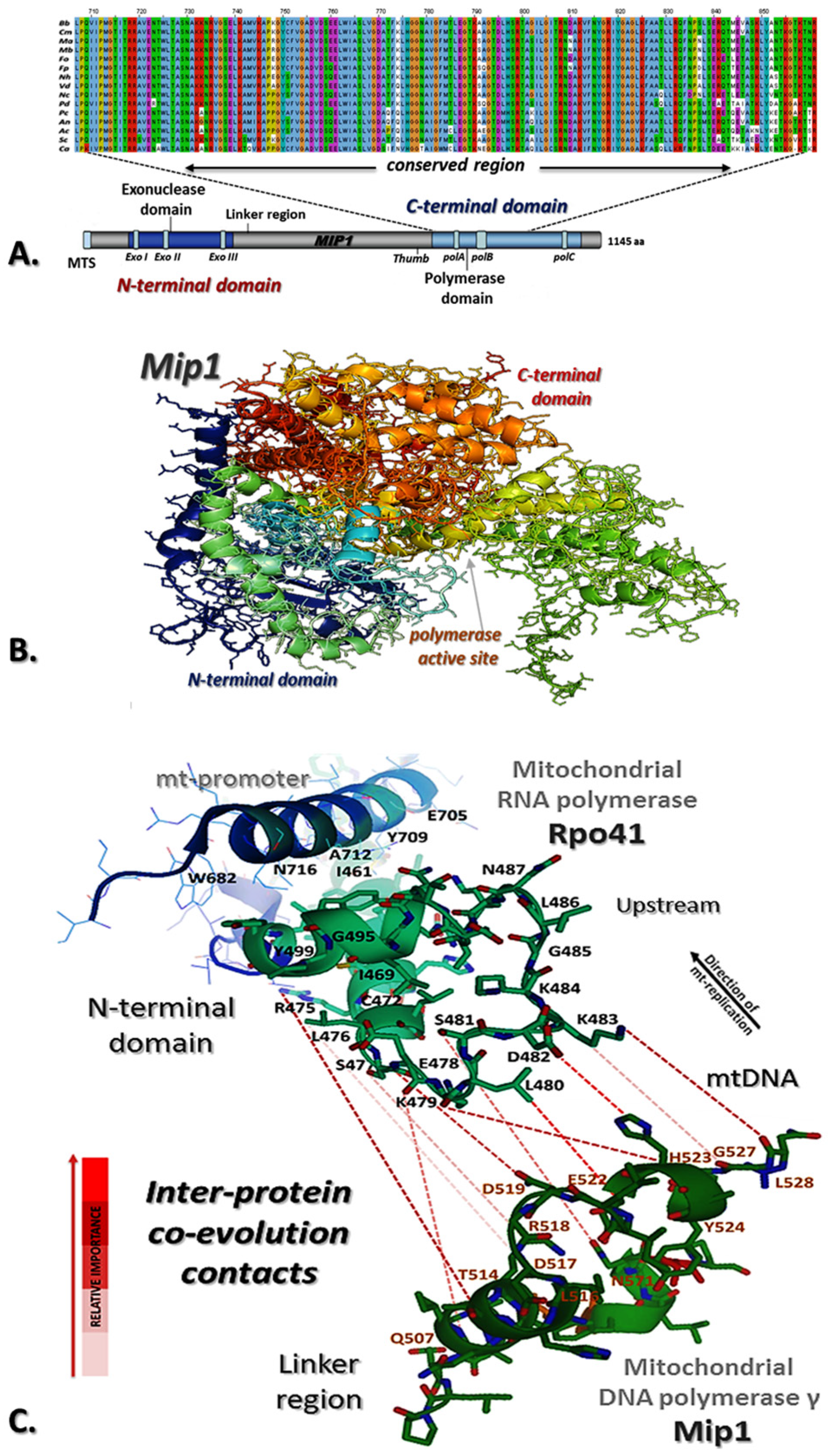
3.1. Identification of M. brunneum Mip1

Using BlastP and the corresponding sequence of the DNA polymerase gamma of S. cerevisiae (NP_014975.2), Mip1 from M. brunneum named as MBR_07687 was identified in NCBI Genome with Accession Number NW_014574705.1 (48% amino acid identity, 92% similarity). The gene was amplified from the genomic DNA of the wild-type strain ARSEF 3297, cloned and sequenced. The size of the gene was found to be 3499 bp, including an intron of 154 bp, thus encoding a protein of 1144 amino acids (Figure 1A, Supplementary Figure S1). The physical and chemical properties of M. brunneum Mip1 were analyzed in silico, showing a protein mass of 129.68 kDa and an isoelectric point of 8.158 (Figure 1B). The MitoProt II (v1.101) program revealed a signal sequence indicating that the identified Mip1 is indeed a mitochondrial matrix protein. Phylogenetic analysis of Mip1 of M. brunneum within Ascomycota (Supplementary Table S1) was also performed to establish evolutionary relationships and to identify conserved regions (Supplementary Figure S2a). The analysis revealed that the Mip1 gene shows similar phylogeny to that based on the ITS1-5.8S-ITS2 region (Supplementary Figure S2b), as species of the same order can be seen to cluster together. This result offered another indication that the M. brunneum Mip1 was correctly identified, since the Mip1 gene is essential for survival and hence conserved throughout time, following the evolution of the organisms carrying it.

3.2. Mip1 Has an Important Role in Fungal Cell Growth, Morphogenesis, Conidiation and Stress Tolerance

In order to study the function of Mip1 and the cellular differentiation caused by mtDNA–related mitochondrial dysfunction, we attempted to generate knock-out mutants, but no mutant was acquired, as complete inactivation of mip1 is most probably lethal for exclusively aerobic species like M. brunneum. Therefore, the knock-down mutants were constructed based on homologous recombination via the transformation of linear DNA cassettes that included, as a selection marker, the gene responsible for hygromycin B resistance (Figure 2A).
The ethanol-inducible, glucose-repressible promoter alcAp, often used for controllable protein overexpression in fungi [31,32], was selected as the candidate regulatable promoter. However, after multiple attempts and analysis of dozens of transformants, we failed to obtain a mutant carrying a stable alcAp integration at the mip1 genomic locus, a result suggesting that wild-type expression levels of mip1 are crucial for survival, and/or that the efficiency of homologous recombination in the wild-type strain is extremely low. Nevertheless, using a similar construct that carried all DNA fragments except the alcAp sequence, we isolated a strain with pronounced altered colony morphology, which proved to carry a functional insertion of hygB between the mip1 ORF and its native promoter region (Figure 2A) that would plausibly alter the expression of mip1. This insertion was verified with PCR amplification (see Supplementary Figure S1). Indeed, this mutant, from onwards designated as Mip1, showed a 60% reduction of expression compared to the wild-type ARSEF 3297, as shown by quantitative qPCR analysis (Figure 2B,C). Interestingly, the gene expression levels of the mitochondrial ATP synthase subunit 9 (atp9) appear less affected (Figure 2C). Both these genes were normalized against the housekeeping gene of β-actin (see Materials and Methods).
The Mip1 mutant exhibited significantly slower growth (p-value: 8.9 × 10⁻⁴), less compact aerial mycelium, and abnormal colony periphery, both on MM and on CM (Figure 3A,B and Supplementary Figure S3A) at pH 6.5 and 25 °C. Colonies of Mip1 grown on CM, PDA and MM media containing various carbon resources (Supplementary Figure S3B) were smaller compared to the wild type. Growth of all the strains of M. brunneum was sparse in nutrient poor media (MM) but denser in nutrient rich media (Supplementary Figure S3C). Furthermore, pigment deposition in the mip1-disrupted strains was delayed by 19 days. The mutant exhibited significant phenotypic alterations regarding its asexual reproductive capacity. Finally, conidiation was significantly lower than in the wild type (p-value: 9.4 × 10⁻⁷; Supplementary Figure S3B) on various carbon sources tested.
In order to further investigate Mip1 disruption phenotypes in the context of altered colony morphology and decreased conidial yield, conidia of the wild-type strain and the Mip1 mutant were observed by microscopy after 4–6 of germination. Overall, Mip1 cells showed rather abnormal cell morphology (Figure 3C). The conidia were elongated and swollen, compared to wild-type cells (rod-like conidia), at pH 6.5 and 25 °C. The wild-type strain produced larger conidia (~7 μm) when compared to the Mip1 strain (~6 μm). On the other hand, the average width of conidia was 3.0 ± 0.5 μm in the Mip1 strain and 2.5 ± 0.4 μm in wild-type cells, exhibiting a marginal difference (Figure 3D). To gain a better understanding of the phenotype in the two strains, the length/width ratio of the mutant conidia was determined. This was 2.14 for Mip1 versus 2.38 for the WT conidia, (swt = 0.41, smut = 0.23 and CVwt = 17% > CVmut = 10%), resulting in smaller, more globose-elliptical conidia (Figure 3C,D).
The optimal growth temperature and pH of the WT strain was 25 °C and 6.5, respectively. It was found that the colony of the Mip1 strain was smaller than that of the WT strain, the relative inhibition rate of growth of the Mip1 strain increased significantly compared to the wild-type strain, and the hyphal growth of the Mip1 strain was significantly decreased at various temperatures (Supplementary Figures S3 and S4A,C) in comparison to the WT strain. The conidial yield of the Mip1 strain was much lower compared with the WT strain (Supplementary Figure S4B,D). Furthermore, the results showed that the germination rate of Mip1 strain was slower compared to the WT strain (Supplementary Figure S5C). More specifically, differences in germination rate occurred mainly at 13 and 15 h, respectively (Supplementary Figure S6). The GT50 (the mean 50% germination time) of the Mip1 strain (15.94 ± 0.19 h) was moderately prolonged compared with the WT (13.45 ± 0.23 h) at 25 °C (Supplementary Figures S5 and S6).

3.3. In Silico Analysis of M. brunneum Mip1 and Interactions with Rpo41-Mtf1 Complex

The in vitro re-constituted mtDNA transcriptional complexes (Rpo41–Mtf1) of S. cerevisiae and humans, aided with high-resolution structures and biochemical characterizations, have provided a deeper understanding of the mechanism and regulation of mitochondrial DNA transcription [30,33,34,35]. However, the study of the mechanism of the initiation of mtDNA replication at the corresponding level is limited [36]. A 3-D model of the Mip1-Rpo41-Mtf1 (see Materials and Methods) may be proposed for the initiation of mitochondrial DNA replication of M. brunneum based on our in silico analysis (Figure 1C, Supplementary Figure S7). This proposition relies (i) on the fact that the mtDNA polymerase requires a primer and cannot initiate synthesis de novo [30,36], and (ii) on the hypothesis that Rpo41 synthesizes primers that are used for primer extension by Mip1 at origins of replication (Supplementary Figure S8).
The predicted Mtf1 interaction and binding with Rpo41 probably caused a change in the conformation of Rpo41 and Mtf1 to facilitate DNA melting, where the mtDNA (Supplementary Figure S7, transcription bubble region shown in yellow) was melted from −4 to +2 [30]. Both Rpo41 and Mip1 of M. brunneum had four amino acids, which may have been responsible for a conserved conformation as our analysis from EV-couplings and ET-viewer indicates (Figure 1C, Supplementary Table S2) and, to an extent, they may play a role in the overall activity of the complex.

4. Discussion

The fungal Mip1 is solely responsible for fast and faithful replication of the mitochondrial genome [2]. In M. brunneum, Mip1 is also identified as the DNA polymerase responsible for replication, and cells lacking a fully expressed Mip1 exhibit a slow-growing “petite-colony” phenotype. Mip1 partially deficient cells not only display abnormal cell morphology but also show reduced growth rate. The Mip1 phenotype in M. brunneum is similar to the phenotypes reported for Schizosaccharomyces pombe and reflects a relative tolerance for loss of mitochondria in both these two organisms, in contrast to S. cerevisiae which can afford losing all mitochondria [37,38]. The yeast S. cerevisiae is one of the few eukaryotic organisms that can either survive in the absence of mtDNA (rho0) or contain deletions in their mtDNA (rho) [39]. Needless to say, Mip1 is important to support normal growth in S. pombe and M. brunneum, and cells bearing a mip1 inactivation exhibit different growth phenotypes and similar cell morphologies [6].
The qPCR experiment of this study showed that gene expression levels of the mitochondrial ATP synthase subunit 9 (atp9), which is co-transcribed with genes nad2, nad3 and the tRNAs lying upstream the gene nad2 in M. brunneum, i.e., the polycistronic transcription unit TU2 of the wild-type strain [10], are comparable to that of the Mip1 mutant (Figure 2). While it seems expected that in a mutant for Mip1, both replication and transcription must be affected, since replication will not provide the same amount of mtDNA with that of the wild-type strain and in extent transcription of mt genes like atp9 will be reduced, it was found that the levels of expression of this mt gene remained significantly the same in both strains. Therefore, the hypothesis is that mt transcription is not affected from the partial inactivation of Mip1, but replication is undermined, as the reduction of the Mip1 expression and the slower growth of the mutant strains showed. This hypothesis can be indirectly supported further, by the mutants produced in the respective Mip1 of S. cerevisiae and more specifically in its C- terminal extension region of this yeast, as these mutants showed not a loss of function but a decrease in polymerization [11,12]. Moreover, it may be suggested that mt transcripts of the Mip1 mutant of M. brunneum may have a longer half-life than those of the WT strain, or that there are other DNA polymerases, which may compensate the reduced function of Mip1. This latter hypothesis was based mostly on the presence of two DNA polymerases, i.e., Rev1p and Pol ζ, in S. cerevisiae, and the possible complementation of function with the existence of Rev1p, when Pol ζ is missed or inactive [40].
Direct interactions between Mip1 and Rpo41, the non-template strand at origins of replication and promoter sequences have been observed in S. cerevisiae [36,41,42,43]. A putative structural model of Mip1-Rpo41-Mtf1 complex was proposed in this work for initiation of mtDNA replication, since origins of replication are located at the same regions with promoters of transcription [36,44]. Mip1 may contact promoter DNA near the transcription start site at origin of replication through interaction with Rpo41-Mtf1 [45,46] and the priming nucleotide (Figure 1, Supplementary Figure S8). This model structure is in agreement with the in vivo replication mechanism in Candida albicans, which involves a combination of the double-stranded break model for replicating the leading strand and Rpo41 as primase to replicate the lagging strand in this yeast [47].

5. Conclusions

This study underscores the pivotal role of the fungal Mip1 in mitochondrial genome replication besides the already known data from S. cerevisiae and other yeasts, as demonstrated through the slow growth phenotype of M. brunneum cells which have a partially impaired functional Mip1. This deficiency affects both the cell morphology and growth rate, akin to observations in other organisms like S. pombe. Interestingly, despite impaired replication in the Mip1-deficient strain, the qPCR analysis revealed comparable expression levels of mitochondrial genes, suggesting that while replication is compromised, transcription remains largely unaffected. The proposed structural model of the Mip1-Rpo41-Mtf1 complex may shed further light on the initiation of mtDNA replication, highlighting potential interactions crucial for this process. However, further research focusing on the structure and function of the mitochondria themselves is needed in the future, and these experiments into compensatory mechanisms and structural insights promise a deeper understanding of mitochondrial biology. This study represents the first significant step towards the usage of the hyphomycete M. brunneum as an alternative and complementary model system for molecular, genetic and biochemical studies of mitochondrial DNA replication in filamentous ascomycetes. Furthermore, this is the first approach to study the cellular function of Mip1 in M. brunneum, an entomopathogenic fungus used as BCA.

Supplementary Materials

The following supporting information can be downloaded at: https://www.mdpi.com/article/10.3390/microorganisms12061052/s1, Figure S1: The mip1 gene structure of M. brunneum and verification strategy of constructing the mutant mip1-strain; Figure S2: The phylogenetic analyses; Figure S3: Role of Mip1 in Growth and Physiology; Figure S4: Disruption of Mip1 weakens tolerance to temperature and pH variation and affects hyphal growth and sporulation; Figure S5: Disruption of mip1 Affected Hyphal Growth; Figure S6: Germination of conidial cells of WT and Mip1 mutant strains at the early developmental stages; Figure S7: The combined structure of Mip1-Rpo41-Mtf1 holoenzyme for initiation of mtDNA replication; Figure S8: in silico model of mitochondrial replication in Ascomycetes; Table S1: The fungi used in the phylogenetic analysis of this study, their taxonomy and the GenBank accession numbers of their ITS1-5.8S-ITS2 rDNA and mip1 gene regions used in the respective gene matrices; Table S2: Evolutionary Couplings (ECs) between Mitochondrial RNA polymerase (Rpo41) of S. cerevisiae and Mitochondrial DNA polymerase gamma (Mip1) of M. brunneum, as found by the EV couplings server.

Author Contributions

Conceptualization, S.P.V., S.A., K.M.P. and V.N.K.; methodology, S.P.V. and S.A; validation, S.A., K.M.P. and V.N.K.; formal analysis, S.P.V.; resources, S.A. and V.N.K.; data curation, S.P.V.; writing—original draft preparation, S.P.V. and S.A.; writing—review and editing, S.P.V., S.A., K.M.P. and V.N.K.; visualization, S.P.V.; supervision, V.N.K.; project administration, V.N.K.; funding acquisition, S.P.V. and V.N.K. All authors have read and agreed to the published version of the manuscript.

Funding

This research work was supported by the Hellenic Foundation for Research and Innovation (HFRI) under the HFRI Ph.D. Fellowship grant (Fellowship No. 16177) for S.P.V. and the Special Account for Research Grand (SARG) of NKUA (Grant No. 11239).

Data Availability Statement

The M. brunneum mutant strain Mip1 is available upon request.

Acknowledgments

The authors thank George Diallinas (National and Kapodistrian University of Athens, Greece) for the discussion and critically reading the manuscript. The authors thank Assoc. Dimitrios Stravopodis (National and Kapodistrian University of Athens, Greece) for providing the qPCR instrument to us for performing our experiments and Margaritis Avgeris (National and Kapodistrian University of Athens, Greece) for his critical help in the qPCR assay. Metarhizium brunneum strain ARSEF 3297 was kindly provided by T. M. Butt (Swansea University, Wales, UK).

Conflicts of Interest

The authors declare no conflicts of interest.

References

  1. Foury, F. Cloning and sequencing of the nuclear gene MIP1 encoding the catalytic subunit of the yeast mitochondrial DNA polymerase. J. Biol. Chem. 1989, 264, 20552–20560. [Google Scholar] [CrossRef]
  2. Viikov, K.; Väljamäe, P.; Sedman, J. Yeast mitochondrial DNA polymerase is a highly processive single-subunit enzyme. Mitochondrion 2011, 11, 119–126. [Google Scholar] [CrossRef]
  3. Lucas, P.; Lasserre, J.P.; Plissonneau, J.; Castroviejo, M. Absence of accessory subunit in the DNA polymerase γ purified from yeast mitochondria. Mitochondrion 2004, 4, 13–20. [Google Scholar] [CrossRef]
  4. Greenleaf, A.L.; Kelly, J.L.; Lehman, I.R. Yeast RPO41 gene product is required for transcription and maintenance of the mitochondrial genome. Proc. Natl. Acad. Sci. USA 1986, 83, 3391–3394. [Google Scholar] [CrossRef]
  5. Schinkel, A.H.; Koerkamp, M.G.; Tabak, H.F. Mitochondrial RNA polymerase of Saccharomyces cerevisiae: Composition and mechanism of promoter recognition. EMBO J. 1988, 7, 3255. [Google Scholar] [CrossRef] [PubMed]
  6. Jiang, H.; Sun, W.; Wang, Z.; Zhang, J.; Chen, D.; Murchie, A.I. Identification and characterization of the mitochondrial RNA polymerase and transcription factor in the fission yeast Schizosaccharomyces pombe. Nucleic Acids Res. 2011, 39, 5119–5130. [Google Scholar] [CrossRef]
  7. Lisowsky, T.; Wilkens, D.; Stein, T.; Hedtke, B.; Börner, T.; Weihe, A. The C-terminal region of mitochondrial single-subunit RNA polymerases contains species-specific determinants for maintenance of intact mitochondrial genomes. Mol. Biol. Cell 2002, 13, 2245–2255. [Google Scholar] [CrossRef]
  8. Cermakian, N.; Ikeda, T.M.; Miramontes, P.; Lang, B.F.; Gray, M.W.; Cedergren, R. On the evolution of the single-subunit RNA polymerases. J. Mol. Evol. 1997, 45, 671–681. [Google Scholar] [CrossRef]
  9. Filée, J.; Forterre, P.; Sen-Lin, T.; Laurent, J. Evolution of DNA polymerase families: Evidences for multiple gene exchange between cellular and viral proteins. J. Mol. Evol. 2002, 54, 763–773. [Google Scholar] [CrossRef]
  10. Varassas, S.P.; Kouvelis, V.N. Mitochondrial Transcription of Entomopathogenic Fungi Reveals Evolutionary Aspects of Mitogenomes. Front. Microbiol. 2022, 13, 821638. [Google Scholar] [CrossRef]
  11. Trasviña-Arenas, C.H.; Hoyos-Gonzalez, N.; Castro-Lara, A.Y.; Rodriguez-Hernandez, A.; Sanchez-Sandoval, M.E.; Jimenez-Sandoval, P.; Ayala-García, V.M.; Díaz-Quezada, C.; Lodi, T.; Baruffini, E.; et al. Amino and carboxy-terminal extensions of yeast mitochondrial DNA polymerase assemble both the polymerization and exonuclease active sites. Mitochondrion 2019, 49, 166–177. [Google Scholar] [CrossRef]
  12. Young, M.J.; Imperial, R.J.; Lakhi, S.; Court, D.A. A non-radioactive DNA synthesis assay demonstrates that elements of the Sigma 1278b Mip1 mitochondrial DNA polymerase domain and C-terminal extension facilitate robust enzyme activity. Yeast 2021, 38, 262–275. [Google Scholar] [CrossRef]
  13. Freel, K.C.; Friedrich, A.; Schacherer, J. Mitochondrial genome evolution in yeasts: An all-encompassing view. FEMS Yeast Res. 2015, 15, fov023. [Google Scholar] [CrossRef]
  14. Kolondra, A.; Labedzka-Dmoch, K.; Wenda, J.M.; Drzewicka, K.; Golik, P. The transcriptome of Candida albicans mitochondria and the evolution of organellar transcription units in yeasts. BMC Genom. 2015, 16, 827. [Google Scholar] [CrossRef]
  15. Christinaki, A.C.; Kanellopoulos, S.G.; Kortsinoglou, A.M.; Andrikopoulos, M.A.; Theelen, B.; Boekhout, T.; Kouvelis, V.N. Mitogenomics and mitochondrial gene phylogeny decipher the evolution of Saccharomycotina yeasts. Genome Biol. Evol. 2022, 14, evac073. [Google Scholar] [CrossRef]
  16. Kortsinoglou, A.M.; Saud, Z.; Eastwood, D.C.; Butt, T.M.; Kouvelis, V.N. The mitochondrial genome contribution to the phylogeny and identification of Metarhizium species and strains. Fungal Biol. 2020, 124, 845–853. [Google Scholar] [CrossRef]
  17. Saud, Z.; Kortsinoglou, A.M.; Kouvelis, V.N.; Butt, T.M. Telomere length de novo assembly of all 7 chromosomes and mitogenome sequencing of the model entomopathogenic fungus, Metarhizium brunneum, by means of a novel assembly pipeline. BMC Genomics 2021, 22, 87. [Google Scholar] [CrossRef]
  18. Butt, T.M.; Jackson, C.; Magan, N. Fungi as Biocontrol Agents: Progress Problems and Potential; CABI: Egham, UK, 2001. [Google Scholar] [CrossRef]
  19. Vega, F.E.; Posada, F.; Aime, M.C.; Pava-Ripoll, M.; Infante, F.; Rehner, S.A. Entomopathogenic fungal endophytes. Biol. Control 2008, 46, 72–82. [Google Scholar] [CrossRef]
  20. Kimberly, M.S.A.; Seow, M.H. Mode of infection of Metarhizium spp. fungus and their potential as biological control agent’s fungi. J. Fungi 2017, 3, 30. [Google Scholar] [CrossRef]
  21. Apostolaki, A.; Harispe, L.; Calcagno-Pizarelli, A.M.; Vangelatos, I.; Sophianopoulou, V.; Arst, H.N., Jr.; Penalva, M.A.; Amillis, S.; Scazzocchio, C. Aspergillus nidulans CkiA is an essential casein kinase I required for delivery of amino acid transporters to the plasma membrane. Mol. Microbiol. 2012, 84, 530–549. [Google Scholar] [CrossRef]
  22. Goldstein, A.L.; McCusker, J.H. Three new dominant drug resistance cassettes for gene disruption in Saccharomyces cerevisiae. Yeast 1999, 15, 1541–1553. [Google Scholar] [CrossRef]
  23. Koukaki, M.; Giannoutsou, E.; Karagouni, A.; Diallinas, G. A novel improved method for Aspergillus nidulans transformation. J. Microbiol. Methods 2003, 55, 687–695. [Google Scholar] [CrossRef]
  24. Aqeel, A.M.; Pasche, J.S.; Gudmestad, N.C. Variability in morphology and aggressiveness among North American vegetative compatibility groups of Colletotrichum coccodes. Phytopathology 2008, 98, 901–909. [Google Scholar] [CrossRef]
  25. Buchan, D.W.; Jones, D.T. The PSIPRED protein analysis workbench: 20 years on. Nucleic Acids Res. 2019, 47, W402–W407. [Google Scholar] [CrossRef]
  26. Zimmermann, L.; Stephens, A.; Nam, S.Z.; Rau, D.; Kübler, J.; Lozajic, M.; Gabler, F.; Söding, J.; Lupas, A.N.; Alva, V. A completely reimplemented MPI bioinformatics toolkit with a new HHpred server at its core. J. Mol. Biol. 2018, 430, 2237–2243. [Google Scholar] [CrossRef]
  27. Webb, B.; Sali, A. Comparative protein structure modeling using MODELLER. Curr. Protoc. Bioinform. 2016, 54, 5–6. [Google Scholar] [CrossRef]
  28. Doublie, S.; Tabor, S.; Long, A.M.; Richardson, C.C.; Ellenberger, T. Crystal structure of a bacteriophage T7 DNA replication complex at 2.2 Å resolution. Nature 1998, 391, 251–258. [Google Scholar] [CrossRef]
  29. Zhang, W.; Bell, E.W.; Yin, M.; Zhang, Y. EDock: Blind protein–ligand docking by replica-exchange monte carlo simulation. J. Cheminform. 2020, 12, 37. [Google Scholar] [CrossRef]
  30. De Wijngaert, B.; Sultana, S.; Singh, A.; Dharia, C.; Vanbuel, H.; Shen, J.; Vasilchuk, D.; Martinez, S.E.; Kandiah, E.; Patel, S.S.; et al. Cryo-EM structures reveal transcription initiation steps by yeast mitochondrial RNA polymerase. Mol. Cell 2021, 81, 268–280. [Google Scholar] [CrossRef]
  31. Felenbok, B. The ethanol utilization regulon of Aspergillus nidulans: The alcA-alcR system as a tool for the expression of recombinant proteins. J. Biotechnol. 1991, 17, 11–17. [Google Scholar] [CrossRef]
  32. Lee, S.; Lee, Y.J.; Choi, S.; Park, S.B.; Tran, Q.G.; Heo, J.; Kim, H.S. Development of an alcohol-inducible gene expression system for recombinant protein expression in Chlamydomonas reinhardtii. J. Appl. Phycol. 2018, 30, 2297–2304. [Google Scholar] [CrossRef]
  33. Yang, X.; Chang, H.R.; Yin, Y.W. Yeast mitochondrial transcription factor Mtf1 determines the precision of promoter-directed initiation of RNA polymerase Rpo41. PLoS ONE 2015, 10, e0136879. [Google Scholar] [CrossRef]
  34. Basu, U.; Bostwick, A.M.; Das, K.; Dittenhafer-Reed, K.E.; Patel, S.S. Structure, mechanism, and regulation of mitochondrial DNA transcription initiation. J. Biol. Chem. 2020, 295, 18406–18425. [Google Scholar] [CrossRef]
  35. Goovaerts, Q.; Shen, J.; De Wijngaert, B.; Basu, U.; Patel, S.S.; Das, K. Structures illustrate step-by-step mitochondrial transcription initiation. Nature 2023, 622, 872–879. [Google Scholar] [CrossRef]
  36. Sanchez-Sandoval, E.; Diaz-Quezada, C.; Velazquez, G.; Arroyo-Navarro, L.F.; Almanza-Martinez, N.; Trasviña-Arenas, C.H.; Brieba, L.G. Yeast mitochondrial RNA polymerase primes mitochondrial DNA polymerase at origins of replication and promoter sequences. Mitochondrion 2015, 24, 22–31. [Google Scholar] [CrossRef]
  37. Stuart, G.R.; Santos, J.H.; Strand, M.K.; Van Houten, B.; Copeland, W.C. Mitochondrial and nuclear DNA defects in Saccharomyces cerevisiae with mutations in DNA polymerase γ associated with progressive external ophthalmoplegia. Hum. Mol. Genet. 2006, 15, 363–374. [Google Scholar] [CrossRef]
  38. Chu, Z.; Li, J.; Eshaghi, M.; Karuturi, R.K.M.; Lin, K.; Liu, J. Adaptive expression responses in the Pol-γ null strain of S. pombe depleted of mitochondrial genome. BMC Genomics 2007, 8, 323. [Google Scholar] [CrossRef]
  39. Dujon, B. Mitochondrial genetics revisited. Yeast 2020, 37, 191–205. [Google Scholar] [CrossRef]
  40. Kalifa, L.; Sia, E.A. Analysis of Rev1p and Pol ζ in mitochondrial mutagenesis suggests an alternative pathway of damage tolerance. DNA Repair 2007, 6, 1732–1739. [Google Scholar] [CrossRef]
  41. Baldacci, G.; Bernardi, G. Replication origins are associated with transcription initiation sequences in the mitochondrial genome of yeast. EMBO J. 1982, 1, 987. [Google Scholar] [CrossRef]
  42. de Zamaroczy, M.; Faugeron-Fonty, G.; Baldacci, G.; Goursot, R.; Bernardi, G. The ori sequences of the mitochondrial genome of a wild-type yeast strain: Number, location, orientation and structure. Gene 1984, 32, 439–457. [Google Scholar] [CrossRef] [PubMed]
  43. Ramachandran, A.; Nandakumar, D.; Deshpande, A.P.; Lucas, T.P.; Ramanagouda, R.; Tang, G.Q.; Raney, K.; Yin, Y.W.; Patel, S.S. The yeast mitochondrial RNA polymerase and transcription factor complex catalyzes efficient priming of DNA synthesis on single-stranded DNA. J. Biol. Chem. 2016, 291, 16828–16839. [Google Scholar] [CrossRef] [PubMed]
  44. Baldacci, G.; Cherif-Zahar, B.; Bernardi, G. The initiation of DNA replication in the mitochondrial genome of yeast. EMBO J. 1984, 3, 2115. [Google Scholar] [CrossRef]
  45. Graves, T.; Dante, M.; Eisenhour, L.; Christianson, T.W. Precise mapping and characterization of the RNA primers of DNA replication for a yeast hypersuppressive petite by in vitro capping with guanylyltransferase. Nucleic Acids Res. 1998, 26, 1309–1316. [Google Scholar] [CrossRef] [PubMed]
  46. Paratkar, S.; Patel, S.S. Mitochondrial Transcription Factor Mtf1 Traps the Unwound Non-template Strand to Facilitate Open Complex Formation. J. Biol. Chem. 2010, 285, 3949–3956. [Google Scholar] [CrossRef]
  47. Gerhold, J.M.; Aun, A.; Sedman, T.; Jõers, P.; Sedman, J. Strand invasion structures in the inverted repeat of Candida albicans mitochondrial DNA reveal a role for homologous recombination in replication. Mol. Cell 2010, 39, 851–861. [Google Scholar] [CrossRef]
Figure 1. Prediction of the structure of mitochondrial DNA polymerase in M. brunneum and inter-protein contacts for the complex of Rpo41-Mip1. (A) The conserved polymerase activity domain as produced in amino acids alignments of the Mip1 (M. brunneum ARSEF 3297) with its orthologues in other fungal species and the location of this domain in the structure of the sequenced gene from ARSEF 3297. (B) Prediction of Mip1 structure in M. brunneum ARSEF 3297, based on crystallographic structure of the DNA polymerase of bacteriophage T7 (PDB_1T7P). (C) Prediction of evolutionary couplings between mt-RNA polymerase (Rpo41, N-terminal domain) and mt-DNA polymerase (Mip1, Linker region). The predicted inter-ECs for this complex were obtained based on a combination of the EV couplings server (Supplementary Table S2), the distance between interacting residues in protein complex (Rpo41-Mip1) from the combined structure of Mip1-Rpo41-Mtf1 complex for initiation of mtDNA replication using PyMol,and the characteristics-properties of these amino acids [monomer subunit in petrol-green (Rpo41, N-terminal domain) and deep-green (Mip1, Linker region), inter-ECs in red dashed lines based on the relative importance of ECs.
Microorganisms 12 01052 g001
Figure 2. Disruption of the mip1 gene in M. brunneum ARSEF 3297. (A) Schematic representation of the Mip1 wild-type (WT) locus and the plasmid pHygB-Mip1 (containing two regions homologous to the mip1 reading frame) that were used for gene disruption through double crossover recombination. Replacement and WT-specific primer combinations and expected fragments are shown as grey lines. (B) Physical map of the mip1, atp9 and β-actin genes of M. brunneum strain ARSEF 3297 with locations of the primer sets R1, R2 and R3 (Table 1) for RT-PCR analysis of the above three genes in the WT and Mip1 strains. Numbers with asterisks (*, **, ***) indicate their positioning in different scaffolds. (C) mip1 and atp9 expression were verified by Real-time RT-PCR using cDNA from wild-type M. brunneum ARSEF 3297 and mutant (Mip1) strains (p-value ≤ 0.01, error bars ±1%-not shown). The β-actin gene was used to normalize the quantification of expression.
Microorganisms 12 01052 g002
Figure 3. Morphological characterization of Mip1 strain as Mip1 regulates conidial differentiation. (A) Colony morphologies of the M. brunneum wild-type isolate (ARSEF 3297) and the mutant Mip1 after growth for 3, 5, 7 and 10 days on MM. (B) Colony diameter of the WT and Mip1 strains on MM medium and cultured at 25 °C for 3–14 days. (C) Conidium differentiation in M. brunneum wild-type (normal/rod-like conidia) and Mip1 mutant (swollen conidia) strains. (D) Average measurements (width and length in μm) from the two strains. All experiments were performed in triplicate. Bars: SD—statistical significance of differences was tested by one-way ANOVA, followed by Tukey’s post hoc test (p ≤ 0.001).
Microorganisms 12 01052 g003
Table 1. Oligonucleotides used in this study for cloning and sequencing purposes. Underlined is the relevant restriction enzyme sequence.
NameSequence
5UTR-MIP1[ApaI]-FCGCGGGGCCCTTCACATTGACATGTCATTAATGACTGCC
5UTR-MIP1[SpeI]-RCGCGACTAGTCAAGAGTTGAACATCAAGTTTGAGCTGTC
HYGrB-pAG32-[XbaI]-FCGCGTCTAGAGACATGGAGGCCCAGAATACCCTC
HYGrB-pAG32-[SpeI]-RCGCGACTAGTCAGTATAGCGACCAGCATTCACATAC
alc-prom-[SpeI]-FCGCGACTAGTCAATGAAGCCCATTCATCTTCTTGTCGACGAGC
alc-prom-[SpeI]-RCGCGACTAGTTTGTCGGCTGTTTCGTGGCAAGTCGTG
MTS-MIP1[SpeI]-[5]FCGCGACTAGTATGAATACGCTTTGTCCTGCTGCTGGTCACG
MTS-MIP1[NotI]-[3]RCGCGGCGGCCGCCATACTCGTATTCCGACGACAGTGTGCC
 β-actin-FCATACATGGTCGAGAACAAGTCC
 β-actin-RAGTCCAGCGCCCCAAATAAC
 mip1-FAGGAGCGGGTGGTTGTTG
 mip1-RTCGCGATTCTTCTTGTGCC
 atp9-FATGTTACAATCTTCAAAAATAATAGGAGC
 atp9-RTTAAGCAACATTTAATAATAATAATGACA
Disclaimer/Publisher’s Note: The statements, opinions and data contained in all publications are solely those of the individual author(s) and contributor(s) and not of MDPI and/or the editor(s). MDPI and/or the editor(s) disclaim responsibility for any injury to people or property resulting from any ideas, methods, instructions or products referred to in the content.

