Article

Insights into Effects of Combined Capric and Lauric Acid on Rumen Bacterial Composition

1
Department of Microbiology, Nutrition and Dietetics, Faculty of Agrobiology, Food and Natural Resources, Czech University of Life Sciences, Kamýcká 129, 165 00 Prague, Czech Republic
2
Department of Nutrition and Feeding of Farm Animals, Institute of Animal Science, Přátelství 815, 104 00 Prague, Czech Republic
*
Author to whom correspondence should be addressed.
Microorganisms 2024, 12(6), 1085; https://doi.org/10.3390/microorganisms12061085
Submission received: 14 May 2024 / Revised: 22 May 2024 / Accepted: 25 May 2024 / Published: 27 May 2024
(This article belongs to the Special Issue Microorganisms: A Way Forward for Sustainable Development?)

Abstract

:
This study used next-generation sequencing to assess the impact of combined capric acid (C10) and lauric acid (C12) on the ruminal bacterial composition. Eight Holstein cows were randomly assigned to two groups using a cross-over design. The cows were fed two silage-based diets with the addition of either 100 g of stearic acid per cow per day (control), or 50 g of capric acid and 50 g of lauric acid per cow per day (C10 + C12). On day 18, 250 mL of rumen fluid was collected from each cow, and DNA was isolated, amplified, and sequenced. Treatment did not alter bacterial diversity indices, the relative abundance of archaea, nor the fiber-degrading microorganisms, except for a decrease in Fibrobacter (from 2.9% to 0.7%; p = 0.04). The relative abundance of Prevotellaceae decreased (from 39.9% to 29.6%; p = 0.009), which is notable because some members help to efficiently utilize ammonia by releasing it slowly into the rumen. Furthermore, the relative abundance of Clostridia increased (from 28.4% to 41.5%; p = 0.008), which may have aided the increased ammonia–nitrogen levels in the rumen, as this class contains hyperammonia-producing members. Our study reveals alterations in bacterial abundances with implications for rumen ammonia levels, offering insights into potential strategies for modulating rumen fermentation processes and methane production in ruminant livestock.

1. Introduction

One of the main targets in the agriculture sector is reducing greenhouse gas (GHG) emissions, specifically those of methane (CH4). Of global CH4 emissions, 31% originate from agriculture [1]. Most of these emissions originate from enteric fermentation in the bovine rumen, and account for 4.3% of the global GHG emissions [2]. In addition to being a potent GHG, CH4 also represents a significant energy loss for the animals (2–12% of gross energy [2]). This energy can be saved and used to increase efficiency and production sustainability [3,4], as the global demand for meat and milk is predicted to increase. To implement sustainable practices in ruminant agriculture, it is essential to understand how anti-methanogenic additives affect the rumen microbiota.
Ruminants have a unique digestive system that heavily relies on symbiotic relationships with their microbiota. Ruminal microorganisms provide the ruminants with up to 70% of their energy requirements in the form of volatile fatty acids (VFAs). Microbial fermentation of feedstuffs also produces carbon dioxide (CO2) and hydrogen (H2), which are the main substrates for CH4 production. Methanogenesis is performed by the methanogenic archaea, which indirectly stabilizes the fermentation process in the rumen by using H2 and reducing the H2 partial pressure [5]. In addition to archaea, the rumen harbors a highly diverse ecosystem of bacteria, protozoa, and fungi that actively degrade and use different feed components. Disturbances in this complex ecosystem in the form of antimethanogenic additives elicit various reactions [5]. Gaining insight into ruminal microbiota modulation and individual variations in ruminal microbial populations may enhance fermentation, decrease CH4 emissions, and potentially help identify and select more efficient animals [6]. To manipulate ruminant efficiency, understanding the role of ruminal microbiota in feed utilization by the host is important.
Numerous studies have modified microbial fermentation and decreased CH4 production through changes in diet composition or supplementation with feed additives (e.g., 3-nitrooxypropanol [3-NOP], Asparagopsis). CH4-inhibiting feed additives vary in their mechanisms of action. For example, a few target methanogens directly (3-NOP), a few provide a competitive electron acceptor (nitrates), and others increase the production of propionate (probiotics) or exert antimicrobial effects (oils and plant compounds [7]). Among the antimicrobial agents, medium-chain fatty acids (MCFAs) have shown promising inhibitory effects on methanogenesis, with a chain-length-dependent impact. Capric acid (C10) and lauric acid (C12) may be toxic to bacteria, protozoa, and methanogens [8]. This toxicity to microbes, especially to fibrolytic bacteria [9] and protozoa, may affect fiber digestion [5]. Previous studies have either not explored the effects of MCFA on microbiota or used insufficiently specific techniques [10,11,12,13]. The exact effect of the combination of C10 + C12 on ruminal bacteria has not been described and, to the best of our knowledge, has not been fully confirmed. In our previous study, with Joch et al. [14], we described the effects of C10 + C12 on rumen fermentation, gas production, and protozoa counts in vivo. The present study aimed to expand upon these findings and used next-generation sequencing to evaluate the effects of C10 + C12 on ruminal bacteria.
We hypothesized that the supplementation with C10 + C12 would (1) decrease the diversity of the bacterial population due to the predicted antimicrobial effect, (2) decrease the relative abundance of bacteria producing H2 (e.g., Ruminococcaeae) and increase the relative abundance of H2 utilizing bacteria (e.g., Succinivibrionaceae), and (3) inhibit fibrolytic bacteria (e.g., Fibrobacter spp.).

2. Materials and Methods

2.1. Ethical Compliance

Animal procedures adhered to Czech legislation (Act No. 246/1992 Coll., on the protection of animals against cruelty) and relevant European directives and regulations (Directive 2010/63/EU, on the protection of animals used for scientific purposes). The cows were housed in an experimental farm located at the Institute of Animal Science (Netluky, Prague, Czech Republic).

2.2. Animals, Diets, and Experimental Design

Detailed information about the cows and their feeding was reported in our previous study [14]. Briefly, eight multiparous dry Holstein cows were randomly assigned to two groups with two dietary treatments in a cross-over design and allowed to acclimate for 17 d before rumen sample collection. Dietary treatments were as follows: (1) control (control), silage-based basal diet + 100 g of stearic acid per cow per day (cow/d), (2) capric/lauric acid mixture (C10 + C12), silage-based basal diet + 50 g of capric acid + 50 g of lauric acid/cow/d. Fatty acids were pelleted using wheat bran (100 g of fatty acids and 900 g of wheat bran/kg of pellets as feed). One kilogram of pellets was divided into two equal individual meals and administered daily at 05:00 h and 17:00 h. Each group of four cows was housed in an airflow-controlled chamber.

2.3. Rumen Fluid Collection

On day 18 of each experimental period, a representative sample (250 mL) of rumen fluid was collected from each cow 3 h after morning feeding using an oral rumen tube technique [15]. The head of the oral-rumen tube fitted with a strainer was inserted to a depth of approximately 180 cm to reach the central rumen, and the sample was collected in a 500 mL Erlenmeyer flask using a vacuum pump. The first 200 mL of rumen fluid was discarded to minimize saliva contamination. The samples were immediately placed on ice and transported to the laboratory. Subsamples were stored at −80 °C for subsequent microbiome analysis.

2.4. DNA Extraction, Polymerase Chain Reaction (PCR), and Amplicon Sequence Variant (ASV) Analysis

DNA extraction was conducted as described previously [16] using a modified method of repeated bead beating and column purification. Briefly, 200 μL of DNA was extracted from each homogenized sample (0.5 mL) of the rumen fluid. A Tecan Infinite M200 spectrometer (Tecan Group, Männedorf, Switzerland) was used to quantify the final DNA yield and quality. The extracted DNA was normalized to the same molecule concentration (5 ng/μL) and stored at −20 °C before the polymerase chain reaction (PCR). The V4 region of the 16S rRNA gene was amplified using the primers 515F (5′-GTGCCAGCMGCCGCGGTAA-3′) and 806R (5′-GGACTACHVGGGTWTCTAAT-3′). The number of PCR cycles was uniform for all samples and maintained as low as possible to prevent the formation of chimeric sequences. The success of the PCR amplification was confirmed through agarose gel (1%) electrophoresis, with the gel stained using SYBR™ Green I Nucleic Acid Gel Stain (Thermo Fisher, Waltham, MA, USA) and then documented with the Gel Doc XR+ System (BioRad, Hercules, CA, USA). All samples were amplified in triplicate.
The PCR products were sequenced using the MiniSeq platform (Illumina, San Diego, CA, USA). Subsequent analysis of the resulting amplicons was performed using the DADA2 pipeline and the SILVA database (v128). The obtained data were normalized to the sample with the lowest sequence depth (30,000 sequences/sample).

2.5. Visualization, Assessment, and Statistical Analysis of the Bacterial Community
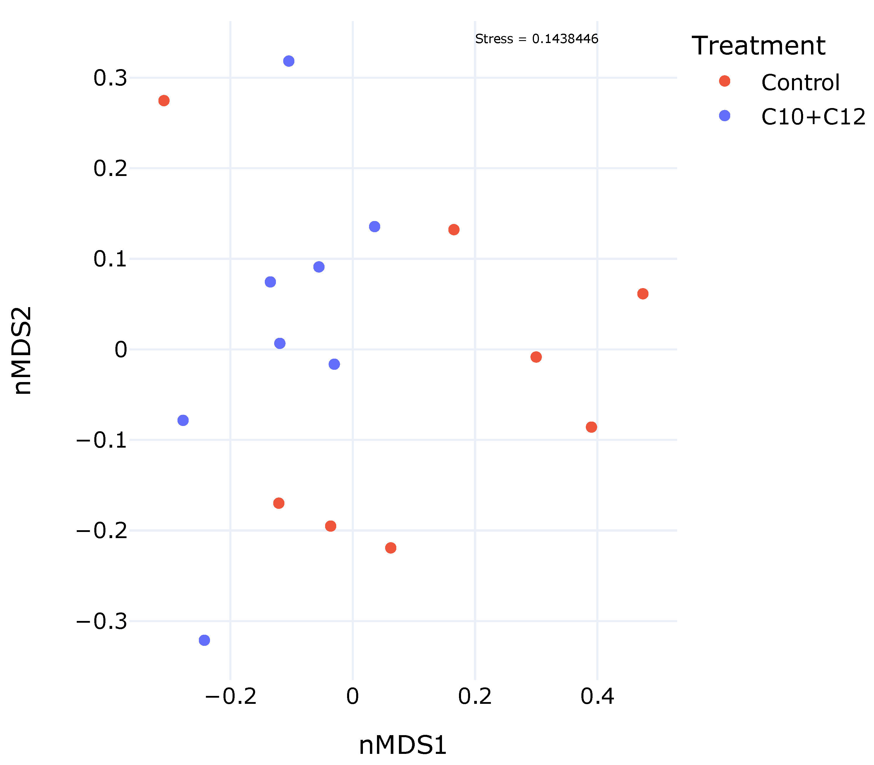
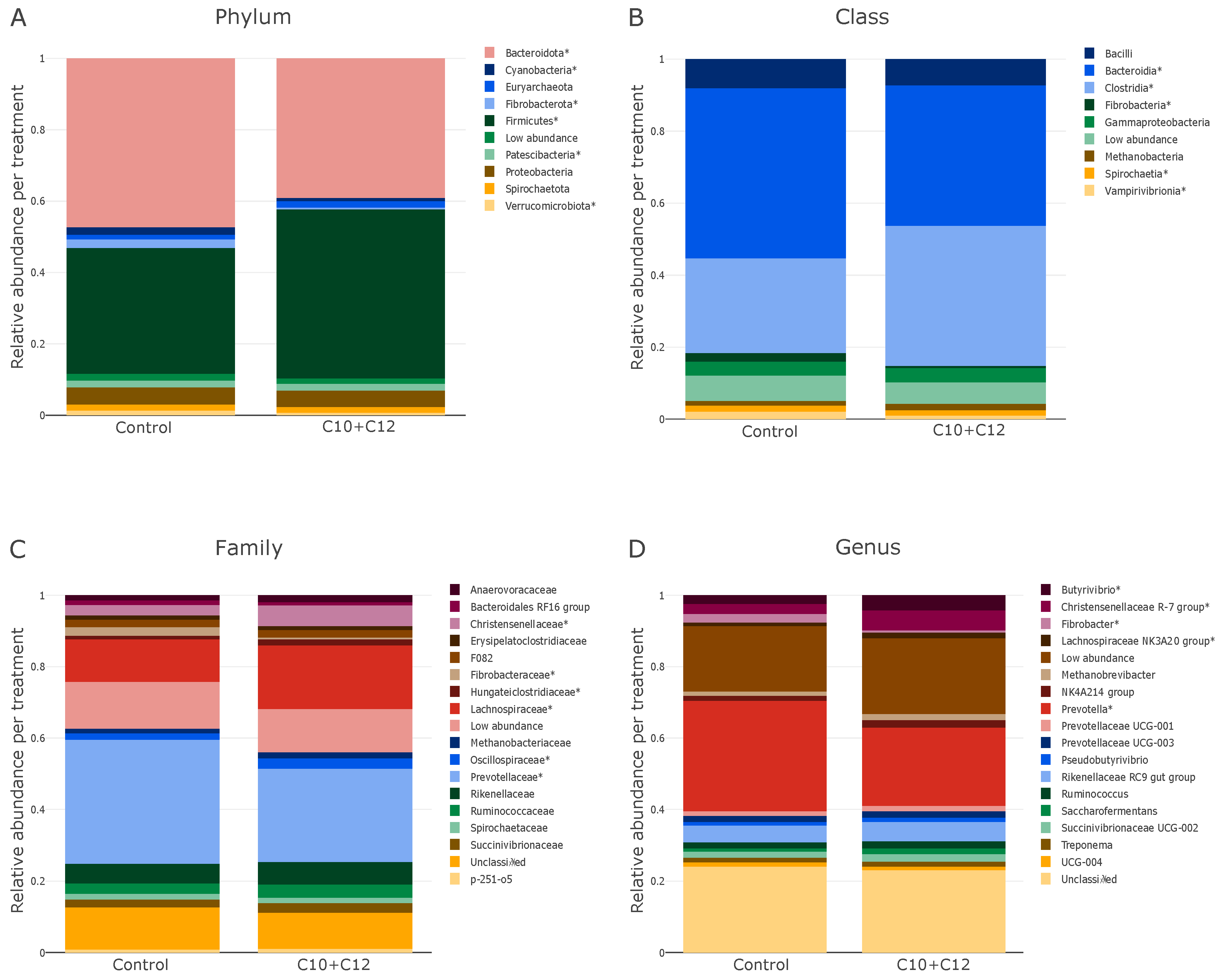
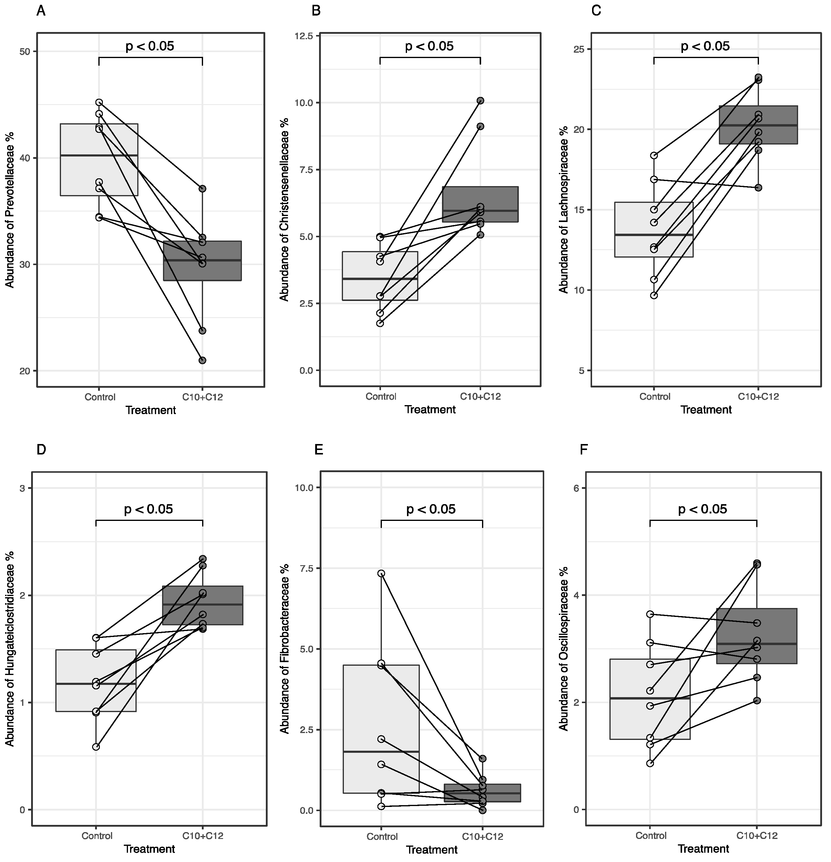
The relative abundance of bacteria was visualized at various taxonomic levels (> 0.1% of the total sequences) in Python using the “plotly” package. Differences in relative abundances were assessed using the Wilcoxon signed-rank test [17]. Differences were considered statistically significant at p < 0.05. Alpha diversity indices (Chao1, Pileou, Shannon, and Simpson) were calculated based on the rarefied amplicon sequence variant (ASV) count table and analyzed using a paired t-test [17]. Boxplots illustrating significant differences in relative abundances at the family level were generated using the “ggplot2”, “ggpubr”, and “ggsignif” packages in R software (version 4.3.2). The compositional differences (beta diversity) were assessed and visualized using the “vegan” package in R. The data were analyzed using the metaMDS function through a nonmetric multidimensional scaling (NMDS). Since the stress values of the two-dimensional NMDS were <0.20 (stress = 0.1438446), the ordination patterns were deemed acceptable. The stress values for NMDS were reported after 100 attempts, with the best solution being repeated 30 times.

3. Results

Diversity, evenness, and richness were not affected by the treatment, as C10 + C12 did not affect the alpha diversity indices (Simpson, Chao1, Shannon, and Pielou) nor the ASV count (p > 0.05; Table 1).
Beta diversity, as visualized using the NMDS (Figure 1), did not show a specific cluster or pattern between the samples. The C10 + C12 samples were located in the upper left quadrant, farther from the control, indicating treatment-dependent differences in bacterial composition. The relative abundances of the control and treatment groups at the phylum, class, family, and genus levels are shown in Figure 2. The highest relative abundance at the phylum level (Figure 2A) was noted for Bacteroidota (48.2%) and Firmicutes (35.9%) in the control group. The treatment decreased the abundance of Bacteroidota to 39.7% and increased that of Firmicutes to 48.1%.
The class level (Figure 2B) matched the phylum level, and the relative abundance of Bacteroidia (phylum Bacteroidota) was higher in the control group (50.9%) than in the treatment group (41.6%). In contrast, the highest relative abundance in the experimental group was that of Clostridia (phylum Firmicutes; 41.5%), which was higher than that in the control group (28.4%). A decrease in the relative abundance of 2.6% to 0.6% was noted for Fibrobacteria in the treatment group.
The significantly different families are shown in Figure 2C, and further details for individual cows are shown in Figure 3. The effect of C10 + C12 was consistent at the family level of microbes between individual animals; in all animals, the treatment significantly decreased and increased the relative abundances of Prevotellaceae, Fibrobacteraceae, and Christensenellaceae, respectively (p < 0.05). The relative abundances of Lachnospiraceae and Oscillospiraceae increased in the treatment group apart from one and two animals, respectively.
On the genus level (Figure 2D), the relative abundances of Butyrivibrio, Christensenellaceae R-7 group, Lachnospiraceae NK3A20 group, and Fibrobacter increased (p < 0.05) at the expense of the Prevotella genus, which decreased (p < 0.05).

4. Discussion

In a previous study, we reported the effects of C10 + C12 on gas production and rumen fermentation characteristics [14]. In summary, the supplementation of C10 + C12 in vivo decreased CH4 production by 11.5% but increased rumen ammonia-N (NH3-N) concentrations (+28.5%) and ammonia gas emissions (+37.2%). The treatment did not affect the total VFA concentration, ruminal pH, or protozoal count. This implies that C10 + C12 has CH4 mitigation potential but may compromise the efficiency of N utilization. However, the mechanism of action remains unclear. We hypothesized that the microbial communities would provide insights into these observed effects.

4.1. Bacterial Diversity, ASVs, and Indices

Our findings show that the C10 + C12 treatment did not alter bacterial diversity, evenness, or richness (Table 1), contradicting our initial hypothesis. The lack of effects on the overall bacterial indices could be explained by the low doses of MCFA (0.8% DM) [18]. Our results further agree with those of Burdick et al. [8], who used comparatively lower doses of MCFA (0.25% DM; blend of C8, C10, and C12). Ruminal diversity was not affected in our study, implying that MCFA did not jeopardize the overall stability of the ruminal community. In contrast, lower microbial richness, diversity, and bacterial abundance, along with a higher abundance of fiber-degrading bacteria, has previously been associated with higher feed efficiency in dairy cows [19,20]. This is because the resulting metabolic network is simpler and leads to higher concentrations of specific components suitable for the host’s energy requirements [21]. In comparison, in our previous in vitro study evaluating the combined effects of nitrate and various MCFAs, the alpha diversity indices decreased. The differences may be because it was an in vitro study, where we used higher concentrations of MCFA, and added nitrate [16]. Another reason could be using a solvent (ethanol) to dissolve MCFA in the medium in vitro, but not in the in vivo trial. This corroborates the theory that saturated fatty acids may require partial dissolution in a buffer or medium to have a significant impact [22].

4.2. Microbial Relative Abundances

At the kingdom level, the relative abundances of bacteria and archaea (Table 1, Figure 4) were not altered by the treatment, which is in line with other studies on MCFA in the rumen [8,23]. The lack of an effect on the relative abundance does not align with our previously reported decrease in methanogenesis (−11.5%) from these animals [14]. However, there have been reports of reduced methanogenesis (and methanogen activity) with no change in the methanogen number [24]. Consequently, it has been suggested that the abundance of methanogens may not be the primary factor affecting CH4 emissions [3], but rather the composition of the archaeal community and the metabolic activity of each methanogenic species [25]. For example, tea saponin did not affect total methanogen numbers, but significantly decreased methanogenesis (by 8%), which was explained by the reduced gene expression (−76%; [24]); specifically, the expression of the gene encoding methyl-coenzyme M reductase (MCR), an enzyme that catalyzes the last step of CH4 production [7]. MCFA may be able to reduce methanogenesis by decreasing the metabolic activity of archaea [26]. Thus, in our study, the reduced methanogenesis may have been caused by the altered composition of the methanogenic community, suppressed metabolism, or both.
The dominant bacterial phyla in both the control and treatment groups were Bacteroidota and Firmicutes (Figure 2A), which is consistent with most ruminant studies [20,27]. These phyla showed an inverse relationship in their relative abundance, as seen in previous research [21,28]; Bacteroidota decreased in the C10 + C12 group, and in parallel, Firmicutes increased. This reciprocal adjustment aligns with the sensitivity of Bacteroidota to altered ruminal environments (e.g., by the diet), and their propensity to be among the first to react to these alterations [6]. The decrease in Bacteroidota resulted in an increased Firmicutes to Bacteroidota (F:B) ratio. This ratio has been identified as a significant parameter for assessing the microbial impact on the host energy requirements. An increased F:B ratio has been shown to contribute to improved bovine performance, including body weight gain and milk fat yield [29], but there are contradictory reports [30]. A greater abundance of Firmicutes indicates a more feed-efficient animal [21,31] with improved structural polysaccharide degradation [32]. Changes in this ratio did not affect digestibility, and we could not assess its effect on production parameters, because the animals in our study were dry cows [14].
We observed a few changes in the relative abundances of bacteria at the family level. The relative abundances of Christensenellaceae and Lachnospiraceae increased; these families are associated with H2 production [33]. In contrast, the bacteria associated with H2 utilization remained either unaffected (Succinivibrionaceae) or decreased (Prevotella) by the treatment [34], which refutes our second hypothesis. The general hypothesis is that CH4 emissions depend on the abundance of H2-producing and H2-utilizing bacteria in the rumen [6,34]. The concentrations of H2 are maintained at low levels primarily through methanogenesis, which maintains favorable conditions for continual fermentation and fiber digestion. The inhibition of CH4 production leads to elevated H2 levels in the rumen [9]. For example, supplementation with a CH4 inhibitor, 3-NOP, resulted in a 37-fold increase in H2 yield [2]. Notably, research shows that even high H2 levels (i.e., 550 μM in comparison with the typical range of 0.2–30 μM) do not compromise fiber breakdown in the rumen [35]. If the accumulated H2 is not incorporated into alternative H2 sinks, some of the excess H2 is simply emitted from the rumen [6,36]. Our results imply there was a surplus of H2 in the rumen. We did not measure H2 emissions nor dissolved H2 in the rumen liquid, but the fiber digestibility was not affected [14]. Thus, our results seem to confirm the hypothesis of Hristov et al. [36] according to which the ruminal microbiota can cope with excess H2, and surplus H2 was most probably emitted. Possibly, ruminal microbiota may have utilized some of the excess H2 for processes such as amino acid and fatty acid synthesis [37]. Microbial biomass fulfills a significant portion protein needs of the host and is thought to be a minor electron sink (0.2–0.3%; [38]). However, we did not measure the microbial protein production in this study.
At the genus level, the relative abundance of the fibrolytic Fibrobacter decreased in the treatment groups, whereas that of Ruminococcus remained unchanged. This may indicate a higher sensitivity of Fibrobacter to C10 + C12, even though it is a Gram-negative bacterium that is reported to be less sensitive to MCFA [11,23]. Gram-negative bacteria are usually thought to be more resilient than Gram-positive bacteria due to the presence of a protective outer membrane [11]. The genera Fibrobacter and Ruminococcus had low relative abundances in the treatment group (0.7% and 2.43%, respectively); however, even taxa with low-abundance taxa may be necessary for the microbial community [20]. The rumen has a highly adaptable microbial community with overlapping metabolic capabilities [21], which probably supports the unaltered digestibility [14].
In our previous study [14], C10 + C12 increased the concentration of ammonium-N (NH3-N) in the rumen fluid and the emission of ammonia (NH3) in dairy cows. Our results suggest that these changes may be explained by the effect of C10 + C12 on the relative abundances of bacteria that metabolize nitrogen. As mentioned previously, the relative abundance of the family Prevotellaceae was decreased. Members of this family, such as Prevotella Ruminicola, are considered to have low deamination activity and release NH3 at a lower rate, thus allowing its high utilization efficiency [39]. In contrast, the relative abundance of the class Clostridia increased during the treatment. A few members of this class (e.g., Clostridium sticklandii and Clostridium aminophilum) have high deamination activities and are referred to as hyperammonia-producing bacteria (HAB; [39,40]). HAB can hydrolyze small peptides and deaminate amino acids [39] and have been shown to produce ammonia at rates up to 20 times higher than those of other ammonia-producing bacteria found in the rumen [37]. The higher abundance of this class may have caused a higher production of NH3 in the rumen (+28.5%; [14]), which other microorganisms could not utilize. Therefore, the higher production of NH3-N in the rumen fluid may have led to lower NH3 utilization efficiency and higher NH3 emissions (+37.2%) when supplying C10 + C12. Inadequate N utilization in the rumen leads to NH3-N excretion and ultimately to nitrous oxide emissions from manure, contributing to air and ground water pollution [39]. Ruminants are responsible for approximately 30% of the total anthropogenic NH3 emissions and more than 70% of the NH3 emissions from the global livestock, which leads to substantial economic losses and adverse effects on human health [41].
The use of NGS in our study has some limitations. Mainly, NGS does not differentiate between live and dead cells [42,43]. Furthermore, to obtain precise functional characterizations of not only bacteria, but also archaea, more advanced techniques would have to be employed, such as metagenomics and metabolomics. Since we report relative abundances, we cannot be sure of the absolute counts of the microorganisms. Another limitation is the data analysis and data interpretation, since studies on microbiota use different sequencing platforms, different groups of closely related DNA sequences (ASVs or OTUs), different software and packages, and statistical tests.

5. Conclusions

Our study highlights the effects of combined capric acid and lauric acid supplementation on rumen microbiota, revealing alterations in bacterial abundances with implications for rumen ammonia levels, offering insights into potential strategies for modulating rumen fermentation processes and methane production in ruminant livestock.

Author Contributions

Conceptualization, M.J. and M.V.; methodology, M.J., M.V. and A.Š.; software, M.V.; formal analysis, M.V.; sample collection, A.V., Y.T. and D.T.; writing—original draft preparation, M.V. and M.J.; writing—review and editing, M.V. and M.J.; visualization, M.V.; supervision, M.J.; project administration, M.J. and M.V.; funding acquisition, M.J. All authors have read and agreed to the published version of the manuscript.

Funding

The Ministry of Agriculture of the Czech Republic supported this study [Grant No. NAZV QK23020011].

Data Availability Statement

The raw data supporting the conclusions of this article will be made available by the authors on request.

Acknowledgments

Computational resources were provided by the e-INFRA CZ project (ID:90254), supported by the Ministry of Education, Youth and Sports of the Czech Republic.

Conflicts of Interest

The authors declare no conflict of interest. The funders had no role in the design of the study; in the collection, analyses, or interpretation of data; in the writing of the manuscript; or in the decision to publish the results.

References

  1. Yamada, K.; Iwamae, K.; Suzuki, Y.; Koike, S.; Kobayashi, Y. Batch Culture Analysis to Identify Potent Organic Acids for Suppressing Ruminal Methane Production. Anim. Sci. J. 2023, 94, e13873. [Google Scholar] [CrossRef] [PubMed]
  2. Gruninger, R.J.; Zhang, X.M.; Smith, M.L.; Kung, L.; Vyas, D.; McGinn, S.M.; Kindermann, M.; Wang, M.; Tan, Z.L.; Beauchemin, K.A. Application of 3-Nitrooxypropanol and Canola Oil to Mitigate Enteric Methane Emissions of Beef Cattle Results in Distinctly Different Effects on the Rumen Microbial Community. Anim. Microbiome 2022, 4, 35. [Google Scholar] [CrossRef]
  3. Choi, Y.; Lee, S.J.; Kim, H.S.; Eom, J.S.; Jo, S.U.; Guan, L.L.; Seo, J.; Kim, H.; Lee, S.S.; Lee, S.S. Effects of Seaweed Extracts on in Vitro Rumen Fermentation Characteristics, Methane Production, and Microbial Abundance. Sci. Rep. 2021, 11, 24092. [Google Scholar] [CrossRef] [PubMed]
  4. Sikiru, A.; Michael, A.O.; John, M.O.; Egena, S.S.A.; Oleforuh-Okoleh, V.U.; Ambali, M.I.; Muhammad, I.R. Methane Emissions in Cattle Production: Biology, Measurement and Mitigation Strategies in Smallholder Farmer Systems. Environ. Dev. Sustain. 2024. [Google Scholar] [CrossRef]
  5. Tapio, I.; Snelling, T.J.; Strozzi, F.; Wallace, R.J. The Ruminal Microbiome Associated with Methane Emissions from Ruminant Livestock. J. Anim. Sci. Biotechnol. 2017, 8, 7. [Google Scholar] [CrossRef] [PubMed]
  6. De Assis Lage, C.F.; Räisänen, S.E.; Melgar, A.; Nedelkov, K.; Chen, X.; Oh, J.; Fetter, M.E.; Indugu, N.; Bender, J.S.; Vecchiarelli, B.; et al. Comparison of Two Sampling Techniques for Evaluating Ruminal Fermentation and Microbiota in the Planktonic Phase of Rumen Digesta in Dairy Cows. Front. Microbiol. 2020, 11, 618032. [Google Scholar] [CrossRef] [PubMed]
  7. Honan, M.; Feng, X.; Tricarico, J.; Kebreab, E. Feed Additives as a Strategic Approach to Reduce Enteric Methane Production in Cattle: Modes of Action, Effectiveness and Safety. Anim. Prod. Sci. 2021, 62, 1303–1317. [Google Scholar] [CrossRef]
  8. Burdick, M.; Zhou, M.; Guan, L.; Oba, M. Effects of Medium-Chain Fatty Acid Supplementation on Performance and Rumen Fermentation of Lactating Holstein Dairy Cows. Animal 2022, 16, 100491. [Google Scholar] [CrossRef] [PubMed]
  9. Kjeldsen, M.H.; Weisbjerg, M.R.; Larsen, M.; Højberg, O.; Ohlsson, C.; Walker, N.; Hellwing, A.L.F.; Lund, P. Gas Exchange, Rumen Hydrogen Sinks, and Nutrient Digestibility and Metabolism in Lactating Dairy Cows Fed 3-NOP and Cracked Rapeseed. J. Dairy Sci. 2023, 107, 2047–2065. [Google Scholar] [CrossRef] [PubMed]
  10. Dohme, F.; Machmüller, A.; Wasserfallen, A.; Kreuzer, M. Ruminal Methanogenesis as Influenced by Individual Fatty Acids Supplemented to Complete Ruminant Diets. Lett. Appl. Microbiol. 2001, 32, 47–51. [Google Scholar] [CrossRef]
  11. Machmüller, A. Medium-Chain Fatty Acids and Their Potential to Reduce Methanogenesis in Domestic Ruminants. Agric. Ecosyst. Environ. 2006, 112, 107–114. [Google Scholar] [CrossRef]
  12. Machmüller, A.; Kreuzer, M. Methane Suppression by Coconut Oil and Associated Effects on Nutrient and Energy Balance in Sheep. Can. J. Anim. Sci. 1999, 79, 65–72. [Google Scholar] [CrossRef]
  13. Machmüller, A.; Soliva, C.R.; Kreuzer, M. Methane-Suppressing Effect of Myristic Acid in Sheep as Affected by Dietary Calcium and Forage Proportion. Br. J. Nutr. 2003, 90, 529–540. [Google Scholar] [CrossRef] [PubMed]
  14. Joch, M.; Vadroňová, M.; Češpiva, M.; Zabloudilová, P.; Výborná, A.; Tyrolová, Y.; Kudrna, V.; Tichá, D.; Plachý, V.; Hroncová, Z. Capric and Lauric Acid Mixture Decreased Rumen Methane Production, While Combination with Nitrate Had No Further Benefit in Methane Reduction. Ann. Anim. Sci. 2023, 23, 799–808. [Google Scholar] [CrossRef]
  15. Muizelaar, W.; Bani, P.; Kuhla, B.; Tapio, I.; Yáñez-Ruiz, D.; van Gastelen, S. Rumen Fluid Sampling via Oral Stomach Tubing Method. In Methods in Cattle Physiology and Behaviour Research; Publisso: Cologne, Germany, 2020. [Google Scholar]
  16. Vadroňová, M.; Šťovíček, A.; Jochová, K.; Výborná, A.; Tyrolová, Y.; Tichá, D.; Homolka, P.; Joch, M. Combined Effects of Nitrate and Medium-Chain Fatty Acids on Methane Production, Rumen Fermentation, and Rumen Bacterial Populations In Vitro. Sci. Rep. 2023, 13, 21961. [Google Scholar] [CrossRef]
  17. Jiang, X.; Xu, H.J.; Cui, Z.Q.; Zhang, Y.G. Effects of Supplementation with Lactobacillus plantarum 299v on the Performance, Blood Metabolites, Rumen Fermentation and Bacterial Communities of Preweaning Calves. Livest. Sci. 2020, 239, 104120. [Google Scholar] [CrossRef]
  18. Cancino-Padilla, N.; Gajardo, F.; Neves, A.L.A.; Kholif, A.E.; Mele, M.; Huws, S.A.; Loor, J.J.; Romero, J.; Vargas-Bello-Pérez, E. Influence of Dietary Oils Rich in Omega-6 or Omega-3 Fatty Acids on Rumen Microbiome of Dairy Cows. Transl. Anim. Sci. 2023, 7, txad074. [Google Scholar] [CrossRef]
  19. Shabat, S.K.B.; Sasson, G.; Doron-Faigenboim, A.; Durman, T.; Yaacoby, S.; Berg Miller, M.E.; White, B.A.; Shterzer, N.; Mizrahi, I. Specific Microbiome-Dependent Mechanisms Underlie the Energy Harvest Efficiency of Ruminants. ISME J. 2016, 10, 2958–2972. [Google Scholar] [CrossRef] [PubMed]
  20. Mizrahi, I.; Wallace, R.J.; Moraïs, S. The Rumen Microbiome: Balancing Food Security and Environmental Impacts. Nat. Rev. Microbiol. 2021, 19, 553–566. [Google Scholar] [CrossRef]
  21. Díaz Carrasco, J.M.; Cabral, C.; Redondo, L.M.; Pin Viso, N.D.; Colombatto, D.; Farber, M.D.; Fernández Miyakawa, M.E. Impact of Chestnut and Quebracho Tannins on Rumen Microbiota of Bovines. BioMed Res. Int. 2017, 2017, 9610810. [Google Scholar] [CrossRef]
  22. Zhou, X.; Meile, L.; Kreuzer, M.; Zeitz, J.O. The Effect of Saturated Fatty Acids on Methanogenesis and Cell Viability of Methanobrevibacter ruminantium. Archaea 2013, 2013, e106916. [Google Scholar] [CrossRef] [PubMed]
  23. Yanza, Y.R.; Szumacher-Strabel, M.; Jayanegara, A.; Kasenta, A.M.; Gao, M.; Huang, H.; Patra, A.K.; Warzych, E.; Cieślak, A. The Effects of Dietary Medium-chain Fatty Acids on Ruminal Methanogenesis and Fermentation In Vitro and In Vivo: A Meta-analysis. J. Anim. Physiol. Anim. Nutr. 2020, 105, 874–889. [Google Scholar] [CrossRef] [PubMed]
  24. Guo, Y.Q.; Liu, J.-X.; Lu, Y.; Zhu, W.Y.; Denman, S.E.; McSweeney, C.S. Effect of Tea Saponin on Methanogenesis, Microbial Community Structure and Expression of mcrA Gene, in Cultures of Rumen Micro-Organisms. Lett. Appl. Microbiol. 2008, 47, 421–426. [Google Scholar] [CrossRef]
  25. Danielsson, R.; Ramin, M.; Bertilsson, J.; Lund, P.; Huhtanen, P. Evaluation of a Gas In Vitro System for Predicting Methane Production In Vivo. J. Dairy Sci. 2017, 100, 8881–8894. [Google Scholar] [CrossRef]
  26. Patra, A.; Park, T.; Kim, M.; Yu, Z. Rumen Methanogens and Mitigation of Methane Emission by Anti-Methanogenic Compounds and Substances. J. Anim. Sci. Biotechnol. 2017, 8, 13. [Google Scholar] [CrossRef] [PubMed]
  27. Zhang, X.; Medrano, R.F.; Wang, M.; Beauchemin, K.A.; Ma, Z.; Wang, R.; Wen, J.; Bernard, L.A.; Tan, Z. Effects of Urea plus Nitrate Pretreated Rice Straw and Corn Oil Supplementation on Fiber Digestibility, Nitrogen Balance, Rumen Fermentation, Microbiota and Methane Emissions in Goats. J. Anim. Sci. Biotechnol. 2019, 10, 6. [Google Scholar] [CrossRef]
  28. Cancino-Padilla, N.; Catalán, N.; Siu-Ting, K.; Creevey, C.J.; Huws, S.A.; Romero, J.; Vargas-Bello-Pérez, E. Long-Term Effects of Dietary Supplementation with Olive Oil and Hydrogenated Vegetable Oil on the Rumen Microbiome of Dairy Cows. Microorganisms 2021, 9, 1121. [Google Scholar] [CrossRef] [PubMed]
  29. Jami, E.; White, B.A.; Mizrahi, I. Potential Role of the Bovine Rumen Microbiome in Modulating Milk Composition and Feed Efficiency. PLoS ONE 2014, 9, e85423. [Google Scholar] [CrossRef] [PubMed]
  30. Lima, F.S.; Oikonomou, G.; Lima, S.F.; Bicalho, M.L.S.; Ganda, E.K.; de Oliveira Filho, J.C.; Lorenzo, G.; Trojacanec, P.; Bicalho, R.C. Prepartum and Postpartum Rumen Fluid Microbiomes: Characterization and Correlation with Production Traits in Dairy Cows. Appl. Environ. Microbiol. 2015, 81, 1327–1337. [Google Scholar] [CrossRef]
  31. Zhang, X.M.; Smith, M.L.; Gruninger, R.J.; Kung, L., Jr.; Vyas, D.; McGinn, S.M.; Kindermann, M.; Wang, M.; Tan, Z.L.; Beauchemin, K.A. Combined Effects of 3-Nitrooxypropanol and Canola Oil Supplementation on Methane Emissions, Rumen Fermentation and Biohydrogenation, and Total Tract Digestibility in Beef Cattle. J. Anim. Sci. 2021, 99, skab081. [Google Scholar] [CrossRef]
  32. Jiao, P.; Wang, Z.; Yang, W. Effect of Clostridium Butyricum Supplementation on In Vitro Rumen Fermentation and Microbiota with High Grain Substrate Varying with Media pH Levels. Front. Microbiol. 2022, 13, 912042. [Google Scholar] [CrossRef]
  33. Huaiquipán, R.; Quiñones, J.; Díaz, R.; Velásquez, C.; Sepúlveda, G.; Velázquez, L.; Paz, E.A.; Tapia, D.; Cancino, D.; Sepúlveda, N. Review: Effect of Experimental Diets on the Microbiome of Productive Animals. Microorganisms 2023, 11, 2219. [Google Scholar] [CrossRef] [PubMed]
  34. Guo, Y.; Hassan, F.; Li, M.; Xie, H.; Peng, L.; Tang, Z.; Yang, C. Effect of Sodium Nitrate and Cysteamine on In Vitro Ruminal Fermentation, Amino Acid Metabolism and Microbiota in Buffalo. Microorganisms 2022, 10, 2038. [Google Scholar] [CrossRef]
  35. Ungerfeld, E.M.; Beauchemin, K.A.; Muñoz, C. Current Perspectives on Achieving Pronounced Enteric Methane Mitigation From Ruminant Production. Front. Anim. Sci. 2022, 2, 795200. [Google Scholar] [CrossRef]
  36. Hristov, A.N.; Oh, J.; Giallongo, F.; Frederick, T.W.; Harper, M.T.; Weeks, H.L.; Branco, A.F.; Moate, P.J.; Deighton, M.H.; Williams, S.R.O. An Inhibitor Persistently Decreased Enteric Methane Emission from Dairy Cows with No Negative Effect on Milk Production. Proc. Natl. Acad. Sci. USA 2015, 112, 10663–10668. [Google Scholar] [CrossRef] [PubMed]
  37. Wang, K.; Xiong, B.; Zhao, X. Could Propionate Formation Be Used to Reduce Enteric Methane Emission in Ruminants? Sci. Total Environ. 2023, 855, 158867. [Google Scholar] [CrossRef] [PubMed]
  38. Guyader, J.; Tavendale, M.; Martin, C.; Muetzel, S. Dose-Response Effect of Nitrate on Hydrogen Distribution between Rumen Fermentation End Products: An In Vitro Approach. Anim. Prod. Sci. 2016, 56, 224–230. [Google Scholar] [CrossRef]
  39. Tan, P.; Liu, H.; Zhao, J.; Gu, X.; Wei, X.; Zhang, X.; Ma, N.; Johnston, L.J.; Bai, Y.; Zhang, W.; et al. Amino Acids Metabolism by Rumen Microorganisms: Nutrition and Ecology Strategies to Reduce Nitrogen Emissions from the inside to the Outside. Sci. Total Environ. 2021, 800, 149596. [Google Scholar] [CrossRef] [PubMed]
  40. Millen, D.D.; Arrigoni, M.D.B.; Pacheco, R.D.L. Rumenology; Springer: Berlin/Heidelberg, Germany, 2016; ISBN 3-319-30533-6. [Google Scholar]
  41. Zhang, Z.; Wei, W.; Yang, S.; Huang, Z.; Li, C.; Yu, X.; Qi, R.; Liu, W.; Loor, J.J.; Wang, M.; et al. Regulation of Dietary Protein Solubility Improves Ruminal Nitrogen Metabolism In Vitro: Role of Bacteria–Protozoa Interactions. Nutrients 2022, 14, 2972. [Google Scholar] [CrossRef]
  42. Pitta, D.; Indugu, N.; Narayan, K.; Hennessy, M. Symposium Review: Understanding the Role of the Rumen Microbiome in Enteric Methane Mitigation and Productivity in Dairy Cows. J. Dairy Sci. 2022, 105, 8569–8585. [Google Scholar] [CrossRef]
  43. Weinroth, M.D.; Belk, A.D.; Dean, C.; Noyes, N.; Dittoe, D.K.; Rothrock, M.J.; Ricke, S.C.; Myer, P.R.; Henniger, M.T.; Ramírez, G.A.; et al. Considerations and Best Practices in Animal Science 16S Ribosomal RNA Gene Sequencing Microbiome Studies. J. Anim. Sci. 2022, 100, skab346. [Google Scholar] [CrossRef] [PubMed]
Figure 1. Supplementation with C10 + C12 shows microbiome variations on the amplicon sequence variant (ASV) level through a nonmetric multidimensional scaling (NMDS) visualization based on the control diet (red) or supplemented diet (blue). The C10 + C12 effect is visible in the sample distribution; however, the samples do not aggregate into clusters.
Microorganisms 12 01085 g001
Figure 2. Bar charts showing the effect of C10 + C12 on relative bacterial abundances at the phylum (A), class (B), family (C), and genus (D) levels. Taxonomic groups accounting for less than 0.1% of total sequences in each sample were grouped together into a low-abundance category. * Asterisks indicate significant differences at p < 0.05.
Microorganisms 12 01085 g002
Figure 3. The addition of C10 + C12 to the experimental diet had consistent effects in individual animals on bacterial families (Prevotellaceae, (A); Christensenellaceae, (B); Lachnospiraceae, (C); Hungateiclostridiaceae, (D); Fibrobacteraceae, (E); Oscillospiraceae, (F)). Boxplots show significantly different relative abundances of bacterial families in individual animals without C10 + C12 (control, light gray) supplementation and with supplementation (dark gray) in each animal. The crossover design allowed us to consecutively feed the cows both the control diet and the C10 + C12-supplemented diet (and vice versa). The solid lines connect the paired values of individual cows.
Microorganisms 12 01085 g003
Figure 4. Supplementation of C10 + C12 in the experimental diet did not affect ruminal archaea. The left boxplot (light gray) shows the relative abundances of archaea when the cows were fed the control diet, and the right (dark gray) shows the relative abundances of archaea after supplementation with C10 + C12. The boxplots show a relatively consistent effect on the relative abundance of archaea apart from the two animals. The solid lines connect the paired values of individual cows.
Microorganisms 12 01085 g004
Table 1. Effect of C10 + C12 on bacterial α-diversity indices, amplicon sequence variant (ASV) count, and methanogen relative abundance in vivo.
TreatmentShannon Diversity
Index
Simpson Diversity IndexChao1 Richness IndexPielou Evenness IndexASV CountArchaeal Relative Abundance
Control9.9255620.99568055690.79733855890.013248
C10 + C129.9829640.99635558300.80631353340.017648
SEM0.053720.000422.261620.0039121.459580.16
p-value0.28280.29180.84920.12080.89560.195312
The treatment did not differ significantly (p < 0.05) from the corresponding control. SEM, standard error of the mean.
Disclaimer/Publisher’s Note: The statements, opinions and data contained in all publications are solely those of the individual author(s) and contributor(s) and not of MDPI and/or the editor(s). MDPI and/or the editor(s) disclaim responsibility for any injury to people or property resulting from any ideas, methods, instructions or products referred to in the content.

