Article

Development and Evaluation of Bioconverted Milk with Anti-Microbial Effect against Periodontal Pathogens and α-Glucosidase Inhibitory Activity

1
Risk Analysis Research Center, Sookmyung Women’s University, Seoul 04310, Republic of Korea
2
Department of Food and Nutrition, Sookmyung Women’s University, Seoul 04310, Republic of Korea
3
Department of Oral Microbiology, College of Dentistry, Wonkwang University, Iksan 54538, Republic of Korea
*
Authors to whom correspondence should be addressed.
These authors contributed equally to this work.
Microorganisms 2024, 12(7), 1290; https://doi.org/10.3390/microorganisms12071290
Submission received: 24 May 2024 / Revised: 19 June 2024 / Accepted: 21 June 2024 / Published: 25 June 2024

Abstract

:
To decrease periodontal pathogens and increase the number of beneficial bacteria, probiotics and bioactive compounds made via microbial bioconversion are recently used. In addition, the interest regarding probiotics-mediated bioconversion with popular medicinal plants is increasing. Artemisia herba-alba, a type of wormwood, has recently been attention as a medicinal plant due to its various bioactive compounds. Therefore, we developed bioconverted milk containing A. herba-alba that effectively inhibited periodontal pathogens and α-glucosidase. To select the appropriate lactic acid bacteria for the probiotic candidate strain, 74 strains of lactic acid bacteria were screened. Among them, Lactiplantibacillus plantarum SMFM2016-RK was chosen as the probiotic due to its beneficial characteristics such as high acid and bile tolerance, antioxidant activity, and α-glucosidase inhibition. Based on the minimal bactericidal concentration against three periodontal pathogens, the following appropriate concentrations of Artemisia herba-alba extract were added to milk: 5 mg/mL of A. herba-alba ethanol extract and 25 mg/mL of A. herba-alba hot-water extract. Four bioconverted milks (BM), BM1, BM2, BM3, and BM4, were produced by combining L. plantarum SMFM2016-RK alone, L. plantarum SMFM2016-RK and ethanol extract, L. plantarum SMFM2016-RK and hot-water extract, and L. plantarum SMFM2016-RK with both extracts. As a result of antimicrobial activity, BM3 inhibited the growth of Aggregatibacter actinomycetemcomitans and Porphyromonas gingivalis the most, and BM4 suppressed the growth of Fusobacterium nucleatum the most. In addition, bioconverted milk containing A. herba-alba (BM2, BM3, and BM4) inhibited α-glucosidase more effectively than BM1. The whole genome of L. plantarum SMFM2016-RK was obtained, and 3135 CDS, 67 tRNA, and 16 RNA were predicted. The genome annotation of L. plantarum SMFM2016-RK revealed 11 CDS related to proteolysis and amino acid metabolism and 2 CDS of phenolic acid-metabolizing enzymes. In conclusion, A. herba-alba-added milk bioconverted by L. plantarum SMFM2016-RK displayed both the growth inhibitory effect on periodontal pathogens and the α-glucosidase inhibitory activity; thus, it necessitates to evaluate the effects on the alleviation of periodontal diseases and glycemic control through future animal experiments.

1. Introduction

Oral disease is a major global health concern that affects a large portion of the world’s population. According to a report by the World Health Organization (WHO), almost half of the world’s population suffers from oral diseases [1]. Furthermore, oral infections and associated inflammatory responses can have a negative impact on blood glucose levels, and a previous study reported that more than 90% of diabetic patients experienced oral complications [2,3]. The α-glucosidase enzyme affects blood glucose levels in people with oral infections. It plays a major role in raising blood sugar, breaks down complex carbohydrates into simple sugars, and then absorbs them into the bloodstream. Therefore, it is known that inhibiting the α-glucosidase enzyme helps to prevent glucose levels from rising rapidly by allowing glucose to be gradually absorbed into the bloodstream [4]. Thus, many researchers have made an effort to identify potent α-glucosidase inhibitors [5,6,7,8]. The treatment and prevention of periodontitis involve various medications and therapeutic approaches. Some of the commonly used medications include antibiotics, antiseptics, and carbamide peroxide topical [9]. However, these treatments have certain limitations such as antibiotic resistance [9]. Due to the increase in antibiotic resistance, the focus of periodontitis treatment is on restoring the balance between the oral microbiota and the host’s periodontal tissue [9]. For this reason, probiotics markets targeting oral health are experiencing significant growth and development. The market size for probiotics specifically for oral health was valued at USD 2.5 billion in 2022 [10]. This growth is driven by research into specific probiotic strains, such as Lactobacillus and Bifidobacterium species, which have shown potential benefits in reducing the risk of dental caries, gum disease, and bad breath [11]. In the food industry, Lactiplantibacillus plantarum (formerly Lactobacillus plantarum) is one of the lactic acid bacteria (LAB) that is often used as a potential probiotic starter. As its main metabolites, lactic acid, succinic acid, and acetic acid are produced by this non-spore-forming bacterium [12]. According to certain research, using L. plantarum specifically was linked to better periodontal health, as seen by smaller pockets and less bleeding when the gums were probed [13,14]. In addition, L. plantarum could reduce oral carriage of Streptococcus mutans [15,16]. Furthermore, various new quality-related components such as phenolic acids, flavones and their glycosides, alkaloids, and terpenoids have been identified due to changes in chemical composition during microbial bioconversion with plants in recent years [17]. Microbial bioconversion showed a synergistic effect compared to simple application of LAB, such as increasing the fermentation ability due to soybean protein and enhancing the inflammatory bowel disease-alleviating effect by boosting the bioactivity of anthocyanin [18,19].
Artemisia herba-alba, commonly known as desert or white wormwood, has been recognized for its potential medicinal properties [20]. Studies on this plant have identified various beneficial compounds such as herbalbin, cis-chryanthenyl acetate, flavonoids (hispidulin and cirsilineol), monoterpenes, and sesquiterpene [20]. It is used in folk medicine for treating a range of diseases [20]. Because of a very low level of toxicity, the aerial portions of A. herba-alba are appropriate for various uses [20]. This plant has shown promise in pharmacological and toxicological properties, but further studies are needed to integrate it more effectively into the healthcare system [20]. Therefore, this study aimed to select LAB as a probiotic strain and develop bioconversion products with A. herba-alba by the selected LAB that alleviate periodontal disease by inhibiting the growth of periodontal pathogenic bacteria and enhance glycemic control by impeding the α-glucosidase activity.

2. Materials and Methods

2.1. Selection of Probiotic Candidate Strain

2.1.1. Preparation of Lactic Acid Bacteria Isolates

Seventy-four LAB isolates obtained from 106 kimchi samples [21] were screened to identify a candidate probiotic strain. The isolates were cultured in 10 mL Lactobacilli MRS broth (Becton, Dickinson and Company, Franklin, NJ, USA) and incubated at 35 °C for 24 h. The cultures were centrifuged at 1912× g (S750–4B swing rotor, Combi 514R, Hanil Science Inc., Gimpo, Republic of Korea) and at 4 °C for 15 min and washed twice with 10 mL phosphate buffered saline (PBS; pH 7.4; 0.2 g KCl, 0.2 g KH2PO4, 8.0 g NaCl, and 1.5 g Na2HPO4·7H2O in 1 L distilled water).

2.1.2. Hemolytic Analysis

Hemolytic activity was analyzed by streaking fresh cultures on Columbia agar (BioMérieux, Marcy l’Etoile, Lyon, France), containing 5% sheep blood (w/v), and incubating them at 35 °C for 48 h. Blood agar plates were examined for signs of α-hemolysis (green-hued zones around colonies), β-hemolysis (clear zones around colonies), and γ-hemolysis (no zone around colonies).

2.1.3. Analysis of β-Glucosidase and β-Glucuronidase Activities

β-glucosidase and β-glucuronidase enzymatic activities were evaluated using the API ZYM test kit (BioMérieux) according to the manufacturer’s instructions. The LAB isolates were diluted until their OD500 reached 1.048; 65 μL of the aliquots was then inoculated into the API ZYM test kit wells and incubated at 35 °C for 4 h. The results were graded from 0 (no activity) to 5 (>40 nanomoles) by comparing color intensity. Results with grades > 2 were considered positive.

2.1.4. Analysis of Acid and Bile Salt Tolerance

For acid tolerance, 500 μL of washed bacterial pellets was inoculated in 500 μL Lactobacilli MRS broth adjusted to pH 2.5 and then incubated at 35 °C. Bacterial cells were counted after 0 and 3 h of exposure to acidic conditions. Aliquots of 100 μL were serially diluted and spread onto Lactobacilli MRS agar plates (Becton, Dickinson and Company). The bacterial colony-forming units were counted after 24 h of incubation at 35 °C. The results of acid tolerance were compared to those of Lacticaseibacillus rhamnosus GG (LGG, ATCC53103). To evaluate bile salt tolerance, 500 μL culture was inoculated in 500 μL Lactobacilli MRS broth, containing 3% oxgall (Becton, Dickinson and Company), in 96-well microplates (SPL Life Sciences, Pocheon, Republic of Korea) and incubated at 35 °C. After 24 h of incubation, 100 μL of the aliquots were serially diluted and plated on Lactobacilli MRS agar. The bacterial cells were counted after 24 h of incubation at 35 °C. These results were compared to those of LGG.

2.1.5. Determination of ABTS-Scavenging Activity

The mixture of 2.6. mM potassium persulfate and 7.4 mM 2,2′-azino-bis (3-ethylbenzothiazoline-6-sulfonic acid) (ABTS; Sigma-Aldrich, St. Louis, MO, USA) solutions was prepared in a 1:1 ratio. The mixture was placed in the dark for 20 h until it became blue-green owing to radical formation. The mixture was diluted until its OD734nm reached 0.7 ± 0.02. Approximately 500 μL of the isolates (9 log CFU/mL) was added to the solution (500 μL) and incubated in the dark at 37 °C for 30 min. The absorbance of the samples was measured at 734 nm using a microplate spectrophotometer (BioTek Instruments Inc., Winooski, VT, USA). The ABTS-scavenging activity of the isolates was calculated as follows; ABTS radical scavenging (%) = {(OD734nm of control − OD734nm of sample)/OD734nm of control} × 100.

2.2. Preparation of Artemisia herba-alba Extracts

Hot water and ethanol were used to extract the hydrophilic and hydrophobic compounds from the dried A. herba-alba leaves (Bedel Food, Incheon, Republic of Korea), respectively. The hydrophobic compounds were extracted from 100 g of dried A. herba-alba in 1 L of 95% ethanol (Samchun, Pyeongtaek, Republic of Korea) at 60 °C for 24 h. The hydrophilic compounds were extracted from 100 g of dried A. herba-alba in 1 L of distilled water at 90 °C for 1 h. The extracts were filtered with Advantec NO. 1 filter paper (Advantec Toyo Kaisha Ltd., Taito-ku, Tokyo, Japan), and the filtrates were concentrated through rotary evaporation (Laborota 4001 WB, Vertrieb, Germany) under vacuum at 80 °C. The concentrates were frozen at −80℃ for 24 h, lyophilized at −80 °C for 3 days using a freeze dryer (FDB-5503, Operon Co Ltd., Gimpo, Republic of Korea), and the powder was collected.

2.2.1. Minimum Bactericidal Concentration of Artemisia herba-alba Extracts against Periodontal Pathogens

To select the optimal concentrations of the two A. herba-alba extracts, the minimum bactericidal concentration (MBC) was analyzed. The standard broth dilution method (CLSI M07-A8) was used to determine the antimicrobial efficacy of A. herba-alba extracts by evaluating the growth of the following periodontal microbial pathogens: Aggregatibacter actinomycetemcomitans ATCC43718, Fusobacterium nucleatum ATCC10953, and Porphyromonas gingivalis ATCC33277. The strains were cultured in 10 mL Wilkins Chalgren Anaerobe Broth (Oxoid, Basingstoke, Hampshire, UK) and incubated at 35 °C for 48 h in a 90% N2, 5% CO2, and 5% H2 atmosphere. The cells were collected through centrifugation at 1912× g and 4 °C for 15 min and washed twice with 10 mL PBS. The two types of A. herba-alba extracts were diluted in Brain Heart Infusion broth (Becton, Dickinson and Company) to concentrations of 102.4–0.4 mg/mL and dispensed into a 96-well microplate. Periodontal bacterial cultures were inoculated into all wells, and the plates were incubated as described above. The culture was streaked on Columbia agar containing 5% sheep blood (w/v) and incubated as described above. The MBC was determined based on colony formation.

2.2.2. Growth of Probiotic Candidate Strains in the Presence of Artemisia herba-alba Extracts

To determine whether A. herba-alba extracts inhibit the growth of LAB, the growing ability of the isolates when cocultured with A. herba-alba was evaluated. The LAB isolates, including Lactilactobacillus curvatus SMFM2016-NK, which was previously identified as an effective anti-periodontitis isolate [22], were diluted to 3.0 ± 0.5 Log CFU/mL using buffered peptone water (Becton, Dickinson and Company). The diluted aliquots were inoculated into four different liquid media—Lactobacilli MRS broth, Lactobacilli MRS broth containing 25 mg/mL A. herba-alba ethanol extract, Lactobacilli MRS broth containing 5 mg/mL A. herba-alba hot-water extract, and Lactobacilli MRS broth containing 25 mg/mL A. herba-alba ethanol extract and 5 mg/mL A. herba-alba hot-water extract. The samples were incubated at 37 °C for 20 h, and they were plated on Lactobacilli MRS agar. The bacterial cells were enumerated after a 24 h incubation at 35 °C. The viable cell counts were used to calculate the growth rate using the following formula:
Growth rate = {LN(D2) − LN(D1)}/(t2 − t1)
where t1 denotes 0 h; t2 denotes 20 h; D1 represents the number of viable cells at 0 h; and D2 represents the number of viable cells at 20 h.
The mean generation time (Td) was calculated as Td = LN(2)/growth rate [23].

2.2.3. Antimicrobial Effects of Artemisia herba-alba Cocultured Broths against Periodontal Pathogens

Coculture broths containing A. herba-alba extracts and LAB were analyzed for their antimicrobial effects against periodontal pathogens. Three types of broth (10 mL) were prepared—Lactobacilli MRS broth, Lactobacilli MRS broth containing 5 mg/mL A. herba-alba ethanol extract, and Lactobacilli MRS broth containing 25 mg/mL A. herba-alba hot-water extract. These broths, along with the probiotic candidate strains, were cultured at 35 °C for 24 h. A. actinomycetemcomitans ATCC43718 and F. nucleatum ATCC10953 were inoculated into 10 mL Wilkins Chalgren Anaerobe Broth (Oxoid) and incubated at 35 °C for 48 h in a 90% N2, 5% CO2, and 5% H2 atmosphere. The cell pellets of periodontal bacteria were harvested through centrifugation at 1912× g at 4 °C for 15 min and washed twice with 10 mL PBS. One hundred microliters of the aliquots were plated on Columbia agar (BioMérieux) and dried for 30 min; then, 10 µL bioconversion broth was spot-inoculated on Columbia agar and incubated at 35 °C for 48 h in a 90% N2, 5% CO2, and 5% H2 atmosphere. The inhibition zone from the edge of the spot to the clear zone was measured.

2.2.4. α-Glucosidase Inhibitory Activity of Artemisia herba-alba Cocultured Broths

To analyze the α-glucosidase inhibitory activity, pre-reaction mixtures comprising 50 μL coculture broth samples, 50 μL of 200 mM phosphate buffer (pH 6.5; Sigma-Aldrich), and 50 μL of 0.75 units/mL α-glucosidase (Sigma-Aldrich) were prepared. The reaction mixture was incubated at 37 °C for 10 min. Subsequently, the reaction was initiated by adding 100 μL p-nitrophenyl α-glucopyranoside (Sigma-Aldrich) diluted 10 times in dimethyl sulfoxide (Duksan Pure Chemicals Co., Ltd., Ansan, Republic of Korea), followed by incubation at 37 °C for 10 min. The reaction was terminated by adding 750 μL of 0.1 M sodium carbonate (Duksan Pure Chemicals Co., Ltd.). p-nitrophenyl release was assessed by measuring the absorbance at 405 nm (BioTek Instruments Inc.). A control sample was prepared by replacing the coculture sample with sterile distilled water. Percentage of α-glucosidase inhibition was calculated using the following formula:
α-glucosidase inhibitory activity (%) = {1 − (OD405nm of sample/OD405nm of control)} × 100.

2.3. Preparation of Bioconverted Milk

Four bioconverted milk (BM) samples, including milk with 9 Log CFU/mL of L. plantarum SMFM2016-RK (BM1), BM1 and 5 mg/mL A. herba-alba ethanol extract (BM2), BM1 and 25 mg/mL A. herba-alba hot-water extract (BM3), and BM1, 5 mg/mL A. herba-alba ethanol extract, and 25 mg/mL A. herba-alba hot-water extract (BM4), were prepared as follows. Approximately 100 mL of 10% skim milk (Becton, Dickinson and Company) containing 0.5% yeast extract (Becton, Dickinson and Company) was pasteurized at 100 °C for 10 min. The pasteurized milk was cooled to 40 °C. Then, 5 mg/mL A. herba-alba ethanol extract, 25 mg/mL A. herba-alba hot-water extract, and both 5 mg/mL A. herba-alba ethanol extract and 25 mg/mL A. herba-alba hot-water extract were added in the milk to prepare BM2, BM3, and BM4, respectively. The milk was stirred for 1 min. Then, 5 mL of the L. plantarum SMFM2016-RK inoculum was inoculated, and the milk was stirred again for 1 min. The inoculated milk samples were incubated at 42 °C; when the pH of the BM fell below 5.0, the samples were stored at 4 °C for 24 h. The BMs were stored at −80 °C for 24 h, and whole frozen BMs were lyophilized at −80 °C for 3 days. The lyophilized BM samples were dissolved in sterile distilled water at a concentration of 1 g/10 mL for further use.

2.3.1. Antimicrobial Effects of Bioconverted Milk against Periodontal Pathogens

The antimicrobial effects of four types of BM samples against periodontal pathogens were analyzed using the paper disc diffusion inhibition method. A. actinomycetemcomitans ATCC43718, F. nucleatum ATCC10953, and P. gingivalis ATCC33277 were prepared as described in Section 2.2.1. Approximately 100 μL of the aliquots was plated on Columbia agar and air-dried for 30 min. Sterilized paper discs were placed at different areas on the surface of the plate. Then, 10 µL BMs were spot-inoculated on the paper disc and dried for 10 min. The dried plates were incubated at 35 °C for 48 h in a 90% N2, 5% CO2, and 5% H2 atmosphere. The inhibition zone, from the edge of the disc to the clear zone, was measured.

2.3.2. Analysis of the α-Glucosidase Inhibitory Activity of Bioconverted Milk

The α-glucosidase inhibitory activity of the four types of BM samples was analyzed as described in Section 2.2.4.

2.4. Whole-Genome Analysis of Novel Probiotics

2.4.1. DNA Extraction and Library Preparation

DNA of the selected LAB was extracted with the DNeasy Blood and Tissue Kit (Qiagen, Hilden, Germany) in accordance with the manufacturer’s instructions. Library was constructed with 5 μg of the extracted DNA sample using the SMRTbell™ Template Prep Kit 1.0 (PN 100-259-100) (Pacific Biosciences, Menlo Park, CA, USA) in accordance with the manufacturer’s instructions. Using the BluePippin size selection technique (Sage Science, Beverly, MA, USA), fragments of the SMRTbell template less than 20 kb were eliminated to create large-insert libraries. For quality control, the generated library was confirmed using an Agilent 2100 Bioanalyzer (Santa Clara, CA, USA).

2.4.2. De Novo Sequencing

The DNA/Polymerase Binding Kit P6 (Pacific Biosciences) was used to bind DNA polymerase to the complex after the SMRTbell libraries were annealed to sequencing primers. The PacBio RS II sequencing platform (Pacific Biosciences) was used to sequence the polymerase-SMRT bell adaptor complex after it was inserted into SMRT cells. De novo assembly was used to create long contigs, and then gene annotation and prediction were carried out to examine their genetic characteristics.

2.4.3. Comparison with Other Lactic Acid Bacteria

The sequence of the final probiotic isolate, identified as L. plantarum, was compared with those of seven other L. plantarum strains (E1, MF1298, SRCM103473, SRCM103472, K259, NCU116, and CNEI-KCA4) that were shown to have high similarity through BLAST analysis by the NCBI GenBank database. Chromosomal characteristics, such as isolated source and location, chromosomal genome size, and the number of tRNAs and rRNAs, of L. plantarum were compared among the strains. The sequence data of seven L. plantarum strains were downloaded from the NCBI to calculate the average nucleotide identity (ANI, %) among the chromosomal DNA of the L. plantarum strains, using the unweighted pair group method with arithmetic mean tree, created using the CLC program (Insilicogen, Yongin, Republic of Korea).

3. Results and Discussion

3.1. Determination of A. herba-alba Extract Concentration

A. herba-alba ethanol extract exhibited the highest antimicrobial effect against the periodontal pathogen P. gingivalis, with an MBC of 1.4 mg/mL. Its MBCs against F. nucleatum and A. actinomycetemcomitans were 4.3 and 5.3 mg/mL, respectively (Table 1). A. herba-alba hot-water extract exhibited the highest antimicrobial effect against F. nucleatum, with an MBC of 5.9 mg/mL. Its MBCs against P. gingivalis and A. actinomycetemcomitans were 26.5 and 10.7 mg/mL, respectively. There was a difference between ethanol extract and hot-water extract as a result of the MBC test of A. herba-alba extracts. A. herba-alba contains various bioactive compounds, including phenolic compounds [24], and there were shown different minimum inhibitory concentrations of compounds from different fractions for Bacillus cereus, Escherichia coli, Staphylococcus aureus, and Proteus vulgaris [25]. Thus, depending on the extraction solvent, different components might have been extracted, resulting in varying the antimicrobial activity in this study. Another cause for the difference in antibacterial activity across extracts could be whether a concentration technique was used to produce the extract. The A. herba-alba ethanol extract was concentrated to remove the solvent after extraction and then lyophilized, whereas the hot-water extract was directly lyophilized. Another possible cause may be the difference between compositions of antimicrobial substances contained in two extracts. The bioactive components extracted from plants generally vary based on the extraction solvent and method. The extraction yields also vary; therefore, desirable antimicrobial activities appear at different concentrations [26]. Accordingly, 5 mg/mL A. herba-alba ethanol extract and 25 mg/mL A. herba-alba hot-water extract efficiently were used to prepare bioconversion products with the chosen probiotic candidate strain.

3.2. Selection of Probiotic Candidate Strain

α-hemolysis and β-hemolysis are major virulence indicators of pathogenic bacteria, while LAB isolates with γ-hemolytic activity are considered safe for consumption, as they exhibit low virulence [27]. Hemolytic analysis is essential to determine whether a strain can be safely used by humans and animals as probiotics [28]. Among the 74 LAB isolates examined in this study, 35 isolates exhibited γ-hemolysis, 36 isolates exhibited α-hemolysis, and 3 isolates exhibited β-hemolysis. The 35 isolates showing γ-hemolysis were selected for further analyses of β-glucuronidase and β-glucosidase activities. Thirteen strains were negative to produce β-glucosidase and β-glucuronidase and were selected as candidate probiotic strains. When the toxic substances enter the body, β-glucoside and glucuronic acid molecules are transferred from the liver to the colon as glucuronic acid conjugates after being detoxified [29]. However, in the colon, β-glucuronidase disrupted this connection, producing amines, poisons, or mutations that could act as carcinogens [29]. β-glucosidase hydrolyzes glycosides, and the undigested glycosides are transported to the colon, where bacterial β-glucosidase further hydrolyzes them [30]. Aglycones formed during this conversion are often toxic and carcinogenic [30]. Therefore, both are considered harmful enzymes [31]. The 13 isolates were assessed for their tolerance to acid and bile salt. The acid and bile salt tolerance of the 13 isolates was 56.6–102.2% and 87.5–110.5%, respectively (Figure 1). The acid tolerance of most isolates, except that of Lactobacillus pentosus SMFM2016-NK7 (66.0%), Leuconostoc citreum SMFM2016-YK (57.3%), and L. curvatus SMFM2016-NK2 (68.6%), was higher than that of LGG (92.6%) (Figure 1A). In the bile tolerance analysis, L. pentosus SMFM2016-NK7 showed the highest tolerance of 110.5%, which was significantly higher (p < 0.05) than that of LGG (107.1%) (Figure 1B). Additionally, all isolates, except L. curvatus SMFM2016-NK2 (87.5%), did not show a significant difference when compared to LGG. Therefore, the L. pentosus SMFM2016-NK7, L. citreum SMFM2016-YK, and L. curvatus SMFM2016-NK2 isolates, which showed significantly lower (p < 0.05) tolerance than LGG, were excluded.
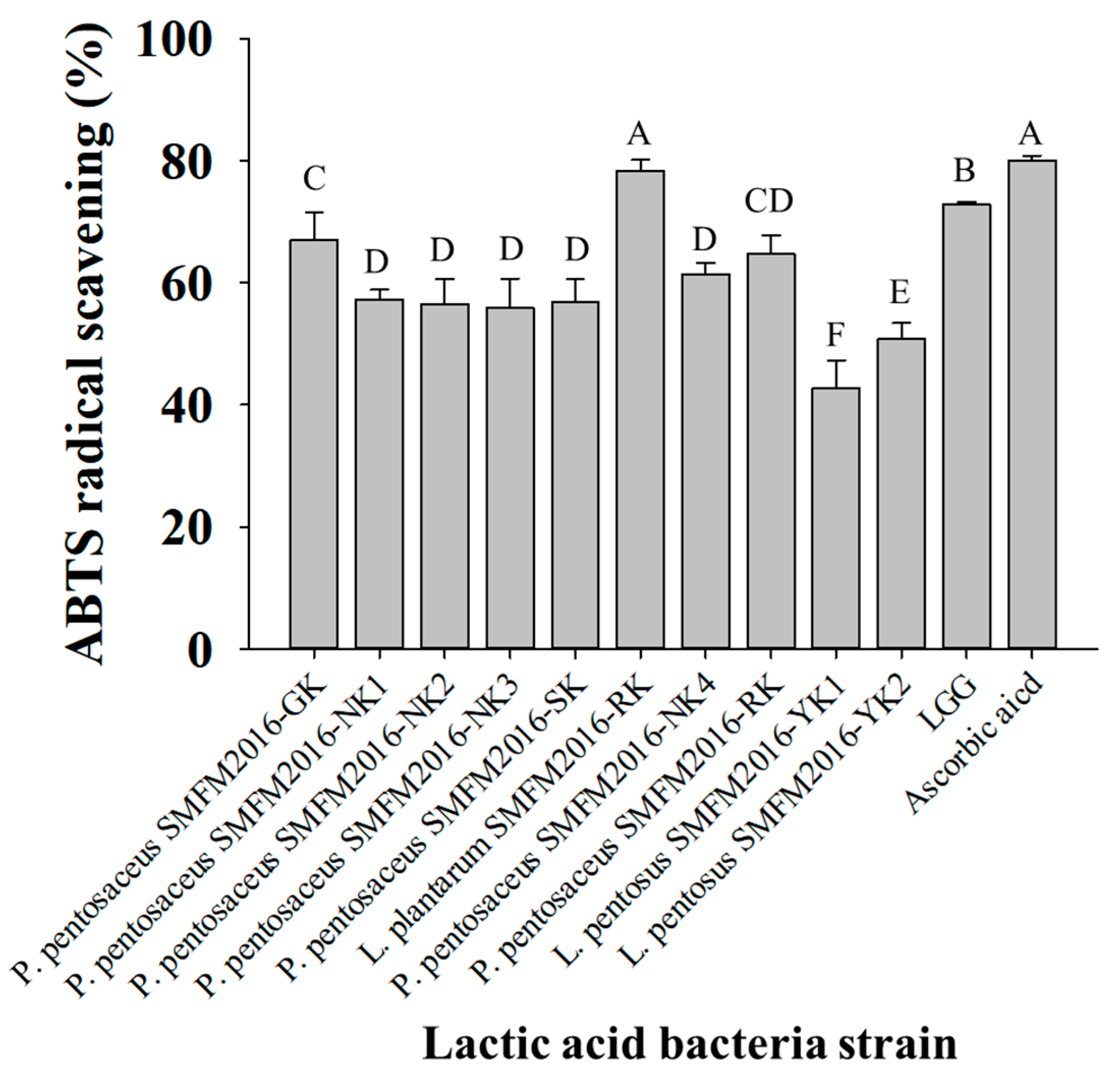
The remaining ten isolates were analyzed for ABTS radical-scavenging activity; their activities were higher than 40% (Figure 2). Among the ten isolates, L. plantarum SMFM2016-RK showed the highest activity (78.4%), with no significant difference in 0.2 mM ascorbic acid (79.8%). L. pentosus SMFM2016-YK1 (42.7%) and L. pentosus SMFM2016-YK2 (50.8%) exhibited significantly lower (p < 0.05) activities than the other bacteria. Therefore, these two strains were excluded from the list of probiotic candidate strains in this study, and the remaining eight isolates were subjected to further analysis.
Among the nine LAB isolates, including L. curvatus SMFM2016-NK, the growth rate averaged 0.051 and the mean generation time was 13.5 h (Table 2 and Table 3). None of the isolates showed significantly altered growth rates upon addition of the two types of A. herba-alba extracts. However, the growth rate of LGG in A. herba-alba ethanol extract was significantly lower, and its generation time in A. herba-alba ethanol extracts was significantly longer (p < 0.05) than that in the MRS broth or A. herba-alba hot-water extract. This indicated that A. herba-alba ethanol extract suppressed the growth of LGG.
The antimicrobial analysis of the ethanol or hot-water extract against the nine isolates revealed that the extracts bioconverted by L. plantarum SMFM2016-RK or L. curvatus SMFM2016-NK exhibited a higher inhibitory effect on A. actinomycetemcomitans ATCC43718 and F. nucleatum ATCC10953 than those bioconverted by other isolates (Table 4). L. plantarum SMFM2016-RK and L. curvatus SMFM2016-NK were more effective on inhibiting the growth of two periodontal pathogens in a coculture broth with A. herba-alba extracts when compared to the isolates grown in MRS broth. Among the cultures, the culture broth of L. plantarum SMFM2016-RK with A. herba-alba ethanol extract showed the highest inhibitory effect against A. actinomycetemcomitans ATCC43718, with an inhibition zone of 2.1 mm (p < 0.05). The culture broth of L. curvatus SMFM2016-NK with A. herba-alba ethanol extract exhibited the highest inhibition size of 4.0 mm against F. nucleatum ATCC10953 (p < 0.05). Although it was difficult to evaluate the antimicrobial effect of LAB and A. herba-alba extracts against P. gingivalis, their effects on the inhibition of P. gingivalis biofilm were observed. Probiotic-mediated bioconversion releases bioactive metabolites, such as antimicrobial peptides, immunopeptides, and bioactive polyphenols, that are effective on maintaining oral health [32,33,34]. Therefore, A. herba-alba coculture broths containing L. plantarum SMFM2016-RK or L. curvatus SMFM2016-NK could be considered for a possible alleviator of periodontal disease, as they inhibit the growth of periodontal pathogens. Hence, L. plantarum SMFM2016-RK and L. curvatus SMFM2016-NK were selected for analyzing α-glucosidase activity. α-glucosidase inhibition disrupts carbohydrate digestion and absorption, thereby alleviating hyperglycemia, and reduces the angiotensin-converting enzyme inhibitory potential [35,36]. Accordingly, the isolate with a high α-glucosidase activity could be effective in alleviating the systemic disease caused by periodontitis. The α-glucosidase inhibitory effects vary from 0 to 85.23% for the different strains of LAB [36,37]. L. plantarum SMFM2016-RK cultured in MRS broth displayed the highest α-glucosidase inhibitory activity of 85.2 ± 0.3% (Table 5). The three coculture broths containing L. curvatus SMFM2016-NK showed significantly lower activities than the other coculture broths (p < 0.05). Since this study examined the α-glucosidase inhibitory activity at a constant concentration of A. herba-alba, it is difficult to compare its activity with that in other studies that showed the concentration of IC50. According to Dar et al. (2024), α-glucosidase inhibitory activity increased with Capsella bursa-pastoris ethanol extract [38]. Thus, it is expected that the inhibitory effect would increase in association with the A. herba-alba concentration. Eventually, L. plantarum SMFM2016-RK was chosen as the probiotic candidate strain to execute the bioconversion of A. herba-alba. Both types of A. herba-alba extracts were effective on different functionalities; therefore, both extracts were used for developing BM.

3.3. Efficacy Evaluation of Bioconverted Milk

When the BM reached an optimum pH of 4.5–5.0, it was considered fermented. The pH of milk containing A. herba-alba extracts and L. plantarum SMFM2016-RK (BM2, BM3, and BM4) was slightly lower than that of milk without addition of A. herba-alba extracts (BM1) at 0 h (Table 6). BM3 and BM4 reached the optimum pH of 4.62 ± 0.30 and 4.58 ± 0.25, respectively, after 6 h of fermentation, and the LAB cell count was 9.2 ± 0.1 Log CFU/mL. Collectively, A. herba-alba hot-water extract exhibited a synergistic effect that improved the growing ability of L. plantarum SMFM2016-RK. BM1 reached a pH of 4.42 ± 0.57 after 24 h, and the LAB cell count was 8.8 ± 0.2 Log CFU/mL. BM2 reached a pH of 4.58 ± 0.50 only after a long fermentation time of 37 h, and the LAB cell count was 8.8 ± 0.3 Log CFU/mL. Therefore, different fermentation conditions were applied to produce each type of BM. The prepared BM samples were analyzed for their antimicrobial effects against periodontal pathogens.
The inhibition zones of A. actinomycetemcomitans ATCC43718, F. nucleatum ATCC10953, and P. gingivalis ATCC33277 were 2.6−3.6, 1.6−2.9, and 1.6−1.9 mm, respectively (Table 7). The average inhibition zones for periodontal pathogens when using BM1, BM2, BM3, and BM4 were 2.0 ± 0.8, 2.4 ± 0.8, 2.5 ± 1.1, and 2.6 ± 1.1 mm, respectively. Bioconversion products with A. herba-alba extracts (BM2, BM3, and BM4) exhibited marginally higher antimicrobial effects than those without the extract (BM1). Among these products, BM3 showed the highest antimicrobial activity against A. actinomycetemcomitans, with an inhibition zone of 3.6 ± 0.8 mm (p < 0.05). BM4 showed the highest inhibition zone of 2.5 ± 0.6 mm for F. nucleatum ATCC10953, which was significantly higher than that achieved with BM1 (1.6 ± 0.5 mm; p < 0.0.5). BM1 and BM3 showed the same inhibition zone of 1.9 ± 0.3 mm for P. gingivalis ATCC33277.
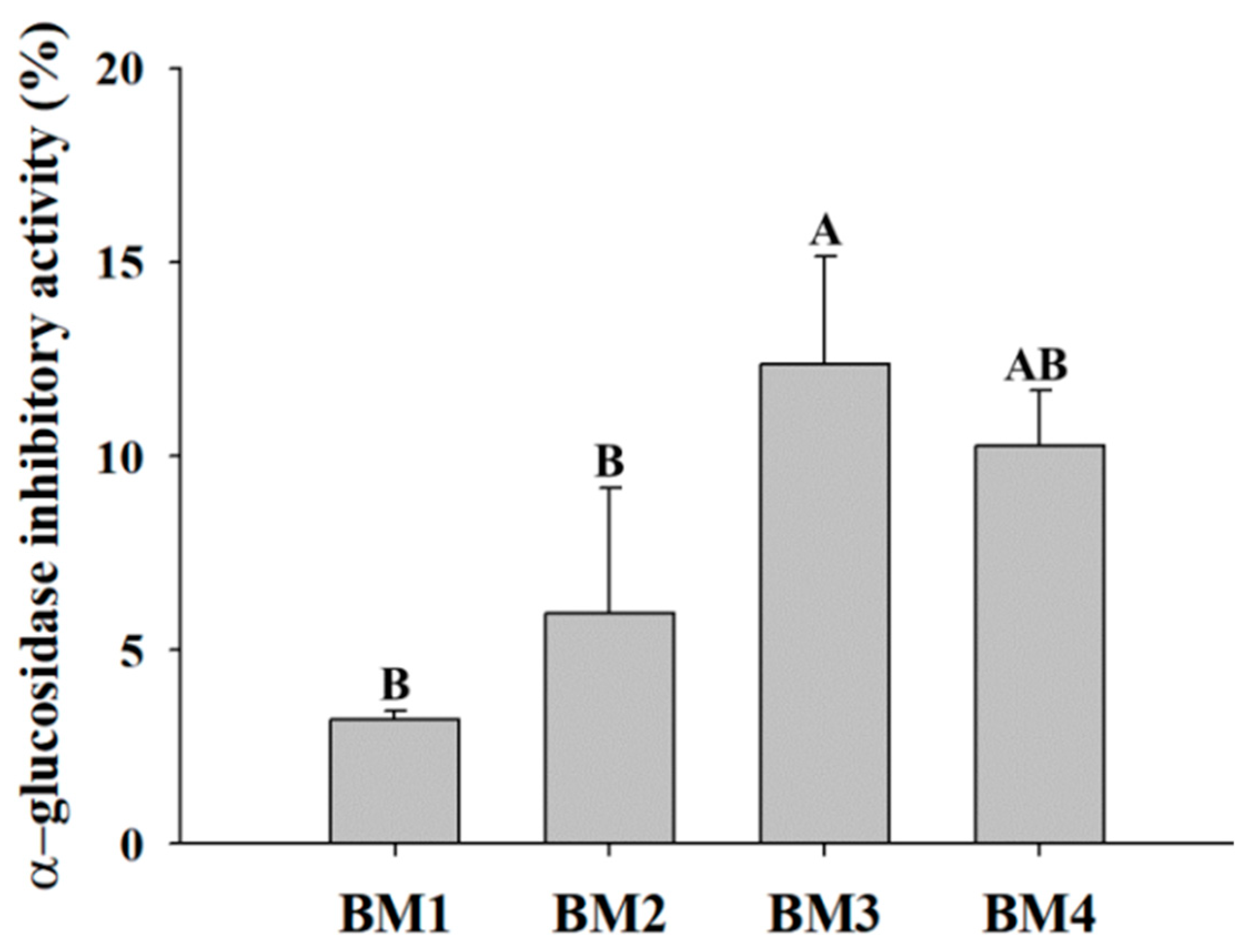
Bioconversion products containing A. herba-alba extracts (BM2, BM3, and BM4) exhibited significantly higher (p < 0.05) α-glucosidase inhibitory activity than BM1 (Figure 3). Notably, BM3 displayed a significantly higher activity (12.4 ± 0.2%) than the other samples (p < 0.05). This study found that the bioconversion process of A. herba-alba hot-water extract and L. plantarum SMFM2016-RK enhanced the fermentation ability of LAB and inhibition activity against α-glucosidase and periodontal pathogens that could be the candidate material to alleviate periodontitis and glycemic control.

3.4. Whole-Genome Analysis of Novel Probiotics

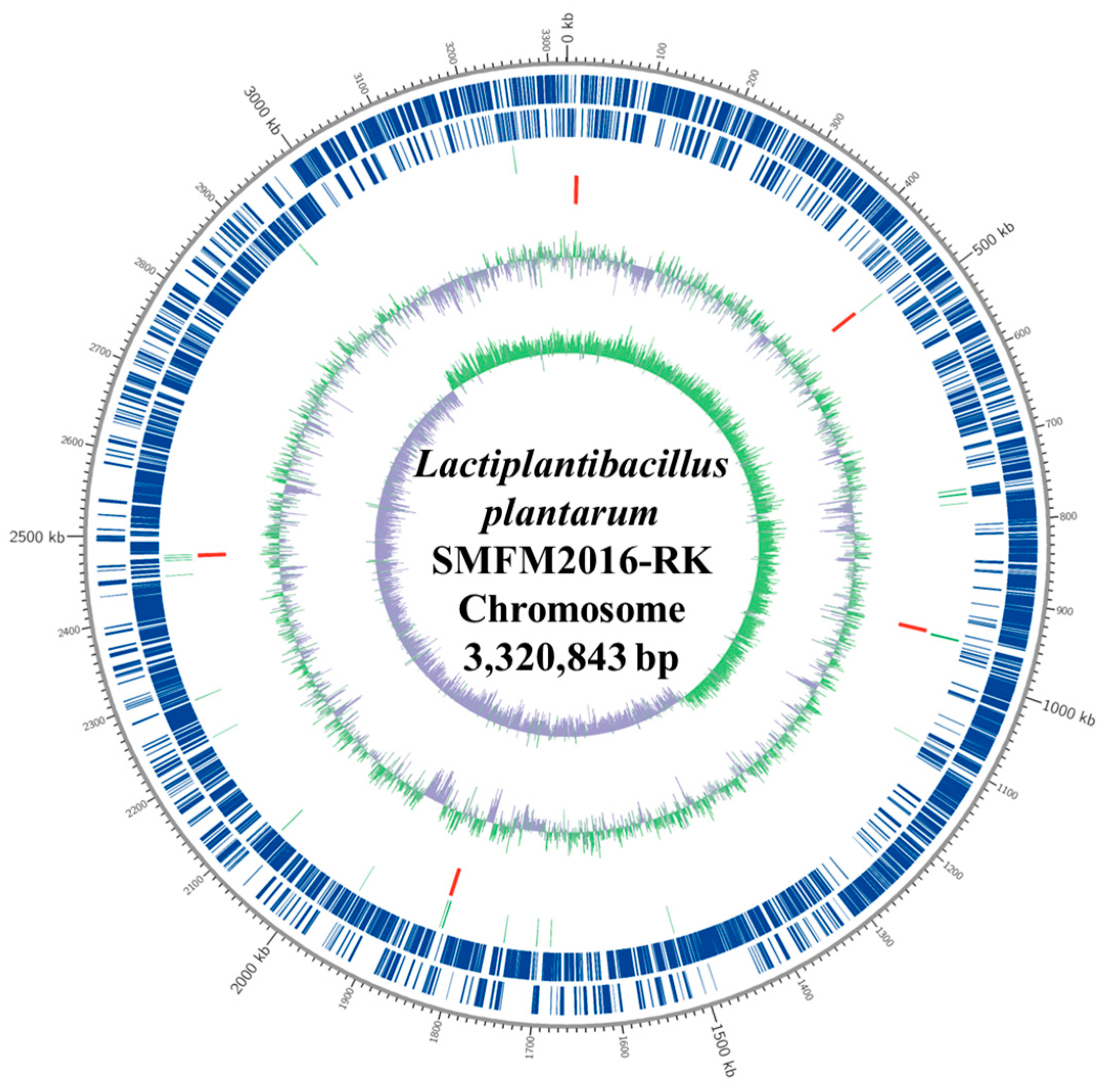
L. plantarum SMFM2016-RK was sequenced using de novo assembly, and the whole genome was obtained. Three contigs—Contig1, Contig2, and Contig3—were assembled, and their lengths were 3,320,843 bp, 60,102 bp, and 39,997 bp, respectively. Using BLAST, the best match for L. plantarum SMFM2016-RK was identified as Lactiplantibacillus plantarum, with a 99.96% identification rate. For gene function analysis, gene annotation and gene prediction were performed using the information provided by the gene ontology database. Contig1 (chromosome) was predicted to contain 3135 coding sequences (CDS). The 67 tRNA and 16 rRNA genes and their locations were predicted from the CDS. The structural and functional properties of Contig1 were described using DNA plots (Figure 4). The predicted functional genes were divided into three primary categories—biological process, molecular function, and cellular component—according to their characteristics. Two thousand nine hundred and twelve transcripts were included under biological process; 2280 transcripts under molecular function; and 1378 transcripts under cellular component (Table 8). In the biological process, metabolic process had the highest number (1089) of transcripts. This included transcripts corresponding to various chemical reactions and pathways of organisms and the processes such as protein synthesis and degradation.
Gene annotation analysis using the evolutionary genealogy of genes: The non-supervised Orthologous Groups (EggNOG) database revealed that L. plantarum SMFM2016-RK had a high ability for carbohydrate transportation and metabolism (9.25%) and amino acid transportation and metabolism (6.57%; Table 9). L. plantarum SMFM2016-RK had some CDS related to proteolysis and amino acid metabolism (Table 10). The genes known as oligopeptide ABC transportation system (oppF, oppD, dtpT, pepF1, and pepO) and the serine-related metabolism (glyA, dsdA, and sdhA) were mostly identified. In addition, the arginosuccinate metabolism operon clustered with aspartate aminotransferase (asp), lyase (argH), and synthase (argG), which can be used for energy production and NADH regeneration, was also observed. These genes might influence various proteins and amino acids present in BM3, thus improving the growth of L. plantarum SMFM2016-RK and reducing the fermentation time. L. plantarum SMFM2016-RK was also shown to possess lpdC (gallate decarboxylase) and padC (phenolic acid decarboxylase), which encode phenolic acid-metabolizing enzymes (Table 11). In relation to this, several studies reported that phenolic acids were the main bioactive ingredients of A. herba-alba [25,39]. The phenolic compounds produced by LAB from precursors improve efficacy in the body or enable the production of bioactive metabolites through changes in gut microbiome [40,41]. According to Sun and Miao (2020), phenolic compounds can lower the glycemic index by altering the digestibility of food, and flavonoids and proanthocyanidins are effective in inhibiting α-glucosidase activity [42]. Likewise, in this study, it can be assumed that the inhibition of α-glucosidase by BM3 is due to the action of these phenolic metabolites produced during bioconversion of milk and A. herba-alba extracts by LAB.
The microbial genome similarity and the molecular phylogenetic relations were determined by analyzing the phylogenetic tree and ANI (%). The ANI of the seven L. plantarum strains in the NCBI GenBank database were compared to that of L. plantarum SMFM2016-RK (Figure 5A). The seven L. plantarum strains were isolated from various foods, and differences in genome size, GC content, and the numbers of tRNA and rRNA were observed. The ANI (%) between L. plantarum strains was 99.07–99.84%, and L. plantarum SMFM2016-RK was closest to the L. plantarum E1 strain, according to the unweighted pair group method with arithmetic mean tree by ANI (Figure 5A,B). Since ANI ≥ 96.5% is common in genomes within the same species, it was necessary to confirm whether L. plantarum SMFM2016-RK was a new strain [43]. Accordingly, whole genome annotation was performed with L. plantarum E1, which showed the highest similarity, using CLC workbench. Differences were observed between the two strains in various regions of the whole genome (Figure 5C). Therefore, L. plantarum SMFM2016-RK was confirmed to be a new strain.

4. Conclusions

This study identified L. plantarum SMFM2016-RK, which might be safe and inhibited the growth of periodontal pathogens and the activity of α-glucosidase. Whole-genome sequence analysis indicated that L. plantarum SMFM2016-RK is a new strain that could be used as a novel probiotic. The A. herba-alba extract added to skim milk was bioconverted by the newly characterized probiotic, and BMs containing A. herba-alba extracts (BM2, BM3, and BM4) showed the effects on impeding the growth of periodontal pathogenic bacteria and the activity of α-glucosidase. Taken together, probiotic-mediated bioconversion of A. herba-alba extract might be effective on periodontal disease and glycemic control. However, since the efficacy of the BM developed in this study was examined in vitro, it is necessary to apply it to animals in the future study to evaluate the efficacy in vivo.

Author Contributions

Conceptualization: K.-H.C. and Y.Y.; methodology: Y.L. and K.-H.C.; investigation: Y.L.; software: Y.L.; writing—original draft: Y.L.; writing—review and editing: Y.L., K.-H.C. and Y.Y. All authors have read and agreed to the published version of the manuscript.

Funding

This research was funded by National Research Foundation of Korea (NRF) grant funded by the Korea government (MSIT) (No. 2020R1A2C1006519).

Data Availability Statement

The original contributions presented in the study are included in the article, further inquiries can be directed to the corresponding authors.

Conflicts of Interest

The authors declare no conflicts of interest.

References

  1. WHO (World Health Organization). WHO Highlights Oral Health Neglect Affecting Nearly Half of the World’s Population. Available online: https://www.who.int/news/item/18-11-2022-who-highlights-oral-health-neglect-affecting-nearly-half-of-the-world-s-population (accessed on 10 August 2022).
  2. Borgnakke, W.S. IDF Diabetes Atlas: Diabetes and oral health—A two-way relationship of clinical importance. Diabetes Res. Clin. Pract. 2019, 157, 107839. [Google Scholar] [CrossRef] [PubMed]
  3. Ahmad, R.; Haque, M. Oral health messiers: Diabetes mellitus relevance. Diabetes Metab. Syndr. Obes. 2021, 14, 3001–3015. [Google Scholar] [CrossRef] [PubMed]
  4. Van de Laar, F.A.; Lucassen, P.L.; Akkermans, R.P.; van de Lisdonk, E.H.; Rutten, G.E.; van Weel, C. α-Glucosidase inhibitors for patients with type 2 diabetes: Results from a Cochrane systematic review and meta-analysis. Diabetes Care 2005, 28, 154–163. [Google Scholar] [CrossRef] [PubMed]
  5. Basri, R.; Ullah, S.; Halim, S.A.; Alharthy, R.D.; Rauf, U.; Khan, A.; Hussain, J.; Al-Ghafri, A.; Al-Harrasi, A.; Shafiq, Z. Synthesis, biological evaluation, and molecular docking study of chromen-linked hydrazine carbothioamides as potent α-glucosidase inhibitors. Drug Dev. Res. 2023, 84, 962–974. [Google Scholar] [CrossRef] [PubMed]
  6. Forozan, R.; Ghomi, M.K.; Iraji, A.; Montazer, M.N.; Noori, M.; Dastyafteh, N.; Mojtabavi, S.; Faramarzi, M.A.; Sadat-Ebrahimi, S.E.; Larijani, B.; et al. Synthesis, in vitro inhibitor screening, structure–activity relationship, and molecular dynamic simulation studies of novel thioquinoline derivatives as potent α-glucosidase inhibitors. Sci. Rep. 2023, 13, 7819. [Google Scholar] [CrossRef] [PubMed]
  7. Ullah, S.; Waqas, M.; Halim, S.A.; Khan, I.; Khalid, A.; Abdalla, A.N.; Makeen, H.A.; Ibrar, A.; Khan, A.; Al-Harrasi, A. Triazolothiadiazoles and triazolothiadiazines as potent α-glucosidase inhibitors: Mechanistic insights from kinetics studies, molecular docking and dynamics simulations. Int. J. Biol. Macromol. 2023, 250, 126227. [Google Scholar] [CrossRef] [PubMed]
  8. Huligere, S.S.; Chandana Kumari, V.B.; Alqadi, T.; Kumar, S.; Cull, C.A.; Amachawadi, R.G.; Ramu, R. Isolation and characterization of lactic acid bacteria with potential probiotic activity and further investigation of their activity by α-amylase and α-glucosidase inhibitions of fermented batters. Front. Microbiol. 2023, 13, 1042263. [Google Scholar] [CrossRef] [PubMed]
  9. Haque, M.M.; Yerex, K.; Kelekis-Cholakis, A.; Duan, K. Advances in novel therapeutic approaches for periodontal diseases. BMC Oral Health 2022, 22, 492. [Google Scholar] [CrossRef] [PubMed]
  10. Rosas-Val, P.; Adhami, M.; Brotons-Canto, A.; Gamazo, C.; Irache, J.M.; Larrañeta, E. 3D printing of microencapsulated Lactobacillus rhamnosus for oral delivery. Int. J. Pharm. 2023, 641, 123058. [Google Scholar] [CrossRef]
  11. Mahasneh, S.A.; Mahasneh, A.M. Probiotics: A promising role in dental health. Dent. J. 2017, 5, 26. [Google Scholar] [CrossRef]
  12. Rejiniemon, T.S.; Hussain, R.R.; Rajamani, B. In-vitro functional properties of Lactobacillus plantarum isolated from fermented ragi malt. South Indian J. Biol. Sci. 2015, 1, 15–23. [Google Scholar] [CrossRef]
  13. Iwasaki, K.; Maeda, K.; Hidaka, K.; Nemoto, K.; Hirose, Y.; Deguchi, S. Daily intake of heat-killed Lactobacillus plantarum L-137 decreases the probing depth in patients undergoing supportive periodontal therapy. Oral Health Prev. Dent. 2016, 14, 207–214. [Google Scholar] [PubMed]
  14. Pudgar, P.; Povšič, K.; Čuk, K.; Seme, K.; Petelin, M.; Gašperšič, R. Probiotic strains of Lactobacillus brevis and Lactobacillus plantarum as adjunct to non-surgical periodontal therapy: 3-month results of a randomized controlled clinical trial. Clin. Oral Investig. 2021, 25, 1411–1422. [Google Scholar] [CrossRef] [PubMed]
  15. Lin, C.W.; Chen, Y.T.; Ho, H.H.; Hsieh, P.S.; Kuo, Y.W.; Lin, J.H.; Liu, C.R.; Huang, Y.F.; Chen, C.W.; Hsu, C.H.; et al. Lozenges with probiotic strains enhance oral immune response and health. Oral Dis. 2022, 28, 1723–1732. [Google Scholar] [CrossRef] [PubMed]
  16. Dadgar, S.; Heydarian, A.; Sobouti, F.; Goli, H.; Rakhshan, V.; Heidari, M. Effects of probiotic and fluoride mouthrinses on Streptococcus mutans in dental plaque around orthodontic brackets: A preliminary explorative randomized placebo-controlled clinical trial. Dent. Res. J. (Isfahan) 2021, 18, 74. [Google Scholar]
  17. Zhu, Y.; Chen, J.; Ji, X.; Hu, X.; Ling, T.; Zhang, Z.; Bao, G.; Wan, X. Changes of major tea polyphenols and production of four new B-ring fission metabolites of catechins from post-fermented Jing-Wei Fu brick tea. Food Chem. 2015, 170, 110–117. [Google Scholar] [CrossRef] [PubMed]
  18. Luo, J.; Liu, S.; Lu, H.; Chen, Q.; Shi, Y. Microbial Community Variations and Bioconversion Improvements during Soybean-Based Fermentation by Kefir Grains. Foods 2023, 12, 1588. [Google Scholar] [CrossRef] [PubMed]
  19. Cosier, D.; Lambert, K.; Batterham, M.; Sanderson-Smith, M.; Mansfield, K.J.; Charlton, K. The INHABIT (synergIstic effect of aNtHocyAnin and proBIoTics in) Inflammatory Bowel Disease trial: A study protocol for a double-blind, randomised, controlled, multi-arm trial. J. Nutr. Sci. 2024, 13, e1. [Google Scholar] [CrossRef]
  20. Moufid, A.; Eddouks, M. Artemisia herba alba: A popular plant with potential medicinal properties. Pak. J. Biol. Sci. 2012, 15, 1152–1159. [Google Scholar] [CrossRef]
  21. Jang, H.J. Potential Use of Lactic Acid Bacteria Isolated from Kimchi as Probiotics. Master’s Thesis, Sookmyung Women’s University, Seoul, Republic of Korea, 2018. Available online: https://www.riss.kr/search/detail/DetailView.do?p_mat_type=be54d9b8bc7cdb09&control_no=2b4278f7fafeb936ffe0bdc3ef48d419&keyword=%EC%9E%A5%ED%98%9C%EC%A7%84%20%20Potential%20Use%20of%20Lactic%20acid%20Bacteria%20Isolated%20from%20Kimchi%20as%20Probiotics (accessed on 20 June 2024).
  22. Choi, Y.; Park, E.; Kim, S.; Ha, J.; Oh, H.; Kim, Y.; Lee, Y.; Seo, Y.; Kang, J.; Lee, S.; et al. Alleviation of periodontal disease using Lactobacillus curvatus SMFM2016-NK. J. Funct. Foods 2021, 83, 104531. [Google Scholar] [CrossRef]
  23. Hunt, R. Plant growth curves. In The Functional Approach to Plant Growth Analysis; Edward Arnold Ltd.: London, UK, 1982. [Google Scholar]
  24. Souhila, T.; Fatma Zohra, B.; Tahar, H.S. Identification and quantification of phenolic compounds of Artemisia herba-alba at three harvest time by HPLC–ESI–Q-TOF–MS. Int. J. Food Prop. 2019, 22, 843–852. [Google Scholar] [CrossRef]
  25. Mohammed, M.J.; Anand, U.; Altemimi, A.B.; Tripathi, V.; Guo, Y.; Pratap-Singh, A. Phenolic composition, antioxidant capacity and antibacterial activity of white wormwood (Artemisia herba-alba). Plants 2021, 10, 164. [Google Scholar] [CrossRef] [PubMed]
  26. Gallego, R.; Montero, L.; Cifuentes, A.; Ibáñez, E.; Herrero, M. Green extraction of bioactive compounds from microalgae. J. Anal. Test. 2018, 2, 109–123. [Google Scholar] [CrossRef]
  27. Mangia, N.P.; Saliba, L.; Deiana, P. Functional and safety characterization of autochthonous Lactobacillus paracasei FS103 isolated from sheep cheese and its survival in sheep and cow fermented milks during cold storage. Ann. Microbiol. 2019, 69, 161–170. [Google Scholar] [CrossRef]
  28. Kim, H.; Kim, J.; Kim, Y.; Jeong, Y.; Kim, J.; Paek, N.; Kang, C. Antioxidant and probiotic properties of Lactobacilli and Bifidobacteria of human origins. Biotechnol. Bioprocess Eng. 2020, 25, 421–430. [Google Scholar] [CrossRef]
  29. Nanno, M.; Morotomi, H.; Takayama, H.; Kuroshima, T.; Tanaka, R.; Mutai, M. Mutagenic activation of biliary metabolites of benzo (a) pyrene by β-glucuronidase-positive bacteria in human faeces. J. Med. Microbiol. 1986, 22, 351–355. [Google Scholar] [CrossRef] [PubMed]
  30. Rafter, J. Lactic acid bacteria and cancer: Mechanistic perspective. Br. J. Nutr. 2002, 88, S89–S94. [Google Scholar] [CrossRef] [PubMed]
  31. Monteagudo-Mera, A.; Caro, I.; Rodríguez-Aparicio, L.B.; Rúa, J.; Ferrero, M.A.; García-Armesto, M.R. Characterization of certain bacterial strains for potential use as starter or probiotic cultures in dairy products. J. Food Prot. 2011, 74, 1379–1386. [Google Scholar] [CrossRef]
  32. Rice-Evans, C.; Miller, N.; Paganga, G. Antioxidant properties of phenolic compounds. Trends Plant Sci. 1997, 2, 152–159. [Google Scholar] [CrossRef]
  33. Ebringer, L.; Ferenčík, M.; Krajčovič, J. Beneficial health effects of milk and fermented dairy products. Folia Microbiol. (Praha) 2008, 53, 378–394. [Google Scholar] [CrossRef]
  34. Ostaff, M.J.; Stange, E.F.; Wehkamp, J. Antimicrobial peptides and gut microbiota in homeostasis and pathology. EMBO Mol. Med. 2013, 5, 1465–1483. [Google Scholar] [CrossRef] [PubMed]
  35. Oboh, G.; Ogunsuyi, O.B.; Ogunbadejo, M.D.; Adefegha, S.A. Influence of gallic acid on α-amylase and α-glucosidase inhibitory properties of acarbose. J. Food Drug Anal. 2016, 24, 627–634. [Google Scholar] [CrossRef] [PubMed]
  36. Ramchandran, L.; Shah, N.P. Proteolytic profiles and angiotensin-I converting enzyme and α-glucosidase inhibitory activities of selected lactic acid bacteria. J. Food Sci. 2008, 73, M75–M81. [Google Scholar] [CrossRef] [PubMed]
  37. Kwun, S.Y.; Bae, Y.W.; Yoon, J.A.; Park, E.H.; Kim, M.D. Isolation of acid tolerant lactic acid bacteria and evaluation of α-glucosidase inhibitory activity. Food Sci. Biotechnol. 2020, 29, 1125–1130. [Google Scholar] [CrossRef] [PubMed]
  38. Dar, M.A.; Siddiqui, N.A.; Mir, S.R.; Akbar, S.; Mothana, R.A.; Masoodi, M.H. Anti-diabetic activity-guided isolation of α-amylase and α-glucosidase inhibitory terpenes from Capsella bursa-pastoris Linn. Open Chem. 2024, 22, 20240025. [Google Scholar] [CrossRef]
  39. Younsi, F.; Trimech, R.; Boulila, A.; Ezzine, O.; Dhahri, S.; Boussaid, M.; Messaoud, C. Essential oil and phenolic compounds of Artemisia herba-alba (Asso.): Composition, antioxidant, antiacetylcholinesterase, and antibacterial activities. Int. J. Food Prop. 2016, 19, 1425–1438. [Google Scholar] [CrossRef]
  40. Loo, Y.T.; Howell, K.; Chan, M.; Zhang, P.; Ng, K. Modulation of the human gut microbiota by phenolics and phenolic fiber-rich foods. Compr. Rev. Food Sci. Food Saf. 2020, 19, 1268–1298. [Google Scholar] [CrossRef]
  41. Leonard, W.; Zhang, P.; Ying, D.; Fang, Z. Hydroxycinnamic acids on gut microbiota and health. Compr. Rev. Food Sci. Food Saf. 2021, 20, 710–737. [Google Scholar] [CrossRef] [PubMed]
  42. Sun, L.; Miao, M. Dietary polyphenols modulate starch digestion and glycaemic level: A review. Crit. Rev. Food Sci. Nutr. 2020, 60, 541–555. [Google Scholar] [CrossRef]
  43. Varghese, N.J.; Mukherjee, S.; Ivanova, N.; Konstantinidis, K.T.; Mavrommatis, K.; Kyrpides, N.C.; Pati, A. Microbial species delineation using whole genome sequences. Nucleic Acids Res. 2015, 43, 6761–6771. [Google Scholar] [CrossRef]
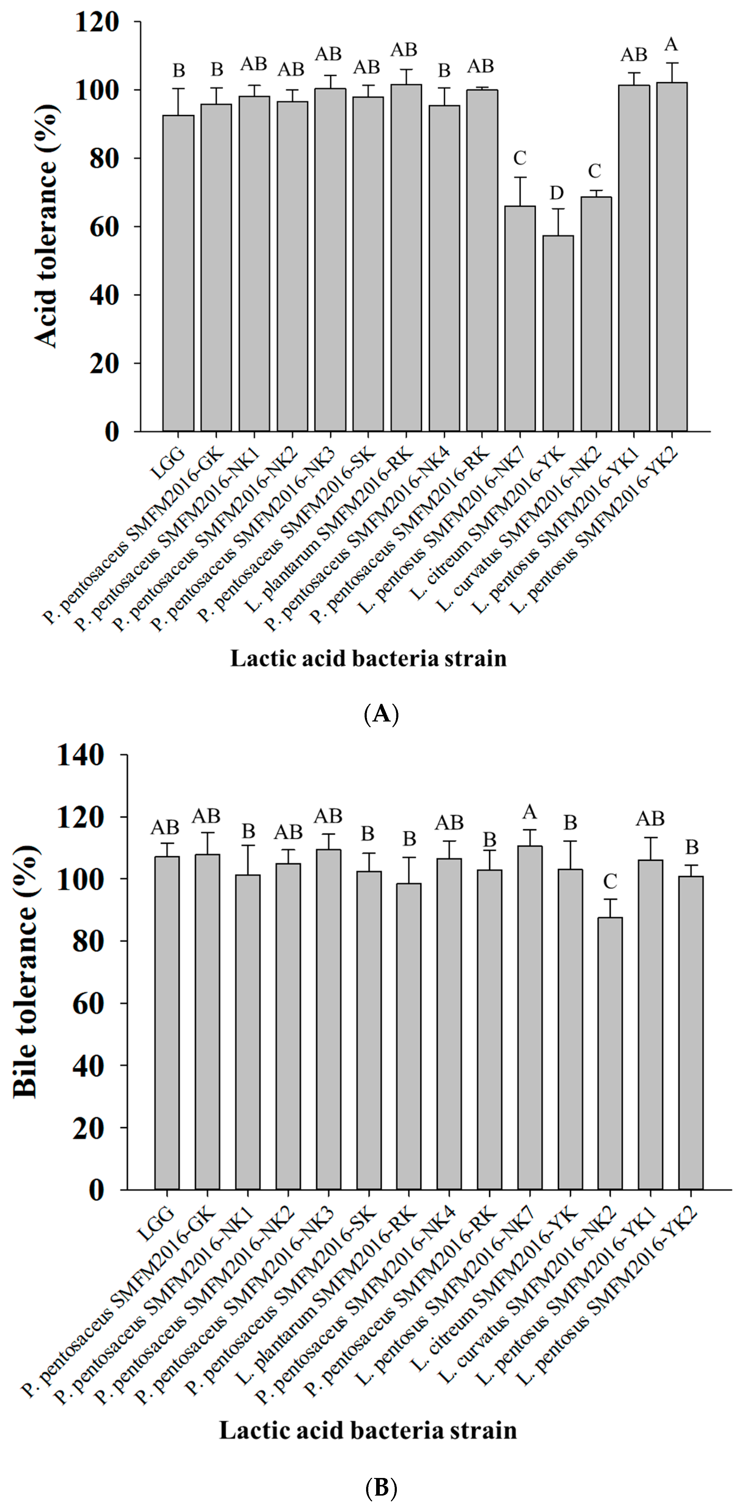
Figure 1. Acid tolerance (A) and bile salt tolerance (B) of the lactic acid bacteria isolates. A–D; different letters indicate a significant difference (p < 0.05).
Microorganisms 12 01290 g001
Figure 2. 2,2′-azinobis (3-ethylbenzothiazoline-6-sulfonic acid; ABTS) scavenging activity of the lactic acid bacteria isolates. A–F; different letters indicate a significant difference (p < 0.05).
Microorganisms 12 01290 g002
Figure 3. Comparison of α-glucosidase inhibitory activity on treatment with bioconverted milk. BM1: bioconverted milk with L. plantarum SMFM2016-RK, BM2: BM1 and 5 mg/mL A. herba-alba ethanol extract, BM3: BM1 and 25 mg/mL A. herba-alba hot-water extract, BM4: BM1, 5 mg/mL A. herba-alba ethanol extract, and 25 mg/mL A. herba-alba hot-water extract. A, B; different letters indicate a significant difference (p < 0.05).
Microorganisms 12 01290 g003
Figure 4. Chromosomal genome properties of Lactiplantibacillus plantarum SMFM2016-RK. Outer scale; base pairs, the first (the outer-most) and second ring; forward and reverse open reading frame (ORF), the third and fourth ring; forward and reverse ORF by gene annotation, the fifth and sixth ring; rRNA and tRNA genes, the seventh and eighth ring; positive and negative GC content values, the inner-most circle; GC skew.
Microorganisms 12 01290 g004
Figure 5. Average nucleotide identity (ANI) (A), unweighted pair group method with arithmetic mean (UPGMA) tree by average nucleotide identity (ANI) (B), and comparison of genome annotation with Lactiplantibacillus plantarum E1 (region 538,067-538,247) (C) of L. plantarum SMFM2016-RK.
Microorganisms 12 01290 g005aMicroorganisms 12 01290 g005b
Table 1. Minimum bactericidal concentrations (mg/mL; mean ± standard deviation) of Artemisia herba-alba extracts against periodontal pathogens.
Artermisia herba-alba
Extracts
Fusobacterium nucleatum ATCC43718Aggregatibacter actinomycetemcomitans ATCC10953Porphyromonas gingivalis ATCC33277Total Average
Ethanol extracts4.3 ± 1.7 C5.3 ± 1.7 C1.4 ± 0.4 C4.0 ± 2.1
Hot-water extracts5.9 ± 1.3 C10.7 ± 3.3 B26.5 ± 2.0 A13.6 ± 9.1
A–C; different letters indicate a significant difference (p < 0.05).
Table 2. Specific growth rate of lactic acid bacteria in MRS broth containing different Artemisia herba-alba extracts (mean ± standard deviation).
Lactic Acid Bacteria StrainMRS BrothMRS Broth +
A. herba-alba
Ethanol Extract (5 mg/mL)
MRS Broth +
A. herba-alba Hot-Water Extract (25 mg/mL)
Pediococcus pentosaceus SMFM2016-GK0.054 ± 0.002 Aa*0.052 ± 0.002 Aab0.052 ± 0.002 Aab
P. pentosaceus SMFM2016-NK10.052 ± 0.002 Aab0.051 ± 0.003 Aab0.050 ± 0.001 Aab
P. pentosaceus SMFM2016-NK20.052 ± 0.001 Aab0.052 ± 0.004 Aab0.052 ± 0.003 Aab
P. pentosaceus SMFM2016-NK30.053 ± 0.002 Aab0.051 ± 0.002 Aab0.051 ± 0.002 Aab
P. pentosaceus SMFM2016-SK0.053 ± 0.001 Aa0.053 ± 0.004 Aab0.052 ± 0.002 Aab
Lactiplantibacillus plantarum SMFM2016-RK0.049 ± 0.001 Ab0.048 ± 0.002 Ab0.049 ± 0.002 Ab
P. pentosaceus SMFM2016-NK40.053 ± 0.002 Aa0.052 ± 0.002 Aab0.052 ± 0.002 Aab
P. pentosaceus SMFM2016-RK0.053 ± 0.002 Aab0.050 ± 0.002 Aab0.050 ± 0.002 Aab
Lactilactobacillus curvatus SMFM2016-NK0.050 ± 0.002 Ab0.050 ± 0.001 Ab0.049 ± 0.002 Ab
Lacticaseibacillus rhamnosus GG0.042 ± 0.004 Ac0.028 ± 0.006 Bc0.44 ± 0.004 Ac
* Means with the same row with different superscript capital letters and same column with different superscript small letters are significantly different (p < 0.05).
Table 3. Generation time of lactic acid bacteria in MRS broth containing different Artemisia herba-alba extracts (h; mean ± standard deviation).
Lactic Acid Bacteria StrainMRS BrothMRS Broth +
A. herba-alba Ethanol Extract (5 mg/mL)
MRS Broth +
A. herba-alba Hot-Water Extract (25 mg/mL)
Pediococcus pentosaceus SMFM2016-GK12.9 ± 0.6 Ac*13.5 ± 0.4 Ab13.3 ± 0.5 Ab
P. pentosaceus SMFM2016-NK113.3 ± 0.4 Aab13.6 ± 0.8 Ab13.9 ± 0.2 Ab
P. pentosaceus SMFM2016-NK213.2 ± 0.3 Aab13.3 ± 0.9 Ab13.3 ± 0.7 Ab
P. pentosaceus SMFM2016-NK313.2 ± 0.4 Aab13.5 ± 0.6 Ab13.7 ± 0.6 Ab
P. pentosaceus SMFM2016-SK13.1 ± 0.4 Aab13.2 ± 0.9 Ab13.3 ± 0.5 Ab
Lactiplantibacillus plantarum SMFM2016-RK14.3 ± 0.3 Ab14.5 ± 0.6 Ab14.2 ± 0.5 Ab
P. pentosaceus SMFM2016-NK413.0 ± 0.5 Aab13.4 ± 0.4 Ab13.2 ± 0.6 Ab
P. pentosaceus SMFM2016-RK13.1 ± 0.5 Aab13.9 ± 0.5 Ab13.8 ± 0.7 Ab
Lactilactobacillus curvatus SMFM2016-NK13.8 ± 0.5 Aab3.9 ± 0.4 Ab14.3 ± 0.5 Ab
Lacticaseibacillus rhamnosus GG16.7 ± 1.6 Ba25.6 ± 6.3 Aa15.7 ± 1.4 Ba
* Means with the same row with different superscript capital letters and same column with different superscript small letters are significantly different (p < 0.05).
Table 4. Sizes of inhibition zones produced by bioconverted broth with lactic acid bacteria (mm; mean ± standard deviation) against Aggregatibacter actinomycetemcomitans ATCC43718 and Fusobacterium nucleatum ATCC10953.
Lactic Acid Bacteria StrainMRS BrothMRS Broth + A. herba-alba Ethanol Extract (5 mg/mL)MRS Broth + A. herba-alba Hot-Water Extract (25 mg/mL)
Aggregatibacter actinomycetemcomitans ATCC43718Fusobacterium nucleatum ATCC10953A. actinomycetemcomitans ATCC43718F. nucleatum ATCC10953A. actinomycetemcomitans ATCC43718F. nucleatum ATCC10953
Pediococcus pentosaceus SMFM2016-GK1.0 ± 0.0 Ab1.5 ± 0.6 ABbc0.5 ± 0.6 Bd1.8 ± 0.3 Ac1.1 ± 0.3 Ab0.7 ± 0.6 Bc
P. pentosaceus SMFM2016-NK11.0 ± 0.0 Ab1.3 ± 0.5 Abc1.0 ± 0.0 Ac1.4 ± 0.5 Ac1.1 ± 0.3 Ab0.3 ± 0.5 Bc
P. pentosaceus SMFM2016-NK21.3 ± 0.3 Ab1.4 ± 0.5 ABbc1.0 ± 0.0 Ac1.8 ± 0.3 Ac1.1 ± 0.3 Ab0.7 ± 0.6 Bc
P. pentosaceus SMFM2016-NK31.0 ± 0.0 Ab1.5 ± 0.9 Abc1.0 ± 0.0 Ac1.2 ± 1.0 Ac1.1 ± 0.3 Ab0.0 ± 0.0 Bc
P. pentosaceus SMFM2016-SK1.0 ± 0.0 Ab2.0 ± 0.5 Ab1.0 ± 0.0 Ac1.6 ± 0.5 Ac0.8 ± 0.8 Ab0.3 ± 0.5 Bc
Lactiplantibacillus plantarum SMFM2016-RK1.4 ± 0.5 Ca3.1 ± 0.3 Aa2.1 ± 0.3 Aa2.8 ± 0.9 Ab2.0 ± 0.0 Ba3.1 ± 0.3 Aa
P. pentosaceus SMFM2016-NK41.0 ± 0.4 Ab0.9 ± 0.6 Ac1.0 ± 0.0 Ac1.1 ± 0.3 Ac0.9 ± 0.3 Ab0.8 ± 0.3 Ac
P. pentosaceus SMFM2016-RK1.0 ± 0.0 Ab0.8 ± 0.5 Ac1.1 ± 0.3 Abc1.0 ± 0.0 Ac1.0 ± 0.0 Ab1.0 ± 0.0 Abc
Lactilactobacillus curvatus SMFM2016-NK1.0 ± 0.0 Bb3.1 ± 1.0 Ba1.9 ± 0.3 Aab4.0 ± 0.9 Aa1.6 ± 0.5 Aa2.6 ± 0.9 Ba
Lacticaseibacillus rhamnosus GG1.0 ± 0.0 Bb1.5 ± 0.6 Abc*1.5 ± 0.6 Ab2.1 ± 0.9 Abc1.8 ± 0.5 Aa1.8 ± 0.5 Ab
* Means with the same row with different superscript capital letters and same column with different superscript small letters are significantly different (p < 0.05).
Table 5. α-glucosidase inhibitory activities (%; mean ± standard deviation) of bioconverted broths with lactic acid bacteria.
Lactic Acid Bacteria StrainMRS BrothMRS Broth+
A. herba-alba Ethanol Extract
(5 mg/mL)
MRS Broth+
A. herba-alba
Hot-Water Extract
(25 mg/mL)
Positive control
(Lacticaseibacillus rhamnosus GG)
83.2 ± 1.7 Aa*60.0 ± 5.5 Ba26.2 ± 0.9 Cb
Lactiplantibacillus plantarum SMFM2016-RK85.2 ± 0.3 Aa60.8 ± 5.0 Ba35.0 ± 0.2 Ca
Lactilactobacillus curvatus SMFM2016-NK0.0 ± 0.0 Bb2.7 ± 1.2 Ab5.9 ± 2.6 Ac
* Means with the same row with different superscript capital letters and same column with different superscript small letters are significantly different (p < 0.05).
Table 6. pH and lactic acid bacteria cell count (mean ± standard deviation) of the bioconverted milk with Lactiplantibacillus plantarum SMFM2016-RK and Artemisia herba-alba extracts during fermentation at 35 °C.
Sample0 h6 h24 h37 h
pHCell Counts (Log CFU/mL)pHCell Counts (Log CFU/mL)pHCell Counts (Log CFU/mL)pHCell Counts (Log CFU/mL)
BM16.57 ± 0.088.3 ± 0.0--4.42 ± 0.578.8 ± 0.2--
BM26.39 ± 0.078.3 ± 0.0----4.58 ± 0.508.8 ± 0.3
BM36.20 ± 0.058.3 ± 0.24.62 ± 0.309.2 ± 0.1----
BM46.11 ± 0.058.2 ± 0.14.58 ± 0.259.2 ± 0.1----
BM1: bioconverted milk with L. plantarum SMFM2016-RK, BM2: BM1 and 5 mg/mL A. herba-alba ethanol extract, BM3: BM1 and 25 mg/mL A. herba-alba hot-water extract, BM4: BM1, 5 mg/mL A. herba-alba ethanol extract, and 25 mg/mL A. herba-alba hot-water extract.
Table 7. Sizes (mm; mean ± standard deviation) of inhibition zones produced by bioconverted milk with Lactiplantibacillus plantarum SMFM2016-RK and Artemisia herba-alba extracts against periodontal pathogens.
SamplePeriodontal PathogenTotal Average
Aggregatibacter actinomycetemcomitans ATCC43718Fusobacterium nucleatum ATCC10953Porphyromonas gingivalis ATCC33277
10%
skim milk
0.0 ± 0.0 D0.0 ± 0.0 D0.0 ± 0.0 D0.0 ± 0.0
BM12.6 ± 1.1 B1.6 ± 0.5 C1.9 ± 0.3 BC2.0 ± 0.8
BM23.0 ± 0.8 AB2.5 ± 0.6 BC1.8 ± 0.5 BC2.4 ± 0.8
BM33.6 ± 0.8 A1.9 ± 1.0 BC1.9 ± 0.3 BC2.5 ± 1.1
BM43.3 ± 1.5 AB2.9 ± 0.6 AB1.6 ± 0.5 C2.6 ± 1.1
BM1: bioconverted milk with L. plantarum SMFM2016-RK, BM2: BM1 and 5 mg/mL A. herba-alba ethanol extract, BM3: BM1 and 25 mg/mL A. herba-alba hot-water extract, BM4: BM1, 5 mg/mL A. herba-alba ethanol extract, and 25 mg/mL A. herba-alba hot-water extract; A–D; different letters indicate a significant difference (p < 0.05).
Table 8. Gene ontology by gene prediction of Lactiplantibacillus plantarum SMFM2016-RK chromosome.
CategoryGene OntologyNumber of Transcripts
Cellular componentCell part263
Cell263
Protein-containing complex120
Organelle61
Extracellular region5
Membrane397
Membrane part249
Extracellular region part4
Organelle part16
Molecular functionCatalytic activity1087
Binding805
Molecular carrier activity2
Transport activity219
Antioxidant activity8
Transcription regulator activity105
Molecular function regulator1
Structural molecule activity51
Molecular transducer activity2
Biological processLocalization317
Response to stimulus104
Metabolic process1089
Cellular process845
Biological regulation223
Regulation of biological process217
Cellular component organization or biogenesis56
Negative regulation of biological process9
Multi-organism process9
Signaling22
Developmental process9
Immune system process1
Biological adhesion8
Detoxification2
Carbon utilization1
Table 9. Distribution of predicted open reading frames (ORFs) over functional class in Lactiplantibacillus plantarum SMFM2016-RK.
DescriptionNumber of ORFsRatio (%)
Translation, ribosomal structure, and biogenesis1504.7847
Transcription2588.2297
Replication, recombination, and repair1966.2520
Cell cycle control, cell division, chromosome partitioning260.8293
Defense mechanisms662.1053
Signal transduction mechanisms702.2329
Cell wall/membrane/envelope biogenesis1795.7097
Cell motility40.1276
Intracellular trafficking, secretion, and vesicular transport250.7974
Posttranslational modification, protein turnover, chaperones692.2010
Energy production and conversion1113.5407
Carbohydrate transport and metabolism2909.2504
Amino acid transport and metabolism2066.5710
Nucleotide transport and metabolism862.7432
Coenzyme transport and metabolism632.0096
Lipid transport and metabolism621.9777
Inorganic ion transport and metabolism1253.9872
Secondary metabolites biosynthesis, transport, and catabolism200.6380
General function prediction only34911.1324
Function unknown78024.8804
Total3135100
Table 10. Coding DNA sequences (DNA) related to proteolysis and amino acid metabolism identified in the gene annotation of Lactiplantibacillus plantarum SMFM2016-RK.
StartEndProductGeneIdentitye-ValueBit Score
1,849,1641,850,129Oligopeptide transport ATP-binding protein OppFoppF77.6360.0518
1,850,1361,851,215Oligopeptide transport ATP-binding protein OppDoppD79.8830.0570
2,470,9572,472,429Di-/tripeptide transporterdtpT66.1890.0648
962,965964,776Oligoendopeptidase F, plasmidpepF157.3330.0728
3,234,3673,236,283Neutral endopeptidasepepO59.5280.0791
50,4486505,796D-serine dehydratasedsdA57.6199.40 × 10−175501
2,530,7762,531,678L-serine dehydratase, alpha chainsdhA72.0695.73 × 10−138397
820,964822,202Serine hydroxymethyltransferaseglyA70.2700.0607
1,173,9451,175,741Aspartate--tRNA ligaseaspS74.6170.0917
2,291,6742,292,909Argininosuccinate synthaseargG73.3500.0634
2,290,2712,291,674Argininosuccinate lyaseargH71.0240.0699
Table 11. Coding DNA sequences (DNA) related to phenolic acid decarboxylase identified in the gene annotation of Lactiplantibacillus plantarum SMFM2016-RK.
StartEndProductGeneIdentitye-ValueBit Score
335,672337,144Gallate decarboxylaselpdC82.8570841
3,008,4853,009,021Phenolic acid decarboxylase PadCpadC87.643.70 × 10−116332
Disclaimer/Publisher’s Note: The statements, opinions and data contained in all publications are solely those of the individual author(s) and contributor(s) and not of MDPI and/or the editor(s). MDPI and/or the editor(s) disclaim responsibility for any injury to people or property resulting from any ideas, methods, instructions or products referred to in the content.