Article

Pangenome Analysis Reveals Novel Contact-Dependent Growth Inhibition System and Phenazine Biosynthesis Operons in Proteus mirabilis BL95 That Are Located in An Integrative and Conjugative Element

1
School of Biological Sciences, University of California, Irvine, CA 92697, USA
2
Department of Microbiology and Immunology, University of Illinois Chicago, Chicago, IL 60612, USA
*
Author to whom correspondence should be addressed.
Microorganisms 2024, 12(7), 1321; https://doi.org/10.3390/microorganisms12071321
Submission received: 7 April 2024 / Revised: 15 June 2024 / Accepted: 25 June 2024 / Published: 28 June 2024
(This article belongs to the Section Molecular Microbiology and Immunology)

Abstract

:
Proteus mirabilis is a leading cause of urinary tract infections and a common commensal of the gastrointestinal tract. Our recent study (JB) showed that P. mirabilis strain BL95 employs a novel contact-dependent killing system against enteric bacteria in the mouse gut and in vitro. To uncover the genetic determinants of this system, we performed whole-genome sequencing of BL95 and compared it with 98 complete genomes of P. mirabilis. BL95 carries 56 coding sequences (CDSs) not found in other P. mirabilis. Over half of these unique genes are located on a novel integrative conjugative element (ICE) named ICEPm2, inserted in tRNA-Phe and exclusive to BL95. ICEPm2 has integration, conjugation, and DNA replication modules nearly identical to ICEPm1 (common in P. mirabilis), but ICEPm2 of BL95 carries two unique operons for P. mirabilis—a phenazine biosynthesis and a contact-dependent growth inhibition (CDI) system. ICEPm2 is absent in the P. mirabilis (AR_0156) closest to BL95 and it is present in the genomes of several Escherichia coli from mouse intestines, indicating its recent horizontal mobilization. BL95 shares over 100 genes of five different secretion systems with other P. mirabilis, mostly poorly studied, making a large pool of candidate genes for the contact-dependent growth inhibition.

1. Introduction

Proteus mirabilis is a gram-negative bacterium best known for swarming motility and urease production. This bacterium is a leading causative agent of catheter-associated urinary tract infections, a common commensal of the gastrointestinal tract, and also widely distributed in soil, stagnant water, and sewage [1,2].
Colonization of the gastrointestinal tract is polymicrobial, and P. mirabilis possesses several systems to outcompete other microorganisms, including distinct strains of its own species [2]. P. mirabilis commonly competes with unrelated conspecific strains using the contact-dependent type VI secretion system (T6SS), which results in Dienes lines when two different strains swarm and encounter one another on an agar plate [3,4,5]. To outcompete other bacterial species during colonization, the genome of P. mirabilis encodes seventeen fimbrial operons and thirteen fimbrial orphan genes, as well as genes involved in the assembly of the type IV pili [6]. Additionally, the genome of P. mirabilis encodes six potential autotransporters of the type V secretion system (T5SS), half of which appear to be adhesins and the other half are potential toxins [6]. The genome also contains genes encoding two-partner secretion systems (TPS) of T5SS that act in a contact-dependent manner [7]; one of these is the HpmB-HpmA system having hemolysin activity and another is the contact-dependent growth inhibition system (CDI; CdiA/CdiB) employed by some gram-negative bacteria to outcompete co-residing bacteria [8,9]. P. mirabilis can also outcompete co-occurring bacteria through the secretion of ammonia and other volatile compounds [10].
The entire set of genes from all representatives of a bacterial species is known as its pangenome [11]. Each isolate of a bacterial species carries a core set of genes ensuring species’ existence, which can be supplemented by accessory genes that confer fitness to a specific niche [12]. The accessory genes are often localized in genomic islands, such as integrative and conjugative elements (ICEs), which are exchanged by horizontal gene transfer between bacteria. ICEs are integrated into a host genome, but they carry genetic apparatus allowing them to excise, produce conjugation machinery, and transfer to other strains and species, moving accessory genes with them [13].
ICEPm1 is an ICE that is common in P. mirabilis, reaching 100% frequency in urinary isolates and 65% in isolates from other body parts [14]. Nearly identical genomic islands were reported in other bacterial species, such as Providencia stuartii and Morganella morganii. The 92.6 kb ICEPm1 from P. mirabilis HI4320 has a highly modular structure typical for ICEs, including integration, conjugation, and replication core modules, as well as interspersed modules for accessory genes such as ones encoding an iron acquisition system and membrane proteins [14]. ICEPm1 integrates into the tRNA-Phe gene, forming 52 bp direct repeats (DR) as flanking regions. P. mirabilis has two copies of tRNA-Phe; experiments by Flannery et al. [12] showed that ICEPm1 preferentially integrates into the second copy of this gene, called tRNA-PheV, but can also integrate into another copy, called tRNA-PheU.
Recently, an isolate of P. mirabilis BL95 isolated from the feces of a lab mouse (Mus musculus strain C57BL/6) was reported to employ a contact-dependent killing system against coexistent Enterobacteriaceae [15]. Several physiological characteristics pointed out that such a system may be novel for Proteus and, possibly, for Enterobacterales. However, no attempts have been made yet to find its genetic underpinning. The objective of our study was to sequence, annotate, and characterize the genome of P. mirabilis BL95, identify unique features of its genome, especially those involved in killing or growth suppression of competing bacteria, and to conduct a comparative analysis of a novel integrative conjugative element (ICE) harboring genes that confer competitive advantage.

2. Materials and Methods

2.1. Library Construction, Sequencing, and Assembly

The whole genome of P. mirabilis BL95 was sequenced using Illumina and Oxford nanopore approaches. Detailed information about methods used can be found in [16]. Briefly, bacteria were grown in 2 mL of tryptic soy broth for 18 h at 35 °C and shaking at 200 rpm and genomic DNA was extracted and purified using phenol/chloroform [17]; after first extraction with phenol/chloroform, RNAse A was added to the sample and incubated at 37 °C for 15 min. DNA quantity and quality were assessed using measurements with Nanodrop (ThermoFisher, Waltham, MA, USA), Qubit (Invitrogen, Carlsbad, CA, USA), and visualization on 0.8% agarose gel. One genomic library was prepared following the Nextera DNA Flex Library Prep protocol (Illumina, San Diego, CA, USA). The library was sequenced on an Illumina MiniSeq sequencer using a 150-bp paired-end sequencing approach. FASTQ file generation pipelines included an adapter trimming option. A total of 628.3 Mb of raw data was generated from 4,160,654 trimmed reads. An Oxford Nanopore Technologies (ONT) (Oxford, UK) library was prepared using SQK-LSK109 and EXP-NBD196 (using the manufacturer’s protocol, without the optional fragmentation step), loaded to a FLO-MIN106 flow cell, and run in a MinION device (Oxford Nanopore Technologies plc, Oxford, UK). In total, 27,847 ONT reads were generated with an average length of 10,517 bp. By using Unicycler v0.4.8-beta [18] with default settings, both sequencing datasets were assembled into a complete circular chromosome.

2.2. Annotation and Identification of Genomic Features

Different annotation methods result in distinct annotations, mainly with regards to naming genetic features, but also in identification of the features themselves. To ensure thorough annotation and gene identification, we used pgap [19], Prokka [20], and BV-BRC Annotation service [21]. We used pgap annotation when referring to specific genetic features by their locus_tag, in order to be consistent with accessions deposited in GenBank. Integrative elements (such as ICE and IME) were identified using ICEFinder in ICEberg 2.0 [22]. Protein secretion systems were identified with TXSScan [23]. Orphan genes of the contact-dependent growth inhibition system (CDI) encoding effector toxins (cdiA-CT) and immunity proteins (cdiI) were identified by searching for GenBank accessions where these genes are a part of the complete CDI system (cdiB/cdiA/cdiI). Naming of the orphan genes followed a system used by Poole et al. [24], by adding the suffix ‘o’ and the index corresponding to the order position of the orphan after complete CDI. Promoters and terminators were predicted with BPROM [25] and ARNold [26] software, respectively, using the default parameters.

2.3. Genomic Comparative Analyses

The Proksee server (https://proksee.ca, accessed on 16 December 2022) was used to generate the annotated map of the strain BL95 chromosome [27]. Similarities between genomes and individual genomic features were shown using EasyFig [28]. Geneious Prime 2022.1.1 (https://www.geneious.com, accessed on 30 June 2023) was used for sequence visualization, navigation, and alignment.
In our comparative genomic analyses, we considered 98 isolates of P. mirabilis (not counting P. mirabilis BL95) with complete genomes from the RefSeq database (Table 1). We did not include partially assembled genomes because we wanted to be sure about the location of genomic features (chromosome vs. plasmid), and relative position of such features in the genome. We also included representatives of three closely related species (P. vulgaris, P. hauseri, and P. penneri) as outgroups for phylogeny reconstruction. Table 1 lists GenBank and RefSeq assembly accessions, species, strains, and location of ICEPm.
The evolutionary tree of P. mirabilis was inferred with concatenated genes of the core genome using Prokka [20] and Roary [29]. First, genomes of all P. mirabilis were annotated by Prokka. Then, homologous protein-encoding genes were identified by similarity and a matrix of genes presence/absence was constructed using Roary. To be considered homologous, we used a threshold of 80% at amino acid level. Finally, genes present in all Proteus strains were individually aligned and concatenated using Roary. Altogether, 2449 genes were individually aligned and then concatenated, for a total of 2,301,814 nucleotide positions in the final dataset. This dataset was used to reconstruct the phylogenetic tree in MegaX [30] using the Neighbor-Joining method with Kimura-2-parameter distance and the bootstrap test (100 replicates).
The matrix of genes presence/absence described above was used to identify genes unique to P. mirabilis BL95.

3. Results

3.1. Phylogenetic Analysis

To better understand the origin of the genomic features of P. mirabilis BL95, we constructed a core-genome phylogeny of P. mirabilis strains from the RefSeq database using all complete genomes (N = 99, including BL95; Table 1). Evolutionary relationships of P. mirabilis based on 2449 genes shared by all 99 isolates are shown in Figure 1. Three well-delineated clades can be distinguished on the tree (shown by Roman numerals above branches). BL95 belongs to the largest clade I, which includes P. mirabilis HI4320 (NCBI RefSeq assembly GCF_000069965.1), often used in experiments and a reference genome for P. mirabilis [6]. The nucleotide identity between BL95 and HI4320 across the core genome is 0.99452. The second clade is represented by strain BB2000, a model organism for self-recognition [31]. The nucleotide identity between BL95 and BB2000 across the core genome is 0.99289. Finally, the third, basal clade III, is made up of five isolates obtained from feces or digestive tract of animals. The nucleotide identity between BL95 and PmBC1123 from clade III is 0.97325.
The closest relative to BL95 is P. mirabilis AR_0156 of unknown origin. AR_0156 and BL95 differ at only 189 nucleotide positions across 2449 core genome genes, resulting in a nucleotide identity of 0.99991 and indicating that these strains share a very recent common ancestor. Assuming mutation rate equals to 10−9—or one substitution in one billion bases per generation—we estimate that the common ancestor of BL95 and AR_0156 lived 42,954 bacterial generations ago, or just about five years ago, if the generation time is one hour. Altogether, P. mirabilis forms a very compact group, well delineated from other Proteus species and exhibiting high nucleotide identity (>97%) even between its most diverged representatives.

3.2. Pangenome Analysis and Comparative Genomics

Due to extensive horizontal gene transfer in bacteria, even the most closely related bacteria may have drastically different phenotypes, if they acquired distinct mobile genetic elements (such as plasmids, ICEs, and transposons). To have a comprehensive view of intraspecies variation, it is thus important to consider the species pangenome, which includes core genome as well as accessory genes. Supplementary Figure S1 summarizes the pangenome of P. mirabilis. While the core genome is made up of 2449 genes, members of this species carry a total of 11,364 genes. Furthermore, more than half of the genes, 7017, can be found in less than 15% of strains. This suggests that despite genetic cohesiveness at the core genome, P. mirabilis is a very polymorphic species due to the presence and absence of accessory genes.
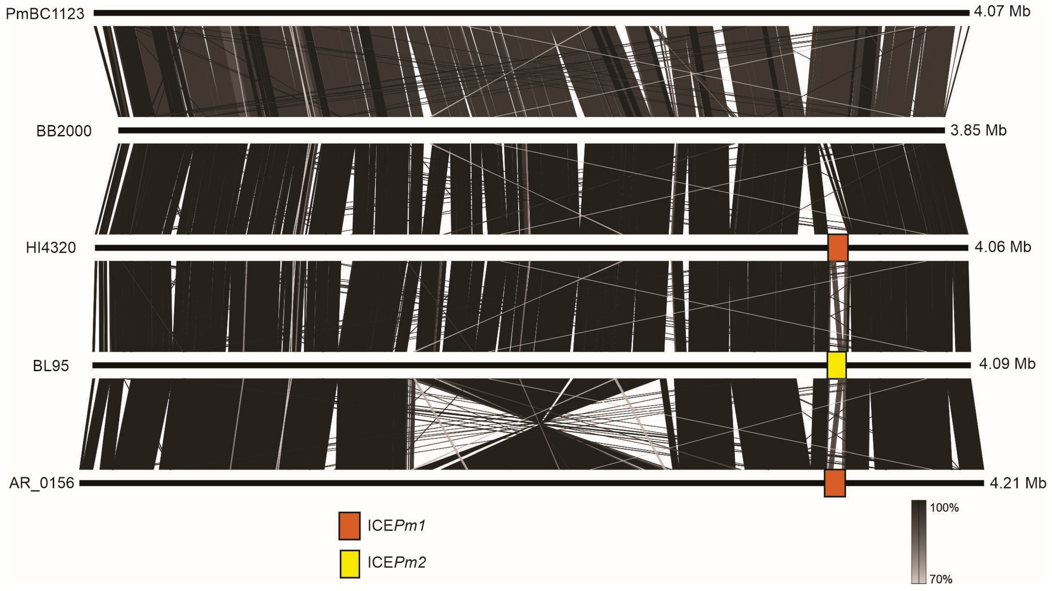
Genomic rearrangements are another potential source of variation. Figure 2 shows the alignment of chromosomes from five representative isolates of P. mirabilis. Overall, the alignment reveals a high degree of synteny (noted previously in [2]). Even strain PmBC1123 from the diverged clade III shows a similar order of genes as most other strains. On the other hand, the strain AR_0156 closely related to BL95, has a large inversion that encompasses 25% of its chromosome. Furthermore, several insertions and deletions are interspersed along the chromosomes in all compared strains, including BL95 and AR_0156. This demonstrates that genomic rearrangements, as well as accumulation and loss of genes, driven largely by mobile genetic elements, can happen on a short evolutionary scale and do not correlate with the number of single nucleotide substitutions.

3.3. General Features of BL95 Genome

The fully assembled genome of BL95 consists of a 4,086,891 bp-long chromosome. No plasmids were found. The chromosome contains 3749 genes, of which 109 are RNA genes and 3640 are coding sequences (CDSs). Among CDSs, 3595 are complete coding genes and 45 are pseudogenes (frameshifted, incomplete, or containing an internal stop). Among RNA genes, 22 are rRNA genes, 83 are tRNAs, and 4 are ncRNAs. The GC content of the complete genome is 39.2%. The GC content of the core genome (2449 protein coding genes for a total length of 2,293,671 bp) is 40.7%. A schematic representation of the P. mirabilis BL95 chromosome is shown in Supplementary Figure S2.

3.4. Unique Features of BL95 Genome

Several genomes of P. mirabilis have been described [31,32,33] with the reference genome of strain HI4320 providing a detailed description of the genomic features typical for this species [6]. Therefore, in our study we focused on genes and genomic features that are unique to BL95. As shown in Table 2, P. mirabilis BL95 carries 56 unique genes (i.e., they are not found in any of the 98 P. mirabilis strains).

3.5. BL95 Carries a Novel ICE, ICEPm2

ICEFinder identified in BL95 an integrative and conjugative element with modules of accessory genes previously unknown in P. mirabilis and not described formally in other species. Below we provide a description of this ICE, which we named ICEPm2, including its structure and key differences from related and well-studied ICEPm1. Furthermore, our comparative bioinformatic analysis uncovered yet another ICE, also related to ICEPm1, which we named ICEPm3.

3.6. Structure of ICEPm2

Over half of the genes unique to BL95 are found within a region identified by ICEFinder as an integrative and conjugative element (nucleotides 3,419,106–3,507,006; Figure S2). This 87.9 kb-long ICE is integrated into the 3′ end of the tRNA-PheV, flanked by nearly identical 52 bp-long direct repeats, and carries genes encoding integrase, relaxase, T4SS, and genes involved in DNA replication. A total of 51 bp of the direct repeats overlap the tRNA-PheV gene. The direct repeat at the left-most end (attL, nucleotides 3,419,106–3,419,157) has one nucleotide difference from the direct repeat at the right-most end (attR, nucleotides 3,506,955–3,507,006). The GC content of this ICE is 50.4%, which is noticeably higher than the GC content of the BL95 genome (39.2%), suggesting that this region was acquired through horizontal gene transfer.
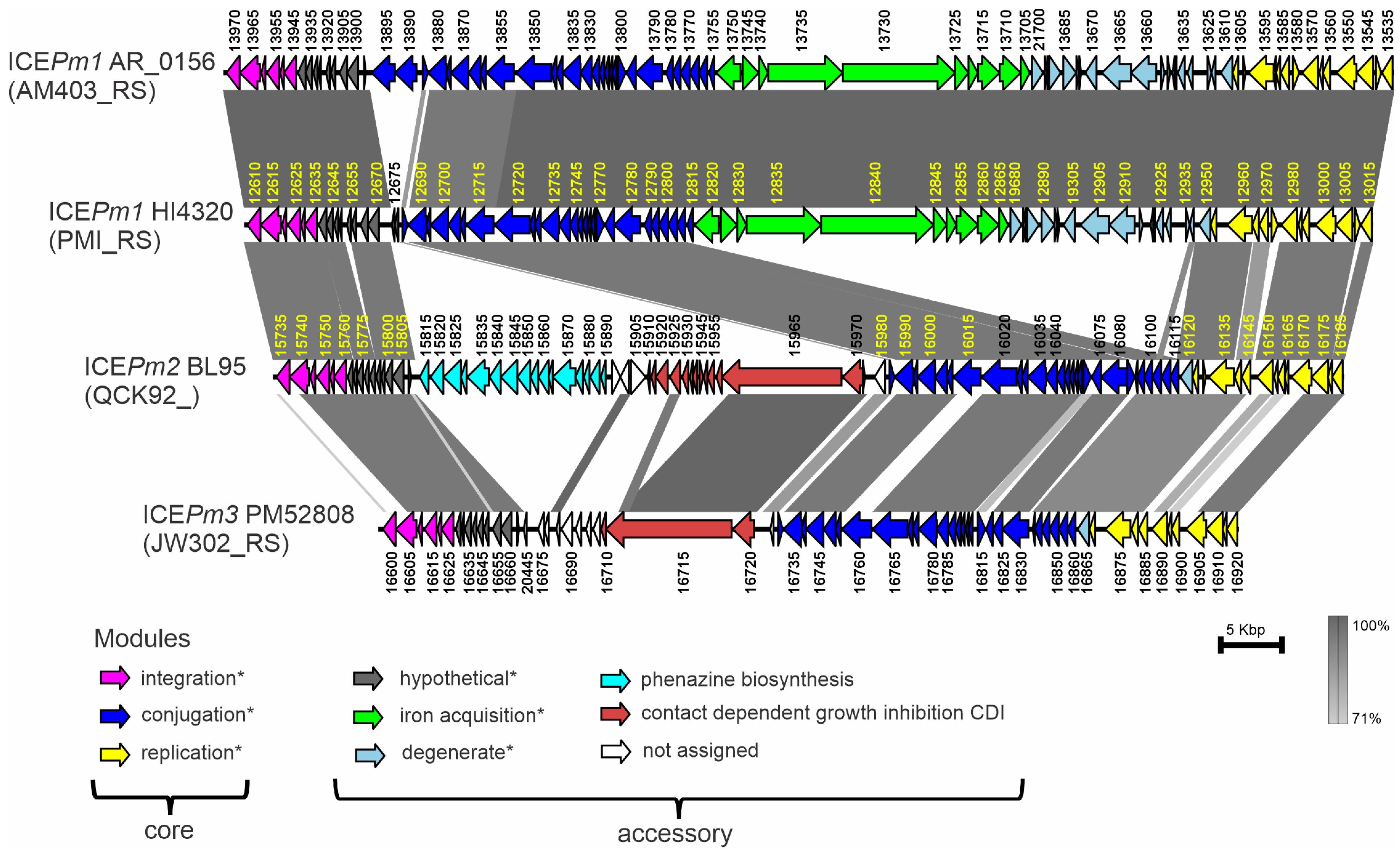
Integration in a tRNA-Phe is a hallmark of ICEPm1, an element initially observed in the first sequenced genome of P. mirabilis HI4320 [6] and analyzed in detail by Flannery et al. [12,14]. A comparison of ICEPm1 and ICE of BL95 showed that they share large blocks of similarity yet have extensive non-shared regions (Figure 3). Considering these substantial differences (see below), from now on, we will refer to this ICE as ICEPm2, a novel ICE in BL95 related to ICEPm1. Flannery et al. [14] distinguished three modules in ICEPm1—integration, conjugation, and DNA replication—that are necessary for its self-transmissibility. Inserted between them are variable modules carrying accessory genes. Our comparative analysis shows that the integration, conjugation, and DNA replication modules of ICEPm1 have corresponding matches in ICEPm2. In contrast, the variable modules differ greatly. ICEPm2 carries a large block of genes, inserted between the integration and conjugation modules, that is absent in ICEPm1 (Figure 3). This block in BL95 ICEPm2 carries unique genes for P. mirabilis that constitute (1) a novel 16 kb phenazine biosynthesis operon (Figure 4a), and (2) an 18.1 kb contact-dependent growth inhibition system operon (Figure 4b). At the same time, the variable modules of ICEPm1 (iron acquisition system and degenerate region), located between its conjugation and DNA replication module, are absent in ICEPm2.

3.7. Distribution of ICEPm1, ICEPm2, and ICEPm3 among P. mirabilis Isolates and Variation in Insertion Sites, tRNA-PheV or tRNA-PheU

To find out the distribution of ICEs similar to ICEPm1 and ICEPm2, to determine their integration sites, and to characterize the main variants of these elements, we conducted a systematic search of complete P. mirabilis genomes (Table 1), as well as other bacteria. Criteria for the search were (1) the identification of the characteristic direct repeats formed by integration into tRNA-Phe, and (2) the presence of modules encoding self-transferability. Of the 99 isolates of P. mirabilis, 35 carried ICEs satisfying these criteria, and some of these isolates had two ICE copies. Comparison of these elements showed that in addition to ICEPm1 and ICEPm2, there is another ICE, sufficiently diverged to classify it under a different name as ICEPm3 (Figure 3). Like ICEPm2, ICEPm3 lacks the variable modules of ICEPm1 and carries genes encoding CDI; unlike ICEPm2, it lacks the module for phenazine biosynthesis. Although ICEPm2 and ICEPm3 have more accessory genes in common, ICEPm2 has higher genetic similarity to ICEPm1 (92.45% ± 0.14%) than to ICEPm3 (89.26% ± 0.17%) in shared genes of the integration, conjugation, and DNA replication modules (Figure 3).
ICEPm1 is found in 34 isolates; in 20 isolates it is integrated into the tRNA-PheV, and in 14 isolates into the tRNA-PheU (Figure 1). ICEPm2 is integrated into the tRNA-PheV and it is found only in BL95. ICEPm3 is found in eight isolates, and all these isolates also have ICEPm1; in two isolates, ICEPm3 is integrated into the tRNA-PheV, and in six isolates into the tRNA-PheU. When an isolate has simultaneously two ICEs (ICEPm1 and ICEPm3), they are integrated in different copies of the tRNA-Phe, with the exception of strain AR379, where both ICEs are associated with the tRNA-PheU (Figure 1). Specifically, ICEPm3 is integrated into the tRNA-PheU, while ICEPm1 is integrated into the direct repeat right (DR attR) of ICEPm3, thus forming a tandem repeat of two ICEs.
Inspection of the distribution of ICEPm1, -2, and -3 on the phylogenetic tree shows that these ICEs have inserted independently multiple times (Figure 1). Furthermore, the frequency of insertions must be high, because closely related BL95 and AR_0156 carry different ICEs (ICEPm2 and ICEPm1, respectively), in the same tRNA-PheV locus. Another pair of closely related isolates, AR_0029 and SCBX1.1, carry ICEPm1, which are integrated in different tRNA-Phe loci (U or V), indicating independent insertion. A similar situation is observed in a cluster of six nearly genetically identical isolates (T18, XH983, T21, FZP2936, FZP3115, L90-1), where ICEPm1 is integrated in different tRNA-Phe loci. These observations strongly suggest that ICEPm1 and ICEPm2 are currently active elements.
Identical copies of ICEPm3 are found in sister-taxa PM52260 and PM52808, but absent in the closely related MPE0346 and in more distantly related strains (Figure 1), indicating a recent integration. The integration likely occurred in the common ancestor of PM52260 and PM52808 after it separated from MPE0346. Moreover, highly similar copies of ICEPm3 (100% coverage and 99.99% identity) are found in six distantly related strains of Proteus (AR379, HURS-181823, HURS-186083, PM52260, PM52808, and AR_0155), and furthermore, integrated in different loci (tRNA-PheU or tRNA-PheV). These observations strongly suggest that ICEPm3 is an active ICE moving between P. mirabilis isolates.
Additional evidence that ICEPm1 and ICEPm2 are active comes from the pattern of distribution and high identity of these ICEs in other bacteria. For example, ICEPm2 in BL95 is nearly identical (100% coverage, 99.91% identity) to such elements in E. coli from mice feces (Accessions CP010221.1, CP010206.1, CP010196.1, CP010186.1, CP010213.1). Highly similar copies of ICEPm1 across the full length (Coverage: ≥95%; Identity: >99%) were found in over 40 g-negative species, including Providencia, Escherichia, Klebsiella, and Morganella. ICEPm3 was not found in other bacteria, suggesting that its movement is limited intra-specifically to P. mirabilis. Considering high similarity between the eight copies of ICEPm3, it may have formed recently, and it did not have time to spread to other bacteria or was not yet found due to low frequency.

3.8. Novel Phenazine Biosynthesis and Contact-Dependent Growth Inhibition System Operons in ICEPm2 in P. mirabilis BL95

3.8.1. Phenazine Biosynthesis Gene Operon in ICEPm2

Embedded in ICEPm2, region 3,431,156–3,446,405 (QCK92_15815–QCK92_15890) of the BL95 chromosome encompasses a cluster of 16 genes involved in phenazine biosynthesis (Figure 4a; Table 3). Six genes in this cluster, phzA/BCDEFG, encode the core phenazine biosynthesis enzymes [34,35], gene ephR encodes resistance to phenazine, and the remaining genes encode enzymes involved in the modification of phenazine tricycles [35]. The canonical phzC gene encodes a 3-deoxy-d-arabino-heptulosonate-7-phosphate (DAHP) synthase, which catalyzes the first step of the shikimate pathway and redirects the intermediates from primary metabolism into phenazine biosynthesis [36]. Although BL95 lacks the DAPH synthase gene, it carries a gene encoding a 3-deoxy-7-phosphoheptulonate synthase which likely performs an analogous function by providing metabolic precursors for the shikimic acid pathway and thus aiding in the synthesis of phenazines [34].
According to BPROM software [25], the genes of the phenazine cluster are regulated by two σ70 promoters, one upstream of ehpR and another within phzG (Figure 4a). The second promoter has probably evolved to increase the transcription level of downstream genes, including phzC that encodes for a core phenazine biosynthesis enzyme. The transcription of the operon is terminated by a Rho-independent terminator as predicted by the ARNold software [26] (Figure 4a).
Even though our results indicate that this operon is found in other bacteria, this is the first report of a phenazine operon in P. mirabilis. This operon is present with the same genes and in the same order in five isolates of E. coli from mice feces (see above, similarity > 99.9%) and two isolates of P. stuartii (CP114580.1, CP114582.1, similarity = 94.8%). Additionally, all the genes of the operon but one (encoding 4′-phosphopantetheinyl transferase) are found on a plasmid of E. coli (CP076707.1, similarity = 95.5%) and on a chromosome of Serratia fontinicola DSM 4576 (CP011254.1, similarity = 76.4%). The high sequence divergence of the S. fontinicola phenazine operon from the rest suggests that this cluster existed in such configuration for a considerable time, and it is likely preserved by natural selection. Leise [35] classified this phenazine cluster as type 2, out of six types distinguished in Enterobacterales, and found that this type was associated with ICE integrated in tRNA-Phe, consistent with our finding of this cluster integrated in tRNA-Phe by ICEPm2.
Phenazines are key contributors to many aspects of the biology of their producers [37]. They exhibit broad antimicrobial properties, making their producers more competitive, increasing virulence and playing a role in biofilm formation and iron acquisition [34,35,38,39,40,41].

3.8.2. Contact-Dependent Growth Inhibition Gene Cluster in ICEPm2

Contact-dependent growth inhibition (CDI) system, first described in E. coli EC93, is encoded by a locus of three genes, cdiB, cdiA, and cdiI [42]. The CdiB-CdiA proteins form a two-partner secretion system: CdiB is an outer membrane protein that presents the CdiA exoprotein on the cell surface [9,24]. The N-terminal part of CdiA is a delivery system, while the polymorphic C-terminal region (CdiA-CT) is a toxin exhibiting a distinct growth inhibition activity on the target bacteria [7,24,43]. The N- and C- termini are demarcated by the VENN motif. CdiI is an immunity protein that binds to cognate CdiA-CT and blocks its activity, thereby protecting the cell from autoinhibition [24]. A comparative genomic analysis showed that many CDI systems contain “orphan” cdiA-CT units not connected to the cdiA N-terminus. Usually, a cdiA-CT unit is linked to a cognate cdiI gene downstream, forming a pair. These pairs are mobile, and can fuse to N-terminus, producing a system with novel inhibition activity [24,43].
Our comparative genomic analysis showed that a cluster of genes that belong to the CDI system is found in the ICEPm2 of P. mirabilis strain BL95, almost immediately upstream of the phenazine biosynthesis operon (Figure 3 and Figure 4b). The CDI system (nucleotides 3449845–3467528, loci QCK92_15910–QCK92_15970) and the phenazine operon are separated by transposases of the IS1 and IS256 families. The CDI system of BL95 has the three central structural genes that form a CDI system, namely cdiB, cdiA, and cdiI (Figure 5). Additionally, as shown in Figure 4b, the CDI system of BL95 has two “orphan” cdiA-CT/cdiI sets that are indicated with the suffixes ‘-o1’ and ‘-o2’. Moreover, our results showed that most of CDI ORFs (except cdiA and cdiB) are found only in BL95 and no other P. mirabilis strains.
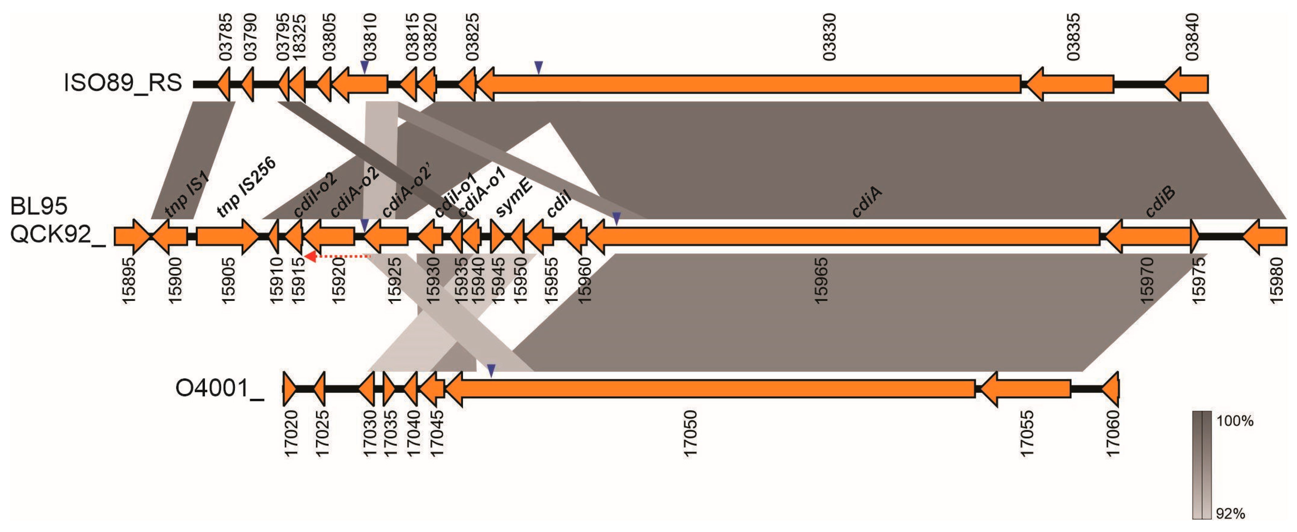
An alignment between ICEPm2 and ICEPm3 showed that the cdiB genes in both ICEs are closely related homologs. Additionally, both ICEs share a closely related cdiA homolog; however, the 3′-ends of cdiA, encoding C-terminus, are non-homologous (Figure 3). The cdiA gene in the ICEPm2 is followed by “orphan” cdiA-CT/cdiI sets that are absent in other P. mirabilis. Some of the genes in these orphan sets have similarity to other genes in the BL95 CDI cluster. For example, locus QCK92_15955 encoding CdiI is 70.1% similar to locus QCK92_15930 encoding the “orphan” CdiI-o1. Additionally, an 830 bp-long portion of cdiA (QCK92_15965) is 95.2% similar to the “orphan” cdiA-o2 (QCK92_15925). Interestingly, one gene in the middle of the CDI cluster in ICEPm2 does not seem to be part of the CDI system but rather represents another toxin system of the SymE family (QCK92_15945).
The genes of the BL95 CDI cluster have numerous correspondences to GenBank sequences, with those most similar ones shown in Figure 5 and Figure S3. The strain BL95 CDI cluster is almost 100% identical to the CDI of E. coli M19 isolated from mice feces (GenBank Accession CP010221). Additionally, a shorter region with high identity is found in another E. coli Mt1B1 (CP028714). It is important to notice that annotations of the CDI system are lacking in these accessions (Figure S3).
Comparison of the BL95 CDI with the P. stuartii strain 2021CK-01196 (CP114580.1) and M. morganii 1810122035 CDIs reveals several matches, which give some insights on how this cluster may have formed in strain BL95. As shown in Figure 5, all these bacteria harbor cdiB and cdiA with an identity higher than 92%. However, the similarity of cdiA only extends to the VENN motif (i.e., cdiA-CT are different) indicated by blue arrowheads in Figure 5. Interestingly, the cdiA-CT and cognate cdiI of P. stuartii are found in BL95, but relative to P. stuartii they translocated a few hundred base pairs downstream of cdiA, forming orphans cdiA-o1 and cdiI-o1. These results indicate that the cdiA in BL95 arose from a progenitor similar to that of P. stuartii by the replacement of cdiA-CT and cognate cdiI with new variants. The displaced cdiA-CT and cognate cdiI were not lost but became “orphans”.
Similarly, the C-terminus of cdiA and cognate cdiI of M. morganii formed the “orphans” cdiA-o2 and cdiI-o2 in P. mirabilis BL95 (Figure 5). Moreover, the cdiA-o2′ (QCK92_15925) and the intergenic region upstream of cdiA-o2 share high similarity with M. morganii, suggesting that in the past, cdiA-o2 and cdiA-o2′ formed a single long ORF that was split into separate ORFs by a single nucleotide insertion (Figure 5).
According to BPROM [25], the cdiB, cdiA, and cdiI are regulated from a single σ70 promoter (Figure 4b), forming an operon. The σ70 promoter perfectly matches the −10-consensus sequence (TATAAT), including the −10-extended (TGN). The ARNold software [26] revealed that the transcription termination for these genes could be achieved by the terminator-2 (Figure 4b). The software predicted three more terminators in the antisense strand that prevent a transcriptional read-through from the antisense strand (Figure 4b). For example, terminator-1 stops the symE gene transcription and prevents any interference with the CDI operon transcription. It is important to mention that the U-tract next to the stem-loop dictates the directionality of the terminators [44].

3.9. Abundance of Secretion Systems in P. mirabilis BL95

A search of the strain BL95 genome with TXSScan resulted in a list of over 100 genes that belong to secretion systems type I, III, IV, V, and VI (Supplementary Table S2). Of these, T3SS is known to be used by bacteria to kill eukaryotic cells [8,15], whereas all other systems are potential candidates for interbacterial competition.
In addition to the CdiB/CdiA system, the secretion systems of strain BL95 include other two-partner systems (TPS). One TPS of T5bSS in nucleotides position 1,198,117–1,205,409 (loci QCK92_05550–QCK92_05560; ShlB/DUF637, T5bSS) is distantly related to the cdiB/A/I of ICEPm2 and is likely involved in competition with other bacteria [6]. The locus encoding that system is not associated with mobile elements, it is found in all P. mirabilis and other members of the genus, and it is likely intrinsic to the genus. Another TPS of T5bSS, called hpmB/hpmA, is in nucleotides position 2,840,233–2,846,685 (loci QCK92_13275 and QCK92_13280). This is a well-known system from P. mirabilis described in 1990 that encodes cytolysin/hemolysin with cytotoxic activity [7,45].

4. Discussion

An earlier study by one of us [15] observed that a commensal P. mirabilis strain BL95 outcompeted commensal E. coli in the gut of neonatal mice and killed E. coli and other enterobacteria in vitro. Experiments showed that killing required direct contact of P. mirabilis and the competing bacteria. Furthermore, P. mirabilis BL95 demonstrated unique features of inhibition, compared to other functionally characterized contact-dependent growth inhibition systems, such as the CDI in E. coli [42]. One such feature was that BL95 developed inhibitory properties when reaching the stationary phase when grown in liquid culture, and not in the exponential phase [42]. Another distinguishing feature was high inhibitory effectiveness in shaking liquid culture, in addition to being effective on solid media [15]. Importantly, the ability to kill enterobacteria extended to other P. mirabilis strains [15], although it is not known if the same systems were involved in the competition by these strains as they were not functionally characterized to the same detail as BL95.
As a first step in identification of candidate genes encoding this contact-dependent inhibition system with novel functional properties, we sequenced and characterized the genome of strain BL95 and compared it with other P. mirabilis with complete genomes. Placing BL95 in a larger phylogenetic framework is a promising approach to narrow down potential candidates. For example, if a CDI with certain properties is scattered non-systematically across the phylogeny, this would indicate that the CDI genes are located on a mobile genetic element, such as a genomic island or a plasmid, prompting the identification of this element to find the genes involved. Alternatively, the CDI may be restricted to a group of related Proteus (clade), suggesting the need to find the common genomic locus present in this clade but not in other Proteus isolates.
Our phylogeny represents the first attempt to characterize the diversity of P. mirabilis using complete genomes and to systematically study the distribution of ICEPm in the species. Overall, P. mirabilis forms a tight-knit genetic group with nucleotide identity higher than 97% between the most diverged members. It is also well separated from the closest species of Proteus, making the species assignment unambiguous. P. mirabilis BL95 belongs to the largest clade, out of three clades, that also includes reference strain HI4320. Despite high similarity of the core genome, P. mirabilis has a vast suite of accessory, or cloud genes, that are found in a subset of strains or even in single strains. For example, P. mirabilis BL95 possesses 56 genes not found in other strains of P. mirabilis. It should be made clear that not all these genes bestow unique functions or features to BL95. Some of these genes may be diverged duplicates of genes present in other isolates and have similar functions. Still, even if the general function is similar (e.g., contact-dependent growth inhibition), the genetic variation might account for unique fine-tuning that may be crucial in some environments (e.g., expanding the range of target species or conferring an ability to establish cell contact in liquid media). On the other hand, some genes are unique to P. mirabilis BL95, such as the phenazine biosynthesis operon present in ICEPm2. However, the functions of the majority of unique genes in strain BL95 are yet unknown.
Phenazines exhibit broad antimicrobial properties by generating toxic reactive oxygen species and helping their producers to outcompete other bacteria in their ecological niche [36]. The broad-spectrum antimicrobials can be particularly beneficial for species occupying a niche with diverse microbiota, so that different potential competitors are targeted [46]. Furthermore, by acting as electron shuttles, phenazines help aerobic bacteria maintain redox homeostasis in low-oxygen environments through their ability to mediate the reoxidation of NADH under oxygen-limiting conditions, explaining the importance of phenazine production for biofilm formation, as mature biofilms are often hypoxic [34,40]. P. mirabilis is notorious for forming biofilms in catheters, being a leading cause of the catheter-associated urinary tract infections [1,2]. Phenazines also regulate diverse physiological functions, serving as intercellular signals and regulators of gene expression [40].
Our analysis revealed the presence of genes potentially encoding multiple secretion systems. Many of these systems have homologies to the reference strain HI4320 or other P. mirabilis. Although a few of these systems were characterized previously [2], the exact function for most of them is not well studied, leaving multiple candidates for contact-dependent killing described by Kiani et al. [15]. For instance, by using a mutant tssM of BB2000 which lacks a functional T6SS, Kiani et al. [15] ruled out T6SS as a candidate system involved in contact-dependent growth inhibition exhibited by BL95 and other P. mirabilis strains. However, the tssM mutation affected only one of T6SS, the same one known to be involved in the contact-dependent competition between strains of Proteus. Other T6SS found in BL95 and other Proteus may have different functions, e.g., they can be involved in interspecies bacteria competition. Pertinent, a genome-wide transposon mutagenesis study of P. mirabilis showed that the known P. mirabilis T6SS operons were overrepresented as fitness factors during coinfection with P. stuartii, indicating a potential role for T6SS in mediating competitive and cooperative interactions during polymicrobial infection [47].
One of the most common ICEs in P. mirabilis is ICEPm1 [14]. In strain HI4320 this element consists of six modules, three of which are the core backbone modules [48] involved in self-replication and self-transmission, and the other three are accessory gene modules. Surprisingly, strain BL95 has an element with the same core backbone modules, but distinct accessory genes modules. Specifically, ICE of BL95 lacked the iron acquisition and degenerate modules of ICEPm1 (located between conjugation and replication modules) but carried the phenazine biosynthesis and CDI modules (located between integration and conjugation modules). Considering the extent of structural and functional changes, we propose that the ICE of BL95 be recognized as a different element which we named ICEPm2. Furthermore, our bioinformatic analysis also showed that the mobilome of P. mirabilis possesses yet another ICE with the same core backbone but distinct accessory genes, which we named ICEPm3. ICEPm2 and ICEPm3 are similar, but the latter lacks the phenazine biosynthesis operon. Furthermore, the cdiA genes of ICEPm2 and ICEPm3 have different cdiA-CT termini, and orphan toxin/immunity gene pairs of the CDI module are unique to ICEPm2 (and BL95).
Another argument for considering ICEPm1, ICEPm2 and ICEPm3 as distinct ICEs is their pattern of presence in some strains. Flannery et al. [12] noted that only one ICEPm1 copy can be present in any single bacterium (inserted in either tRNA-pheU or -pheV). Our analyses showed that several strains carry ICEs in both tRNA-Phe, but in all such strains the inserted ICEs are of different types (ICEPm1 and ICEPm3), and never are they of the same type. All three ICEs have essentially the same core backbone modules determining their integration. Therefore, the lack of strains carrying simultaneously ICEs of the same type is likely not because such integration does not occur, but rather due to evolutionary pressure removing such combinations. Possession of an ICE might implicate both advantages (new functions due to accessory genes) and disadvantages (e.g., metabolic burden to replicate the ICE) [13]. ICEs of different types carry distinct accessory modules that confer distinct advantageous properties, whereas possession of two copies of the same ICE is redundant, leading to the elimination of strains with such combination. We found one strain, P. mirabilis AR379, that had ICEPm1 and ICEPm3 tandemly arranged in tRNA-pheU, which shows that accumulation of ICEs by accretion may be a way for bacteria to create a larger, more complex mobile element [49]. Accumulation by accretion is common for many composite transposons, such as those bound by IS26 [50], but such mechanism is less known for ICEs [51,52]. Some authors proposed that accumulation by accretion may be an important mechanism to increase ICEs diversity [13,53].
ICEPm1-2-3 are somewhat unusual among ICEs because they can integrate into either tRNA-Phe locus. Wozniak and Waldor [49] report that most ICEs targeting tRNA loci integrate into only one of a few tRNA loci, with only two known exceptions, ICEclc and PAPI-1, that can integrate into either tRNA locus. ICEPm1-2-3 not only can integrate into either locus, but they can be found in both loci simultaneously and even integrate into the same locus repeatedly by accretion, forming tandems.
One complication for the study of CDI systems is the lack of specialized software or databases for their identification in a genome. Annotating genes encoding contact-dependent inhibition systems is not trivial, because of their similarity to other better-characterized TPS, such as hemolysins and hemagglutinins. As a result, cdiA genes are often annotated as hemagglutinins, and cdiB genes as ShlB/FhaC/HecB hemolysins [54]. However, CdiB/CdiA pairs constitute a distinct subfamily of the two- partner secretion proteins [55]. CdiA is a large filamentous protein whose N-terminal region is homologous to that of Bordetella pertussis filamentous hemagglutinin (FHA). Indeed, the N-terminal regions of CdiA and FHA contain the typical hemagglutinin repeats and a hemagglutination activity domain [55]. In contrast to FHA, the CdiA N-terminal region ends with a conserved domain designated PT-VENN, helping its identification.
The cdiB genes can be correctly identified by their proximity to and association with cdiA, as well as by sequence comparison with representatives of TPS subfamilies. In our study, locus QCK92_15970 was incorrectly annotated by the NCBI as a ‘ShlB/FhaC/HecB family hemolysin secretion/activation protein CDS’, but our phylogeny using proteins from UniProt showed that it is most closely related to CdiB2 from Burkholderia pseudomallei. The position of QCK92_15970 immediately upstream of cdiA containing VENN motif further confirms that this gene should be annotated as cdiB, rather than ShlB/FhaC/HecB.
Annotating orphan genes is even more challenging because orphan cdi-CTs are short ORFs that do not start with methionine initiation codon. Moreover, cdi-CT and cognate cdiI genes are highly variable and little studied [43]. As a result, these orphan genes are either annotated as hypothetical, or they are not detected by annotation software. We also observed that different annotation software can suggest different ORFs in the same region, with some ORFs corresponding to actual ‘orphan’ genes and others being spurious. One way to unambiguously identify orphan genes is to find them as part of a complete CDI system (cdiB/cdiA/cdiI). Using this approach, we identified orphan genes in the CDI cluster of ICEPm2 of strain BL95. Specifically, one pair of BL95 orphans was found in the complete CDI system of P. stuartii, and another pair in the complete CDI of M. morganii (Figure 5).
Comparison of CDIs in P. mirabilis BL95 and M. morganii also demonstrated how orphan genes are shaped in the course of evolution (Figure 5). This comparison shows that in the past cdiA-o2 and cdiA-o2’ formed one continuous ORF, perhaps making a longer orphan cdiA-CT-o2. Single nucleotide insertion interrupted this ancestral ORF, resulting in two ORFs. One of these ORFs became a shorter orphan encoding toxin (cdiA-o2), while the other ORF is likely a non-functional pseudogene (cdiA-o2’), because it does not have C-terminus and thus it cannot be used for displacing C-termini in cdiA genes to produce new toxin variants. As noted previously for T6SS [56], understanding the importance of orphan genes in bacterial competition will require their careful detection, curation, and systematic analysis. It is essential to create databases of CDI genes, including orphan genes, similar to ISFinder that identifies insertion sequences [57] or services of the CGE (https://www.genomicepidemiology.org, accessed on 1 April 2024).
Previous work established that P. mirabilis BL95 outcompetes co-occurring enterobacteria in vivo and in vitro in a contact-dependent manner. Here we sequenced and analyzed the fully assembled genome of BL95 and showed that it carries (1) a phenazine biosynthesis operon unique to P. mirabilis and (2) a contact-dependent growth inhibition system operon, previously not reported in this species. These operons are adjacent to each other and embedded in an ICE that we named ICEPm2. Future studies will establish whether the phenazine and/or CDI operons in ICEPm2 are responsible for the competitive features displayed by strain BL95. It may be significant that ICEPm2 carrying CDI operon originates from P. mirabilis isolated from mice intestines. Gastrointestinal (GI) tract has a rich and competitive microbiota, compared to the urinary tract which normally harbors a smaller number of microorganisms (mainly Lactobacillus and other Firmicutes) [58]. The presence of ICEPm2 with the CDI operon in BL95 may be connected to the highly diverse microenvironment of the GI tract. Some previous studies suggested that CDI systems are particularly effective when direct cell-cell contact between competing strains is common giving an advantage to their hosts [9,59]. Indicative, that other bacterial species carrying ICEPm2 were also of GI origin. ICEPm2 is a novel ICE that shares core modules—integration, conjugation, and replication—with ICEPm1, but carries distinct accessory modules. Our bioinformatic analyses of P. mirabilis isolates from the GenBank revealed yet another previously undescribed element named ICEPm3, similar to ICEPm2, but lacking the phenazine biosynthesis operon. The distribution of ICEPm1-2-3 on the phylogeny of P. mirabilis shows their multiple independent integration in different Proteus lineages and indicates that these elements are currently active. Notably, ICEPm1 and ICEPm3 can be found simultaneously in the same strain, but none of them (and ICEPm2) are found duplicated. One interpretation of this observation is that these elements co-exist when they confer distinct advantages due to non-overlapping accessory modules; however, isolates containing duplicate copies of the same ICE are selected against due to redundancy and metabolic burden.

Supplementary Materials

The following supporting information can be downloaded at: https://www.mdpi.com/article/10.3390/microorganisms12071321/s1, Figure S1: Pangenome of P. mirabilis. Overall, the pangenome consists of 11,364 genes identified in 99 P. mirabilis strains; Figure S2: Schematic representation of the P. mirabilis BL95 chromosome using Proksee (https://proksee.ca, accessed on 16 December 2022); Figure S3: Similarity of the CDI region from ICEPm2 in P. mirabilis to homologous regions from other species; Table S1: Genetic organization of Integrative and Conjugative Elements ICEPm1, ICEPm2, and ICEPm3 shown in Figure 5; Table S2: Genes in P. mirabilis BL95 corresponding to secretion systems in gram-negative bacteria.

Author Contributions

Conceptualization, A.T. and L.M.-B.; Data curation, A.T., I.M.-G. and L.M.-B.; Formal analysis, A.T. and I.M.-G.; Funding acquisition, L.M.-B.; Investigation, A.T., I.M.-G., I.V. and L.M.-B.; Methodology, A.T., I.M.-G. and L.M.-B.; Project administration, L.M.-B.; Resources, J.B.; Supervision, L.M.-B.; Validation, A.T. and L.M.-B.; Visualization, A.T., I.M.-G. and I.V.; Writing—original draft, A.T.; Writing—review & editing, A.T., I.M.-G., I.V., J.B. and L.M.-B. All authors have read and agreed to the published version of the manuscript.

Funding

This research was funded by NIH grant GM136498 awarded to L.M-B.

Data Availability Statement

The genome sequence data of P. mirabilis BL95 were deposited in NCBI GenBank under BioProject accession number PRJNA954452, BioSample accession number SAMN34145880, and assembled chromosome CP122400.

Conflicts of Interest

The authors declare no conflicts of interest.

References

  1. Armbruster, C.E.; Mobley, H.L. Merging mythology and morphology: The multifaceted lifestyle of Proteus mirabilis. Nat. Rev. Microbiol. 2012, 10, 743–754. [Google Scholar] [CrossRef] [PubMed]
  2. Armbruster, C.E.; Mobley, H.L.T.; Pearson, M.M. Pathogenesis of Proteus mirabilis infection. EcoSal Plus 2018, 8. [Google Scholar] [CrossRef]
  3. Gibbs, K.A.; Urbanowski, M.L.; Greenberg, E.P. Genetic determinants of self identity and social recognition in bacteria. Science 2008, 321, 256–259. [Google Scholar] [CrossRef] [PubMed]
  4. Alteri, C.J.; Himpsl, S.D.; Pickens, S.R.; Lindner, J.R.; Zora, J.S.; Miller, J.E.; Arno, P.D.; Straight, S.W.; Mobley, H.L. Multicellular bacteria deploy the type VI secretion system to preemptively strike neighboring cells. PLoS Pathog. 2013, 9, e1003608. [Google Scholar] [CrossRef] [PubMed]
  5. Mobley, H.L.T. Proteus mirabilis overview. In Proteus mirabilis: Methods and Protocols; Pearson, M.M., Ed.; Humana Press: New York, NY, USA, 2021; pp. 1–4. [Google Scholar] [CrossRef]
  6. Pearson, M.M.; Sebaihia, M.; Churcher, C.; Quail, M.A.; Seshasayee, A.S.; Luscombe, N.M.; Abdellah, Z.; Arrosmith, C.; Atkin, B.; Chillingworth, T.; et al. Complete genome sequence of uropathogenic Proteus mirabilis, a master of both adherence and motility. J. Bacteriol. 2008, 190, 4027–4037. [Google Scholar] [CrossRef] [PubMed]
  7. Guerin, J.; Bigot, S.; Schneider, R.; Buchanan, S.K.; Jacob-Dubuisson, F. Two-partner secretion: Combining efficiency and simplicity in the secretion of large proteins for bacteria-host and bacteria-bacteria interactions. Front. Cell. Infect. Microbiol. 2017, 7, 148. [Google Scholar] [CrossRef] [PubMed]
  8. Hayes, C.S.; Aoki, S.K.; Low, D.A. Bacterial contact-dependent delivery systems. Annu. Rev. Genet. 2010, 44, 71–90. [Google Scholar] [CrossRef]
  9. Willett, J.L.; Ruhe, Z.C.; Goulding, C.W.; Low, D.A.; Hayes, C.S. Contact-dependent growth inhibition (CDI) and CdiB/CdiA two-partner secretion proteins. J. Mol. Biol. 2015, 427, 3754–3765. [Google Scholar] [CrossRef] [PubMed]
  10. Juarez, G.E.; Mateyca, C.; Galvan, E.M. Proteus mirabilis outcompetes Klebsiella pneumoniae in artificial urine medium through secretion of ammonia and other volatile compounds. Heliyon 2020, 6, e03361. [Google Scholar] [CrossRef]
  11. Guimaraes, L.C.; de Jesus, L.B.; Viana, M.V.C.; Silva, A.; Ramos, R.T.J.; Soares, S.D.; Azevedo, V. Inside the pan-genome—Methods and software overview. Curr. Genom. 2015, 16, 245–252. [Google Scholar] [CrossRef]
  12. Flannery, E.L.; Antczak, S.M.; Mobley, H.L. Self-transmissibility of the integrative and conjugative element ICEPm1 between clinical isolates requires a functional integrase, relaxase, and type IV secretion system. J. Bacteriol 2011, 193, 4104–4112. [Google Scholar] [CrossRef] [PubMed]
  13. Johnson, C.M.; Grossman, A.D. Integrative and Conjugative Elements (ICEs): What they do and how they work. Annu. Rev. Genet. 2015, 49, 577–601. [Google Scholar] [CrossRef]
  14. Flannery, E.L.; Mody, L.; Mobley, H.L. Identification of a modular pathogenicity island that is widespread among urease-producing uropathogens and shares features with a diverse group of mobile elements. Infect. Immun. 2009, 77, 4887–4894. [Google Scholar] [CrossRef] [PubMed]
  15. Kiani, D.; Santus, W.; Kiernan, K.A.; Behnsen, J. Proteus mirabilis employs a contact-dependent killing system against competing Enterobacteriaceae. mSphere 2021, 6, e0032121. [Google Scholar] [CrossRef] [PubMed]
  16. Argueta, F.; Tatarenkov, A.; Mota-Bravo, L. Multiple copies of a qnrB19 gene are carried by tandem repeats of an IS26 composite transposon in an Escherichia coli plasmid. Microbiol. Resour. Announc. 2022, 11, e0066122. [Google Scholar] [CrossRef]
  17. Maniatis, T.; Fritsch, E.F.; Sambrook, J.K. Molecular Cloning: A Laboratory Manual; Cold Spring Harbor Laboratory Press: Cold Spring Harbor, NY, USA, 1982; pp. 191–195. [Google Scholar]
  18. Wick, R.R.; Judd, L.M.; Gorrie, C.L.; Holt, K.E. Unicycler: Resolving bacterial genome assemblies from short and long sequencing reads. PLoS Comput. Biol. 2017, 13, e1005595. [Google Scholar] [CrossRef]
  19. Tatusova, T.; DiCuccio, M.; Badretdin, A.; Chetvernin, V.; Nawrocki, E.P.; Zaslavsky, L.; Lomsadze, A.; Pruitt, K.D.; Borodovsky, M.; Ostell, J. NCBI prokaryotic genome annotation pipeline. Nucleic Acids Res. 2016, 44, 6614–6624. [Google Scholar] [CrossRef]
  20. Seemann, T. Prokka: Rapid prokaryotic genome annotation. Bioinformatics 2014, 30, 2068–2069. [Google Scholar] [CrossRef]
  21. Olson, R.D.; Assaf, R.; Brettin, T.; Conrad, N.; Cucinell, C.; Davis, J.J.; Dempsey, D.M.; Dickerman, A.; Dietrich, E.M.; Kenyon, R.W.; et al. Introducing the Bacterial and Viral Bioinformatics Resource Center (BV-BRC): A resource combining PATRIC, IRD and ViPR. Nucleic Acids Res. 2023, 51, D678–D689. [Google Scholar] [CrossRef]
  22. Liu, M.; Li, X.; Xie, Y.; Bi, D.; Sun, J.; Li, J.; Tai, C.; Deng, Z.; Ou, H.Y. ICEberg 2.0: An updated database of bacterial integrative and conjugative elements. Nucleic Acids Res. 2019, 47, D660–D665. [Google Scholar] [CrossRef]
  23. Abby, S.S.; Cury, J.; Guglielmini, J.; Neron, B.; Touchon, M.; Rocha, E.P. Identification of protein secretion systems in bacterial genomes. Sci. Rep. 2016, 6, 23080. [Google Scholar] [CrossRef] [PubMed]
  24. Poole, S.J.; Diner, E.J.; Aoki, S.K.; Braaten, B.A.; t’Kint de Roodenbeke, C.; Low, D.A.; Hayes, C.S. Identification of functional toxin/immunity genes linked to contact-dependent growth inhibition (CDI) and rearrangement hotspot (Rhs) systems. PLoS Genet. 2011, 7, e1002217. [Google Scholar] [CrossRef] [PubMed]
  25. Solovyev, V.; Salamov, A. Automatic annotation of microbial genomes and metagenomic sequences. In Metagenomics and Its Applications in Agriculture, Biomedicine and Environmental Studies; Li, R.W., Ed.; Nova Science Publishers: Hauppauge, NY, USA, 2011; pp. 61–78. [Google Scholar]
  26. Naville, M.; Ghuillot-Gaudeffroy, A.; Marchais, A.; Gautheret, D. ARNold: A web tool for the prediction of Rho-independent transcription terminators. RNA Biol. 2011, 8, 11–13. [Google Scholar] [CrossRef] [PubMed]
  27. Stothard, P.; Grant, J.R.; Van Domselaar, G. Visualizing and comparing circular genomes using the CGView family of tools. Brief Bioinform. 2019, 20, 1576–1582. [Google Scholar] [CrossRef]
  28. Sullivan, M.J.; Petty, N.K.; Beatson, S.A. Easyfig: A genome comparison visualizer. Bioinformatics 2011, 27, 1009–1010. [Google Scholar] [CrossRef] [PubMed]
  29. Page, A.J.; Cummins, C.A.; Hunt, M.; Wong, V.K.; Reuter, S.; Holden, M.T.; Fookes, M.; Falush, D.; Keane, J.A.; Parkhill, J. Roary: Rapid large-scale prokaryote pan genome analysis. Bioinformatics 2015, 31, 3691–3693. [Google Scholar] [CrossRef] [PubMed]
  30. Kumar, S.; Stecher, G.; Li, M.; Knyaz, C.; Tamura, K. MEGA X: Molecular Evolutionary Genetics Analysis across computing platforms. Mol. Biol. Evol. 2018, 35, 1547–1549. [Google Scholar] [CrossRef]
  31. Sullivan, N.L.; Septer, A.N.; Fields, A.T.; Wenren, L.M.; Gibbs, K.A. The complete genome sequence of Proteus mirabilis strain BB2000 reveals differences from the P. mirabilis reference strain. Genome Announc. 2013, 1, e00024-13. [Google Scholar] [CrossRef]
  32. Di Pilato, V.; Chiarelli, A.; Boinett, C.J.; Riccobono, E.; Harris, S.R.; D’Andrea, M.M.; Thomson, N.R.; Rossolini, G.M.; Giani, T. Complete genome sequence of the first KPC-Type carbapenemase-positive Proteus mirabilis strain from a bloodstream infection. Genome Announc. 2016, 4, e00607-16. [Google Scholar] [CrossRef]
  33. Bonnin, R.A.; Girlich, D.; Jousset, A.B.; Gauthier, L.; Cuzon, G.; Bogaerts, P.; Haenni, M.; Madec, J.Y.; Couve-Deacon, E.; Barraud, O.; et al. A single Proteus mirabilis lineage from human and animal sources: A hidden reservoir of OXA-23 or OXA-58 carbapenemases in Enterobacterales. Sci. Rep. 2020, 10, 9160. [Google Scholar] [CrossRef]
  34. Hendry, S.; Steinke, S.; Wittstein, K.; Stadler, M.; Harmrolfs, K.; Adewunmi, Y.; Sahukhal, G.; Elasri, M.; Thomashow, L.; Weller, D.; et al. Functional analysis of phenazine biosynthesis genes in Burkholderia spp. Appl. Environ. Microbiol. 2021, 87, e02348-20. [Google Scholar] [CrossRef] [PubMed]
  35. Leise, C. The Diversity and Evolution of Phenazine Biosynthesis Pathways in Enterobacterales. Bachelor’s Honor Thesis, University of Southern Mississippi, Hattiesburg, MI, USA, 2021. [Google Scholar]
  36. Mavrodi, D.V.; Blankenfeldt, W.; Thomashow, L.S. Phenazine compounds in fluorescent Pseudomonas spp. biosynthesis and regulation. Annu. Rev. Phytopathol. 2006, 44, 417–445. [Google Scholar] [CrossRef] [PubMed]
  37. Price-Whelan, A.; Dietrich, L.E.; Newman, D.K. Rethinking ‘secondary’ metabolism: Physiological roles for phenazine antibiotics. Nat. Chem. Biol. 2006, 2, 71–78. [Google Scholar] [CrossRef] [PubMed]
  38. Mazzola, M.; Cook, R.J.; Thomashow, L.S.; Weller, D.M.; Pierson, L.S. Contribution of phenazine antibiotic biosynthesis to the ecological competence of fluorescent pseudomonads in soil habitats. Appl. Environ. Microbiol. 1992, 58, 2616–2624. [Google Scholar] [CrossRef] [PubMed]
  39. Lau, G.W.; Ran, H.; Kong, F.; Hassett, D.J.; Mavrodi, D. Pseudomonas aeruginosa pyocyanin is critical for lung infection in mice. Infect. Immun. 2004, 72, 4275–4278. [Google Scholar] [CrossRef] [PubMed]
  40. Dietrich, L.E.; Teal, T.K.; Price-Whelan, A.; Newman, D.K. Redox-active antibiotics control gene expression and community behavior in divergent bacteria. Science 2008, 321, 1203–1206. [Google Scholar] [CrossRef] [PubMed]
  41. Wang, Y.; Wilks, J.C.; Danhorn, T.; Ramos, I.; Croal, L.; Newman, D.K. Phenazine-1-carboxylic acid promotes bacterial biofilm development via ferrous iron acquisition. J. Bacteriol. 2011, 193, 3606–3617. [Google Scholar] [CrossRef] [PubMed]
  42. Aoki, S.K.; Pamma, R.; Hernday, A.D.; Bickham, J.E.; Braaten, B.A.; Low, D.A. Contact-dependent inhibition of growth in Escherichia coli. Science 2005, 309, 1245–1248. [Google Scholar] [CrossRef]
  43. Cuthbert, B.J.; Hayes, C.S.; Goulding, C.W. Functional and structural diversity of bacterial contact-dependent growth inhibition effectors. Front. Mol. Biosci. 2022, 9, 866854. [Google Scholar] [CrossRef]
  44. Peters, J.M.; Vangeloff, A.D.; Landick, R. Bacterial transcription terminators: The RNA 3′-end chronicles. J. Mol. Biol. 2011, 412, 793–813. [Google Scholar] [CrossRef]
  45. Uphoff, T.S.; Welch, R.A. Nucleotide sequencing of the Proteus mirabilis calcium-independent hemolysin genes (hpmA and hpmB) reveals sequence similarity with the Serratia marcescens hemolysin genes (shlA and shlB). J. Bacteriol. 1990, 172, 1206–1216. [Google Scholar] [CrossRef] [PubMed]
  46. Hibbing, M.E.; Fuqua, C.; Parsek, M.R.; Peterson, S.B. Bacterial competition: Surviving and thriving in the microbial jungle. Nat. Rev. Microbiol. 2010, 8, 15–25. [Google Scholar] [CrossRef]
  47. Armbruster, C.E.; Forsyth-DeOrnellas, V.; Johnson, A.O.; Smith, S.N.; Zhao, L.; Wu, W.; Mobley, H.L.T. Genome-wide transposon mutagenesis of Proteus mirabilis: Essential genes, fitness factors for catheter-associated urinary tract infection, and the impact of polymicrobial infection on fitness requirements. PLoS Pathog. 2017, 13, e1006434. [Google Scholar] [CrossRef] [PubMed]
  48. Beaber, J.W.; Burrus, V.; Hochhut, B.; Waldor, M.K. Comparison of SXT and R391, two conjugative integrating elements: Definition of a genetic backbone for the mobilization of resistance determinants. Cell. Mol. Life Sci. 2002, 59, 2065–2070. [Google Scholar] [CrossRef]
  49. Wozniak, R.A.; Waldor, M.K. Integrative and conjugative elements: Mosaic mobile genetic elements enabling dynamic lateral gene flow. Nat. Rev. Microbiol. 2010, 8, 552–563. [Google Scholar] [CrossRef]
  50. Bennett, P.M. Plasmid encoded antibiotic resistance: Acquisition and transfer of antibiotic resistance genes in bacteria. Br. J. Pharmacol. 2008, 153 (Suppl. S1), S347–S357. [Google Scholar] [CrossRef]
  51. Hochhut, B.; Beaber, J.W.; Woodgate, R.; Waldor, M.K. Formation of chromosomal tandem arrays of the SXT element and R391, two conjugative chromosomally integrating elements that share an attachment site. J. Bacteriol. 2001, 183, 1124–1132. [Google Scholar] [CrossRef] [PubMed]
  52. Pavlovic, G.; Burrus, V.; Gintz, B.; Decaris, B.; Guedon, G. Evolution of genomic islands by deletion and tandem accretion by site-specific recombination: ICESt1-related elements from Streptococcus thermophilus. Microbiology 2004, 150, 759–774. [Google Scholar] [CrossRef]
  53. Bellanger, X.; Payot, S.; Leblond-Bourget, N.; Guedon, G. Conjugative and mobilizable genomic islands in bacteria: Evolution and diversity. FEMS Microbiol. Rev. 2014, 38, 720–760. [Google Scholar] [CrossRef]
  54. Nikolakakis, K.; Amber, S.; Wilbur, J.S.; Diner, E.J.; Aoki, S.K.; Poole, S.J.; Tuanyok, A.; Keim, P.S.; Peacock, S.; Hayes, C.S.; et al. The toxin/immunity network of Burkholderia pseudomallei contact-dependent growth inhibition (CDI) systems. Mol. Microbiol. 2012, 84, 516–529. [Google Scholar] [CrossRef]
  55. Jamet, A.; Nassif, X. New players in the toxin field: Polymorphic toxin systems in bacteria. mBio 2015, 6, e00285-15. [Google Scholar] [CrossRef] [PubMed]
  56. Robitaille, S.; Trus, E.; Ross, B.D. Bacterial defense against the type VI secretion system. Trends Microbiol. 2021, 29, 187–190. [Google Scholar] [CrossRef] [PubMed]
  57. Siguier, P.; Perochon, J.; Lestrade, L.; Mahillon, J.; Chandler, M. ISfinder: The reference centre for bacterial insertion sequences. Nucleic Acids Res. 2006, 34, D32–D36. [Google Scholar] [CrossRef] [PubMed]
  58. Perez-Carrasco, V.; Soriano-Lerma, A.; Soriano, M.; Gutierrez-Fernandez, J.; Garcia-Salcedo, J.A. Urinary microbiome: Yin and yang of the urinary tract. Front. Cell. Infect. Microbiol. 2021, 11, 617002. [Google Scholar] [CrossRef]
  59. Booth, S.C.; Smith, W.P.J.; Foster, K.R. The evolution of short- and long-range weapons for bacterial competition. Nat. Ecol. Evol. 2023, 7, 2080–2091. [Google Scholar] [CrossRef]
Figure 1. Phylogeny of P. mirabilis. Isolates are named after RefSeq Assembly Accession. Additionally, isolates mentioned in the text have strain name shown. Roman numerals above branches designate three major clades. The chart on the right shows type (ICEPm1, ICEPm2, and ICEPm3), distribution among isolates, and insertion locus (tRNA-PheU or tRNA-PheV) of integrative and conjugative elements. Arrows point to isolates used for the genome comparison in Figure 2.
Microorganisms 12 01321 g001
Figure 2. Alignment of chromosomes in representative P. mirabilis. Included are BL95, its closest sister taxon, AR_0156, and representatives of the three major clades shown in Figure 1. Gray rectangles indicate regions of homology, and their shades show sequence similarity according to the vertical scale bar. Colored rectangles indicate ICEPm1 and ICEPm2. Comparison was performed using EasyFig [28].
Microorganisms 12 01321 g002
Figure 3. Gene organization of ICEPm1, ICEPm2, and ICEPm3 from representative strains of P. mirabilis and their comparison using Easyfig [28]. Genes are colored with different colors according to the module to which they belong. Modules marked with asterisks are after [14], the rest are according to the present study. Genes are labeled using locus number of the locus tags; unique for each gene locus number is shown next to gene, while locus name, unique for GenBank accession, is shown in parentheses on the left (e.g., the full locus tag for the first gene in ICEPm1 from strain AR_0156 is AM403_RS13970). Complete lists of locus tags are shown in Supplementary Table S1. Homologous regions are outlined by gray rectangles, with their shades showing similarity according to the vertical scale bar.
Microorganisms 12 01321 g003
Figure 4. Genetic context of P. mirabilis BL95 phenazine (a) and CDI (b) operons. BPROM and ARNold software predicted the σ70 promoters and the terminators respectively, using the default parameters. The −35 and −10 promoter regions are underlined. Every match to the σ70 promoter consensus is indicated by a colon (:). The U-tract of the terminators is underlined, and the free energy of the stem-loop regions (dG) is shown next to the stem. (a) The genes predicted to be part of the phenazine operon are shown in cyan. The putative σ70 promoter-1 is 292 nucleotides upstream of ehpR, and the putative σ70 promoter-2 is 16 nucleotides downstream of phzG’s start codon. The putative terminator is 374 nucleotides downstream of the last hypothetical protein gene that belongs to the operon. (b) The genes predicted to be part of the CDI operon are shown in brown. The putative σ70 promoter is 642 nucleotides upstream of cdiB. The positions of the putative terminators are as follows: the terminator-1 is six nucleotides downstream of symE, the terminator-2 is next to the stop codon of cdiI-o1, the terminator-3 is 90 nucleotides downstream of cdiI-o1, and the terminator-4 is 15 nucleotides downstream of cdiI-o2.
Microorganisms 12 01321 g004
Figure 5. Gene alignment of CDIs to identify orphan gene pairs cdiA-CT/cdiI in the CDI region from ICEPm2 in P. mirabilis. Orphans were identified by finding homologous regions in the full cdiB/cdiA/cdiI loci in selected bacterial species. Homologous regions are outlined by gray rectangles, with their shades showing similarity according to the vertical scale bar. CDI regions are labeled according to locus name: ISO89_RS, M. morganii 1810122035 (GenBank accession no. NZ_JADICD010000001); QCK92_, P. mirabilis BL95 (CP122400); O4001_, P. stuartii 2021CK-01196 (CP114580). Genetic features are labeled by locus number; additionally, gene names are shown for BL95. Probable orphan genes are indicated by the suffix ‘-o’. Blue triangles indicate VENN motif. Orphans ‘-o1’ are homologous to cdiI and C-terminus of cdiA in P. stuartii, but region of similarity does not include the VENN motif, and thus these orphans are likely non-functional. Orphans ‘-o2’ are homologous to cdiI and C-terminus of cdiA in M. morganii. The red dotted arrow shows the alternative extent of ORF for cdiA-o2, which overlaps the VENN motif. Comparison was performed using EasyFig [28].
Microorganisms 12 01321 g005
Table 1. Ninety-nine P. mirabilis strains with complete genomes used in the phylogeny reconstruction and comparative analyses. Three additional species of Proteus were used as outgroups.
RefSeq Assembly AccessionGenBank AccessionSpeciesStrainInsertion Locus of ICEPm1Insertion Locus of ICEPm2Insertion Locus of ICEPm3
BL95_P_mirabilisCP122400Proteus mirabilisBL95 tRNA-PheV
GCF_000069965.1NC_010554.1Proteus mirabilisHI4320tRNA-PheV
GCF_000444425.1NC_022000.1Proteus mirabilisBB2000
GCF_000783575.2NZ_CP026062.1Proteus mirabilisFDAARGOS_81tRNA-PheU
GCF_000783595.2NZ_CP026059.1Proteus mirabilisFDAARGOS_80tRNA-PheV
GCF_000783875.2NZ_CP026051.1Proteus mirabilisFDAARGOS_67tRNA-PheV
GCF_000784015.2NZ_CP026044.1Proteus mirabilisFDAARGOS_60tRNA-PheV
GCF_001281545.1NZ_CP012674.1Proteus mirabilisCYPM1
GCF_001281565.1NZ_CP012675.1Proteus mirabilisCYPV1
GCF_001640985.1NZ_CP015347.1Proteus mirabilisAOUC-001tRNA-PheU
GCF_002055685.1NZ_CP020052.1Proteus mirabilisAR_0059tRNA-PheV
GCF_002180115.1NZ_CP021550.1Proteus mirabilisAR_0159tRNA-PheU
GCF_002180235.1NZ_CP021694.1Proteus mirabilisAR_0155tRNA-PheV tRNA-PheU
GCF_002197405.1NZ_CP021852.1Proteus mirabilisAR_0156tRNA-PheV
GCF_002310875.1NZ_CP017082.1Proteus mirabilisT21tRNA-PheV
GCF_002310895.1NZ_CP017085.1Proteus mirabilisT18tRNA-PheV
GCF_002944495.1NZ_CP026571.1Proteus mirabilisBC11-24
GCF_002945235.1NZ_CP026581.1Proteus mirabilisGN2
GCF_003073935.1NZ_CP029133.1Proteus mirabilisAR379tRNA-PheU tRNA-PheU
GCF_003204115.1NZ_CP029725.1Proteus mirabilisAR_0029tRNA-PheU
GCF_003855615.1NZ_CP034091.1Proteus mirabilisPmBC1123
GCF_003855635.1NZ_CP034090.1Proteus mirabilisPmSC1111
GCF_008041895.1NZ_CP042907.1Proteus mirabilisVACtRNA-PheV
GCF_008195605.1NZ_CP043332.1Proteus mirabilisCRPM10tRNA-PheU
GCF_008705195.1NZ_CP044028.1Proteus mirabilisK817
GCF_009429045.2NZ_CP045538.2Proteus mirabilisCRE14IB
GCF_009684665.1NZ_CP044136.1Proteus mirabilisENT1157tRNA-PheV tRNA-PheU
GCF_009806715.1NZ_CP047112.1Proteus mirabilisSCBX1.1tRNA-PheV tRNA-PheU
GCF_010442675.1NZ_CP048404.1Proteus mirabilisN18-00201
GCF_010692865.1NZ_CP048787.1Proteus mirabilisCC15031
GCF_011045575.1NZ_CP042857.1Proteus mirabilis1701092
GCF_011045855.1NZ_CP047352.1Proteus mirabilisZA25
GCF_011149675.1NZ_CP049753.1Proteus mirabilisPmBR607tRNA-PheU
GCF_011383025.1NZ_CP049941.1Proteus mirabilisXH1568tRNA-PheV
GCF_011383045.1NZ_CP049942.1Proteus mirabilisXH1569tRNA-PheV
GCF_012516515.1NZ_CP051260.1Proteus mirabilisSTP3
GCF_013255765.1NZ_CP053894.1Proteus mirabilisJPM24
GCF_013256075.1NZ_CP053898.1Proteus mirabilisYPM35
GCF_013343255.1NZ_CP045257.1Proteus mirabilisL90-1tRNA-PheV
GCF_013357405.1NZ_CP053615.1Proteus mirabilisMPE0734
GCF_013357425.1NZ_CP053616.1Proteus mirabilisMPE0767
GCF_013357445.1NZ_CP053614.1Proteus mirabilisS74-1(++)-2
GCF_013357465.1NZ_CP053681.1Proteus mirabilisM3-1-17
GCF_013357485.1NZ_CP053682.1Proteus mirabilisMPE0156
GCF_013357505.1NZ_CP053683.1Proteus mirabilisMPE0027
GCF_013357525.1NZ_CP053718.1Proteus mirabilisMPE4069
GCF_013357545.1NZ_CP053684.1Proteus mirabilisMPE5139
GCF_013357565.1NZ_CP053685.1Proteus mirabilisMPE5203
GCF_013357585.1NZ_CP053719.1Proteus mirabilisMPE0346tRNA-PheU
GCF_013358795.1NZ_CP046048.1Proteus mirabilisHN2p
GCF_014843115.1NZ_CP062146.1Proteus mirabilisS012
GCF_014931585.1NZ_CP047929.1Proteus mirabilisChSC1905
GCF_015169015.1NZ_CP063440.1Proteus mirabilisYak_2019
GCF_015693865.1NZ_CP065147.1Proteus mirabilisPmBJ015-2
GCF_015693965.1NZ_CP065148.1Proteus mirabilisPmBJ012-2
GCF_015693985.1NZ_CP065144.1Proteus mirabilisPmBJ024-1
GCF_015694005.1NZ_CP065145.1Proteus mirabilisPmBJ023-2
GCF_015694025.1NZ_CP065146.1Proteus mirabilisPmBJ020-1
GCF_016725905.1NZ_CP068152.1Proteus mirabilisFDAARGOS_1079tRNA-PheU
GCF_016772335.1NZ_CP044436.1Proteus mirabilisC55
GCF_016772355.1NZ_CP044437.1Proteus mirabilisC74
GCF_016939715.1NZ_CP070569.1Proteus mirabilisPM52260tRNA-PheU tRNA-PheV
GCF_016939735.1NZ_CP070572.1Proteus mirabilisPM52808tRNA-PheU tRNA-PheV
GCF_017161055.1NZ_CP046049.1Proteus mirabilisDY.F1.2
GCF_017808555.1NZ_CP066833.1Proteus mirabilisRGF134-1
GCF_017901195.1NZ_CP072779.1Proteus mirabilis1035
GCF_018138945.1NZ_CP073248.1Proteus mirabilisN292
GCF_018138985.1NZ_CP073246.1Proteus mirabilisN639-2X
GCF_018139005.1NZ_CP073247.1Proteus mirabilisS62-3-2-2
GCF_018139105.1NZ_CP073245.1Proteus mirabilisS74-3-2
GCF_018336495.1NZ_CP047589.1Proteus mirabilisSNYG35
GCF_018972025.2NZ_CP065039.2Proteus mirabilisXH1653
GCF_019192645.1NZ_CP077963.1Proteus mirabilis6Pmi283
GCF_019443785.1NZ_CP048692.1Proteus mirabilisHNS2p
GCF_021228935.1NZ_CP089317.1Proteus mirabilisPM1162tRNA-PheV
GCF_022353845.1NZ_CP055009.1Proteus mirabilisSTIN_74
GCF_022354605.1NZ_CP055095.1Proteus mirabilisSWHIN_109tRNA-PheU
GCF_022453625.1NZ_CP092652.1Proteus mirabilisPM8762
GCF_023093855.1NZ_CP095765.1Proteus mirabilisT1010
GCF_023242175.1NZ_CP096775.1Proteus mirabilisHURS-181823tRNA-PheV tRNA-PheU
GCF_023242195.1NZ_CP096776.1Proteus mirabilisHURS-186083tRNA-PheV tRNA-PheU
GCF_023702555.1NZ_CP098446.1Proteus mirabilisFZP2826
GCF_023702575.1NZ_CP098447.1Proteus mirabilisFZP2936tRNA-PheU
GCF_023702595.1NZ_CP098450.1Proteus mirabilisFZP3115tRNA-PheU
GCF_024138795.1NZ_CP071773.1Proteus mirabilisswupm1
GCF_024138815.1NZ_CP071777.1Proteus mirabilisswupm2
GCF_024138835.1NZ_CP071780.1Proteus mirabilisswupm3
GCF_025264285.1NZ_CP031846.1Proteus mirabilisXH983tRNA-PheV
GCF_025398955.1NZ_CP104698.1Proteus mirabilisNG-ABK-32
GCF_025490355.1NZ_CP104986.1Proteus mirabilisW47
GCF_025998255.1NZ_AP026827.1Proteus mirabilisNUITM-VP1
GCF_026016045.1NZ_CP110371.1Proteus mirabilisCZP17
GCF_026016065.1NZ_CP110372.1Proteus mirabilisCZP44
GCF_026016085.1NZ_CP110373.1Proteus mirabilisCZP26
GCF_026016105.1NZ_CP110376.1Proteus mirabilisNYP69
GCF_026016125.1NZ_CP110377.1Proteus mirabilisNYP73
GCF_026016145.1NZ_CP110375.1Proteus mirabilisNYP6
GCF_026167565.1NZ_CP110673.1Proteus mirabilisDP2019
GCF_900635965.1NZ_LR134205.1Proteus mirabilisNCTC4199tRNA-PheV
GCF_000754995.1NZ_KN150745.1Proteus vulgarisATCC_49132
GCF_004116975.1NZ_CP026364.1Proteus hauseri15H5D-4a
GCF_022369495.1NZ_CP059690.1Proteus penneriS178-2
Table 2. Unique genes identified in P. mirabilis BL95, relative to P. mirabilis with complete genomes (Table 1). Some of these genes have paralogs in the genome of BL95 and other Proteus strains and may encode similar features or functions.
Locus_TagGenome Nucleotide PositionsLengthDirectionProduct (GenBank Annotation)Product (Prokka Annotation)Genes *
QCK92_0017029,331–29,576246reverseAlpA family phage regulatory proteinhypothetical protein
QCK92_0017529,665–30,540876reversehypothetical proteinhypothetical protein
QCK92_0018030,633–31,8981266reversetyrosine-type recombinase/integraseprophage integrase IntA
QCK92_068101,482,955–1,483,173219reverseHEAT repeat domain-containing proteinhypothetical protein
QCK92_104102,229,236–2,229,460225reversetype I toxin-antitoxin system ptaRNA1 family toxinhypothetical protein
QCK92_104152,229,524–2,229,772249reversehypothetical proteinhypothetical protein
QCK92_104202,229,777–2,230,7901014reverseP-type conjugative transfer protein TrbLhypothetical protein
QCK92_104252,230,694–2,231,203510reversetype IV secretion system proteinhypothetical protein
QCK92_104302,231,214–2,231,417204reverseentry exclusion lipoprotein TrbKnot annotated
QCK92_104352,231,461–2,232,240780reverseP-type conjugative transfer protein TrbJhypothetical protein
QCK92_104402,232,381–2,232,569189reversestabilization proteinhypothetical protein
QCK92_104452,233,600–2,234,480881reversereplication protein C, IncQ-typehypothetical protein
QCK92_104502,234,467–2,235,293827reversehelicase RepA family proteinregulatory protein RepA
QCK92_104552,235,298–2,235,519222reverseAlpA family phage regulatory proteinhypothetical protein
QCK92_104602,235,667–2,236,8691203reversetyrosine-type recombinase/integraseprophage integrase IntA
QCK92_125252,679,658–2,679,966309reversehelix-turn-helix domain-containing proteinhypothetical protein
QCK92_125302,680,018–2,680,206189reverseDNA-binding proteinhypothetical protein
QCK92_125352,680,233–2,680,589357forwardhypothetical proteinhypothetical protein
QCK92_125402,680,907–2,681,245339forwardhelix-turn-helix domain-containing proteinhypothetical protein
QCK92_125452,681,381–2,682,151771reverseDNA adenine methylasehypothetical protein
QCK92_125502,682,335–2,682,811477reverseABC transporter ATPasehypothetical protein
QCK92_125552,682,943–2,683,293351forwardputative holinhypothetical protein
QCK92_126452,695,862–2,696,143282forwardhypothetical proteinhypothetical protein
QCK92_127002,704,416–2,705,5071092forwardphage tail proteinhypothetical protein
QCK92_127052,705,507–2,706,286780forwardDUF4376 domain-containing proteinhypothetical protein
QCK92_158153,431,156–3,431,878723reverse4′-phosphopantetheinyl transferase superfamily proteinhypothetical proteinHP
QCK92_158203,432,045–3,433,0911047reverse3-deoxy-7-phosphoheptulonate synthasephospho-2-dehydro-3-deoxyheptonate aldolase, Trp-sensitivephzC
QCK92_158253,433,123–3,434,5351413reverseFAD-dependent oxidoreductasehypothetical proteinHP
QCK92_158303,434,532–3,434,990459reversehypothetical proteinhypothetical proteinHP
QCK92_158353,435,058–3,436,8271770reversenon-ribosomal peptide synthetasedimodular nonribosomal peptide synthasedhbF
QCK92_158403,436,876–3,437,802927reversehypothetical proteinhypothetical proteinHP
QCK92_158453,437,799–3,439,1691371reversealdehyde dehydrogenase family proteinhypothetical proteinehpG
QCK92_158503,439,172–3,440,2331062reverseAMP-binding proteinhypothetical proteinehpF
QCK92_158553,440,246–3,440,881636reversepyridoxal 5′-phosphate synthasephenazine biosynthesis protein PhzGphzG
QCK92_158603,440,895–3,441,740846reversePhzF family phenazine biosynthesis proteintrans-2,3-dihydro-3-hydroxyanthranilate isomerasephzF
QCK92_158653,441,719–3,442,072354reversehypothetical proteinhypothetical proteinHP
QCK92_158703,442,069–3,443,9341866reverseanthranilate synthase family proteinisochorismate synthase MenFphzE
QCK92_158753,443,931–3,444,551621reverseisochorismatase family proteinPhenazine biosynthesis protein PhzDphzD
QCK92_158803,444,627–3,445,085459reversePhzA/PhzB family proteinphenazine biosynthesis protein PhzBphzA/B
QCK92_158853,445,132–3,445,914783reverseSDR family NAD(P)-dependent oxidoreductase3-oxoacyl-[acyl-carrier-protein] reductase FabGehpK, fabG
QCK92_158903,446,019–3,446,405387reverseVOC family proteinphenazine antibiotic resistance protein EhpRehpR
QCK92_158953,446,919–3,447,617699forwardDUF2461 domain-containing proteinhypothetical proteinHP
QCK92_159053,448,489–3,449,6971209forwardIS256 family transposaseIS256 family transposase ISEic2tnp_IS256
QCK92_159103,449,845–3,450,042198reversehypothetical proteinnot annotatedHP
QCK92_159153,450,152–3,450,505354reversehypothetical proteinhypothetical proteincdiI-o2
QCK92_159203,450,518–3,451,501984reversecysteine peptidase family C39 domain-containing proteinhypothetical proteincdiA-o2
QCK92_159253,451,663–3,452,433771reverseVENN motif pre-toxin domain-containing proteindeoxyribonuclease CdiAcdiA-o2′
QCK92_159303,452,688–3,453,185498reversecontact-dependent growth inhibition system immunity proteinimmunity protein CdiI-o11cdiI-o1
QCK92_159353,453,316–3,453,561246reversehypothetical proteindeoxyribonuclease CdiA-o11cdiA-o1
QCK92_159403,453,536–3,453,922387reversehypothetical proteindeoxyribonuclease CdiA-o11cdiA-o1′
QCK92_159453,454,113–3,454,409297forwardSymE family type I addiction module toxinnot annotatedsymE
QCK92_159503,454,471–3,454,740270reversehypothetical proteinhypothetical proteinHP
QCK92_159553,454,771–3,455,304534reversecontact-dependent growth inhibition system immunity proteinimmunity protein CdiI-YPIIIcdiI
QCK92_159603,455,502–3,455,945444reversehypothetical proteinhypothetical proteinHP
QCK92_159653,455,942–3,465,7579816reversepolymorphic toxin type 25 domain-containing proteintRNA nuclease CdiAcdiA
QCK92_161303,495,436–3,495,606171forwardhypothetical proteinhypothetical protein
* Shown are gene names as used in this work.
Table 3. Genes and encoded proteins in the phenazine biosynthesis operon of P. mirabilis BL95. Different functional gene categories are indicated as follows: A, core phenazine biosynthesis; B, modification of phenazine trycycle; C; resistance to phenazines. For additional/alternative protein names, see Table 2.
Locus_TagProtein NameCommonly Used Gene NameGene LengthFunctional Category
QCK92_158154′-phosphopantetheinyl transferase superfamily protein 723B
QCK92_158203-deoxy-7-phosphoheptulonate synthasephzC, aroH1047A
QCK92_15825FAD-dependent oxidoreductase 1413B
QCK92_15830hypothetical protein 459B
QCK92_15835non-ribosomal peptide synthetasedhbF1770B
QCK92_15840hypothetical protein 927B
QCK92_15845aldehyde dehydrogenase family proteinehpG1371B
QCK92_15850AMP-binding proteinehpF1062B
QCK92_15855pyridoxal 5′-phosphate synthasephzG636A
QCK92_15860PhzF family phenazine biosynthesis proteinphzF846A
QCK92_15865hypothetical protein * 354A
QCK92_15870anthranilate synthase family proteinphzE, menF21866A
QCK92_15875isochorismatase family proteinphzD621A
QCK92_15880PhzA/PhzB family proteinphzA/B459A
QCK92_15885SDR family NAD(P)-dependent oxidoreductaseehpK, fabG783B
QCK92_15890VOC family proteinehpR387C
* Although the function of this protein is currently unknown, the presence of its homologs in diverse phenazine biosynthesis systems suggests that it may belong to the core phenazine biosynthesis enzymes.
Disclaimer/Publisher’s Note: The statements, opinions and data contained in all publications are solely those of the individual author(s) and contributor(s) and not of MDPI and/or the editor(s). MDPI and/or the editor(s) disclaim responsibility for any injury to people or property resulting from any ideas, methods, instructions or products referred to in the content.