Article

In Silico Evaluation of Lawsonia intracellularis Genes Orthologous to Genes Associated with Pathogenesis in Other Intracellular Bacteria

by
Mirtha E. Suarez-Duarte
1,
Renato L. Santos
1,
Carlos E. R. Pereira
2,
Talita P. Resende
3,
Matheus D. Araujo
1,
Paula A. Correia
1,
Jessica C. R. Barbosa
1,
Ricardo P. Laub
1,
Diego L. N. Rodrigues
4,
Flavia F. Aburjaile
4 and
Roberto M. C. Guedes
1,*
1
Department of Clinic and Surgery, Veterinary School, Universidade Federal de Minas Gerais, Belo Horizonte 31270-901, Minas Gerais, Brazil
2
Department of Veterinary, Universidade Federal de Viçosa, Viçosa 36570-900, Minas Gerais, Brazil
3
Department of Animal Science, College of Food, Agriculture and Environmental Sciences, Ohio State University, Columbus, OH 43210, USA
4
Department of Preventive Veterinary Medicine, Universidade Federal de Minas Gerais, Belo Horizonte 31270-901, Minas Gerais, Brazil
*
Author to whom correspondence should be addressed.
Microorganisms 2024, 12(8), 1596; https://doi.org/10.3390/microorganisms12081596
Submission received: 20 June 2024 / Revised: 20 July 2024 / Accepted: 1 August 2024 / Published: 6 August 2024
(This article belongs to the Section Veterinary Microbiology)

Abstract

:
Proliferative enteropathy is an enteric disease caused by the bacterium Lawsonia intracellularis, which affects several species of domestic and wild animals. The mechanisms underlying the mechanisms employed by L. intracellularis to cause host cell proliferation are poorly understood, mostly because this bacterium is extremely difficult to isolate and propagate in vitro. Comparative genomics methods for searching for genes orthologous to genes known to be associated with pathogenesis allow identification of genes potentially involved in pathogenesis by the pathogen of interest. The goal of this study was to carry out in silico research on L. intracellularis genes orthologous to genes required for intracellular invasion and survival present in other pathogenic bacteria, particularly Brucella abortus, B. melitensis, B. suis, Listeria monocytogenes, Mycobacterium tuberculosis, Mycobacterium avium subspecies paratuberculosis, Salmonella enterica, Yersinia pestis, Y. enterocolitica, and Y. pseudotuberculosis. A total of 127 genes associated with invasion and intracellular survival from five known intracellular bacteria were mapped against the predicted proteomes of all L. intracellularis strains publicly available on GenBank, using the OrthoFinder program. A total of 45 L. intracellularis genes were orthologous to genes associated with pathogenesis of other intracellular bacteria. Genes putatively associated with signal the transduction of chemotaxis and cell motility were identified. Genes related to DNA binding and repair were also identified, with some of them supporting a possible association of bacteria with macrophages or inducing pro-inflammatory responses. The homology-based identification of these genes suggests their potential involvement in the virulence and pathogenicity of L. intracellularis, opening avenues for future research and insights into the molecular mechanisms of Lawsonia-elicited proliferative enteropathy.

1. Introduction

Lawsonia intracellularis is the etiological agent of proliferative enteropathy, an enteric disease characterized by the thickening of the intestinal mucosa as a result of hyperplasia of intestinal epithelial cells and that affects several animal species, mainly pigs [1]. However, there is little information about the genetic mechanisms underlying the pathogenesis of L. intracellullaris, and a knowledge gap about its mechanisms for host cell invasion and intracellular survival persists.
Since 1974, Rowland and Lawson [2] have associated L. intracellularis with intestinal epithelial hyperplasia. However, the initial names of the bacteria were Campylobacter-like organism, Ileal symbiont intracellularis and Ileobacter intracellularis [2]. It was only in 1993 that this bacterium was formally classified as a member of the Delta subdivision of Proteobacteria, belonging to the family Desulfovibrionaceae, being the only member of the genus Lawsonia [1].
Based on studies of the DNA sequences of the 16S ribosomal gene and the groE operon, L. intracellularis can be taxonomically classified differently concerning other intracellular pathogens, such as the Rickettsiae and Chlamydiae families. Desulfovibrio desulfuricans, a sulfate-reducing bacterium, is the genetically closest bacterium to L. intracellularis, with 91% genetic similarity [2]. Morphologically, L. intracellularis has a sigmoid shape, a cell wall structure with an external trilaminar envelope, a single and mobile flagellum, and an approximate length of 1.25 to 1.75 µm and 0.25 to 0.43 µm in width [3,4]. Upon analyzing the 16S ribosomal DNA (rDNA) sequence of bacterial isolates obtained from various animal species, it was observed that there is no discernible difference among them. This observation characterizes L. intracellularis as the consistent etiological agent of the disease across all affected species, without any significative variation amongst isolates [5,6,7].
L. intracellularis isolate PHE/MN1-00 genome was sequenced and annotated in GenBank (accession: locus AM180252, bio project PRJNA183) and has a total of 1,719,014 base pairs distributed on one chromosome of 1.4 Mb and three plasmids of 27, 39, and 194 kbp. The absence of potential genes encoding virulence factors identified via comparative sequence analysis and hypothetical proteins suggests that L. intracellularis has adopted mechanisms of survival and pathogenesis that are unique among bacterial pathogens [8].
A study comparing L. intracellularis isolate N343 with other L. intracellularis pathogenic isolates from pigs and other animal species revealed the consistent presence of chromosomes and plasmids previously identified in pigs, revealing limited genetic differences [9]. Although multiple variable numbers of tandem repeat (VNTR) sequences in the L. intracellularis genome have shown identical genotypes between low-passage and high-passage of a specific L. intracellularis isolate [10], successive in vitro passages have resulted in a phenotypical change in L. intracellularis pathogenic capacity [11]. The molecular mechanisms involved in that phenotypical change have yet to be identified, although it was hypothesized that it could be associated with bacterial adaptation to in vitro conditions [12].
It is known that several intracellular bacteria use or share some genetic mechanisms that contribute to essential functions for pathogenicity, such as cell invasion, increased or decreased apoptosis, intracellular survival, and suppression of innate defenses [13]. Among these genetic mechanisms, there is the type III secretion system (T3SS), a protein complex responsible for the translocation of bacterial effector proteins to the host cell cytoplasm.
Levels of gene expression between homologous pathogenic and attenuated L. intracellularis isolates were compared, and high levels of expression of genes encoding ABC transporters and specific transcriptional regulators were identified exclusively in the pathogenic variant [14]. These results suggest a specific metabolic adaptation of L. intracellularis, including the acquisition of substrate that allows its efficient proliferation in the infected host [14].
It is believed that orthologous genes may exist in enteroinvasive bacteria and have not yet been described in L. intracellularis which could play a significant role in its pathogenicity. Consequently, it has been postulated that Brucella abortus, B. melitensis, B. suis, Listeria monocytogenes, Mycobacterium tuberculosis, M. avium subspecies paratuberculosis, Salmonella enterica serovar Typhimurium, Yersinia pestis, Y. enterocolitica, and Y. pseudotuberculosis could present similar mechanisms in the infection process when compared to L. intracellularis, specially related to host cell adhesion and invasion stages, as well as its intracellular survival, intracellular multiplication, and subsequent extracellular proliferation.
Despite previous efforts to elucidate the genetic mechanisms underlying L. intracellularis pathogenesis [15], a knowledge gap persists. Therefore, this study aimed to assess, through in silico analysis, the presence of orthologous genes in L. intracellularis, previously identified as crucial for intracellular invasion and survival, in the genomes of five intracellular pathogenic bacteria. In conjunction with this analysis, a comparison of gene expression was conducted between pathogenic and non-pathogenic strains of L. intracellularis [14].

2. Materials and Methods

2.1. Comparison of Lawsonia intracellularis Genomes Available at GenBank Database

All L. intracellularis genomes available at GenBank database [16] by 27 October 2022, were included in this study (Supplementary Table S1). Genomes were obtained in FASTA format and subsequently annotated using the PROKKA version 1.14.6 pipeline [17] with all minimum parameters by default. Then, genomes were compared to each other to check the degree of similarity between them. For this, the pyANI version 0.2.12 program [18] was used for the development of average nucleotide identity analysis and global similarity assessment. A heatmap analysis was performed to show the percentage of similarity between the annotated genomes using the Morpheus platform (https://software.broadinstitute.org/morpheus/, accessed on 15 November 2023).
Based on a broad literature review, all genes described as important for cellular invasion and intracellular survival of other intracellular bacteria, namely, Brucella abortus, B. melitensis, B. suis, Listeria monocytogenes, Mycobacterium tuberculosis, M. avium subspecies paratuberculosis, S. enterica serovar Typhimurium, Yersinia pestis, Y. enterocolitica, and Y. pseudotuberculosis were selected for further analyses. UniProt [19] was used to search genes and predict their encoded proteins and their respective function. The genes selected for each bacterium are listed in Supplementary Tables S2.1 and S2.2.

2.2. Comparison of Orthologous Genes In Silico and Expression Assessment

For the in silico analysis of orthologous genes of intracellular bacteria involved in the cell invasion process of L. intracellularis, an enrichment of the pathway of interest was carried out in L. intracellularis. To this end, an ad hoc database was created based on genes encoding proteins known to be involved in cellular invasion processes of selected entero-pathogenic bacterial species. These target genes were prospected from articles searched in public databases. The gene sequences were manually extracted from the UniProt database, prioritizing sequences found in bacterial species relevant to this work (Supplementary Table S1). The database formed was mapped against the predicted proteomes of nine L. intracellularis isolates publicly available on the NCBI database [16].
OrthoFinder [20] searched for orthologs between the ad hoc database and L. intracellularis proteomes. The criterion used to infer orthology was an e-value lower than 5 × 10⁻⁶ (% alignment being random) [20]. The results obtained were analyzed and the function of the orthologous genes present in each lineage was investigated based on the information available in the scientific literature.
In addition, all the genes associated with the pathogenesis were concatenated and analyzed phylogenetically with the homologous genes of the species that were orthologous, and a second phylogenetic analysis was carried out based on classical regions conserved for the same species. For this, a phylogenomic tree was constructed using orthogroups predicted by OrthoFinder version 2.5.4. Pairwise alignments were performed using DIAMOND version 0.8.36. Multiple sequence alignments were conducted with MAFFT version 7.505. The phylogenomic tree was generated using the FastTree version 2.1.11 algorithm. Furthermore, a set of invasion genes was extracted from the genome of the reference strain PHE_MN1-00. The genetic sequences were incorporated into an ad hoc database and used for a comparative analysis with all strains under investigation. Sequence alignment was performed using DIAMOND version 0.8.36, and the resulting data for each strain were extracted. These proteomes were then used as input to OrthoFinder version 2.5.4, and a phylogenetic tree was constructed using FastTree 2.1.11.

2.3. Comparison of Orthologous Genes Observed in Other Enteropathogens Related to Genes Expressed in Pathogenic and Non-Pathogenic Strains of Lawsonia intracellularis

A research paper published by Vannucci and collaborators [15] on transcriptional profiling of two strains of L. intracellularis, pathogenic and non-pathogenic [15], was used to evaluate the presence and expression level of identified orthologous genes.

3. Results

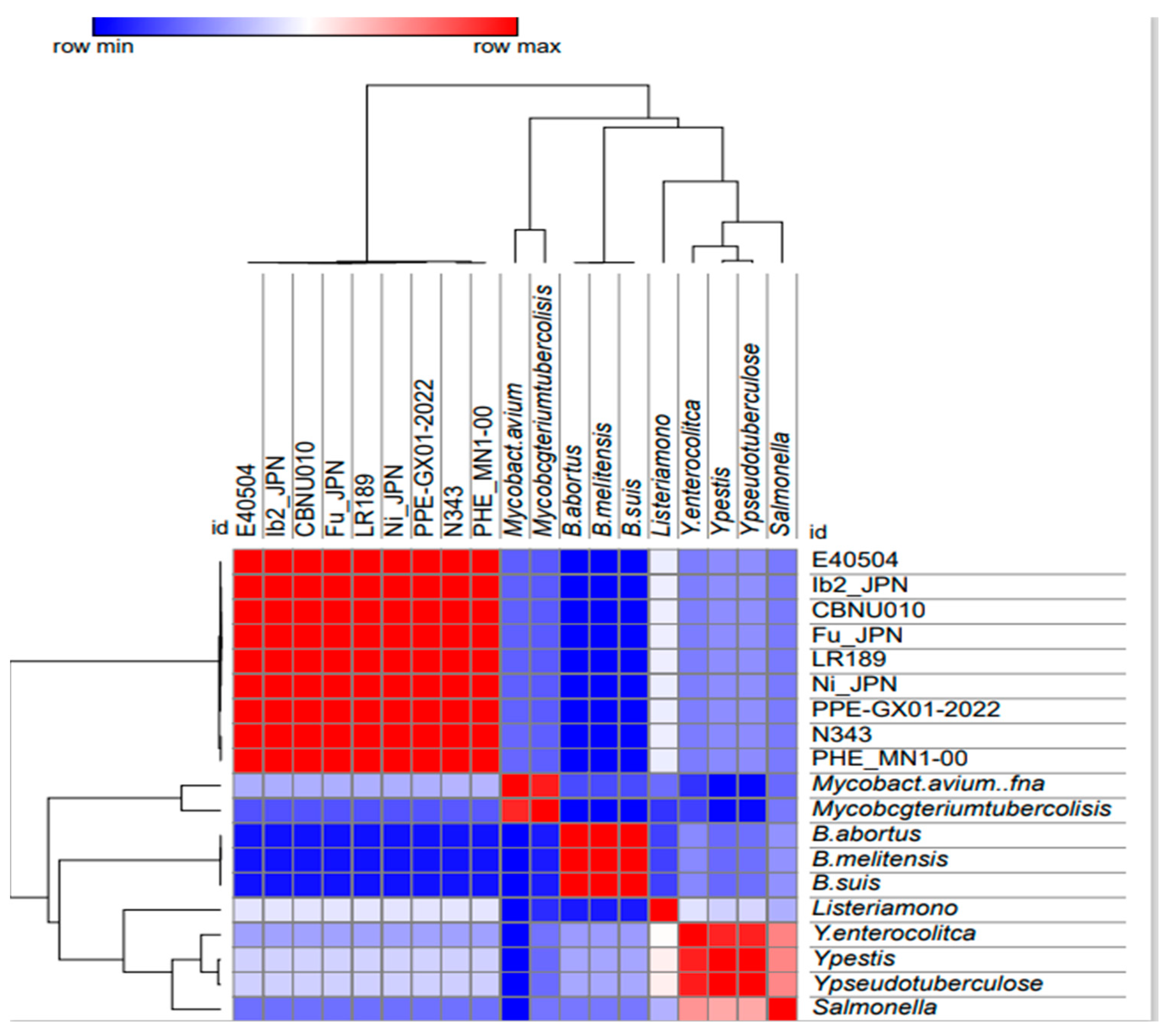
3.1. Similarity Assessment of L. intracellularis Genomes in GenBank

Ten genome assemblies were found for the L. intracellularis genome in GenBank, with one of them not publicly assessable. Therefore, nine genomes were used for subsequent analyses, with the genomes PHE_MN1-00 (USA), N343 (USA), and PPE-GX01-2022 (China) as complete assemblies and six as draft assemblies: Ib2_JPN (Japan), E40504 (Equine-USA), CBNU010 (Korea), Ni_JPN (Japan), LR189 (United Kingdom), and Fu_JPN (Japan). Even considering the incomplete genomes, a very high percentage of similarity was observed between them, to the point of being considered clonal (Supplementary Table S1, Figure 1).

3.2. L. intracellularis Orthologous Genes to Genes Associated with Pathogenesis in Other Intracellular Bacteria

Given the high similarity among the nine L. intracellularis genomes annotated in the system, only two complete genomes (PHE_MN1-00 and N343) and one draft genome (E40504) were chosen for searching orthologous genes. Considering these three genomes, an initial screening found the same orthologous genes associated with pathogenicity of different intracellular bacteria in all of them, and thereafter, the genome identified as PHE_MN1-00 became the reference genome used for the subsequent analyses (Figure 2).
Fifty-two genes previously described as important in bacterial invasion were used for the orthology evaluation. Of these 52 genes, 18 were identified in the PHE_MN1-00 genome and within them, 7 belonged to S. enterica serovar Typhimurium, 5 belonged to Yersinia sp., 4 to Brucella sp., 1 to L. monocytogenes, and 1 to Mycobacterium sp. Three orthologous genes were found in more than one bacterium (Table 1).
Orthologous invasion genes and their functions are presented in Supplementary Table S2.3. Of the total invasion orthologous genes (52), 39% were identified in L. intracellularis (PHE_MN1-00) to be orthologous to S. enterica serovar Typhimurium, while Mycobacterium sp. and L. monocytogenes had a lower proportion of orthology, with 5% and 6% of orthologous genes, respectively (Figure 3).
As for the characterized genes important for intracellular survival, 75 genes were evaluated, of which a total of 27 genes were orthologous to PHE_MN1-00. Among these 27 genes, 14 were present in more than one bacterium species. Twelve L. intracellularis genes were found to be exclusively orthologous to Brucella sp., ten exclusively with S. enterica serovar Typhimurium, five with Mycobacterium sp., six with Yersinia sp., and two with L. monocytogenes (Table 2, Figure 4). Orthologous genes related to survival and their functions are presented in Supplementary Table S2.4.

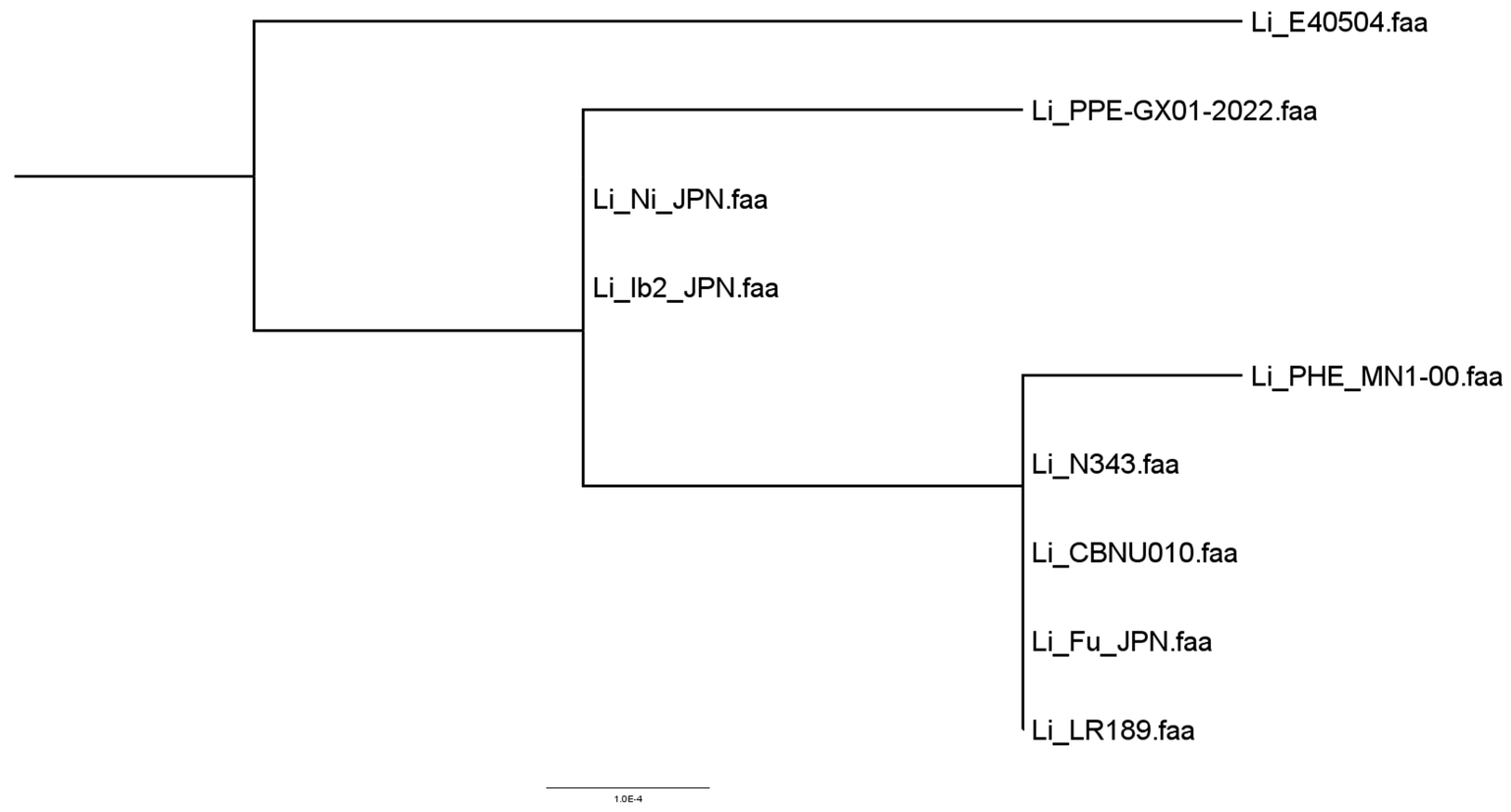
3.3. Results of Concatenated and Phylogenetically Analyzed Genes

From 1422 orthogroups predicted by the program, the phylogenomic tree was constructed, and the paired and multiple alignments of the sequences are presented (Figure 5). Also presented is the sequence alignment result for a set of 15 invasion genes extracted from the genome of the PHE_MN1-00 strain. The genetic sequences were incorporated into an ad hoc database and used for a comparative analysis with all strains under investigation (Figure 6).

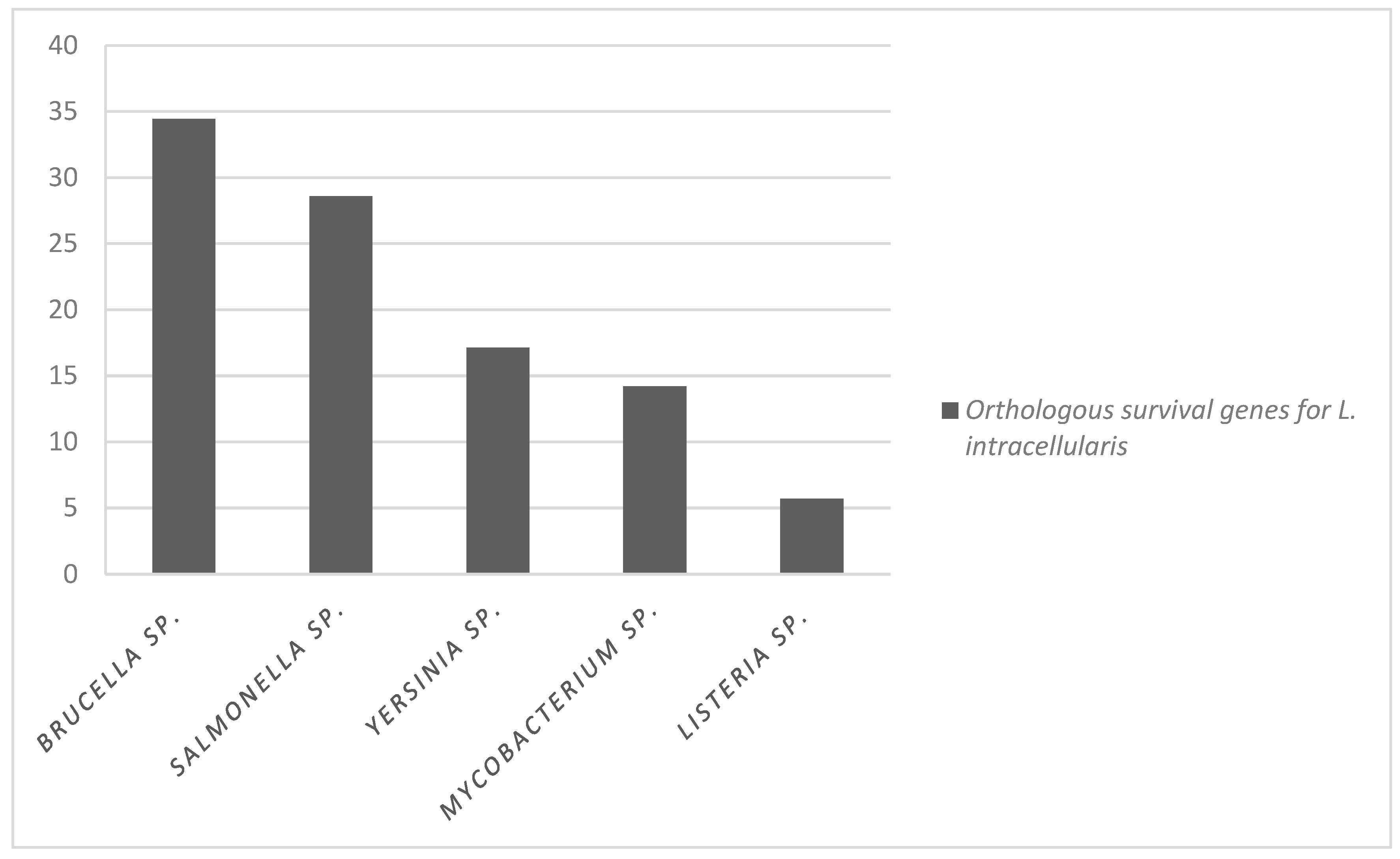
3.4. Comparison Results of Orthologous Genes Observed in Other Enteropathogens Related to Genes Expressed in Pathogenic and Non-Pathogenic Strains of Lawsonia intracellularis

Based on data published in the article by Vannucci et al. [15], comparisons of associated orthologous genes of pathogenesis in other enteropathogens and observed in L. intracellularis was carried out with genes that were expressed or not in pathogenic L. intracellularis and attenuated L. intracellularis. When comparing the orthologous invasion genes and the genes expressed by the pathogenic and non-pathogenic strains, one orthologous gene expressed in the pathogenic L. intracellularis, two orthologous genes expressed in the attenuated L. intracellularis, and one gene expressed in both variants were identified. The orthologous gene expressed in pathogenic L. intracellularis corresponds to the cheW gene, which encodes a chemotaxis signal transduction protein involved in the transmission of sensory signals from chemoreceptors to flagellar motors. This gene is present in S. enterica serovar Typhimurium.
Two orthologous genes were observed in non-pathogenic L. intracellularis: sctN (yscN) and the flgK gene. The sctN gene encodes a yscN type III ATPase secretion system. This component, also called injectosome, is used to inject bacterial effector proteins into eukaryotic host cells and is described in S. enterica serovar Typhimurium and Yersinia sp. The flgK gene, which encodes a protein associated with the flagellar hook and functions to aid cell motility, was observed in S. enterica serovar Typhimurium.
Finally, it was observed that the groEL gene was highly expressed by the pathogenic and attenuated variants of L. intracellularis. This gene encodes proteins that help adherence to other proteins and are involved in the association of bacteria with macrophages, in addition to acting as an adhesin, binding to CD43 on the surface of the host macrophage [21]. These genes are observed in Mycobacterium tuberculosis (Table 3).
For intracellular survival orthologous genes, eight were found to be expressed at a high level in the chromosome of pathogenic L. intracellularis, none of which were highly expressed in non-pathogenic L. intracellularis. The genes observed were ribH, livH, rplW, hypA, sfsA, recO, fur, and rpoN, being considered important genes such as ABC transporters of amino acids, in addition to being important proteins in DNA binding and repair, or acting in the regulation of iron and zinc, important in oxidation reduction (Table 4).
Considering the results presented here, we can see three candidate genes to be considered for future studies, the genes bvrR, cpdR, and phoQ, as they are genes that regulate the expression of mechanisms involved in virulence and adaptation to acidic environments, which is why we created a table, presenting the exact nucleotide positions of the proposed genes from the reference genome of L. intracelullaris (Table 5).

4. Discussion

Due to its intracellular nature and restrictive in vitro growth conditions, studies aiming to investigate L. intracellularis molecular pathogenesis are very scarce. In this in silico study, the presence of genes involved in the invasion and cell survival of better understood enteroinvasive bacterial pathogens were found to be orthologous to L. intracelullaris, several of which with well demonstrated functions. The reference bacteria of the present study were Brucella abortus, B. melitensis, B. suis, Listeria monocytogenes, Mycobacterium tuberculosis, M. avium subspecies paratuberculosis, S. enterica serovar Typhimurium, Yersinia pestis, Y. enterocolitica, and Y. pseudotuberculosis, bacteria that have already shown different molecular mechanisms used to invade the host cell and survive in it.
Comparing all nine available genomes of L. intracellularis amongst each other, a very high percentage of similarity was observed to the point of considering them clonal, as previously found by Bengston et al. [5]. In this study, metagenomic sequencing of clinical samples was carried out, where comparative genomic and phylogenetic analyses of the population structure of L. intracellularis revealed a genetically monomorphic clone responsible for infections in swine and distinct subtypes associated with infections in horses [5].
Based on previous research data, we collected information on all genes of interest, both invasion and survival, and these were noted in a database formed and mapped against the predicted proteomes of the nine L. intracellularis strains publicly available on the NCBI platform [16], aiming to determine the possible orthology relationship between the genes available in the ad hoc database and the proteomes of L. intracellularis strains. As a result, 18 orthologous invasion genes were found, out of a total of 52 genes submitted for evaluation. Among the 18 genes, the sctN gene is one of the most notable. It is present in S. enterica serovar Typhimurium, Y. enterocolitica, and Y. pseudotuberculosis, making it orthologous to L. intracellularis. This gene is an ATPase component of the type III secretion system (T3SS), called injectosome, which is used to inject bacterial effector proteins into host cells, which favors the alteration of several cellular processes. The expression of this gene was demonstrated for the first time by Alberdi et al. [22], where they detected that the T3SS components of L. intracellularis are expressed during infection.
flgK (flaS, flaW) and flhA genes, both necessary for the formation of the rod structure of the flagellar apparatus, were also found. These genes constitute part of the flagellin export apparatus, important in cell motility, as observed in S. enterica serovar Typhimurium, which expresses this protein in the membrane-bound compartment. Flagellin is translocated to the cytoplasm of the host cell, where it is detected by cytosolic receptors that can mount an innate immune response [23,24,25]. In L. intracellularis, the presence of a unipolar flagellum has already been demonstrated extracellularly in cultured organisms [4], but there are no further studies on its functionality in infection. However, expressions of this gene were observed in both the pathogenic and attenuated L. intracellularis strains [4]. Therefore, although this may be a gene related to its virulence, we hypothesize that it is not its absence or presence that determines the ability of L. intracellularis to cause proliferative enteropathy.
The cheW gene, present in L. intracellularis and S. enterica serovar Typhimurium, encodes a receptor kinase coupling protein, called che. che is involved in the transmission of sensory signals from bacterial chemoreceptors to flagellar motors; so, these chemotactic signaling systems allow bacteria to track favorable chemical gradients in the environment [26,27]. This gene, due to its functions, could favor L. intracellularis to track the gradients of both attractive and repellent chemo effectors and to move towards ideal environments for its invasion.
L. intracellularis gene bvrR is also present in B. abortus. This gene, which is part of a two-component regulatory system, controls cell invasion and intracellular survival, playing a role in controlling the bacterial surface and interactions with the host cell, being conclusively implicated in the virulence of Brucella. Studies have shown that bvrR/bvrS mutants of Brucella are avirulent in mice, although they have reduced invasiveness in cells and are unable to inhibit lysosome fusion and replicate intracellularly, an important fact during the escape of Brucella from the host cell response. Furthermore, when there is a bvrR/bvrS dysfunction, there is a decrease in Brucella’s characteristic resistance to bactericidal polycations and an increase in its permeability to surfactants [28,29]. However, this gene was not observed with high expression in the pathogenic isolate nor the non-pathogenic isolate of L. intracellularis [14]. Therefore, the expression of this gene by L. intracellularis strains should be studied under experimental conditions different from those tested by Vannucci et al. [14].
GroEL 1 and 2 genes, present in Mycobacterium sp., were highly expressed by pathogenic and non-pathogenic variants of L. intracellularis. These genes encode molecular chaperones, a group of envelope proteins involved in processes that assist in the folding of other proteins. Some studies have shown its performance in the association of bacteria with macrophages, in addition to acting as an adhesin, binding to CD43 on the surface of the host macrophage [30]. Furthermore, the full-length groEL protein 1 and 2 induce pro-inflammatory responses in dendritic cells (DCs), promoting their maturation and antigen presentation to T cells. When DCs are exposed to the GroEL protein, they induce strong antigen interferon gamma responses, specifically IFN-gamma, interleukin-2 (IL-2), and IL-17A of CD4+ T cells [31]. All this information is of great importance considering that the direct interaction between L. intracellularis and macrophages has already been demonstrated and that L. intracellularis can survive the phagolysosomal environment of macrophages [32]. Therefore, these genes could initially favor the association and adhesion of L. intracellularis to the host’s macrophages.
The phoQ gene (phoZ), also present in S. typhimurium, regulates the expression of genes involved in virulence, adaptation to acidic environments and low Mg2+ content, and resistance to antimicrobial defense peptides of the host. Furthermore, this gene has a negative regulatory function for the prgH gene, which is necessary for the invasion of epithelial cells and is also involved in bacterial tolerance to acidic media, essential for the intra-macrophage survival of S. Typhimurium [33,34]. This gene could be one of the genes that help in the tolerance of L. intracellularis for acidic environments, as the presence of free L. intracellularis was observed in the cytoplasm of macrophages [32].
Regarding the intracellular survival genes of bacteria, among the 75 genes identified in the bacterial species used as reference, 27 genes orthologous to PHE_MN1-00 were observed. Of these 27 genes, 8 were highly expressed in L. intracellularis, both in the pathogenic and non-pathogenic (attenuated) isolate [14]. These genes are responsible for encoding proteins mainly related to cell signaling and molecular biosynthesis, among which the proteins LivH, SfsA, and RecO stand out. LivH is a protein that is part of the binding protein-dependent transport system, responsible for the translocation of substrates across the bacterial membrane, while SfsA is a protein with a DNA-binding function and recO is a DNA repair protein. The genes encoding these three proteins were identified in all bacteria selected as references for this study [35,36,37,38,39], which indicates its high potential for involvement with intracellular and pathogenic bacteria.
Other interesting genes identified in the genome of L. intracellularis as highly expressed in the pathogenic isolate are in orthology with the genome of other bacteria used as references, namely, rplW, ribH, fur, and rpoN. The rplW gene encodes an assembly protein that forms the main docking site for the binding of the triggering factor to the ribosome, encoded by the ribH gene, which acts in the penultimate step in riboflavin biosynthesis. The fur gene determines the production of the ferric uptake regulatory protein, and rpoN is related to bacterial adaptation and stress response [15]. All these genes could be favoring the adaptation of L. intracellularis to the intracellular environment and response to stress as observed in other bacteria [40].
Regarding the analysis of intracellular survival genes, it was observed that of the eight orthologous genes expressed in the pathogenic isolate of L intracellularis, and six were orthologous to S. enterica serovar Typhimurium. This represents 75% of the expressed genes and suggests the genetic proximity of L. intracellularis and S. enterica serovar Typhimurium, indicating that many of Salmonella intracellular survival mechanisms could be observed in L. intracellularis.
Other interesting findings in relation to intracellular survival genes were those of the cpdR gene. This gene is part of the two-component system, which encodes a response regulatory receptor protein, which fulfills the important function of regulating and controlling growth, intracellular division, and survival of B. abortus within mammalian host cells [41]. The rsh gene, in turn, functions as an essential protein for intracellular growth and expression of the type IV secretion system (virB), playing a role in the adaptation of Brucella to its intracellular environment [42]. The presence of cpdR gene, thus, suggests that L. intracellularis might use the same pathways for its growth and adaptation to the intracellular environment.
Evaluating all 20 orthologous intracellular survival genes in this study, whether found or not, was highly expressed by L. intracellularis [15], and a higher proportion (34%) of orthologous genes with Brucella sp. was observed. In this evaluation, it was observed that L. intracellularis presented more genes forming orthology with mechanisms used by Brucella for its intracellular survival than with the other bacteria in the comparison. The genes expressed in L. intracellularis pathogenic and Brucella sp. were the genes ribh, recO, rplw, and fur.
Genes important for chemotaxis, cell motility, DNA binding and repair, and association of bacteria with macrophages or inducers of pro-inflammatory responses, as observed in the present study, provide new target genes to be further studied about regarding L. intracellularis pathogenesis. Studying the evolutionary history of organisms based on the use of mathematical methods helps us to deduce the past of the analyzed species, considering the identification of homologues between different organisms [43].
When evolutionary events occur, such as vertical descent, gene duplication, and gene loss, among others, they usually mark the history of genes and are the main events in genomic evolution. Thus, when a divergence occurs after a speciation event, the relationship between the sequences occurs, and this is what we call orthology [43]. Bringing in L. intracellularis orthology information is important for the advancement of our understanding of which genes this bacterium and a common ancestor to other intracellular pathogenic bacteria share and whether they have the same functions, bringing to light different mechanisms of pathogenesis likely involved in L. intracellularis pathogenesis.

5. Conclusions

Through an in silico evaluation, the present study is the first to provide a comparison of the genomic orthology for L. intracellularis, i.e., the list of homologous or duplicated genes from a common ancestor that could be shared between well-known enteroinvasive intracellular bacteria when compared to L. intracellularis. Based on the results presented here, the main candidates to be considered for future studies would be the genes bvrR, cpdR, and phoQ, as they are genes that regulate the expression of mechanisms involved in virulence and adaptation to acidic environments. New studies could indicate their importance in the virulence and invasion of epithelial cells, due to their involvement in bacterial tolerance, favoring their survival in acidic environments within macrophages.

Supplementary Materials

The following supporting information can be downloaded at: https://www.mdpi.com/article/10.3390/microorganisms12081596/s1, Table S1. Similarity results between Lawsonia intracellularis genomes annotated in Genbank; Table S2.1. List of genes select for invasion comparison; Table S2.2. List of genes selected for intracellular survival comparison; Table S2.3. Orthologous invasion genes and their functions; Table S2.4. Orthologous survival genes and their functions.

Author Contributions

Conceptualization, M.E.S.-D., F.F.A. and R.M.C.G.; Methodology, M.E.S.-D., R.L.S., C.E.R.P., T.P.R., M.D.A., J.C.R.B., F.F.A. and R.M.C.G.; Formal analysis, M.E.S.-D., M.D.A., D.L.N.R. and F.F.A.; Investigation, P.A.C., R.P.L. and D.L.N.R.; Data curation, M.E.S.-D., D.L.N.R. and F.F.A.; Writing—original draft, M.E.S.-D.; Writing—review & editing, R.L.S., C.E.R.P., T.P.R. and R.M.C.G.; Supervision, F.F.A. and R.M.C.G.; Project administration, R.M.C.G. All authors have read and agreed to the published version of the manuscript.

Funding

This work was funded by FAPEMIG, CAPES, and CNPq. RMCG and RLS are beneficiaries of grants from CNPq, Brazil. MESD was supported by the Capes Foundation, Ministry of Education, Brazil.

Data Availability Statement

The original contributions presented in the study are included in the article and Supplementary Materials, further inquiries can be directed to the corresponding author.

Acknowledgments

We thank the Laboratory of Integrative Bioinformatics, department of Preventive Veterinary Medicine, UFMG.

Conflicts of Interest

The authors declare no conflicts of interest.

References

  1. McOrist, S.; Gebhart, C.J.; Boid, R.; Barns, S.M. Characterization of Lawsonia intracellularis gen. nov., sp. nov., the obligately intracellular bacterium of porcine proliferative enteropathy. Int. J. Syst. Bacteriol. 1995, 45, 820–825. [Google Scholar] [CrossRef] [PubMed]
  2. Rowland, A.C.; Lawson, G.H. Intestinal adenomatosis in the pig: Immunofluorescent and electron microscopic studies. Res. Vet. Sci. 1974, 17, 323–330. [Google Scholar] [CrossRef] [PubMed]
  3. Lawson, G.H.K.; McOrist, S.; Jasni, S.; Mackie, R.A. Intracellular bacteria of porcine proliferative enteropathy: Cultivation and maintenance in vitro. J. Clin. Microbiol. 1993, 31, 1136–1142. [Google Scholar] [CrossRef] [PubMed]
  4. Lawson, G.H.; Gebhart, C.J. Proliferative enteropathy. J. Comp. Pathol. 2000, 122, 77–100. [Google Scholar] [CrossRef] [PubMed]
  5. Bengtsson, R.J.; Wee, B.A.; Yebra, G.; Bacigalupe, R.; Watson, E.; Guedes, R.M.C.; Jacobson, M.; Stadejek, T.; Archibald, A.L.; Fitzgerald, J.R.; et al. Metagenomic sequencing of clinical samples reveals a single widespread clone of Lawsonia intracellularis responsible for porcine proliferative enteropathy. Microb. Genom. 2020, 6, e000358. [Google Scholar] [CrossRef] [PubMed]
  6. Cooper, D.M.; Swanson, D.L.; Barns, S.M.; Gebhart, C.J. Comparison of the 16S ribosomal DNA sequences from the intracellular agents of proliferative enteritis in a hamster, deer, and ostrich with the sequence of a porcine isolate of Lawsonia intracellularis. Int. J. Syst. Bacteriol. 1997, 47, 635–639. [Google Scholar] [CrossRef] [PubMed]
  7. Levy, M.; Thaiss, C.A.; Zeevi, D.; Dohnalová, L.; Zilberman-Schapira, G.; Mahdi, J.A.; David, E.; Savidor, A.; Korem, T.; Herzig, Y.; et al. Microbiota-Modulated Metabolites Shape the Intestinal Microenvironment by Regulating NLRP6 Inflammasome Signaling. Cell 2015, 163, 1428–1443. [Google Scholar] [CrossRef] [PubMed]
  8. Kaur, K.; Zhang, Q.; Beckler, D.; Munir, S.; Li, L.; Kinsley, K.; Herron, L.; Peterson, A.; May, B.; Singh, S.; et al. The Complete Genome Sequence of Lawsonia intracellularis: The Causative Agent of Proliferative Enteropathy. Available online: https://www.ncbi.nlm.nih.gov/nuccore/AM180252.1 (accessed on 10 July 2022).
  9. Sait, M.; Aitchison, K.; Wheelhouse, N.; Wilson, K.; Lainson, F.A.; Longbottom, D.; Smith, D.G.E. Genome sequence of Lawsonia intracellularis strain N343, isolated from a sow with hemorrhagic proliferative enteropathy. Genome Announc. 2013, 1, 1–2. [Google Scholar] [CrossRef] [PubMed]
  10. Oliveira, A.R.G.C. Differentiation of Lawsonia intracellularis Isolates from Different Animal Species Using Pulsed Field Gel Electrophoresis (PFGE). U.S. Patent Application No. 2014/0017268 A1, 16 January 2014. [Google Scholar]
  11. Sampieri, F.; Vannucci, F.A.; Allen, A.L.; Pusterla, N.; Antonopoulos, A.J.; Ball, K.R.; Thompson, J.; Dowling, P.M.; Hamilton, D.L.; Gebhart, C.J. Species-specificity of equine and porcine Lawsonia intracellularis isolates in laboratory animals. Can. J. Vet. Res. 2013, 77, 261–272. [Google Scholar]
  12. Vannucci, F.A.; Beckler, D.; Pusterla, N.; Mapes, S.M.; Gebhart, C.J. Attenuation of virulence of Lawsonia intracellularis after in vitro passages and its effects on the experimental reproduction of porcine proliferative enteropathy. Vet. Microbiol. 2013, 162, 265–269. [Google Scholar] [CrossRef]
  13. Worley, M.J. Immune evasion and persistence in enteric bacterial pathogens. Gut Microbes 2023, 15, 2163839. [Google Scholar] [CrossRef] [PubMed]
  14. Vannucci, F.A.; Pusterla, N.; Mapes, S.M.; Gebhart, C. Evidence of host adaptation in Lawsonia intracellularis infections. Vet. Res. 2012, 43, 53. [Google Scholar] [CrossRef] [PubMed]
  15. Vannucci, F.A.; Foster, D.N.; Gebhart, C.J. Comparative Transcriptional Analysis of Homologous Pathogenic and Non-Pathogenic Lawsonia intracellularis Isolates in Infected Porcine Cells. PLoS ONE 2012, 7, e46708. [Google Scholar] [CrossRef] [PubMed]
  16. Pruitt, K.D.; Tatusova, T.; Maglott, D.R. NCBI reference sequences (RefSeq): A curated non-redundant sequence database of genomes, transcripts and proteins. Nucleic Acids Res. 2007, 35, D61–D65. [Google Scholar] [CrossRef] [PubMed]
  17. Seemann, T. Prokka: Rapid prokaryotic genome annotation. Bioinformatics 2014, 30, 2068–2069. [Google Scholar] [CrossRef] [PubMed]
  18. Pritchard, L.; Glover, R.; Humphris, S.; Elphinstone, J.; Toth, I. Genomics and Taxonomy in Diagnostics for Food Security: Soft-rotting enterobacterial plant pathogens. Anal. Methods 2015, 8, 12–24. [Google Scholar] [CrossRef]
  19. Bateman, A.; Martin, M.J.; Orchard, S.; Magrane, M.; Agivetova, R.; Ahmad, S.; Alpi, E.; Bowler-Barnett, E.H.; Britto, R.; Bursteinas, B.; et al. UniProt: The universal protein knowledgebase in 2021. Nucleic Acids Res. 2021, 49, D480–D489. [Google Scholar] [CrossRef]
  20. Emms, D.M.; Kelly, S. OrthoFinder: Phylogenetic orthology inference for comparative genomics. Genome Biol. 2019, 20, 238. [Google Scholar] [CrossRef] [PubMed]
  21. Hickey, T.B.M.; Ziltener, H.J.; Speert, D.P.; Stokes, R.W. Mycobacterium tuberculosis employs Cpn60.2 as an adhesin that binds CD43 on the macrophage surface. Cell Microbiol. 2010, 12, 1634–1647. [Google Scholar] [CrossRef]
  22. Alberdi, M.P.; Watson, E.; McAllister, G.E.; Harris, J.D.; Paxton, E.A.; Thomson, J.R.; Smith, D.G. Expression by Lawsonia intracellularis of type III secretion system components during infection. Vet. Microbiol. 2009, 139, 298–303. [Google Scholar] [CrossRef]
  23. Homma, M.; DeRosier, D.J.; Macnab, R.M. Flagellar hook and hook-associated proteins of Salmonella typhimurium and their relationship to other axial components of the flagellum. J. Mol. Biol. 1990, 213, 819–832. [Google Scholar] [CrossRef] [PubMed]
  24. Inoue, Y.; Kinoshita, M.; Kida, M.; Takekawa, N.; Namba, K.; Imada, K.; Minamino, T. The FlhA linker mediates flagellar protein export switching during flagellar assembly. Commun. Biol. 2021, 4, 646. [Google Scholar] [CrossRef] [PubMed]
  25. Minamino, T.; Iino, T.; Kutuskake, K. Molecular characterization of the Salmonella typhimurium flhB operon and its protein products. J. Bacteriol. 1994, 176, 7630–7637. [Google Scholar] [CrossRef] [PubMed]
  26. Hazelbauer, G.L.; Falke, J.J.; Parkinson, J.S. Bacterial chemoreceptors: High-performance signaling in networked arrays. Trends Biochem. Sci. 2008, 33, 9–19. [Google Scholar] [CrossRef] [PubMed]
  27. Stock, A.; Mottonen, J.; Chen, T.; Stock, J. Identification of a possible nucleotide binding site in CheW, a protein required for sensory transduction in bacterial chemotaxis. J. Biol. Chem. 1987, 262, 535–537. [Google Scholar] [CrossRef] [PubMed]
  28. Freer, E.; Moreno, E.; Moriyón, I.; Pizarro-Cerdá, J.; Weintraub, A.; Gorvel, J.P. Brucella-Salmonella lipopolysaccharide chimeras are less permeable to hydrophobic probes and more sensitive to cationic peptides and EDTA than are their native Brucella sp. counterparts. J. Bacteriol. 1996, 178, 5867–5876. [Google Scholar] [CrossRef] [PubMed]
  29. López-Goñi, I.; Guzmán-Verri, C.; Manterola, L.; Sola-Landa, A.; Moriyón, I.; Moreno, E. Regulation of Brucella virulence by the two-component system BvrR/BvrS. Vet. Microbiol. 2002, 90, 329–339. [Google Scholar] [CrossRef] [PubMed]
  30. Hickey, T.B.M.; Thorson, L.M.; Speert, D.P.; Daffé, M.; Stokes, R.W. Mycobacterium tuberculosis Cpn60.2 and DnaK are located on the bacterial surface, where Cpn60.2 facilitates efficient bacterial association with macrophages. Infect. Immun. 2009, 77, 3389–3401. [Google Scholar] [CrossRef] [PubMed]
  31. Georgieva, M.; Sia, J.K.; Bizzell, E.; Madan-Lala, R.; Rengarajan, J. Mycobacterium tuberculosis GroEL2 Modulates Dendritic Cell Responses. Infect. Immun. 2018, 86. [Google Scholar] [CrossRef] [PubMed]
  32. Pereira, C.E.R.; Resende, T.P.; Armién, A.G.; Laub, R.P.; Vannucci, F.A.; Santos, R.L.; Gebhart, C.J.; Guedes, R.M.C. Survival of Lawsonia intracellularis in porcine peripheral blood monocyte-derived macrophages. PLoS ONE 2020, 15, e0236887. [Google Scholar] [CrossRef]
  33. Miller, S.I.; Kukral, A.M.; Mekalanos, J.J. A two-component regulatory system (phoP phoQ) controls Salmonella typhimurium virulence. Proc. Natl. Acad. Sci. USA 1989, 86, 5054–5058. [Google Scholar] [CrossRef] [PubMed]
  34. Sanowar, S.; Le Moual, H. Functional reconstitution of the Salmonella typhimurium PhoQ histidine kinase sensor in proteoliposomes. Biochem. J. 2005, 390 Pt 3, 769–776. [Google Scholar] [CrossRef] [PubMed]
  35. Anderson, P.E.; Matsunaga, J.; Simons, E.L.; Simons, R.W. Structure and regulation of the Salmonella typhimurium rnc-era-recO operon. Biochimie 1996, 78, 1025–1034. [Google Scholar] [CrossRef] [PubMed]
  36. Chain, P.S.G.; Carniel, E.; Larimer, F.W.; Lamerdin, J.; Stoutland, P.O.; Regala, W.M.; Georgescu, A.M.; Vergez, L.M.; Land, M.L.; Motin, V.L.; et al. Insights into the evolution of Yersinia pestis through whole-genome comparison with Yersinia pseudotuberculosis. Proc. Natl. Acad. Sci. USA 2004, 101, 13826–13831. [Google Scholar] [CrossRef]
  37. DelVecchio, V.G.; Kapatral, V.; Redkar, R.J.; Patra, G.; Mujer, C.; Los, T.; Ivanova, N.; Anderson, I.; Bhattacharyya, A.; Lykidis, A.; et al. The genome sequence of the facultative intracellular pathogen Brucella melitensis. Proc. Natl. Acad. Sci. USA 2002, 99, 443–448. [Google Scholar] [CrossRef]
  38. Glaser, P.; Frangeul, L.; Buchrieser, C.; Rusniok, C.; Amend, A.; Baquero, F.; Berche, P.; Bloecker, H.; Brandt, P.; Chakraborty, T.; et al. Comparative genomics of Listeria species. Science 2001, 294, 849–852. [Google Scholar] [CrossRef]
  39. Zheng, H.; Lu, L.; Wang, B.; Pu, S.; Zhang, X.; Zhu, G.; Shi, W.; Zhang, L.; Wang, H.; Wang, S.; et al. Genetic basis of virulence attenuation revealed by comparative genomic analysis of Mycobacterium tuberculosis strain H37Ra versus H37Rv. PLoS ONE 2008, 3, e2375. [Google Scholar] [CrossRef]
  40. Bagg, A.; Neilands, J.B. Ferric uptake regulation protein acts as a repressor, employing iron (II) as a cofactor to bind the operator of an iron transport operon in Escherichia coli. Biochemistry 1987, 26, 5471–5477. [Google Scholar] [CrossRef]
  41. Willett, J.W.; Herrou, J.; Briegel, A.; Rotskoff, G.; Crosson, S. Structural asymmetry in a conserved signaling system that regulates division, replication, and virulence of an intracellular pathogen. Proc. Natl. Acad. Sci. USA 2015, 112, E3709–E3718. [Google Scholar] [CrossRef]
  42. Dozot, M.; Boigegrain, R.-A.; Delrue, R.-M.; Hallez, R.; Ouahrani-Bettache, S.; Danese, I.; Letesson, J.-J.; De Bolle, X.; Köhler, S. The stringent response mediator Rsh is required for Brucella melitensis and Brucella suis virulence, and for expression of the type IV secretion system virB. Cell Microbiol. 2006, 8, 1791–1802. [Google Scholar] [CrossRef]
  43. Snel, B.; Huynen, M.A.; Dutilh, B.E. Genome trees and the nature of genome evolution. Annu. Rev. Microbiol. 2005, 59, 191. [Google Scholar] [CrossRef] [PubMed]
Figure 1. A heatmap of the analysis was carried out to show the percentage of similarity, demonstrating very high similarity between the annotated L. intracellularis genomes.
Microorganisms 12 01596 g001
Figure 2. A heatmap (pangenome) of the analysis was performed to show the percentage of similarity between the annotated genomes of L. intracellularis against the genomes of the other bacteria in the analysis.
Microorganisms 12 01596 g002
Figure 3. The proportion of orthologous invasion genes common between intracellular bacteria and L. intracellularis PHE_MN1-00 of the total of 52 genes.
Microorganisms 12 01596 g003
Figure 4. The proportion of orthologous survival genes common between species of intracellular bacteria with L. intracellularis PHE-MN-00.
Microorganisms 12 01596 g004
Figure 5. Phylogenomic tree with pairwise and multiple alignments of the sequences of all orthologous genes extracted from the L. intracellularis genome.
Microorganisms 12 01596 g005
Figure 6. Phylogenomic tree with alignment of invasion gene sequences extracted from the L. intracellularis genome.
Microorganisms 12 01596 g006
Table 1. Orthologous genes in different intracellular bacteria were found in the analysis of the occurrence of invasion genes of L. intracellularis.
Locus_TagSpeciesGeneDescription
PHE_MN1-00_00947Salmonella enterica serovar TyphimuriumsctN1 (invC, spaI, spaL)SPI-1 ATPase of type 3 secretion system
PHE_MN1-00_00595Yersinia pseudotuberculosis serotype I and Yersinia enterocoliticasctN (yscN)Type 3 secretion system ATPase
PHE_MN1-00_00605Yersinia enterocoliticaLCDLow calcium response locus protein D
PHE_MN1-00_00585Yersinia enterocoliticaflhAFlagellar biosynthesis protein FlhA
PHE_MN1-00_01288PHE_MN1-00_00596Yersinia pseudotuberculosis serotype IsctL (lcrKC, yscL)Type 3 secretion system stator protein
PHE_MN1-00_01265Brucella abortusbvrRFlagellar transcriptional regulator FtcR
PHE_MN1-00_00693Mycobacterium avium sub paratuberculosis, Mycobacterium tuberculosis.groEL1,2 (groL2, hsp65, mopA).Chaperonin GroEL 1,2
PHE_MN1-00_01294Salmonella enterica sorovar TyphimuriumcheWCheW protein chemotaxis
PHE_MN1-00_00296Brucella melitensis. Listeria monocytogenes serovar 1/2arhoRho transcription termination factor
PHE_MN1-00_00828Salmonella enterica sorovar TyphimuriumflgK (flaS, flaW)Flagellar hook-associated protein 1
PHE_MN1-00_00510Yersinia enterocoliticayadB (gluQ) (unreviewed)Glutamyl-Q tRNA(Asp) synthetase
PHE_MN1-00_00430Salmonella enterica sorovar Typhimuriumrsep (yaeL)RseP sigma-E protease regulator
PHE_MN1-00_00972Salmonella enterica sorovar TyphimuriumphoQ (phoZ)Virulence sensor histidine kinase PhoQ
PHE_MN1-00_01197Brucella abortusbvrS (not revised)Histidine kinase
PHE_MN1-00_01283Salmonella enterica sorovar TyphimuriumsctC2 (spiA, ssaC)SPI-2 type 3 secretion system secretin
Table 2. Bacteria and their orthologous intracellular survival genes found in L. intracellularis.
Locus_Tag.SpeciesGeneDescription
PHE_MN1-00_01458
PHE_MN1-00_00396
Mycobacterium tuberculosis., Salmonella TyphimuriumdnaBReplicative DNA helicase
PHE_MN1-00_00545Yersinia pestis, Salmonella enterica sorovar Typhimurium,
Brucella abortus, Brucella suis biovar 1. Brucella melitensis biotype 1
ssbSingle-stranded DNA-binding protein
PHE_MN1-00_00974Salmonella enterica sorovar Typhimurium. Yersinia pseudotuberculosis serotype IsfsASugar-fermentation-stimulating protein A
PHE_MN1-00_00191Brucella melitensis biotype 1. Brucella abortus. Brucella suis biovar 1rshGTP pyrophosphokinase rsh
PHE_MN1-00_00172Brucella suis biovar 1. Brucella abortus. Brucella melitensis biotype 1, Yersinia enterocolitica.
Mycobacterium tuberculosis. Salmonella enterica sorovar Typhimurium
ribH6,7-dimethyl-8-ribothylumazine synthase 2
PHE_MN1-00_00339Salmonella enterica sorovar Typhimurium. Brucella melitensis biotype 1 Yersinia pseudotuberculosis. Mycobacterium tuberculosis. Listeria monocytogenesrecORecO DNA repair protein
PHE_MN1-00_00988Salmonella enterica sorovar Typhimurium. Brucella melitensis biotype 1lnt (cutE)Apolipoprotein N-acyltransferase
PHE_MN1-00_00378Salmonella TyphimuriumlivHHigh-affinity branched-chain amino acid transport system permease protein
PHE_MN1-00_01265Brucella abortusctrACell cycle response regulator CtrA
PHE_MN1-00_01068Yersinia enterocolitica. Mycobacterium tuberculosis.
Brucella melitensis
rplW50S ribosomal protein L23
PHE_MN1-00_01312Mycobacterium tuberculosiseccA1ESX-1 secretion system protein EccA1
PHE_MN1-00_00581Brucella abortuscpdRCpdR response regulator receptor protein
PHE_MN1-00_00498Brucella melitensis biotype 1pyrGCTP synthase
PHE_MN1-00_00072Mycobacterium tuberculosiseccCa1 (snm1)ESX-1 secretion system protein EccCa1
PHE_MN1-00_00278Salmonella TyphimuriumhypAHypA hydrogenase maturation factor
PHE_MN1-00_01140Salmonella TyphimuriumepmA (genX, yjeA)Elongation factor P--(R)-beta-lysine ligase
PHE_MN1-00_01197Brucella abortuscckACckA sensor kinase
PHE_MN1-00_00036Brucella abortus biovar 1. Brucella melitensis biotype 1 Yersinia pestisfurFerric uptake regulatory protein
PHE_MN1-00_01306Brucella abortusrecARecA protein
PHE_MN1-00_00504Salmonella Typhimurium rpoNRNA polymerase factor sigma-54
Table 3. Comparison of orthologous invasion genes to genes expressed in pathogenic and non-pathogenic Lawsonia intracellularis strains.
Locus TagOrthologous GenesGenes Highly Expressed in
Pathogenic L. intracellularis
Genes Highly Expressed in
Non-Pathogenic L. intracellularis
PHE_MN1-00_01294cheWcheW chemotaxis signal transduction protein____
PHE_MN1-00_00595sctN (yscN)____yscN type III secretion system ATPase
PHE_MN1-00_00828flgK (flaS, flaW)____flgK flagellar hook-associated protein
PHE_MN1-00_00693groEL1 and 2 (groL2, hsp65, mopA).GroEL (Chaperonin)GroEL (Chaperonin)
Table 4. Comparison of orthologous intracellular survival genes to genes expressed in pathogenic and non-pathogenic Lawsonia intracellularis strains.
Locus_Tag.Orthologous GenesGenes Expressed in Pathogenic
L. intracellularis
Genes Expressed in Non-Pathogenic L. intracellularis
PHE_MN1-00_00172ribHribH riboflavin synthase beta-chain____
PHE_MN1-00_00378livHlivH branched-chain amino acid ABC transporter (permease)____
PHE_MN1-00_01068rplWrplW. 50S ribosomal protein L23____
PHE_MN1-00_00278hypAhypA zinc finger protein____
PHE_MN1-00_00974sfsAsfsA DNA-binding protein, stimulates sugar fermentation____
PHE_MN1-00_00339recOrecO DNA repair protein RecO (recombination protein O)____
PHE_MN1-00_00036Furfur Fe2+/Zn2+ uptake regulation proteins____
PHE_MN1-00_00504rpoNrpoN Sigma54-like protein____
Table 5. Results of the exact nucleotide positions of the proposed genes from the reference genome of L. intracelullaris.
StrainAssembly AccessionGeneStrandStartStop
PHE_MN1-00GCF_000055945.1bvrR+14001721400540
PHE_MN1-00GCF_000055945.1cpdR644060644440
PHE_MN1-00GCF_000055945.1phoQ10918271093269
Disclaimer/Publisher’s Note: The statements, opinions and data contained in all publications are solely those of the individual author(s) and contributor(s) and not of MDPI and/or the editor(s). MDPI and/or the editor(s) disclaim responsibility for any injury to people or property resulting from any ideas, methods, instructions or products referred to in the content.

