Article

The Functional Network of PrkC and Its Interaction Proteins in Bacillus subtilis Spores

1
Key Laboratory of Fruits and Vegetables Processing, Ministry of Agriculture, Engineering Research Centre for Fruits and Vegetables Processing, Ministry of Education, National, Engineering Research Center for Fruit and Vegetable Processing, College of Food Science and Nutritional Engineering, China Agricultural University, Beijing 100083, China
2
College of Food Science and Technology, Henan Agricultural University, Zhengzhou 450002, China
*
Author to whom correspondence should be addressed.
Microorganisms 2025, 13(4), 744; https://doi.org/10.3390/microorganisms13040744
Submission received: 11 March 2025 / Revised: 21 March 2025 / Accepted: 24 March 2025 / Published: 26 March 2025
(This article belongs to the Section Food Microbiology)

Abstract

:
In the food industry, food spoilage caused by spores is a pressing scientific challenge that needs to be addressed urgently, and spore germination is a key approach to solving this problem. Studies have shown that peptidoglycan-induced spore germination represents a novel mechanism of action, which can bind to the PASTA domain of the serine/threonine kinase PrkC. However, the signaling mechanism of peptidoglycan-induced spore germination remains unclear. This study focuses on Bacillus subtilis, using pull-down experiments to screen for proteins interacting with PrkC. There are 80 interaction proteins of PrkC that were identified in the spore. GO analysis reveals that PrkC-interacting proteins in the spore are mainly involved in metabolic processes, cell part and catalysis. KEGG results indicate that PrkC-interacting proteins in the spore are mainly involved in RNA degradation, quorum sensing, oxidative phosphorylation, etc. Additionally, proteins are categorized into six groups by function based on events that may be associated with post-germination triggered by peptidoglycan-induced activation of the PrkC signaling pathway, including “stimulate translation initiation” and “ATP synthesis and energy metabolism”. The experimental results provide a theoretical basis for further elucidating the signaling mechanism of PrkC, revealing the signaling pathway of peptidoglycan-induced spore germination, and identifying targeted inducers and repressors of spore germination.

1. Introduction

The spores of Bacillus, as the resting bodies, exhibit no metabolism and possess strong resistance to conditions and substances such as heat, radiation, dryness, and toxic chemicals. They can persist in the environment for extended periods, even years [1]. However, spores sense the environment and respond to favorable conditions by germinating and restoring cell growth [2]. Currently, research on spores primarily focuses on their germination mechanism, as germinated spores lose their resistance and become more susceptible to killing. Many Bacillus spores can cause food spoilage, food poisoning, and human diseases [3,4]. The dormant spores are non-pathogenic, and only synthesize toxins when they germinate and grow into vegetative cells [5]. Therefore, inhibiting spore germination or rapidly inducing germination of all spores holds significant research importance.
When the external environment becomes suitable, nutrients or non-nutrients can be utilized, and the spore exits dormancy to rapidly form a vegetative reproductive body [6]. In this process, spore germination is known as the key step. Spore germination is induced by a series of nutrient molecules, including amino acids, sugars, and cell wall peptides, which bind to receptors on the spore inner membrane and interact to activate downstream signaling substances, guiding spore resuscitation [7]. In the spore germination pathway, germination is induced mainly by nutrients and non-nutrients.
Previous studies have shown that spores possess a non-nutrient germination pathway through the PrkC protein kinase, which located on the inner membrane of B. subtilis spores can induce spore germination through binding to peptidoglycan (PGN) [8]. This germination pathway exists in the genera Bacillus and Clostridium, and is a new signal transmission pathway that is relatively independent of nutritional germination. Studies have shown that ΔprkC spores still responded to the nutrient germinant L-alanine and to the chemical germinant Ca2+-dipicolinic acid (Ca-DPA). Spores with low expression levels of germination receptors (GRs), activated by L-alanine and AGFK (a mixture of l-asparagine, d-glucose, d-fructose, and potassium ions), can germinate via the PrkC pathway [9]. This indicates that during the germination process of spores, the PrkC receptor germination pathway is independent and can work together with the traditional nutritional receptor germination pathway. Although superdormant spores cannot be induced to germinate by GRs, they can be induced to germinate by PrkC receptors [10]. The PrkC-mediated germination pathway only a single identified receptor, represents a promising and tractable target for controlling spore outgrowth in food industry applications. Therefore, the study of the mechanism of action of the PrkC germination pathway is of great significance, which can improve the mechanism of spore germination and provide a theoretical basis for more effective spore killing in food industry.
PGN is a crucial cell wall polymer for bacteria, consisting of N-acetylglucosamine (GlcNac) and N-acetylmuramic acid (MurNar) sugar chains linked by β-1,4 glycosidic bonds and connected by short peptides [11]. Depending on the third amino acid in its peptide chain, PGN is classified into Lys-type and meso-diaminopimelic acid (DAP)-type. The muropeptide fragment of PGN can bind to the PASTA (consisting of 3 β sheets and an α helix) [12] domain of the serine/threonine kinase PrkC, activating the intracellular kinase functional domain and inducing spore germination [8,13]. Notably, it is the DAP-type muropeptide, rather than the Lys-type muropeptide, that plays a role [8]. The recognition process of PrkC for DAP-type peptidoglycan fragments is semi-mediated by DAP. DAP-type peptidoglycan can interact with an arginine (Arg) residue located at position 500 on the PASTA domain of the C-terminal of PrkC. Furthermore, mutation of the arginine residue completely inhibits the binding of muropeptide to PrkC [13]. However, only a few studies have reported molecules downstream of PrkC-mediated spore germination. The elongation factor G (EF-G) in B. subtilis possesses ribosomal GTPase activity and can be phosphorylated by PrkC, thereby affecting the germination process of spores, and this modification is likely necessary for germination in response to PG. Elongation Factor Tu (Ef-Tu) also as a substrate of PrkC and phosphorylated by PrkC [14,15]. In Bacillus anthracis spores, the enolase (Eno) is phosphorylated by the conserved PrkC which decreases the catalytic activity of Eno, thereby controlling the overall spore germination process [16]. Another study showed that phosphoglycerate mutase (Pgm) is also regulated by PrkC phosphorylation, which is important for spore germination [17]. The specific details of the downstream signaling pathway mediated by PrkC are currently unclear. Therefore, this study will use pull-down experiments to screen interaction proteins of PrkC kinase in spores, and establish the interaction network of PrkC kinase. These results could provide reference for revealing the mechanism of PrkC protein in inducing the spore germination pathway.

2. Materials and Methods

2.1. Strain and Plasmid

The Bacillus subtilis 168 and PY79 strains are preserved in our laboratory. The prkC gene was amplified from Bacillus subtilis str. 168 genomic DNA using primers PrkC-F (5′-CAGCAAATGGGTCGCCTCGAGGTGCTAATCGGCAAGCGGATCAGC-3′) and PrkC-R (5′-GTGGTGGTGGTGGTGGCGGCCGCTTCATCTTTCGGATACTCAATGGT-3′). Both the purified PCR product and pGEX-4T-1 vector were digested with NcoI and XhoI. Ligate prkC gene and plasmid by Gibson assembly, and validated by Sanger sequencing.
B. subtilis 168 were cultured in LB broth medium until an optical density at 600 nm (OD600) of 1–2. Spores were grown for around 1 day on DSM medium and purified using the Histodenz gradient centrifugation method [18]. Spores were kept at −20 °C.

2.2. Induced Expression of PrkC Protein

The correctly sequenced plasmids were transferred into E. coli expression strain BL21, respectively. Positive monoclonal plaques were selected and inoculated into 5 mL of LB medium containing the corresponding antibiotics. The culture was incubated overnight at 37 °C and 220 rpm. The bacterial solution was inoculated into 200 mL of LB liquid medium containing the corresponding antibiotics at a volume ratio of 1:100, and incubated at 37 °C and 220 rpm until the OD600 reached 0.6–0.8. IPTG was added to the bacterial solution to a final concentration of 0.1 mM, and the culture was induced at 22 °C for 6–7 h. The bacterial solution was transferred to a new centrifuge tube, centrifuged for 10 min at 4 °C, and the supernatant was discarded. A buffer solution (100 mM Tris-HCl pH 7.5, 150 mM NaCl) was added to wash the bacterial cells, which were then centrifuged at 5000× g for 10 min at 4 °C, and the supernatant was discarded.

2.3. Purification of PrkC Protein

The precipitate was resuspended in 3 mL of lysis buffer (100 mM Tris-HCl pH 7.5, 150 mM NaCl, 1 mM PMSF). Following complete resuspension, ultrasonic disruption was performed at an output power of 270 W using 6–8 cycles of 20 s pulses interspersed with 40 s cooling intervals. The lysate was centrifuged at 12,000× g for 30 min at 4 °C, and the supernatant was transferred to a fresh centrifuge tube. For His-tagged protein purification, a Chelating Sepharose Fast Flow nickel affinity chromatography column was employed. Briefly, 2 mL of resin slurry was packed into a column and equilibrated with 10 mL of lysis buffer. The clarified supernatant was loaded onto the equilibrated column and incubated with the resin at 4 °C for 2 h. The flow-through was collected, and the column was sequentially washed with 10 column volumes of wash buffer (100 mM Tris-HCl pH 7.5, 150 mM NaCl, 50 mM imidazole) to remove nonspecifically bound impurities. Target protein elution was achieved using elution buffer (100 mM Tris-HCl pH 7.5, 150 mM NaCl, 300 mM imidazole), with five 2-mL fractions collected. Protein concentrations in the eluted fractions were quantified via Bradford assay (Coomassie Brilliant Blue G-250), and purification efficiency was assessed by SDS-PAGE analysis (12% resolving gel, Coomassie R-250 staining).

2.4. Pull-Down Experiment

Fusion-expressed GST empty-vector and GST-PRKC bacteria were obtained, treated with PDS solution, vortexed, and briefly centrifuged. After removal of the supernatant, the pellet was rinsed, resuspended in lysis buffer containing protease inhibitors, and lysed by ultrasonication. The lysate was centrifuged, and the supernatant was collected as the input control. Pre-equilibrated glutathione resin was incubated with different protein mixtures to assess interaction specificity. GST resin was incubated with spore lysate as a negative control for nonspecific binding, GST-PRKC resin was incubated with lysis buffer only to confirm that binding requires spore proteins, and GST-PRKC resin was incubated with spore lysate as the experimental group. After incubation at 4 °C, spores were lysed separately under similar conditions, and their lysate was collected. Following incubation, the resin was washed thoroughly to remove unbound proteins, and bound proteins were eluted using 20 mM freshly prepared reduced glutathione. The eluate was stored at −80 °C as the pull-down product. A portion was mixed with loading buffer, heated, and analyzed by Western blot.

2.5. Western Blot Analysis

The input and pull-down products were mixed with an appropriate volume of 4× loading buffer and incubated in a boiling water bath for 10 min. The mixture was centrifuged at 12,000× g for 3 min at 4 °C, and the supernatant was subjected to Western blot analysis. Electrophoresis was performed at a constant current of 16 mA per gel until the bromophenol blue tracking dye migrated to the bottom of the gel. Following electrophoresis, the gel was carefully removed, and a transfer sandwich was assembled for electroblotting. Protein transfer was conducted at a constant voltage of 100 V for 1 h in an ice-water-cooled electrotransfer tank. Upon completion of transfer, the membrane was retrieved and placed in separate antibody incubation chambers. Blocking was performed by incubating the membrane with 5% (w/v) skimmed milk powder or 2% (w/v) bovine serum albumin (BSA) in 1 × TBST for 1 h on a horizontal shaker. After discarding the blocking solution, the membrane was briefly rinsed with 1 × TBST and transferred to an antibody incubation tray. The GST-specific primary antibody (1:6000 dilution in 1 × TBST) was applied, and the membrane was incubated overnight at 4 °C. The primary antibody was removed, and the membrane was washed three times with 1 × TBST (10 min per wash). A horseradish peroxidase (HRP)-conjugated secondary antibody (1:8000 dilution in 1 × TBST) was then added, followed by incubation at room temperature for 2 h. After three additional TBST washes (10 min each), residual buffer was absorbed using filter paper. The membrane was exposed to X-ray film in a light-tight cassette for signal detection.

2.6. LC–MS/MS Analysis

The collected protein solution was digested with trypsin to generate peptide segments, which were then dried. Each dried peptide sample was re-dissolved in a sample solution containing 0.1% formic acid and 2% acetonitrile and subjected to LC–MS/MS analysis with a 100-min LC gradient for protein identification and label-free quantification (LFQ). Mass spectrometry (MS) data acquisition was performed using an Orbitrap Fusion Lumos Tribrid Mass Spectrometer (Thermo Fisher Scientific, San Jose, CA, USA) coupled to an Ultimate 3000 RSLCnano UHPLC system Peptide trapping was conducted on an Acclaim PepMap 100 nanoViper column (75 μm × 20 mm) at a 5 µL/min flow rate for 5 min, followed by separation on an Acclaim PepMap RSLC nanoViper column (75 μm × 500 mm, C-18, 2 μm, 100 Å, Thermo Fisher) at 300 nL/min and 40 °C. The mobile phases consisted of 0.1% formic acid in water (solvent A) and 0.1% formic acid in acetonitrile (solvent B). A multi-step linear gradient was applied as follows: 5% B (0–4 min), 5% B (5 min), 50% B (45 min), 90% B (50 min), 90% B (55 min), and re-equilibration to 5% B (65 min). A data-dependent acquisition (DDA) strategy was employed with 3 s cycles, utilizing specific MS scan parameters. Full MS scans were performed in the Q Exactive analyzer at 70,000 resolving power to precisely determine peptide mass-to-charge ratios (m/z), Scan range: 350–1800 m/z. MS2 analysis targeted precursor ions at 70,000 resolving power. Selected peptide ions were isolated via quadrupole (0.7 m/z window), fragmented using high-energy collision dissociation (HCD, 27% normalized collision energy). Additionally, a dynamic exclusion window of 30 s was applied to improve spectral acquisition.

2.7. MS Data Analysis and Peptide Identification

Acquired raw MS data were analyzed using the Bacillus subtilis UniProt database (strain-specific entries) with Sequest HT (Proteome Discoverer 2.5) and MaxQuant (v2.0.3). Full tryptic peptides were searched with up to two missed cleavages allowed, and fixed modifications included phosphorylation, ubiquitination, methylation, acetylation, and glycosylation. Data were filtered for precursor ions with a signal-to-noise (S/N) ratio of at least 1.5, and peptide spectrum matches (PSMs) were refined using a delta Cn threshold of 0.05.
To control the false discovery rate (FDR), q-values were calculated at the PSM level using Percolator and subsequently at the peptide level using the Qvality algorithm. For protein identification, explicit statistical criteria were applied: candidate proteins were validated using Sum PEP protein scores and accepted as significant if they met a strict FDR threshold of 0.01, with an alternative relaxed threshold of 0.05 provided in order not to filter proteins.

2.8. Bioinformatics Analysis

Identified proteins were annotated using DAVID (v6.8) with Gene Ontology (GO) terms (biological process, molecular function, cellular component, and only result FDR p < 0.05 and raw p values < 0.05). Proteins were functionally annotated and categorized using the SubtiWiki database. (https://subtiwiki.uni-goettingen.de/, accessed on 15 February 2025). Pathway enrichment analysis was subsequently conducted via the KEGG database (https://www.kegg.jp/, accessed 20 January 2025).

3. Results

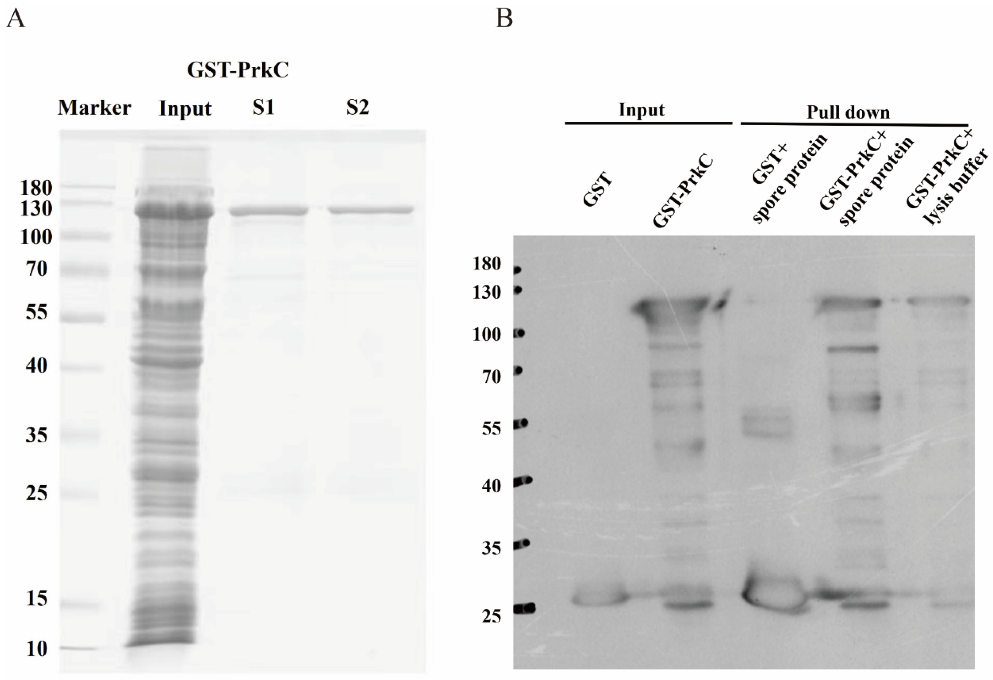
3.1. In Vitro Expression and Purification of PrkC Protein

A prokaryotic expression vector of GST-tagged PrkC protein was constructed, and GST-PrkC protein was purified (Figure 1A). The total proteins of B. subtilis spores were extracted, hybridized with purified GST-PrkC protein, and the proteins bound to PrkC protein were collected using GST antibody protein (Figure 1B). Then, the collected proteins were analyzed by liquid chromatography-mass spectrometry (LC–MS), and the data of these proteins were compared to analyze the signaling pathways and biological functions involved in them.

3.2. Screening of PrkC-Interacting Proteins in B. subtilis Spores

The LC–MS data were analyzed using ProteomeDiscoverer 2.4 against a target-decoy database of B. subtilis 168 (UniProt taxonomy ID 224308) with stringent filtering (1% FDR, ≥2 unique peptides). The data showed that in addition to PrkC protein, there were 79 interacting proteins in the spore (Table 1). Gene and protein names were annotated in the table.

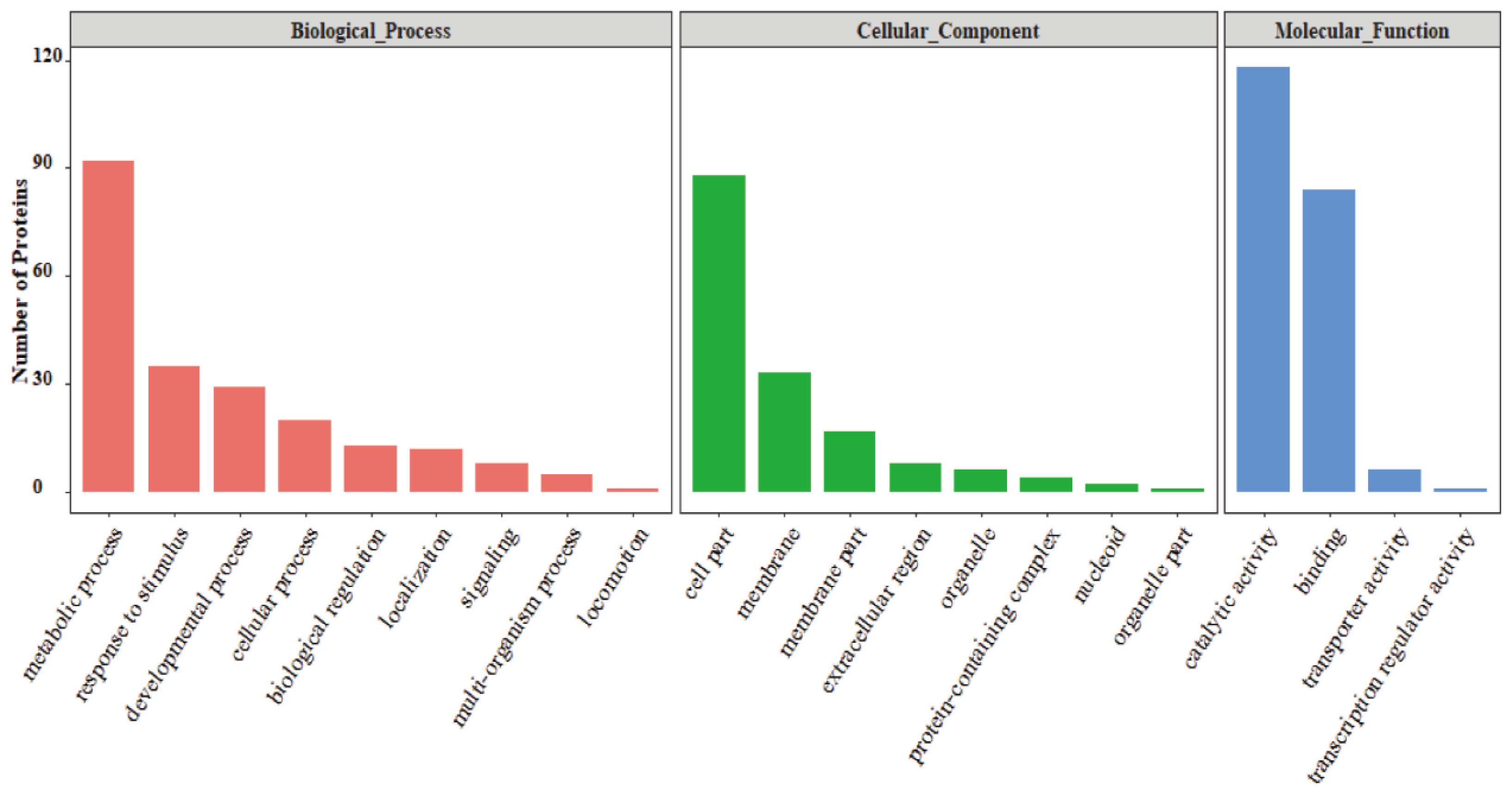
3.3. GO Analysis of PrkC-Interacting Proteins in B. subtilis Spores

To determine what biological characteristics these interacting proteins have, we performed GO analysis on the identified interacting proteins in the spores. The results showed (Figure 2) that the PrkC-interacting proteins in the spores were involved in numerous molecular pathways. The GO functional annotation mainly includes biological processes (BP), molecular functions (MF), and cellular components (CC). In biological processes, the interacting proteins in spore are involved in metabolic processes, stress responses, developmental processes, cellular processes, biological regulation, localization, signaling pathways, multi-tissue processes and movement. Among them, there are more PrkC-interacting proteins involved in the metabolic process in the spore, including energy metabolism, nucleotide metabolism, and biosynthesis (categorized via SubitiWiki), which may be related to the restoration of physiological activity after germination. In cellular component, enriched in cell part, membrane and membrane part. PrkC localizes to the spore inner membrane and have a membrane-spanning sequence. Membrane proteins are more likely to interact with PrkC under dormant conditions. Molecular function analysis indicated that catalytic activity and binding described the majority of proteins in all four comparisons. The detailed functional classification analysis is systematically elaborated in the Discussion below.

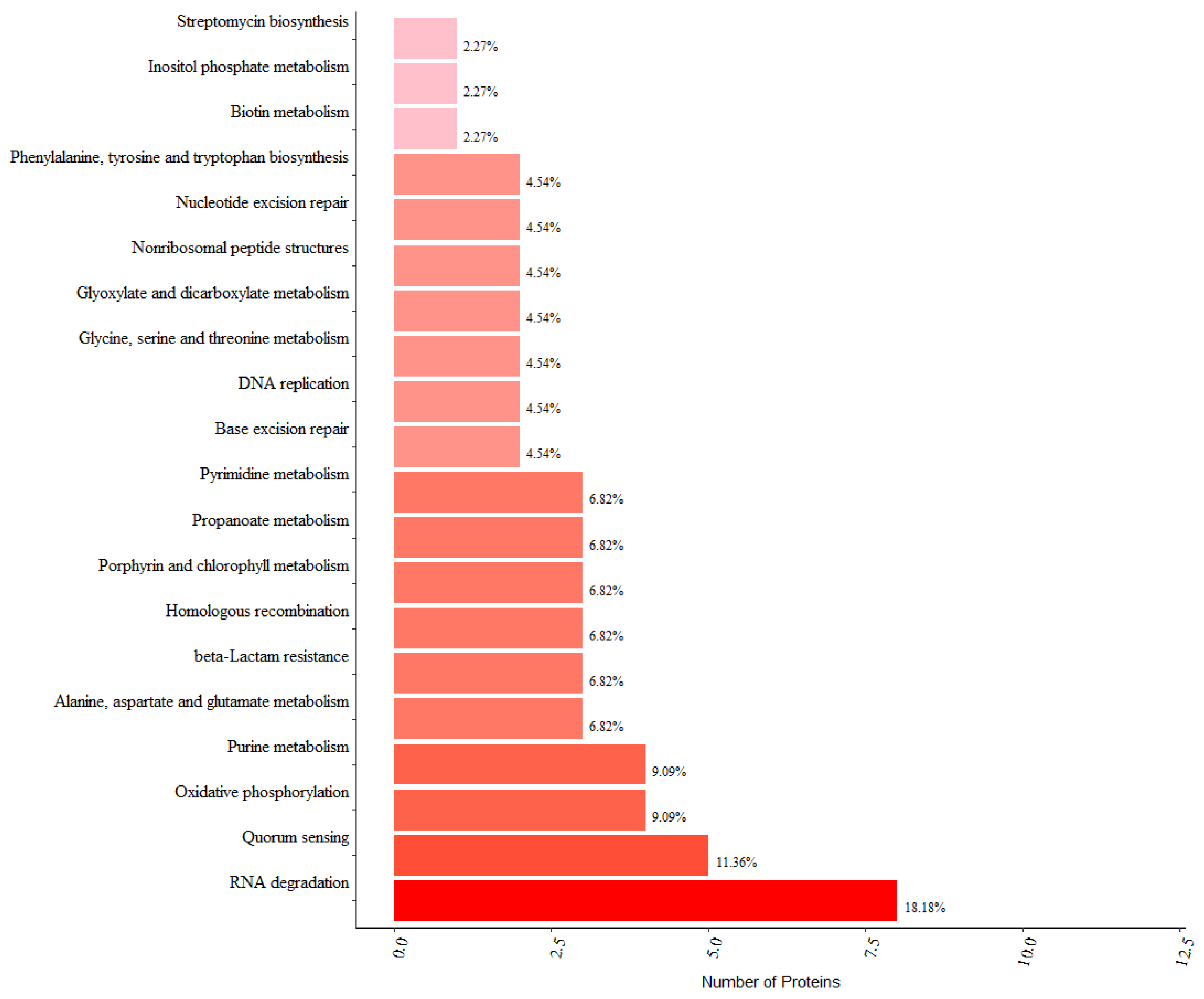
3.4. KEGG Pathway Analysis and Protein–Protein Interaction Analysis of PrkC-Interacting Proteins in B. subtilis Spores

In order to deeply analyze the mechanism of action of PrkC-interacting proteins, KEGG pathway analysis was carried out. The results showed (Figure 3) that in spores, interaction proteins of PrkC kinase are mainly involved in RNA degradation, population effect, oxidative phosphorylation, etc. More pathways are directed to metabolic processes such as amino acid metabolism and biotin metabolism. Consistent with GO’s analysis, suggesting that metabolism is important in this process. Association analysis of PrkC-interacting proteins in spores showed that prkC had direct interactions with ftsW, sbcC, pksJ and dnaK, respectively (Figure 4A). These interactions may occur more in vegetative cells or in the process of sporulation. Through optimizing the protein interaction network by removing non-central proteins and those functionally unrelated to dormant spore recovery activity, we identified recA and dnaK interacting with prkC. And these two also had some direct interactions, and both of them could interact with Tuf protein, while RecA protein could also interact with SrfA1 protein (Figure 4B). The functions of RecA and DnaK proteins have been extensively studied in Bacillus subtilis; however, their roles during spore germination remain poorly characterized. Both DNA repair and chaperone-mediated folding of newly synthesized proteins are important processes for restoring physiological activity following spore germination. These proteins may regulate the process of spore germination and thus deserve further investigation.

4. Discussion

Previous studies showed that PrkC mutant spores could not germinate under peptidoglycan-induced conditions, while GRs mutant spores could germinate, indicating that PrkC and GRs receptors were relatively independent in the spore germination pathway. In food sterilization, the two can interact with each other, which may produce a better sporicidal effect, and this result has been confirmed by the previous study of our group [19].Therefore study the downstream mechanism of PrkC-mediated germination is important for the guidance of food sterilization of spores. In this study, pull-down assay was used to screen the interaction protein of PrkC protein in the spores of B. subtilis.
The identification and mapping of protein interactions are critical for elucidation of their role in unraveling signaling pathways, metabolic networks, and regulatory cascades [20]. There are two main approaches for detecting interacting proteins, one measuring direct physical interactions and the second measuring interactions among groups of proteins. Co-immunoprecipitation (Co-IP) and pull-down assays are commonly employed to investigate proteomic interactions [21]. However, in the absence of specific protein antibodies, pull-down assays can serve as in vitro screening tools to initially identify unknown protein–protein interactions.
In spore germination, interaction proteins of PrkC kinase can be involved. Previous studies have shown that peptidoglycan can bind to the PASTA region of PrkC protein and activate downstream signaling molecules, so that the spore germination signal can be transmitted and promote spore germination. Functional annotation of the detected proteins reveals that the events that may be associated with post-germination triggered by peptidoglycan-induced activation of the PrkC signaling pathway can be categorized into the following categories. (i) “stimulate translation initiation”. Previous studies have found that the ribosomal GTPase EF-G is phosphorylated by PrkC, which stimulates translation of dormant spores and initiates spore germination. EF-G catalyzes the ribosome translocation of the mRNA–tRNA complex from the A and P sites to the P and E sites. Before that, aminoacyl-tRNA (aa-tRNA) is transported to the A site of the ribosome by the GTPase EF-Tu [22]. EF-Tu and EF-G exhibit sequential and essential functional coordination during ribosomal cycling. Moreover, in vitro PrkC specifically phosphorylates EF-Tu on T384 was previously reported [15]. The identification of the interaction of tuf (EF-Tu) with PrkC in our data suggests that PrkC-stimulated ribosomal activation is critical for spore germination. Several studies have found that dormant cells contain large amounts of mRNA and ribosomes [23,24]. Our data support that translation stimulation might be a fundamental mechanism of exit from dormancy [24]. (ii) “peptidoglycan metabolism”. Peptidoglycan cortex-degrading enzymes are typically released during the early phase of spore germination. Notably, we detected the presence of peptidoglycan synthesis-related proteins (e.g., PbpF, PdaC, and YrrL) [25,26] that may be activated by PrkC. A large PrkC-dependent effect of the padC gene has been demonstrated in vegetative cells [27]. We also detected a protein involved in the process of peptidoglycan peptide recovery, NagZ [28]. But no proteins associated with peptidoglycan degradation. Spore cortex-lytic enzymes essential for germination, such as SleB, CwlJ have been alternately demonstrated localized to the outer cortex (B) or the spore coat (J). They cannot directly interact with PrkC localized on the inner membrane. Thus, the signaling molecules for peptidoglycan degradation in the cortex after germination may not interact directly with PrkC. The mechanism inducing cortex degradation during the second stage of germination may be similar to nutrient-induced germination. (iii) “ATP synthesis and energy metabolism”. The NADH formation, oxygen uptake, and RNA synthesis were initiated immediately after germination [29]. We detected several proteins associated with energy metabolism (e.g., Ldh, AcoL, PrpB, YvoA, and Gdh) [30,31] and ATP synthase AtpA. PrkC has been shown to phosphorylate several metabolic enzymes in vitro [32], and initiation of metabolic processes with scarce resources in the spore is a prerequisite for spore germination. With the progress of germination, the spore requires substantial metabolic supplements. Multiple substrates may be involved in energy metabolism. Notably, Gdh is a glucose dehydrogenase that is directly involved in glucose metabolism and has the possibility of providing direct energy during the early metabolism of germination. Although studies have shown that neither of the two major glucose catabolic pathways is required for triggering Bacillus subtilis spore germination [30], Gdh may be important for energy provision. (iv) “nucleotide metabolism”. Accompanied by nucleotide metabolism in the early stages of germination [33]. We similarly detected a number of proteins involved in nucleotide metabolism (e.g., PurF and PyrAB). (v) “DNA repair”. Oxidative stress due to full hydration of the spore core during germination and activation of metabolism in spore outgrowth can lead to the production of reactive oxygen species (ROS) [34]. So DNA repair proteins in the spore may be extremely important in spore germination. We detected some DNA repair functional proteins including PolA, RecA, YprA (MrfA), YycJ, and SbcC. (vi) “ABC transporter”. During the early stages of spore germination, the involvement of transporter proteins is typically limited due to energy constraints. However, in the later stages of germination, numerous transporter proteins are recruited to facilitate the restoration of metabolic activity [33,35]. We detected ABC transporter AppD, YdbJ, and MntB. AppD, an oligopeptide transporter, may be activated by PrkC and potentially involved in protein transport processes during the late stages of spore germination.
The IM channel for Ca-DPA, composed of multiple SpoVA proteins (A, C, D, Eb, Ea, and F), opens to trigger a rapid release of Ca-DPA following spore germination [36]. Previous studies have suggested that SpoVAD acts as a similar cytoplasmic plug in the DPA transport complex [37]. During nutrient-induced germination, germinant receptor (GR) activation initiates cation efflux, which subsequently activates Ca-DPA channel opening [38]. SpoVAC acts as a mechanosensitive channel and allows the release of Ca-DPA and amino acids [39]. However, the mechanism by which muropeptide and PrkC binding leads to downstream Ca-DPA release remains unclear. We detected SpoVAD protein interacting with PrkC, though phosphorylation of SpoVAD has not been reported. Moreover, we detected YqhR (recently designated FigP), which oligomerizes with SpoVAF to form ion channels and serves as an essential cofactor for 5AF activity [40]. Protein phosphorylation, a critical signal transduction process, can induce conformational changes and plays a significant role in regulating spore germination [41,42]. Further studies are required to determine whether PrkC directly phosphorylates DPA channel components.
We also identified several spore-specific proteins, including SpoIIIE, YhcQ, CotI, and CotA. SpoIIIE is known to mediate chromosome translocation during sporulation [43], and its role in germination has not been reported. YhcQ, CotI and CotA are located on the spore coat. Notably, CotI is known as a bacterial spore kinase (BSK). They may be able to interact with specific substrates [44]. The tight association between some BSKs and glycosyl transferases and predicted nucleotide sugar metabolizing enzymes suggests that they may bind or phosphorylate one of these reactants [44]. Furthermore, as the earliest protein to contact with the signaling molecule, spore coat proteins could play a role in the inward transmission of the signaling molecule. However, this interaction should not occur on germination because of differences in localization in the spore. Additionally, we detected several proteins of particular interest in bacterial spores, such as the heat shock protein and molecular chaperone DnaK. Chaperones have been shown to participate in protein folding during early germination stages [45]. And DnaK is phosphorylated and dephosphorylated by PtkA and PtpZ in vegetative cells, respectively [46]. DnaK may also have a function in regulating protein quality in spore formation and germination.
The mechanism by which muropeptide binding to PrkC activates downstream germination events remains largely enigmatic, and our study offers critical clues to unravel this process.

5. Conclusions

In summary, this study successfully identified key proteins interacting with PrkC in Bacillus subtilis spores through pull-down assays, mass spectrometry, and comprehensive bioinformatics analyses. The findings revealed that these interacting proteins are involved in critical biological processes, such as metabolic process and are also enriched in many metabolic pathways. They were further categorized into six functional groups associated with post-germination events triggered by the peptidoglycan-induced PrkC signaling pathway, including “stimulate translation initiation”, “peptidoglycan metabolism”, “ATP synthesis and energy metabolism”, “nucleotide metabolism”, “DNA repair”, and “ABC transporter”. In addition, the discussion covers signal mechanisms that may induce Ca-DPA release and several proteins that, while crucial in vegetative cells, have been less extensively studied in the context of spore reactivation. These results enhance the understanding of the mechanisms underlying PrkC-mediated germination and provide a foundation for future investigations into specific signaling pathways.

Author Contributions

Conceptualization, K.M., L.D. and X.H.; methodology, K.M. and T.C; software, K.M. and T.C.; validation, K.M. and Z.Z.; formal analysis, K.M.; investigation, Y.S.; resources, C.F.; writing—original draft, K.M., L.D. and X.H.; writing—review and editing, L.D. and X.H.; visualization, K.M.; supervision, C.F.; project administration, L.D. and X.H.; funding acquisition, C.F. and X.H. All authors have read and agreed to the published version of the manuscript.

Funding

This work was supported by the National Key Research and Development Program of China (2022YFD2100404) and National Natural Science Foundation of China (31601563).

Institutional Review Board Statement

Not applicable.

Informed Consent Statement

Not applicable.

Data Availability Statement

The original contributions presented in this study are included in this article. Further inquiries can be directed to the corresponding author.

Conflicts of Interest

The authors declare no conflicts of interest.

References

  1. Setlow, P. Spores of Bacillus subtilis: Their Resistance to and Killing by Radiation, Heat and Chemicals. J. Appl. Microbiol. 2006, 101, 514–525. [Google Scholar] [PubMed]
  2. Setlow, P. Germination of Spores of Bacillus Species: What we Know and Do Not Know. J. Bacteriol. 2014, 196, 1297–1305. [Google Scholar] [PubMed]
  3. Li, J.; Yang, J.; Xin, W.; Wu, S.; Wang, X.; Wang, C.; Zhang, Z. Inactivation of Bacillus subtilis Spores by a Combination of High-Pressure Thermal Treatment and Potassium Sorbate. Food Microbiol. 2023, 115, 104345. [Google Scholar]
  4. Wells-Bennik, M.; Eijlander, R.T.; Den Besten, H.M.; Berendsen, E.M.; Warda, A.K.; Krawczyk, A.O.; Nierop Groot, M.N.; Xiao, Y.; Zwietering, M.H.; Kuipers, O.P.; et al. Annual Review of Food Science and Technology: Bacterial Spores in Food: Survival, Emergence, and Outgrowth; Doyle, M.P., Klaenhammer, T.R., Eds.; Annual Reviews; PALO ALTO: Santa Clara, CA, USA, 2016; pp. 457–482. [Google Scholar]
  5. Zhang, T.; Gong, Z.; Zhou, B.; Rao, L.; Liao, X. Recent Progress in Proteins Regulating the Germination of Bacillus subtilis Spores. J. Bacteriol. 2025, 207, e0028524. [Google Scholar]
  6. Setlow, P.; Wang, S.; Li, Y. Germination of Spores of the Orders Bacillales and Clostridiales. Annu. Rev. Microbiol. 2017, 71, 459–477. [Google Scholar]
  7. Kohler, L.J.; Quirk, A.V.; Welkos, S.L.; Cote, C.K. Incorporating Germination-Induction Into Decontamination Strategies for Bacterial Spores. J. Appl. Microbiol. 2018, 124, 2–14. [Google Scholar]
  8. Shah, I.M.; Laaberki, M.; Popham, D.L.; Dworkin, J. A Eukaryotic-Like Ser/Thr Kinase Signals Bacteria to Exit Dormancy in Response to Peptidoglycan Fragments. Cell 2008, 135, 486–496. [Google Scholar]
  9. Wei, J.; Shah, I.M.; Ghosh, S.; Dworkin, J.; Hoover, D.G.; Setlow, P. Superdormant Spores of Bacillus Species Germinate Normally with High Pressure, Peptidoglycan Fragments, and Bryostatin. J. Bacteriol. 2010, 192, 1455–1458. [Google Scholar]
  10. Chen, Y.; Ray, W.K.; Helm, R.F.; Melville, S.B.; Popham, D.L. Levels of Germination Proteins in Bacillus subtilis Dormant, Superdormant, and Germinating Spores. PLoS ONE 2014, 9, e957814. [Google Scholar]
  11. Humann, J.; Lenz, L.L. Bacterial Peptidoglycan-Degrading Enzymes and their Impact On Host Muropeptide Detection. J. Innate Immun. 2009, 1, 88–97. [Google Scholar]
  12. Young, T.A.; Delagoutte, B.; Endrizzi, J.A.; Falick, A.M.; Alber, T. Structure of Mycobacterium tuberculosis Pknb Supports a Universal Activation Mechanism for Ser/Thr Protein Kinases. Nat. Struct. Biol. 2003, 10, 168–174. [Google Scholar] [PubMed]
  13. Squeglia, F.; Marchetti, R.; Ruggiero, A.; Lanzetta, R.; Marasco, D.; Dworkin, J.; Petoukhov, M.; Molinaro, A.; Berisio, R.; Silipo, A. Chemical Basis of Peptidoglycan Discrimination by Prkc, a Key Kinase Involved in Bacterial Resuscitation From Dormancy. J. Am. Chem. Soc. 2011, 133, 20676–20679. [Google Scholar] [PubMed]
  14. Arora, G.; Sajid, A.; Arulanandh, M.D.; Misra, R.; Singhal, A.; Kumar, S.; Singh, L.K.; Mattoo, A.R.; Raj, R.; Maiti, S.; et al. Zinc Regulates the Activity of Kinase-Phosphatase Pair (Basprkc/Basprpc) in Bacillus anthracis. Biometals 2013, 26, 715–730. [Google Scholar] [PubMed]
  15. Absalon, C.; Obuchowski, M.; Madec, E.; Delattre, D.; Holland, I.B.; Seror, S.J. Cpga, Ef-Tu and the Stressosome Protein Yezb are Substrates of the Ser/Thr Kinase/Phosphatase Couple, Prkc/Prpc, in Bacillus subtilis. Microbiology 2009, 155, 932–943. [Google Scholar]
  16. Virmani, R.; Sajid, A.; Singhal, A.; Gaur, M.; Joshi, J.; Bothra, A.; Garg, R.; Misra, R.; Singh, V.P.; Molle, V.; et al. The Ser/Thr Protein Kinase Prkc Imprints Phenotypic Memory in Bacillus anthracis Spores by Phosphorylating the Glycolytic Enzyme Enolase. J. Biol. Chem. 2019, 294, 8930–8941. [Google Scholar]
  17. Virmani, R.; Pradhan, P.; Joshi, J.; Wang, A.L.; Joshi, H.C.; Sajid, A.; Singh, A.; Sharma, V.; Kundu, B.; Blankenberg, D.; et al. Phosphorylation-Mediated Regulation of the Bacillus anthracis Phosphoglycerate Mutase by the Ser/Thr Protein Kinase Prkc. Biochem. Biophys. Res. Commun. 2023, 665, 88–97. [Google Scholar]
  18. Pang, Y.; Wu, R.; Cui, T.; Zhang, Z.; Dong, L.; Chen, F.; Hu, X. Proteomic Response of Bacillus subtilis Spores Under High Pressure Combined with Moderate Temperature and Random Peptide Mixture Lk Treatment. Foods 2022, 11, 1123. [Google Scholar] [CrossRef]
  19. Liang, D.; Wang, X.; Wu, X.; Liao, X.; Chen, F.; Hu, X. The Effect of High Pressure Combined with Moderate Temperature and Peptidoglycan Fragments On Spore Inactivation. Food Res. Int. 2021, 148, 110615. [Google Scholar]
  20. Lalonde, S.; Ehrhardt, D.W.; Loque, D.; Chen, J.; Rhee, S.Y.; Frommer, W.B. Molecular and Cellular Approaches for the Detection of Protein-Protein Interactions: Latest Techniques and Current Limitations. Plant J. 2008, 53, 610–635. [Google Scholar]
  21. Velasco-Garcia, R.; Vargas-Martinez, R. The Study of Protein-Protein Interactions in Bacteria. Can. J. Microbiol. 2012, 58, 1241–1257. [Google Scholar]
  22. Agirrezabala, X.; Frank, J. Elongation in Translation as a Dynamic Interaction Among the Ribosome, Trna, and Elongation Factors Ef-G and Ef-Tu. Q. Rev. Biophys. 2009, 42, 159–200. [Google Scholar] [CrossRef] [PubMed]
  23. Belland, R.J.; Zhong, G.M.; Crane, D.D.; Hogan, D.; Sturdevant, D.; Sharma, J.; Beatty, W.L.; Caldwell, H.D. Genomic Transcriptional Profiling of the Developmental Cycle of Chlamydia trachomatis. Proc. Natl. Acad. Sci. USA 2003, 100, 8478–8483. [Google Scholar] [PubMed]
  24. Dworkin, J.; Shah, I.M. Exit From Dormancy in Microbial Organisms. Nat. Rev. Microbiol. 2010, 8, 890–896. [Google Scholar] [CrossRef] [PubMed]
  25. Popham, D.L.; Bernhards, C.B. Spore Peptidoglycan. Microbiol. Spectr. 2015, 3, 157–177. [Google Scholar] [CrossRef]
  26. Yunck, R.; Cho, H.; Bernhardt, T.G. Identification of Mltg as a Potential Terminase for Peptidoglycan Polymerization in Bacteria. Mol. Microbiol. 2016, 99, 700–718. [Google Scholar]
  27. Libby, E.A.; Goss, L.A.; Dworkin, J. The Eukaryotic-Like Ser/Thr Kinase Prkc Regulates the Essential Walrk Two-Component System in Bacillus subtilis. PLoS Genet. 2015, 11, e10052756. [Google Scholar]
  28. Litzinger, S.; Duckworth, A.; Nitzsche, K.; Risinger, C.; Wittmann, V.; Mayer, C. Muropeptide Rescue in Bacillus subtilis Involves Sequential Hydrolysis by β-N-Acetylglucosaminidase and N-Acetylmuramyl-L-Alanine Amidase. J. Bacteriol. 2010, 192, 3132–3143. [Google Scholar]
  29. Sano, K.; Otani, M.; Uehara, R.; Kimura, M.; Umezawa, C. Primary Role of Nadh Formed by Glucose-Dehydrogenase in Atp Provision at the Early Stage of Spore Germination in Bacillus-Megaterium Qm B1551. Microbiol. Immunol. 1988, 32, 877–885. [Google Scholar] [CrossRef]
  30. Sano, K.; Otani, M.; Umezawa, C.; Nakatani, Y. Role of Glucose-Dehydrogenase and Glucose Catabolism in Triggering of Spore Germination in Bacillus-subtilis. Biosci. Biotechnol. Biochem. 1994, 58, 931–933. [Google Scholar]
  31. Ali, N.O.; Bignon, J.; Rapoport, G.; Debarbouille, M. Regulation of the Acetoin Catabolic Pathway is Controlled by Sigma L in Bacillus subtilis. J. Bacteriol. 2001, 183, 2497–2504. [Google Scholar]
  32. Pietack, N.; Becher, D.; Schmidl, S.R.; Saier, M.H., Jr.; Hecker, M.; Commichau, F.M.; Stülke, J. In Vitro Phosphorylation of Key Metabolic Enzymes From Bacillus subtilis: Prkc Phosphorylates Enzymes From Different Branches of Basic Metabolism. J. Mol. Microbiol. Biotechnol. 2010, 18, 129–140. [Google Scholar] [PubMed]
  33. Pospisil, J.; Sax, A.; Hubalek, M.; Krasny, L.; Vohradsky, J. Whole Proteome Analysis of Germinating and Outgrowing Bacillus subtilis 168. Proteomics 2024, 24, e2400031. [Google Scholar] [CrossRef] [PubMed]
  34. Ibarra, J.R.; Orozco, A.D.; Rojas, J.A.; López, K.; Setlow, P.; Yasbin, R.E.; Pedraza-Reyes, M. Role of the Nfo and Exoa Apurinic/Apyrimidinic Endonucleases in Repair of Dna Damage During Outgrowth of Bacillus subtilis Spores. J. Bacteriol. 2008, 190, 2031–2038. [Google Scholar] [PubMed]
  35. Swarge, B.; Abhyankar, W.; Jonker, M.; Hoefsloot, H.; Kramer, G.; Setlow, P.; Brul, S.; de Koning, L.J. Integrative Analysis of Proteome and Transcriptome Dynamics During Bacillus subtilis Spore Revival. Msphere 2020, 5, e00463-204. [Google Scholar]
  36. Francis, M.B.; Sorg, J.A. Dipicolinic Acid Release by Germinating Clostridium difficile Spores Occurs through a Mechanosensing Mechanism. Msphere 2016, 1, e00306-166. [Google Scholar]
  37. Gao, Y.; Barajas-Ornelas, R.D.C.; Amon, J.D.; Ramírez-Guadiana, F.H.; Alon, A.; Brock, K.P.; Marks, D.S.; Kruse, A.C.; Rudner, D.Z. The Spova Membrane Complex is Required for Dipicolinic Acid Import During Sporulation and Export During Germination. Gene Dev. 2022, 36, 634–646. [Google Scholar]
  38. Christie, G.; Setlow, P.; Germination, B.S. Unknowns and What we Need to Learn. Cell Signal 2020, 74, 109729. [Google Scholar]
  39. Velasquez, J.; Schuurman-Wolters, G.; Birkner, J.P.; Abee, T.; Poolman, B. Bacillus subtilis Spore Protein Spovac Functions as a Mechanosensitive Channel. Mol. Microbiol. 2014, 92, 813–823. [Google Scholar] [CrossRef]
  40. Gao, Y.; Amon, J.D.; Brogan, A.P.; Artzi, L.; Ramírez-Guadiana, F.H.; Cofsky, J.C.; Kruse, A.C.; Rudner, D.Z. Spovaf and Figp Assemble Into Oligomeric Ion Channels that Enhance Spore Germination. Gene Dev. 2024, 38, 31–45. [Google Scholar]
  41. Pompeo, F.; Foulquier, E.; Galinier, A. Impact of Serine/Threonine Protein Kinases On the Regulation of Sporulation in Bacillus subtilis. Front. Microbiol. 2016, 7, 568. [Google Scholar]
  42. Zhou, B.; Semanjski, M.; Orlovetskie, N.; Bhattacharya, S.; Alon, S.; Argaman, L.; Jarrous, N.; Zhang, Y.; Macek, B.; Sinai, L.; et al. Arginine Dephosphorylation Propels Spore Germination in Bacteria. Proc. Natl. Acad. Sci. USA 2019, 116, 14228–14237. [Google Scholar] [PubMed]
  43. Lopez-Garrido, J.; Ojkic, N.; Khanna, K.; Wagner, F.R.; Villa, E.; Endres, R.G.; Pogliano, K. Chromosome Translocation Inflates Bacillus Forespores and Impacts Cellular Morphology. Cell 2018, 172, 758. [Google Scholar] [PubMed]
  44. Scheeff, E.D.; Axelrod, H.L.; Miller, M.D.; Chiu, H.; Deacon, A.M.; Wilson, I.A.; Manning, G. Genomics, Evolution, and Crystal Structure of a New Family of Bacterial Spore Kinases. Proteins 2010, 78, 1470–1482. [Google Scholar] [PubMed]
  45. Sinai, L.; Rosenberg, A.; Smith, Y.; Segev, E.; Ben-Yehuda, S. The Molecular Timeline of a Reviving Bacterial Spore. Mol. Cell 2015, 57, 695–707. [Google Scholar]
  46. Shi, L.; Ravikumar, V.; Derouiche, A.; Macek, B.; Mijakovic, I. Tyrosine 601 of Bacillus subtilis Dnak Undergoes Phosphorylation and is Crucial for Chaperone Activity and Heat Shock Survival. Front. Microbiol. 2016, 7, 533. [Google Scholar]
Figure 1. Purification of GST-PrkC and GST pull-down assay. (A) The SDS-PAGE of GST- PrkC. Lane Marker: Protein molecular weight marker; Lane 2: Input control; Lane 3 and 4: Elution once and twice. (B) Proteins were expressed in E. coli BL21(DE3) cells, and bacterial extracts were applied onto a glutathione resin. Empty plasmid GST+spore protein and GST-PrkC+ lysis buffer act as negative controls. Eluted proteins were probed with an anti-GST antibody by Western blotting.
Microorganisms 13 00744 g001
Figure 2. GO analysis of PrkC interaction proteins in B. subtilis spores.
Microorganisms 13 00744 g002
Figure 3. KEGG analysis of PrkC interaction proteins in B. subtilis spores.
Microorganisms 13 00744 g003
Figure 4. The role network diagram of PrkC and its interaction proteins in B. subtilis spores. (A) A protein–protein interaction (PPI) network was reconstructed using the STRING database. (B) Proteins unrelated to germination were pruned. The refined network revealed interaction between PrkC and two proteins.
Microorganisms 13 00744 g004
Table 1. PrkC interaction protein in B. subtilis spores.
UniprotKBcGene NamesProtein Names
P45867acdAAcyl-CoA dehydrogenase (EC 1.3.99.-)
O34324acoLDihydrolipoyl dehydrogenase (EC 1.8.1.4) (Dihydrolipoamide dehydrogenase) (E3 component of acetoin cleaving system)
P42064appDOligopeptide transport ATP-binding protein AppD
O31788aprXSerine protease AprX (EC 3.4.21.-)
P35146aroD3-dehydroquinate dehydratase (3-dehydroquinase) (EC 4.2.1.10) (Type I DHQase) (Type I dehydroquinase) (DHQ1)
P37808atpAATP synthase subunit alpha (EC 7.1.2.2) (ATP synthase F1 sector subunit alpha) (F-ATPase subunit alpha) (Vegetative protein 100) (VEG100)
P07788cotALaccase (EC 1.10.3.2) (Bilirubin oxidase) (EC 1.3.3.5) (Spore coat protein A)
O34656cotISpore coat protein I
P17820dnaKChaperone protein DnaK (HSP70) (Heat shock 70 kDa protein) (Heat shock protein 70)
P51831fabG3-oxoacyl-[acyl-carrier-protein] reductase FabG (EC 1.1.1.100) (3-ketoacyl-acyl carrier protein reductase) (Beta-Ketoacyl-acyl carrier protein reductase) (Beta-ketoacyl-ACP reductase)
O07639ftsWProbable peptidoglycan glycosyltransferase FtsW (PGT) (EC 2.4.99.28) (Cell division protein FtsW) (Cell wall polymerase) (Peptidoglycan polymerase) (PG polymerase)
P54376gcvPAProbable glycine dehydrogenase (decarboxylating) subunit 1 (EC 1.4.4.2) (Glycine cleavage system P-protein subunit 1) (Glycine decarboxylase subunit 1) (Glycine dehydrogenase (aminomethyl-transferring) subunit 1)
P12310gdhGlucose 1-dehydrogenase (EC 1.1.1.47)
O07621hemATHeme-based aerotactic transducer HemAT
P21248hemDUroporphyrinogen-III synthase (UROS) (EC 4.2.1.75) (Hydroxymethylbilane hydrolyase [cyclizing]) (Uroporphyrinogen-III cosynthase)
P13714ldhL-lactate dehydrogenase (L-LDH) (EC 1.1.1.27)
O34338mntBManganese transport system ATP-binding protein MntB
O31667mtnX2-hydroxy-3-keto-5-methylthiopentenyl-1-phosphate phosphatase (HK-MTPenyl-1-P phosphatase) (EC 3.1.3.87)
P40406nagZBeta-hexosaminidase (EC 3.2.1.52) (Beta-N-acetylhexosaminidase) (N-acetyl-beta-glucosaminidase) (N-acetylglucosaminidase) (ORF1)
P38050pbpFPenicillin-binding protein 1F (PBP-1F) (Penicillin-binding protein F) (Peptidoglycan TGase); Penicillin-sensitive transpeptidase (DD-transpeptidase)]
O34798pdaCPeptidoglycan-N-acetylmuramic acid deacetylase PdaC (Peptidoglycan MurNAc deacetylase) (EC 3.5.1.-) (Polysaccharide deacetylase PdaC)
O34819pelBPectin lyase (PNL) (EC 4.2.2.10)
O07617phoEUncharacterized phosphatase PhoE (EC 3.1.3.-)
P40806pksJPolyketide synthase PksJ (PKS)
Q05470pksLPolyketide synthase PksL (PKS)
P40872pksMPolyketide synthase PksM
O34996polADNA polymerase I (POL I) (EC 2.7.7.7)
P39846ppsBPlipastatin synthase subunit B (EC 2.3.1.-) (Peptide synthase 2) [Includes ATP-dependent tyrosine adenylase 1 (TyrA 1) (Tyrosine activase 1); ATP-dependent threonine adenylase (ThrA) (Threonine activase)]
O34507prkCSerine/threonine-protein kinase PrkC (Ser/Thr-protein kinase PrkC) (EC 2.7.11.1)
O31755proSProline-tRNA ligase (EC 6.1.1.15) (Prolyl-tRNA synthetase) (ProRS)
P54528prpB2-methylisocitrate lyase (2-MIC) (MICL) (EC 4.1.3.-)
P00497purFAmidophosphoribosyltransferase (ATase) (EC 2.4.2.14) (Glutamine phosphoribosylpyrophosphate amidotransferase) (GPATase)
P25994pyrABCarbamoyl phosphate synthase pyrimidine-specific large chain (EC 6.3.4.16) (EC 6.3.5.5) (Carbamoyl phosphate synthetase ammonia chain)
P16971recAProtein RecA (Recombinase A)
P17894recNDNA repair protein RecN (Recombination protein N)
P35161resBCytochrome c biogenesis protein ResB
Q03222rhoTranscription termination factor Rho (EC 3.6.4.-) (ATP-dependent helicase Rho)
O31466rtpATryptophan RNA-binding attenuator protein inhibitory protein (Anti-TRAP protein) (AT)
O06714sbcCNuclease SbcCD subunit C
P21458spoIIIEDNA translocase SpoIIIE
P17867spoIVCAPutative DNA recombinase (Stage IV sporulation protein CA)
P40869spoVADStage V sporulation protein AD
P39627spsGSpore coat polysaccharide biosynthesis protein SpsG
P27206srfA1Surfactin synthase subunit 1
P40401ssuCPutative aliphatic sulfonates transport permease protein SsuC
O32165sufDIron-sulfur cluster assembly protein SufD
P33166tufElongation factor Tu (EF-Tu) (P-40)
P54334xkdOPhage-like element PBSX protein XkdO
P37557yabORQC P-site tRNA stabilizing factor (RqcP) (Hsp15) (Ribosome-associated protein quality control protein P)
O34772ycdCUncharacterized protein YcdC
P96605ydbJUncharacterized ABC transporter ATP-binding protein YdbJ
O31557yfjBUncharacterized protein YfjB
P54601yhcQSpore coat protein F-like protein YhcQ
O07529yhdAFMN-dependent NADPH-azoreductase (EC 1.7.-.-) (Azobenzene reductase)
C0SP94yhfQPutative ABC transporter substrate-binding lipoprotein YhfQ
O31607yjbIGroup 2 truncated hemoglobin YjbI (Truncated Hb) (trHbO) (Hemoglobin-like protein YjbI) (Truncated BHb)
O31649yjdHUncharacterized protein YjdH
O34798yjeAPeptidoglycan-N-acetylmuramic acid deacetylase PdaC (Peptidoglycan MurNAc deacetylase) (EC 3.5.1.-) (Polysaccharide deacetylase PdaC)
O07638ylaNUPF0358 protein YlaN
O34549ylbOUncharacterized protein YlbO
O34569yoaAUncharacterized N-acetyltransferase YoaA (EC 2.3.1.-)
O34748yocIATP-dependent DNA helicase RecQ (DNA 3′-5′ helicase RecQ)
O31993yolBSPbeta prophage-derived uncharacterized protein YolB
P50830yprAUncharacterized ATP-dependent helicase YprA (EC 3.6.4.-)
P45931yqbOUncharacterized protein YqbO
P45942yqcGToxin YqcG (DNase YqcG)
P54463yqeWUncharacterized protein YqeW
P54516yqhRUncharacterized protein YqhR
O05389yrbEUncharacterized oxidoreductase YrbE (EC 1.-.-.-)
O34758yrrLEndolytic murein transglycosylase (EC 4.2.2.29) (Peptidoglycan lytic transglycosylase) (Peptidoglycan polymerization terminase)
O34496ytpQUPF0354 protein YtpQ
C0SPA7yukBESX secretion system protein YukB
O05267yumBNADH dehydrogenase-like protein YumB (EC 1.6.-.-)
O34817yvoAHTH-type transcriptional repressor NagR (N-acetylglucosamine utilization regulator)
P96726ywqNPutative NAD(P)H-dependent FMN-containing oxidoreductase YwqN (EC 1.-.-.-)
P42296yxiDToxin YxiD (DNase YxiD)
P42299yxiGUncharacterized protein YxiG
P55183yxjMSensor histidine kinase YxjM (EC 2.7.13.3)
Q07835yxxFUncharacterized transporter YxxF
C0SP91yycJExodeoxyribonuclease YycJ (EC 3.1.11.-)
P45867acdAAcyl-CoA dehydrogenase (EC 1.3.99.-)
O34324acoLDihydrolipoyl dehydrogenase (EC 1.8.1.4) (Dihydrolipoamide dehydrogenase) (E3 component of acetoin cleaving system)
P42064appDOligopeptide transport ATP-binding protein AppD
Disclaimer/Publisher’s Note: The statements, opinions and data contained in all publications are solely those of the individual author(s) and contributor(s) and not of MDPI and/or the editor(s). MDPI and/or the editor(s) disclaim responsibility for any injury to people or property resulting from any ideas, methods, instructions or products referred to in the content.

