Article

Inhibition–Disruption of Candida glabrata Biofilms: Symmetrical Selenoesters as Potential Anti-Biofilm Agents

by
María L. De la Cruz-Claure
1,*,
Ariel A. Cèspedes-Llave
2,
María T. Ulloa
3,
Miguel Benito-Lama
4,
Enrique Domínguez-Álvarez
4 and
Agatha Bastida
4,*
1
Facultad de Odontología, Facultad de Ciencias de Bioquímica, Química Farmacéutica y Biología, Universidad Mayor, Real y Pontificia de San Francisco Xavier de Chuquisaca, Sucre, Bolivia Calle Dalence Casilla 497, Sucre 685041, Bolivia
2
Facultad de Ciencias de Bioquímica, Química Farmacéutica y Biología, Universidad Mayor, Real y Pontificia de San Francisco Xavier de Chuquisaca, Sucre, Bolivia Calle Dalence Casilla 497, Sucre 685041, Bolivia
3
Programa de Microbiología-Micología, Facultad de Medicina, Universidad de Chile (Av. Independencia 1027, Comuna Independencia), Santiago 7591538, Chile
4
Consejo Superior de Investigaciones Científicas, Instituto de Química Orgánica General, (IQOG-CSIC) c/ Juan de la Cierva 3, 28006 Madrid, Spain
*
Authors to whom correspondence should be addressed.
Microorganisms 2019, 7(12), 664; https://doi.org/10.3390/microorganisms7120664
Submission received: 11 November 2019 / Revised: 4 December 2019 / Accepted: 5 December 2019 / Published: 9 December 2019
(This article belongs to the Section Antimicrobial Agents and Resistance)

Abstract

:
Candida glabrata is one of the most prevalent pathogenic Candida species in dental plaque on tooth surfaces. Candida biofilms exhibit an enhanced resistance against most antifungal agents. Thus, the development of alternative more potent and effective antimicrobials is required to overcome this resistance. In this study, three novel fluorinated derivatives and nine selenoester compounds were screened as novel antifungal and antibiofilm agents against C. krusei, C. parapsilosis, and C. glabrata (N = 81 dental isolates). C. glabrata strains were susceptible only to fluorinated compounds while C. krusei, C. parapsilosis, and C. glabrata were susceptible to the action of the selenoesters. The evaluated symmetrical selenoester compounds presented very good antifungal activity against all the tested C. glabrata dental isolates (1–4 μg/mL of minimum inhibitory concentration-MIC). The most active compound (Se-5) was able to inhibit and disperse C. glabrata biofilms. These results demonstrated that selenoesters may be novel and promising biocide agents against C. glabrata clinical dental isolates.

1. Introduction

The oral cavity can be colonized by different groups of microorganisms, such as bacteria, viruses, and fungi [1,2]. Bacterial adhesion to surfaces and the subsequent production of extracellular polymeric substances prompts the formation of surface-related bacterial communities called biofilms [3]. Biofilm infections can be caused by a single microbial species or by a mixture of species [4,5]. The most common infection by Candida is denture stomatitis, which is caused by the formation of biofilm on the surface of the acrylic denture [6]. The composition of the denture biofilm microbial community is similar to that of dental plaque, with the exception of an increase in Candida spp. [7]. Dental biofilms are composed of microorganisms embedded in an extracellular matrix that consists of organic constituents (proteins, lipids, carbohydrates, and glycoproteins) and inorganic constituents (calcium, phosphorus, sodium, potassium, and fluoride) derived from saliva [8,9] (Figure 1). The microorganisms growing in biofilms have a considerable impediment to antimicrobial therapy due to the specific architecture of the biofilm, nutrient limitation, slow growth, and overexpression of multidrug-resistance (MDR) efflux pumps [10,11,12].
Candida glabrata has been isolated in removable dental prostheses as one of the most prevalent pathogenic yeast species among the Candida genus because it presents a higher affinity for the acrylic surface of dentures [13,14]. This yeast is able to adhere to the mucous surfaces and to stick to the acrylic resins of dental prostheses, being this adherence a crucial step in the generation of biofilms [15,16]. The biofilm acts as a protective barrier that confers to its embedded fungal cells the ability to develop resistance towards the majority of the antifungal drugs commonly used in the treatment of chronic fungal infections [17,18,19].
The probability of developing candidiasis is higher in older patients because they present weaker defense mechanisms. Consequently, dentures predispose these patients to Candida infection, which was observed in 65% of patients in a previous study [20,21,22]. This pathogenesis is multifactorial because of its dependency of the host and of the yeast’s capability to adhere and proliferate in the host epithelial tissues. The antibiotics available to date are ineffective in the treatment of biofilm due to their high values of minimum inhibitory concentration (MIC) against Candida strains [23,24,25]. The reduced inhibitory or disruption effects against Candida biofilm exerted by current antifungal drugs and the rapid development of drug resistance have highlighted the need for the discovery of new antifungal agents [4,26]. A few selenium drugs have been described to present antimicrobial activity [27], but none of them has been described as a Candida glabrata biofilm inhibitor.
Regarding selenium, certain selenium derivatives or selenium nanoparticles have shown antifungal activity [28,29,30]. In this context, our group has reported in previous works the activity of selenoesters and selenoanhydrides against cancer cells [31], cancer multi-drug resistance [32], and resistant bacteria as Staphylococcus aureus and Chlamydia trachomatis [33], among other biological effects. The mechanisms of action of the selenocompounds are very diverse [34]. In the case of the selenium derivatives with anticancer properties, it has been reported, among other effects, that they can trigger apoptotic events [35], and have the ability to modulate the redox thiolstat thanks to the unique redox properties of this element [36]. Selenium can act, depending on the cellular environment, as an oxidant or a pro-oxidant. In contrast, not all microorganisms have enzymes to metabolize the selenocompounds. This lack of selenium metabolism enzymes can lead to the precipitation of nanoparticles of elemental selenium, which exert a toxic effect against pathogenic bacteria or fungus [37]. The formation of these nanoparticles is hindered in human cells because selenocompounds are metabolized within the cells, leading to the formation of H2Se. This key metabolite, depending on the cell requirements, can evolve to HSePO32-, which is used to incorporate selenium in selenoproteins in the form of the selenocysteine aminoacid [38]. Alternatively, if there is an excess of selenium, it can be methylated to form dimethylselenide (excreted through lungs) or the cation trimethylselenonium, which is excreted through the urine [38]. This differential metabolism of selenium in human cells and in fungal/bacterial cells may enable the discovery of selective drugs towards these pathogenic microbial species.
The aim of this study was to describe novel fluoride and selenoesters drugs with antifungal and antibiofim activity against Candida strains commonly present in dentures. Thus, three fluoride derivatives and nine selenoester compounds were evaluated in vitro as inhibition/disruption biofilm agents against Candida glabrata isolates from dental prostheses of an adult care home in Bolivia. These selenoesters are structural derivatives of bioactive selenoesters reported in previous works.

2. Materials and Methods

2.1. Tested Compounds

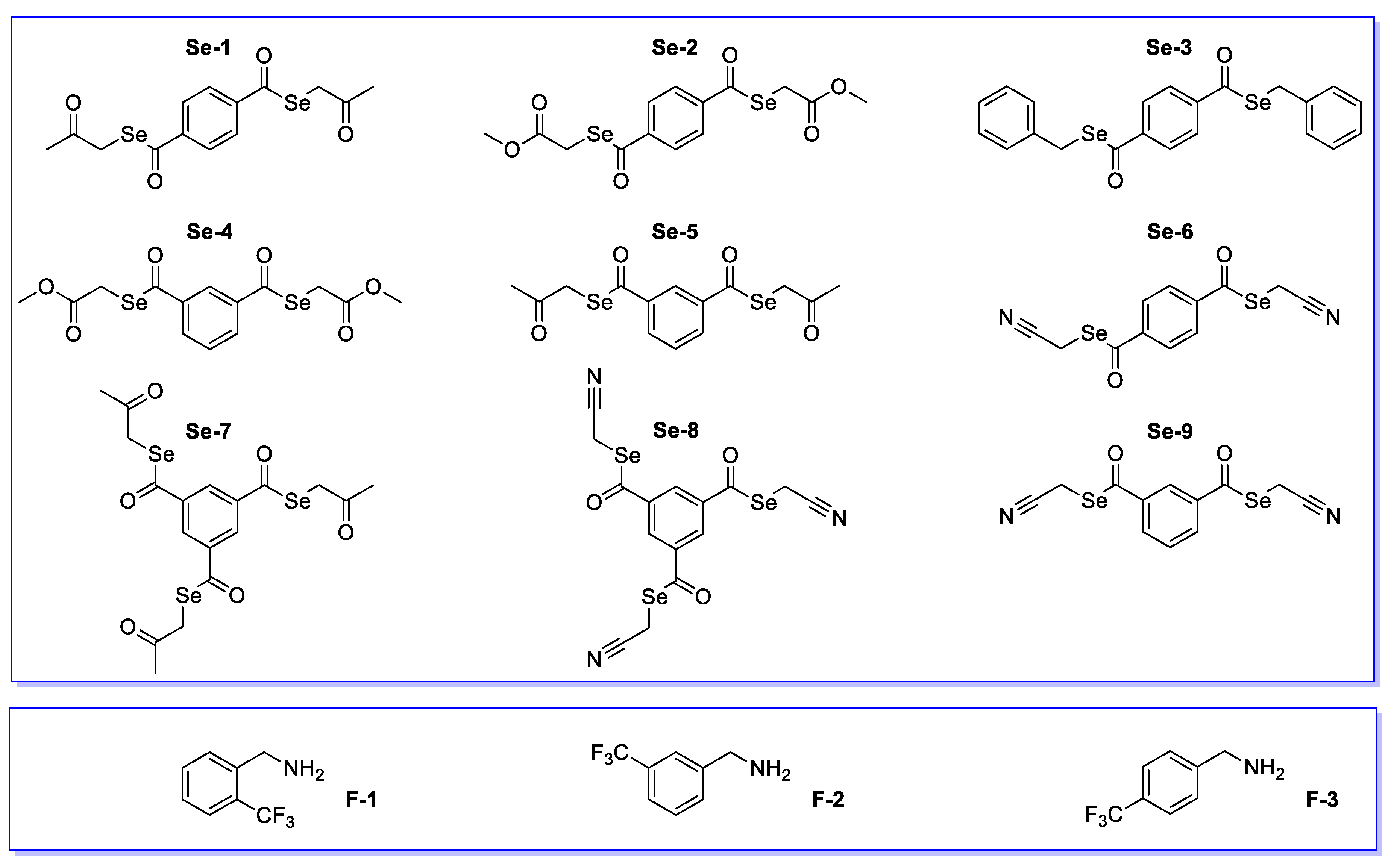
The fluoride-containing compounds were supplied by Sigma-Aldrich España (Madrid, Spain) F-1 (o-(trifluoromethyl) phenethylamine) F-2 (m-(trifluoromethyl)phenethylamine) and F-3 (p-(trifluoromethyl) phenethylamine). Selenoesters were synthesized by Domínguez-Álvarez et al. [39]: Se-1 (Se,Se-bis(2-oxopropyl)benzene-1,4-bis(carboselenoate), Se-2 Se,Se-bis(methyloxycarbonyl methyl)benzene-1,4-bis(carboselenoate), Se-3 Se,Se-dibenzylbenzene-1,4-bis(carbo selenoate), Se-4 (Se,Se-bis(methyloxy carbonylmethyl)benzene-1,3-bis(carboselenoate), Se-5 (Se,Se-bis(2-oxopropyl) benzene-1,3-bis (carboselenoate), Se-6 (Se,Se-bis(cyanomethyl)benzene-1,4-bis(carboselenoate), Se-7 (Se,Se,Se-tris(cyanomethyl)benzene-1,3,5-tris (carboselenoate), Se-8 (Se,Se,Se-tris(cyanomethyl) benzene-1,3,5-tris(carboselenoate), and Se-9 (Se,Se-bis(cyanomethyl)benzene-1,3-bis(carboselenoate).

2.2. Growth Conditions

For the isolation of C. glabrata, Sabouraud Agar (DIFCO, NJ, USA) was used (glucose 40 g, agar 20 g, 0.05 g of chloramphenicol, and 1 L of H2O, at pH 6.5). CHROMID® Candida bioMerieux microculture and API/ID32 Candida (bioMèrieux, France) and MALDI-TOF MS (bioMèrieux, France) were used. Susceptibility testing by disc diffusion agar used Mueller-Hinton agar supplemented with glucose 2% and methylene blue (5 µg/mL). Susceptibility of C. glabrata with the bioactive compounds was determined by the microdilution method in (Roswell Park Memorial Institute) RPMI medium -1640 (supplemented with MOPS, (3-(N-morpholino)propanesulfonic acid) at pH 7). FBS (fetal bovine serum 50%, bovine IgG <1 mg/mL hemoglobin <20 mg/dl), PBS (phosphate-buffered saline 0.01 M pH 7.4 Sigma-Aldrich, MO, USA), medium M 138 (peptone 2.5 g, glucose 5 g, yeast extract 2.5 g, malt extract 1.5 g, and chloramphenicol 0.05 g, 500 mL, and pH 6.5, adjusted with 0.1 N NaOH and 0.1 N HCl), Violet Gentian Solution (Sigma Aldrich, MO, USA), bleaching solution (25% acetone/75% propanol), and 24-well polystyrene plates (Biofilm, CA, USA) were used in the formation of biofilms. The microplate reader used was SpectraMax Plus 384 Microplate Reader. The optical density (OD) of the biofilms formed at 36 °C was measured after 24 h at OD 620 nm.

2.3. Isolation and Identification of Candida Strains

Three selected strains among the most pathogenic species of Candida genus were used as a control from the American Type Culture Collection (ATCC): C. glabrata ATCC 2001, C. krusei ATCC 6258, and C. parapsilosis ATCC 22019.
In total, Candida isolates from 81 adults from care homes (“Mercedes”, “Santa Rita” and “25 de Mayo”) from Sucre, Bolivia, each patient with informed consent] were employed in this study. The Candida strains were isolated by swabbing the surface of dentures of the upper or lower arch. [40 For the isolation of C. glabrata, agar dextrose Sabouraud and in CROMagar was used, in which C. glabrata had a mauve-pink color. The identification was carried out with API/ID32 Candida and MALDI-TOF MS.

2.4. Antifungal Assay

The antifungal activity of the tested compounds was evaluated by the agar disc diffusion (CLSI) and minimal inhibitory concentrations (MICs) methods [31]. The microorganisms kept at −80 °C were seeded onto Petri dishes with Sabouraoud dextrose agar culture medium supplemented with chloramphenicol (50 µg/L) and incubated at 37 °C for 48 h. The standardization of the inoculum for the susceptibility tests was carried out in 0.5 of McFarland, from five colonies of each microorganism (five reference strains of Candida species or the 37 isolated of dentures), which were streaked on Muller-Hinton agar supplemented with glucose 2% and methylene blue (5 µg/mL). Then, filter paper discs (6–10 mm), containing the tested compounds at appropriate concentrations (fluorinated compounds: 50–500 µg/mL; selenium compounds: 1–4 µg/mL), were placed on the agar surface. The inoculated plates were incubated for 24 h at 37 °C overnight for C. glabrata and C. krusei or 48 h for C. parapsilosis. The tested antimicrobial agents diffused into the agar and their inhibitions of Candida spp. growth were measured (Figure S1, Table 1). Sensidisks of voriconazole, caspofungin, and fluconazole were used as a control. The standardization of the inoculum for the susceptibility tests was carried out in 0.5 Mc Farland (0.5–2.5 × 10⁶ UFC/mL).

2.5. Minimal Inhibitory Concentrations (MICs)

The tested concentrations of selenoesters, voriconazole (VOR), fluconazole (FLZ), and caspofungin (CAS) were prepared in RPMI-1640 (pH = 7). The inoculum was prepared by suspending five distinct colonies (> 6–10 mm diameter) from 24-h cultures, in at least 3 mL of sterile distilled water. The inoculum was then suspended by vigorous shaking on a vortex mixer for 15 s, the cell density was adjusted to the density of a 0.5 McFarland standard, with the addition of sterile distilled water if required, to render a yeast suspension of 1 to 5 × 10⁶ colony forming units (CFUs)/mL. A working suspension was prepared by the dilution of the standardized suspension in sterile distilled water to yield 1 to 5 × 10⁵ CFU/mL. The 24-wellplate was prepared with 200 µL of cell suspension and 20 µL of selenoesters (1–100 µg/mL) or antifungal agents (0.2–10 µg/L for VOR, CAS, and FLZ) and incubated at 37 °C during 24 to 48 h. MIC was determined as the lowest concentration of the drug that inhibits the growth of the yeasts. The results were visualized by spectrometry at 530 nm.

2.6. Biofilm Inhibition Assay

The biofilm-forming ability of the Candida strains was measured using the crystal violet assay (CV), which is based on the ability of this dye to color the polysaccharide matrix [40,41]. In order to determine the ability of compounds to inhibit biofilms, the tested cultures were left growing in 24-well polystyrene microtiter plates in the presence of the respective fluoride or selenoester compounds at different concentrations. Briefly, each Candida spp. isolated from adult care homes (37 samples) and C. glabrata ATCC 2001, C. krusei ATCC 6258, and C. parapsilosis ATCC 22019 wild type were grown in 10 mL of rich M130 medium at 36 °C overnight in anorbital shakers at 150 rpm. Those cultures were diluted with M130 medium in order to obtain a final density of 1.0 × 10⁶ CFU/mL. Then, 0.2 mL of each strain were grown in a 24-well microtiter plate and incubated for 2 h at 36 °C until OD 620 nm was equal to 0.5 and added to the evaluated compounds (3 fluoride derivatives and 9 selenoester compounds) at different concentrations and sealed and left overnight at 36 °C. Three assays were performed for each strain in each plate. We included three controls: Negative control (vehicle, medium M138), without growth and without biofilm formation; positive control (vehicle, medium M138; and biofilm-forming strain); and positive control with antifungal drug (vehicle, medium M138; biofilm-forming strain and caspofungin). After biofilm surface priming, the medium in each well was removed carefully with a multichannel pipette, taking care to not disrupt the biofilm; each well was subsequently washed three times with PBS. The formed biofilm at the bottom of plate was stained with 0.5% aqueous solution of crystal violet for 3 min. After staining, plates were washed three times with sterile water and dried at room temperature. The optical density (OD) of each well was measured at 620 nm with an automated microplate reader SpectraMax Plus 384.

2.7. Biofilm Disruption Assay

In order to determine the ability of the fluoride or selenoester-containing compounds to disrupt pre-formed biofilms at the bottom of plate, the test cultures were formed in microtiter plate wells for 24 h. The pre-formed biofilms were then treated with the compounds at different concentrations for 24 h and the residual biofilm was estimated using the crystal violet assay. Microtiter plate wells containing fungal cells without antimicrobial compounds were used as controls during the experimentation. The data related to these experiments are depicted as the average values of the triplicate observations and error bars indicate the standard deviation (Figures S2–S7). The average OD from the control wells was subtracted from the OD of all test wells. The biofilm inhibition assay and biofilm disruption assay were performed three times for all Candida isolate from the adult care home.
BBU (Biofilm Biomass Unit) = ODb 620 nm – ODc 620/ODb 620 nm
BBU (%) = Biofilm Biomass Unit by 100
ODb = Absorbance without tested compound
ODc = Absorbance with tested compound
BBU (t = 0) Biofilm Inhibition Units by 100
BBU (t = 24) Biofilm Disruption Unit by 100

2.8. Data Analysis

The normality test was performed using the Shapiro Wilk test with a p value >0.05 with a 95% confidence level. Then, the results obtained were compared with the Tukey test using an ANOVA; the variance analyses were contrasted with the treatments of the fluoride and selenoester compounds according to the concentration and the time of formation of biofilms of the clinical strains of C. glabrata. Statistical comparisons among the different strains were performed by using Sigma Stat statistical software (SPSS, IBM, Armonk, NY, USA).

3. Results and Discussion

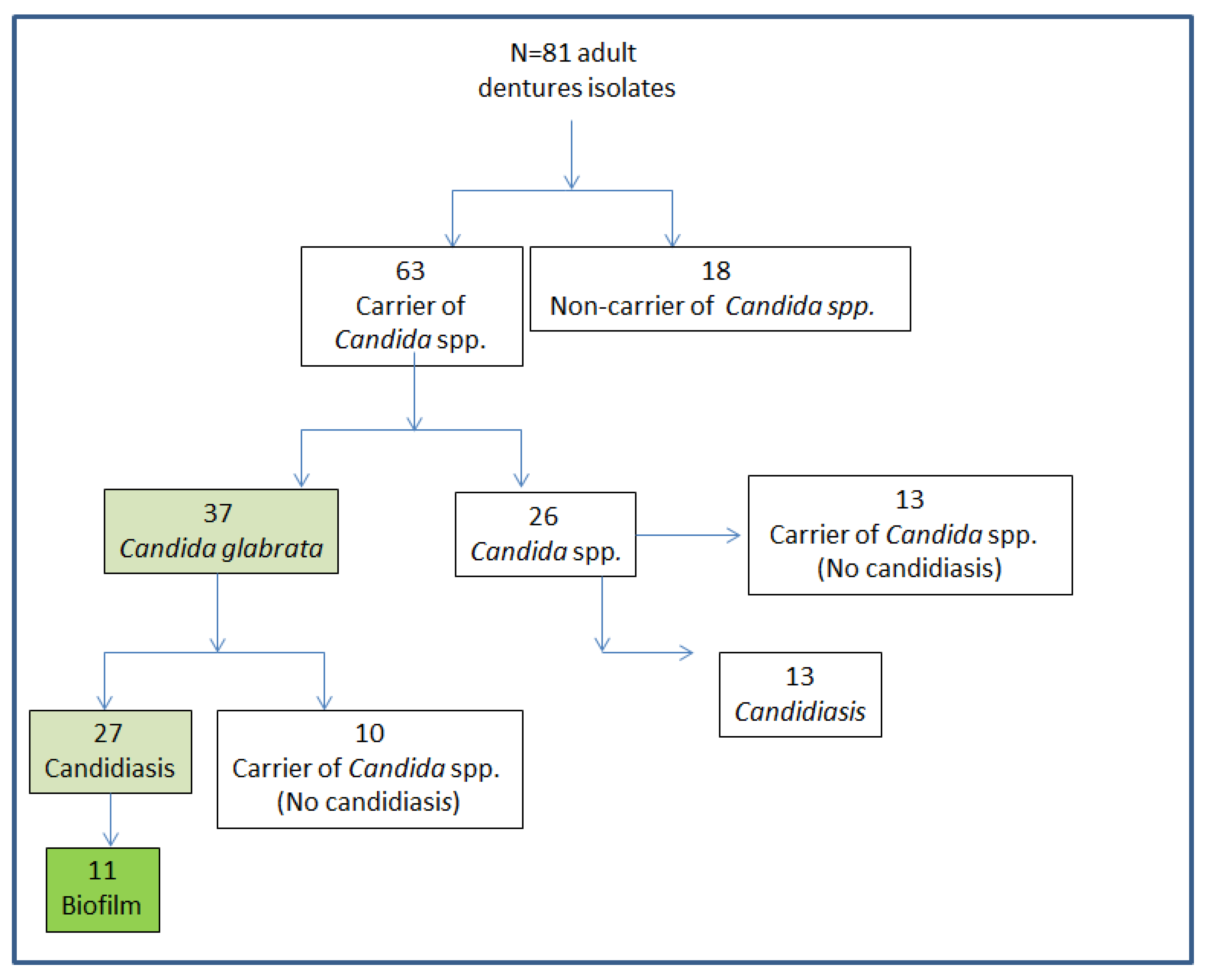
Among the 81 dental samples from the adult care home (>80 year (25%), 60–80 years (55%), and <59 years (20%)), the evaluated Candida species were found in 80% (Scheme 1).
Of the samples with Candida, 37 samples (58%) were identified as Candida glabrata and 70% of them had candidiasis (Scheme 1). In addition, among those 27 isolates with candidiasis by C. glabrata, 11 were able to form biofilm (Scheme 1, Table S1). According to our data, older adults presented a 4.4-fold higher risk of developing candidiasis if their dentures were colonized by these microorganisms (prevalence 64,8%, IC 95% 1.727616–11.218251, and p = 0.0015) and more likely to cause candidiasis if colonized by C. glabrata. We observed an interesting behavior in mixed biofilms of C. albicans and C. glabrata, when compared to single biofilms. A 77% reduction of C. albicans was observed when this fungus formed mixed biofilms with C. glabrata. This reveals that there are competitive interactions in which C. glabrata predominates over C. albicans during the formation of biofilm. Those results verify that Candida glabrata is an emergent pathogen in the oral cavity present in dental stomatitis [42] and that it plays an important role in the formation of dental biofilms.

3.1. Antifungal Screening

Herein, we tested two types of compounds: (i) Fluorinated compounds and (ii) symmetrical selenoesters (Scheme 2). On one hand, the fluorinated compounds are small molecules bearing aromatic rings, trifluoromethyl, and amine groups. On the other hand, the symmetrical selenoesters contain a phenyl core substituted by two or three identical aliphatic moieties, each containing a selenoester and a functional group (methylketone, methyl oxygen ester, phenyl, or cyano) bound to selenium through a methylene linker. The chemical structure of the halogenated and selenated compounds is shown in Scheme 2. The fluoride compounds were available commercially, whereas the selenoesters were synthesized according to known literature methods [39].
The behavior of the fluoride compounds and the selenoesters against Candida spp. according to the agar diffusion method is reported in Table 1 (Figure S1).
To evaluate their antifungal activity, these molecules were challenged against drug-sensitive yeast C. glabrata and drug-resistant yeasts, such as C. krusei or C. parapsilosis. Results showed that fluorinated compounds were active against C. glabrata while Se-5 (ketone selenodiester), Se-7 (ketone selenotriester), and Se-8 (cyano selenotriester) had antifungal activity against all Candida species. Caspofungin (CAS), fluconazole (FLZ), and voriconazole (VOR) were used as a control.
The antifungal efficacy is expressed as the minimum inhibitory concentration (MIC), that is, the minimum concentration of the molecules required to inhibit the growth of the yeast (Table 2 and Table S2). MIC values of selenoesters were determined against five reference strains and 37 isolate dentures. The obtained MIC values were compared against antifungal agents (voriconazole, caspofungin, micafungin, amphotericin B, and fluconazole) (Table 2 and Table S2). MIC values of 37 dental isolates with the selenoesters ranged from 1 to 64 μg/mL against Candida spp.
In general, all selenoester compounds showed a broad-spectrum activity against various drug-resistant fungi (Table 2). The range of MIC values for the selenocompounds Se-5, Se-7, and Se-8 (-1,3-COSeCH2COCH3, -1,3,5-COSeCH2COCH3, or -1,3,5-COSeCH2CN) was 0.5 to 5 µg/mL against all commercial Candida species. Besides, Se-1 (-1,4-COSeCH2COCH3) exerted antifungal activity against the selected C. glabrata reference strain, with an MIC value of 8 μg/mL.
As mentioned before, 11 dental isolates were able to form biofilms. Table 3 shows the MIC values of Se-5, Se-7, and Se-8 against those isolates and C. glabrata ATCC 2001. Standard antifungal agents, voriconazole and caspofungin were used for comparison.
Selenocompounds Se-5 and Se-7 showed very good antifungal activity, with an MIC of 1 to 4 μg/mL or 1 to 16 μg/mL, respectively, for the 11 dental isolates whereas Se-8 showed a moderate antifungal activity with an MIC value of 16 to 32 μg/mL (except with isolates 3 and 18, where MIC values were 4 and 8 μg/mL, respectively).
The above results suggest that an optimum substitution in the aromatic ring of di- or tri-esters is required for maximum activity. Thus, the symmetrical selenodiesters or selenotriesters could be good candidates as antifungal agents against Candida present in dentures. Among them, methylketone-containing selenoesters showed the most promising activities.

3.2. Inhibition of Biofilm Growth and Disruption of Preformed Biofilm

Candida species present the ability to form biofilms, which protects them from the action of antifungal drugs. As is well known, cells in biofilms on an abiotic surface are 1000-fold more resistant to conventional antibiotics than in planktonic cultures [44]. The general protocol used for the inhibition or disruption of the biofilm by fluoride derivatives or selenoester compounds against C. glabrata is represented in Figure 2. Here, IC50 or IC90 are defined as the concentration of the compound that inhibits the biofilm development by 50% or 90% (BBU = 0.5 or 0.9) while EC50 is defined as the concentration of compound that disperses 50% of a preformed biofilm (Table S3).
All fluoride compounds (F-1, F-2, and F-3) presented a similar behavior against the biofilm formation generated by C. glabrata ATCC 2001 or dental isolates (Figure 2). The IC50 values [45,46] of F-1, F-2, and F-3 against C. glabrata reference strains and dental isolate biofilm formation were found to be 50, 100, and 150 μg/mL (t = 0), respectively (Figure 2 and Figure 3, the mean values of BBU for C. glabrata ATCC2001 with those compounds are shown in Figures S2–S4 in the Supplementary Material). The IC50 value of caspofungin against C. glabrata dental isolates was found to be 1 μg/mL (red line, Figure 2). In the presence of fluoride compounds, the biofilm formation by C. glabrata decreased by 83% (p < 0.05), with most of the dental isolates at a concentration of 160 μg/mL (t = 0). This is consistent with the value described in the literature for fluconazole at the same concentration [47]. The IC90 value of caspofungin (t = 0) was 2 μg/mL and F-1, F-2, and F-3 were found to be 150, 300, and 320 μg/mL, respectively (Figure 2 and Figure 3). IC90 values at t = 24 h for all fluoride compounds were >320 μg/mL.
The discovery of novel antifungal agents with the ability to not only inhibit biofilm formation but also to disperse established biofilms would be a significant advance to tackle infections caused by Candida spp [48]. In order to evaluate the efficiency of fluoride compounds to eradicate preformed biofilms, F-1, F-2 and F-3 were used against C. glabrata ATCC2001 and against 11 dental isolates biofilms. Pre-formed biofilms of C. glabrata ATCC2001 were treated with six different concentrations of fluoride compounds, 50 to 320 μg/mL (Figure 3 and Figure 4; range 50–320 μg/mL). The EC50 values for F-1, F-2, F-3, and caspofungin (red line) were 50, 150, >320, and 2 μg/mL, respectively.
The same study of biofilm inhibition or disruption was carried out with the active Se-5, Se-7, and Se-8 selenoester compounds (Figure 5, Figure 6 and Figure 7). Se-5 was found to be an effective inhibitor of C. glabrata biofilm formation as shown in Figure 5.
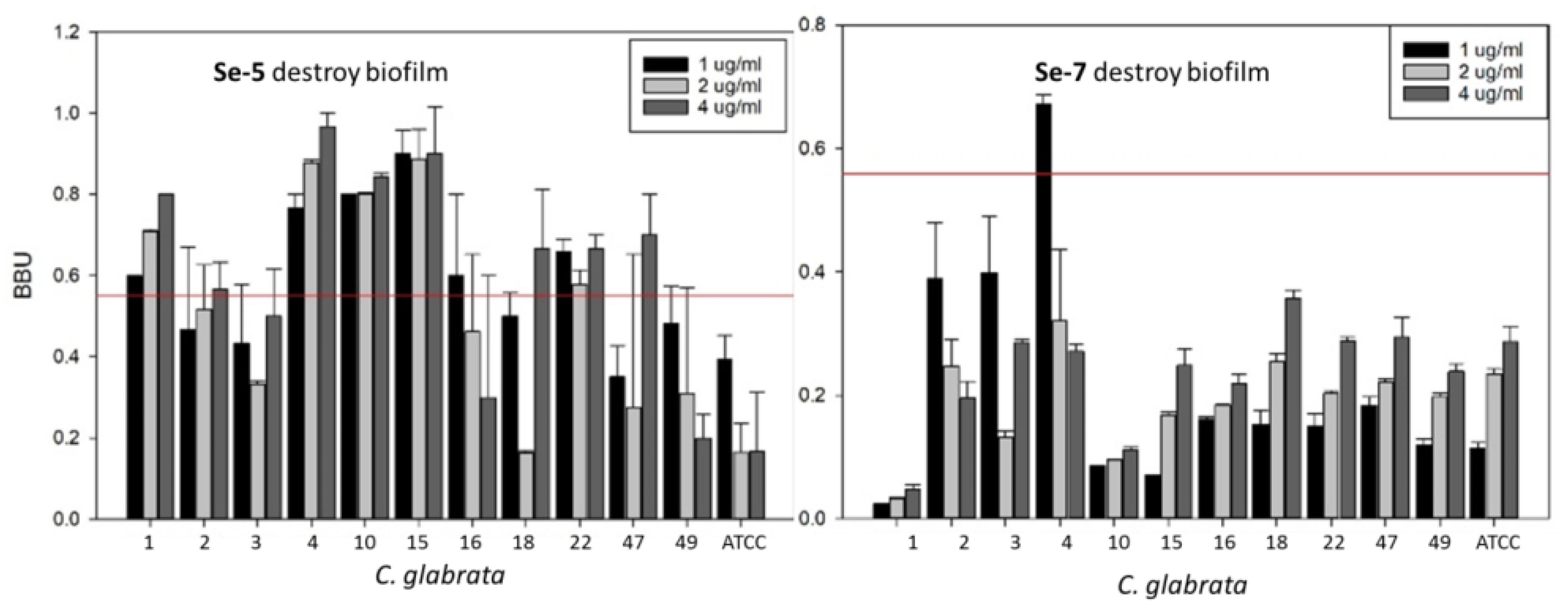
The IC50 and IC90 values at t = 0 of Se-5 and Se-7 against C. glabrata ATCC2001 and the dental isolates biofilm formation were found to be similar at 3 μg/mL (Figure 6, Table S3). The IC50 and IC90 values at t = 24 h of Se-5 and Se-7 against C. glabrata ATCC2001 and dental isolate biofilm formation were found to be 1 to 4 and >4 μg/mL respectively. Mean values of BBU for C. glabrata with those compounds are shown in Figures S5 and S6 in the Supplementary Material.
The selenoester Se-5 presents better biofilm inhibition or disruption than caspofungin or the other selenoesters (Figure 6, Figure 7). Se-5, in many cases, dispersed up to 85% of the biofilm at 2 μg/mL, when caspofungin and Se-7 dispersed 58% or 30%, respectively (Figure 7). The above results indicate thus the ability of symmetrical selenoesters to disperse biofilms. The effect of Se-5 on the formation of C. glabrata biofilms at t = 0 h and 4 μg/mL was reduced by 33.33% (0.59 ± 0.12–0.97 ± 0.02 BBU) biofilm formation compared to caspofungin at 2 ug/mL (0.93 BBU ± 0.03) (positive control), being statistically significant (p < 0.0001) in the two biofilms. After 24 h, Se-5 at 4 ug/mL reduced the biofilm formation (0.16 ± 0.25–0.96 ± 0.05BBU) by 66.66%, with a greater effect compared to the positive control of caspofungin (0.56 ± 0.17 BBU), with a p < 0.0001 in eight biofilms and p > 0.05 in four biofilms (Figure S6).
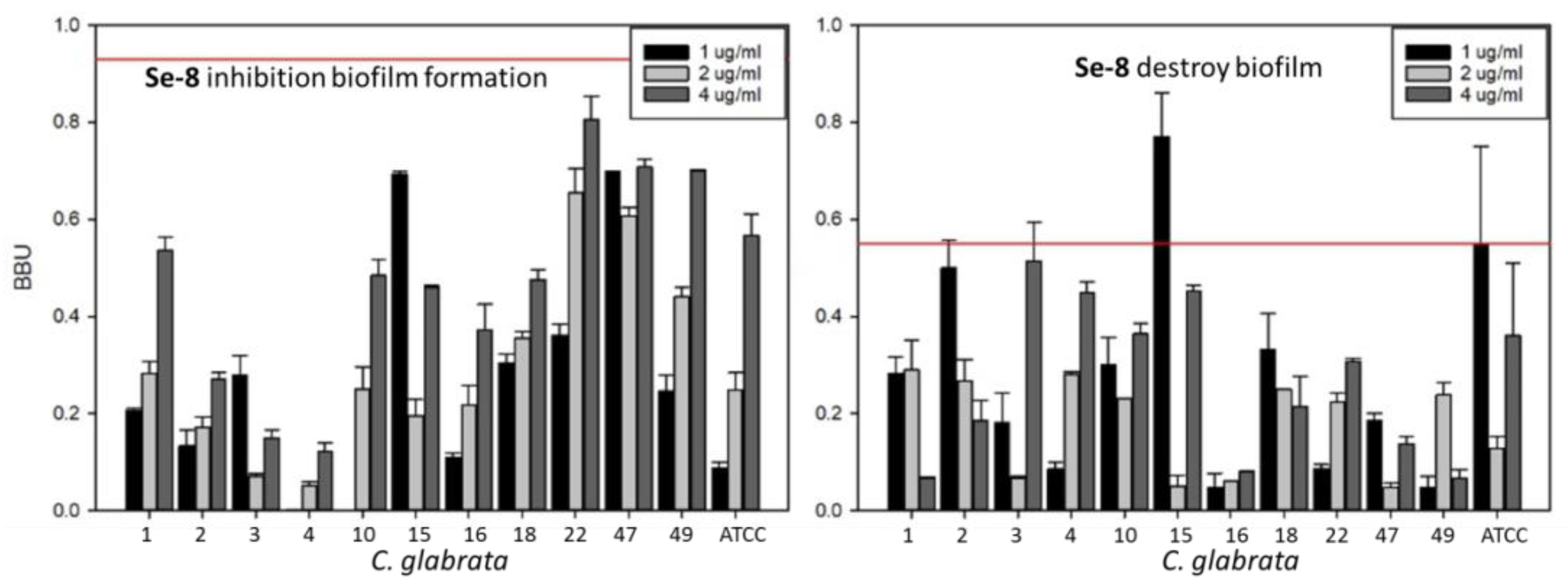
On the other hand, Se-7 (t = 0, at 2 μg/mL) reduced biofilm formation on C. glabrata with a greater effect in 91.66% (0.80 ± 0.12–1.10 ± 0.01 BBU) in comparison to caspofungin at 2 μg/mL (0.93 BBU ± 0.03) (positive control), being statistically significant (p < 0.0001). After 24 h at 4 ug/mL, the Se-7 compound did not reduce the formation of biofilms (0.05 ± 0.01–0.36 ± 0.02 BBU) in 100% of the biofilms compared to caspofungin (0.56 ± 0.17 BBU), with a p-value < 0.0001. The IC50 value of Se-8 against C. glabrata ATCC2001 and dental isolate biofilm formation was 10 μg/mL. The mean value of BBU for C. glabrata with Se-8 is shown in Figure S7 in the Supplementary Material. The IC90 value of Se-8 against C. glabrata dental isolates was 20 μg/mL. Se-8 presented poorer biofilm inhibition or disruption when compared to the other selenoesters evaluated in antibiofilm assay or to caspofungin (red line, Figure 8).
The effect of Se-8 on the formation of biofilms generated by C. glabrata dental isolates at t = 0 or 24 h and 4 μg/mL was very weak (0.15 ± 0.03–0.80 ± 0.08 BBU, and 0.07 ± 0.03–0.51 ± 0.14 BBU, respectively), compared to caspofungin (0.93 BBU ± 0.03, 0.56 ± 0.17 BBU, respectively), being statistically significant, p < 0.0001. Selenocompound had the lowest biofilm-reducing effect at 0 and 24 h among the four selenocompounds evaluated in this anti-biofilm assay.
If we compare caspofungin and selenoesters at the same concentration of 2 μg/mL, Se-5 and Se-7 present a similar biofilm inhibition but Se-5 presets better biofilm disruption against the tested C. glabrata dental isolates. At this stage, the underlying mechanism of action of these novel selenoesters against C. glabrata is unknown and more experiments are needed to determine the ultimate mechanism behind the antifungal activity described herein. Selenocompounds and among them, selenoesters, as briefly mentioned in the introduction [31,32,33,34,35,36,37,38,39], have been proven in previous works to be able to exert a wide range of interesting biological activities, both in the anticancer and antimicrobial fields; for example, the modulation of the redox cellular state, scavenging of free radicals, increase of the oxidative stress, induction of apoptosis, and inhibition of efflux pumps, among others [31,32,33,34,35,36,37,38,39]. With the caveat that future works would need to prove it, we could hypothesize that one of several of these mechanisms could explain the observed activities for the symmetrical selenoesters presented in this work.
The abovementioned biofilm inhibition exerted by the Se-5 compound makes it a promising antibiofilm agent. Thus, it could be a good starting point in the design of new antibiofilm agents against Candida spp. present in dental prosthesis. Besides, it will be necessary to carry out additional studies to elucidate the mechanism through which the active selenoester Se-5 exerts its promising antifungal and biofilm inhibition activity. The inhibition and disruption of the biofilm with all test cultures was statistically significant, with p < 0.05 in the case of the treated cells compared to the untreated controls.

4. Conclusions

In summary, our findings support that Candida glabrata isolated in dental isolates is one of the most prevalent pathogenic yeast species of the Candida spp. Novel inhibitor and disruptor biofilm compounds against C. glabrata based upon the symmetrical selenoesters scaffold were evaluated herein. Among them, Se-5 and Se-7 showed good antifungal activity against all species studied as well as against all dental isolates. Fluoride-containing compounds presented IC50 values of 50 to 150 μg/mL and EC50 > 320 μg/mL. The most active compound, Se-5, presented an IC50 value of 2 μg/mL and an EC50 value of 2 to 4 μg/mL. These active compounds not only inhibited the formation of biofilm but also were able to disperse the established biofilms. Consequently, symmetrical selenoester compounds could be a novel promising target of research in terms of the development of novel anti-biofilms drugs.

Supplementary Materials

The following are available online at https://www.mdpi.com/2076-2607/7/12/664/s1, Table S1; Biofilm formation of 11 C. glabrata dental isolates and C. glabrata ATCC2001 control strain, Figure S1; Zone diameter wholes (mm) for casponfungin, fluoride and selenoester compounds against Candida spp. according to the agar diffusion method, Table S2; MIC values of voriconazole (VOR), caspofungin (CAS), micafungin (MIC), amphotericin B (APH) and fluconazole (FLZ) against C. glabrata (N= 37 dental isolates)., Table S3; Biofilm inhibition IC50 and dispersion EC50 of fluoride and seleno-esters compounds against C. glabrata Figure S2: IC50 and EC50 of F-1 and mean values of BBU for C. glabrata ATCC 2001, Figure S3; Mean values of BBU for C. glabrata ATCC 2001 with F-2 (50–320 µg/mL) at t = 0 and 24 h, Figure S4; Mean values of BBU for C. glabrata ATCC 2001 with F-3 (50–320 µg/mL) at t = 0 and 24 h, Figure S5; Mean values of BBU for C. glabrata ATCC 2001 with Se-5 (50–320 µg/mL) at t = 0 and 24 h, Figure S6; Mean values of BBU for C. glabrata ATCC 2001 with Se-7 (50–320 µg/mL) at t = 0 and 24 h, Figure S7; Mean values of BBU for C. glabrata ATCC 2001 with Se-8 (50–320 µg/mL) at t = 0 and 24 h.

Author Contributions

M.L.D.l.C.-C. was involved in the conceptualization of the study; M.L.D.l.C.-C. and A.A.C.-L., were involved in the data analysis and validation; A.B. and M.T.U. were involved in the investigative aspects of the study and in project administration and supervised; M.B.-L. were involved in formal analysis; A.B., E.D.-Á. and M.T.U. wrote and edited the manuscript; A.B., M.L.D.l.C.-C., E.D.-Á. and M.T.U. were involved in resolved funding acquisition and investigation. All authors read and approved the manuscript and agree to be accountable for all aspects of the research in ensuring that the accuracy or integrity of any part of the work are appropriately investigated.

Funding

This research was funded by Consejo Superior de Investigaciones Científicas (CSIC), grant numbers COOPB20237 (A.B.) and 201780I027 (E.D.A).

Conflicts of Interest

The authors declare no conflict of interest.

References

  1. Dewhirst, F.E.; Chen, T.; Izard, J.; Paster, B.J.; Tanner, A.C.; Yu, W.H.; Wade, W.G. The human oral Microbiome. J. Bacteriol. 2010, 192, 5002–5017. [Google Scholar] [CrossRef] [Green Version]
  2. Paster, B.J.; Olsen, I.; Aas, J.A.; Dewhirst, F.E. The breadth of bacterial diversity in the human periodontal pocket and other oral sites. Periodontology 2000 2006, 42, 80–87. [Google Scholar] [CrossRef] [PubMed]
  3. Corte, L.; Pierantoni, D.C.; Tascini, C.; Roscini, L.; Cardinali, G. Biofilm Specific Activity: A Measure to Quantify Microbial Biofilm. Microorganisms 2019, 7, 73. [Google Scholar] [CrossRef] [PubMed] [Green Version]
  4. Costerton, J.W.; Stewart, P.S.; Greenberg, E.P. Bacterial Biofilms: A Common Cause of Persistent Infections. Science 1999, 284, 1318–1322. [Google Scholar] [CrossRef] [PubMed] [Green Version]
  5. Jenkinson, H.F.; Douglas, L.J. Interactions between Candida species and bacteria in mixed infections. In Polymicrobial Diseases, 2nd ed.; Brogden, K.A., Guthmiller, J.M., Eds.; ASM Press: Washington, DC, USA, 2002; Chapter 18; pp. 357–373. [Google Scholar]
  6. Budtz-Jorgensen, E. Candida-associated denture stomatitis and angular cheilitis. In Oral Candidosis; Samaranayake, L.P., MacFarlane, T.W., Eds.; John Wright: Guilford, UK, 1990; pp. 156–183. ISBN 0723609837. [Google Scholar]
  7. Nikawa, H.; Hamada, T.; Yamamoto, T. Denture plaque past and recent concerns. J. Dent. 1998, 26, 299–304. [Google Scholar] [CrossRef]
  8. Vu, B.; Chen, M.; Crawford, R.J.; Ivanova, E.P. Bacterial extracellular polysaccharides involved in biofilm formation. Molecules 2009, 14, 2535–2554. [Google Scholar] [CrossRef]
  9. Mukherjee, P.K.; Chandra, J. Candida biofilm resistance. Drug Resist. Updates 2004, 7, 301–309. [Google Scholar] [CrossRef]
  10. Lang, N.P.; Lindhe, J. (Eds.) Clinical Periodontology and Implant Dentistry, 2nd Volume Book, 6th ed.; Blackwell Publishing Company: Oxford, UK, 1998; ISBN 978-0-470-67248-8. [Google Scholar]
  11. Ramage, G.; Rajendran, R.; Sherry, L.; Williams, C. Fungal biofilm resistance. Int. J. Microbiol. 2012, 2012, 528521. [Google Scholar] [CrossRef]
  12. Mah, T.-F.C.; O’toole, G.A. Mechanisms of biofilm resistance to antimicrobial agents. Trends Microbiol. 2001, 9, 34–39. [Google Scholar] [CrossRef]
  13. Sampaio-Maia, B.; Figueiral, M.H.; Sousa-Rodrigues, P.; Fernandes, M.H.; Scully, C. The effect of denture adhesives on Candida albicans growth in vitro. Gerodontology 2012, 29, 348–356. [Google Scholar] [CrossRef]
  14. Lee, M.X.; Cajas, C.N.; Gómez, C.L.; Vergara, N.C.; Ivankovic, S.M.; Astorga, B.E. Ocurrencia de levaduras del género Cándida y estomatitis protésica antes y después del tratamiento rehabilitador basado en prótesis removible. Rev. Clin. Periodoncia Implantol. Rehabil. Oral. 2015, 8, 31–37. [Google Scholar] [CrossRef]
  15. Loster, B.W.; Loster, J.; Wieczorek, A.; Ryniewicz, W. Mycological analysis of the oral cavity of patients using acrylic removable dentures. Gastroenterol. Res. Pract. 2012, 2012, 951572. [Google Scholar] [CrossRef] [PubMed] [Green Version]
  16. Marra, J.; Paleari, A.G.; Rodriguez, L.S.; Leite, A.R.P.; Pero, A.C.; Compagnoni, M.A. Effect of an acrylic resin combined with an antimicrobial polymer on biofilm formation. J. Appl. Oral Sci. 2012, 20, 643–648. [Google Scholar] [CrossRef] [PubMed] [Green Version]
  17. Gjodsbol, K.; Christensen, J.J.; Karlsmark, T.; Jorgensen, B.; Klein, B.M.; Krogfelt, K.A. Multiple bacterial species reside in chronic wounds: A longitudinal study. Int. Wound J. 2006, 3, 225–231. [Google Scholar] [CrossRef] [PubMed]
  18. Kirketerp-Moller, K.; Jensen, P.O.; Fazli, M.; Madsen, K.G.; Pedersen, J.; Moser, C.; Tolker-Nielsen, T.; Høiby, N.; Givskov, M.; Bjarnsholt, T. Distribution, organization, and ecology of bacteria in chronic wounds. J. Clin. Microbiol. 2008, 46, 2717–2722. [Google Scholar] [CrossRef] [Green Version]
  19. Castrillon, L.; Palma, A.; Padilla, M. Biopelículas fúngicas. Dermatol. Rev. Mex. 2013, 57, 350–361. [Google Scholar]
  20. Salerno, C.; Pascale, M.; Contaldo, M.; Esposito, V.; Busciolano, M.; Milillo, G.A.; Petruzzi, M.; Serpico, R. Cándida-associated denture stomatitis. Med. Oral Patol. Oral Cir. Bucal. 2011, 16, e139–e143. [Google Scholar] [CrossRef]
  21. Abraham, C.M. Advances and emerging techniques in the identification, diagnosis and treatment of oral candidiasis. Open. Pathol. J. 2011, 5, 8–12. [Google Scholar] [CrossRef] [Green Version]
  22. Dangi, Y.S.; Soni, M.L.; Namdeo, K.P. Oral candidiasis: A review. Int. J. Pharm. Pharm. Sci. 2010, 2, 3641. [Google Scholar]
  23. Vediyappan, G.; Rossignol, T.; d’Enfert, C. Interaction of Candida albicans biofilms with antifungals: Transcriptional response and binding of antifungals to beta-glucans. Antimicrob. Agents Chemother. 2010, 54, 2096–2111. [Google Scholar] [CrossRef] [Green Version]
  24. Hengzhuang, W.; Wu, H.; Ciofu, O.; Song, Z.; Hoiby, N. Pharmacokinetics/Pharmacodynamics of colistin and imipenem on mucoid and nonmucoid Pseudomonas aeruginosa biofilms. Antimicrob. Agents Chemother. 2011, 55, 4469–4474. [Google Scholar] [CrossRef] [PubMed] [Green Version]
  25. Hoiby, N.; Ciofu, O.; Johansen, H.K.; Song, Z.J.; Moser, C.; Jensen, P.O.; Molin, S.; Givskov, M.; Tolker-Nielsen, T.; Bjarnsholt, T. The clinical impact of bacterial biofilms. Int. J. Oral Sci. 2011, 3, 55–65. [Google Scholar] [CrossRef] [PubMed] [Green Version]
  26. Flemming, H.-C.; Wingender, J. The Biofilm Matrix. Nat. Rev. Microbiol. 2010, 8, 623–633. [Google Scholar] [CrossRef] [PubMed]
  27. Rendekova, J.; Vlasakova, D.; Arsenyan, P.; Vasiljeva, J.; Nasim, M.J.; Witek, K.; Dominguez-Alvarez, E.; Zeslawska, E.; Manikova, D.; Tejchman, W.; et al. The Selenium-Nitrogen Bond as Basis for Reactive Selenium Species with Pronounced Antimicrobial Activity. Curr. Org. Synth. 2017, 14, 1082–1090. [Google Scholar] [CrossRef]
  28. Gularte, M.S.; Anghinoni, J.M.; Abenante, L.; Voss, G.T.; de Oliveira, R.L.; Vaucher, R.A.; Luchese, C.; Wilhelm, E.A.; Lenardão, E.J.; Fajardo, A.R. Synthesis of chitosan derivatives with organoselenium and organosulfur compounds: Characterization, antimicrobial properties and application as biomaterials. Carbohydr. Polym. 2019, 219, 240–250. [Google Scholar] [CrossRef]
  29. Guisbiers, G.; Lara, H.H.; Mendoza-Cruz, R.; Naranjo, G.; Vincent, B.A.; Peralta, X.G.; Nash, K.L. Inhibition of Candida albicans biofilm by pure selenium nanoparticles synthesized by pulsed laser ablation in liquids. Nanomedicine 2017, 13, 1095–1103. [Google Scholar] [CrossRef] [Green Version]
  30. Salin, H.; Fardeau, V.; Piccini, E.; Lelandais, G.; Tanty, V.; Lemoine, S.; Jacq, C.; Devaux, F. Structure and properties of transcriptional networks driving selenite stress response in yeasts. BMC Genom. 2008, 9, 333. [Google Scholar] [CrossRef] [Green Version]
  31. Csonka, A.; Kincses, A.; Nové, M.; Vadas, Z.; Sanmartín, C.; Domínguez-Álvarez, E.; Spengler, G. Selenoesters and Selenoanhydrides as Novel Agents Against Resistant Breast Cancer. Anticancer Res. 2019, 39, 3777–3783. [Google Scholar] [CrossRef]
  32. Spengler, G.; Gajdács, M.; Marć, M.A.; Domínguez-Álvarez, E.; Sanmartín, C. Organoselenium Compounds as Novel Adjuvants of Chemotherapy Drugs-A Promising Approach to Fight Cancer Drug Resistance. Molecules 2019, 24, 336. [Google Scholar] [CrossRef] [Green Version]
  33. Mosolygó, T.; Kincses, A.; Csonka, A.; Tönki, Á.S.; Witek, K.; Sanmartín, C.; Marć, M.A.; Handzlik, J.; Kieć-Kononowicz, K.; Domínguez-Álvarez, E.; et al. Selenocompounds as Novel Antibacterial Agents and Bacterial Efflux Pump Inhibitors. Molecules 2019, 24, 1487. [Google Scholar] [CrossRef] [Green Version]
  34. Ali, W.; Álvarez-Pérez, M.; Marć, M.A.; Salardón-Jiménez, N.; Handzlik, J.; Domínguez-Álvarez, E. The anticancer and chemopreventive activity of selenocyanate-containing compounds. Curr. Pharmacol. Rep. 2018, 4, 468–481. [Google Scholar] [CrossRef]
  35. Zhang, T.; Zhao, G.; Zhu, X.; Jiang, K.; Wu, H.; Deng, G.; Qiu, C. Sodium selenite induces apoptosis via ROS-mediated NF-κB signaling and activation of the Bax-caspase-9-caspase-3 axis in 4T1 cells. J. Cell. Physiol. 2019, 234, 2511–2522. [Google Scholar] [CrossRef] [PubMed]
  36. Jamier, V.; Ba, L.A.; Jacob, C. Selenium- and tellurium-containing multifunctional redox agents as biochemical redox modulators with selective cytotoxicity. Chemistry 2010, 16, 10920–10928. [Google Scholar] [CrossRef] [PubMed]
  37. Staicu, L.; Oremland, R.; Tobe, R.; Mihara, H. Bacteria Versus Selenium. In Selenium in Plants. Plant Ecophysiology; Pilon-Smits, E., Winkel, L., Lin, Z.Q., Eds.; Springer: Berlin/Heidelberg, Germany, 2017; Volume 11, Chapter 6. [Google Scholar] [CrossRef]
  38. Roman, M.; Jitaru, P.; Barbante, C. Selenium biochemistry and its role for human health. Metallomics 2014, 6, 25–54. [Google Scholar] [CrossRef] [PubMed]
  39. Domínguez-Álvarez, E.; Spengler, G.; Jacob, C.; Sanmartín, C. Seleno Ester-Containing Compounds for Use in the Treatment of Microbial Infections or Colorectal Cancer. European Patent Application EP17382693, 28 September 2018. [Google Scholar]
  40. CLSI. Performance Standards for Antimicrobial Disk Susceptibility Tests, Approved Standard, 7th ed.; CLSI Document M02-A11; Clinical and Laboratory Standards Institute: Wayne, PA, USA, 2012. [Google Scholar]
  41. O’Toole, G.; Kolter, R. Initiation of biofilm formation in Pseudomona florescens WCS365 proceeds via múltiple, convergent signalling pathways: A genetic analysis. Mol. Microbiol. 1998, 28, 3. [Google Scholar] [CrossRef]
  42. Ramage, G.; Vande, W.; Wickes, B.; Lopez-Ribot, J. Standardized Method for in vitro Antifungal Susceptibility Testing of Cándida albicans Biofilms. Antibiocrob. Agents Chemother. 2001, 45, 2475–2479. [Google Scholar] [CrossRef] [PubMed] [Green Version]
  43. De la Cruz, M. Factores de Riesgo Asociados a la Resistencia de Cándida spp. a Fluconazol y Voriconazol en el Tratamiento de Candidiasis Subprotesica, en Adultos Portadores de Protesis Dental que Residen en el Hogar “25 de Mayo” Sucre 2012. Tesis Para Optar a Magister Farmacologia Básica y Clínica. Master’s Thesis, Farmacologia básica y Clínica, Universidad Andina Simón Bolivar, Sucre, Bolivia, 2013. [Google Scholar]
  44. Budtz-Jorgensen, E. Ecology of Candida-associated denture stomatitis. Microb. Ecol. Health Dis. 2000, 12, 170–185. [Google Scholar] [CrossRef] [Green Version]
  45. Canuto, M.M.; Rodero, F.G. Antifungal drug resistance to azoles and polyenes. Lancet Infect. Dis. 2002, 2, 550–563. [Google Scholar] [CrossRef]
  46. Pfaller, M.A.; Diekema, D.J. Epidemiology of invasive candidiasis: A persistent public health problem. Clin. Microbiol. Rev. 2007, 20, 133–163. [Google Scholar] [CrossRef] [Green Version]
  47. Davies, D. Understanding Biofilm Resistance to Antibacterial. Agents. Nat. Rev. Drug Discov. 2003, 2, 114–122. [Google Scholar] [CrossRef]
  48. Rogers, S.A.; Huigens, R.W., III; Melander, C. A 2-Aminobenzimidazole That Inhibits and Disperses Gram-Positive Biofilms through a Zinc-Dependent Mechanism. J. Am. Chem. Soc. 2009, 131, 9868–9869. [Google Scholar] [CrossRef] [PubMed]
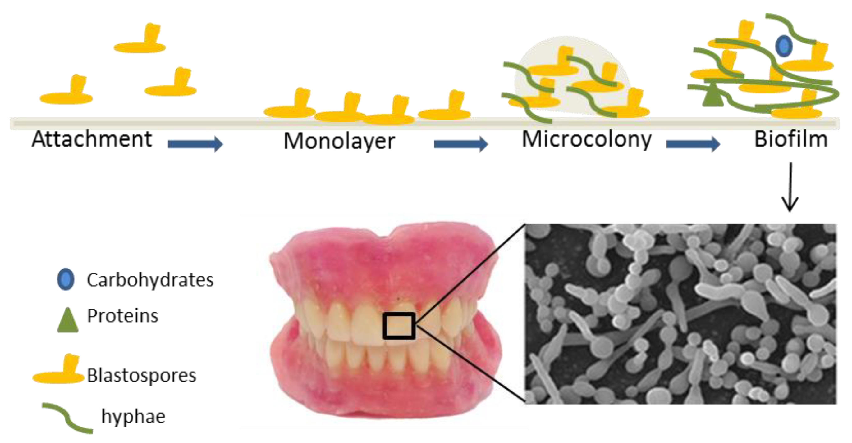
Figure 1. Schematic overview of the formation of fungal biofilm. Attachment, formation of monolayer, microcolony, and biofilm.
Microorganisms 07 00664 g001
Scheme 1. Samples were isolated from an adult care home and analyzed in the laboratory of Universidad Mayor Real y Pontificia de San Francisco Xavier de Chuquisaca in Sucre (Bolivia) (UMRPSFXCH).
Microorganisms 07 00664 sch001
Scheme 2. Chemical structures of selenoester and fluoride compounds used as antifungal and anti-biofilm agents against Candida glabrata ATCC2001 and dental isolates.
Microorganisms 07 00664 sch002
Figure 2. General (A) inhibition or (B) dispersion of preformed biofilm exerted by fluoride or selenoester compounds against C. glabrata.
Microorganisms 07 00664 g002
Figure 3. Effect of fluoride compounds F-1 and F-2 against C. glabrata dental isolates and C. glabrata ATCC 2001 in the biofilm formation (t = 0) and disruption biofilm at different concentrations. Red line value obtained with caspofungin at 2 μg/mL. The BBU (biofilm biomass units) units were monitored at 24 h. BBU = 1 inhibition or disruption 100% of biofilm, BBU = 0 no inhibition or disruption the biofilm by the fungal agents.
Microorganisms 07 00664 g003
Figure 4. Effect of F-3 compound against C. glabrata dental isolates and C. glabrata ATCC2001 in the biofilm formation (t = 0) and disruption (t = 24 h) at different concentrations. Red line value obtained with caspofungin at 2 μg/mL. The BBU units were monitored at 24 or 48 h. BBU = 1 biofilm inhibition or disruption 100%, BBU = 0 no inhibition or killing the biofilm by the agents.
Microorganisms 07 00664 g004
Figure 5. C. glabrata biofilm inhibition treated with selenoester Se-5, Se-7, and Se-8 at three concentrations (1, 2, and 4 μg/mL) and not treated (0 μg/mL).
Microorganisms 07 00664 g005
Figure 6. Effect of Se-5 or Se-7 against C. glabrata dental isolates (11) and C. glabrata ATCC 2001 in biofilm formation (t = 0) at different concentrations (1–4 μg/mL). Caspofungin was used as a control (2 μg/mL, red line). The BBU units were monitored at 24 h. BBU = 1 corresponds with a 100% biofilm inhibition, whereas BBU = 0 indicates lack of inhibition of the biofilm by the agents.
Microorganisms 07 00664 g006
Figure 7. Effect of Se-5 or Se-7 against C. glabrata dental isolates and C. glabrata ATCC2001 at t = 24 h (destroy effect) at different concentrations (1–4 μg/mL). Red line value obtained with caspofungin (2 μg/mL). The BBU units were monitored at 24 or 48 h. BBU = 1 means biofilm inhibition or dispersion 100%, and BBU = 0 means lack of inhibition or dispersion the biofilm by the agents.
Microorganisms 07 00664 g007
Figure 8. Effect of Se-8 compound against C. glabrata dental isolates and C. glabrata ATCC2001 in biofilm formation (t = 0) and dispersion (t = 24 h) at different concentrations (1–4 μg/mL). Red line value obtained with caspofungin (2 μg/mL). The BBU units were monitored at 24 or 48 h. BBU = 1 means 100% biofilm inhibition or dispersion, BBU = 0 means lack of inhibition or dispersion of the biofilm.
Microorganisms 07 00664 g008
Table 1. Diameter for select antifungal agents against Candida spp. after 24 h of incubation. FLZ = Fluconazole (C. krusei 6 mm), C. parapsilosis (22–23 mm), VOR = Voriconazole (C. krusei 16–25 mm, C. parapsilosis 28–37 mm); CAS = Caspofungin (C. krusei 19–26 mm, C. parapsilipsis 14–23).
SpeciesInterpretive Categories and Zone Diameter (mm)
Antifungal AgentsFluoride CompoundsSelenoesters
FLZVORCASF-1F-2F-3Se-5Se-7Se-8
(25 μg)(1 μg)(5 μg/mL)(500 μg)(2 μg)
C. glabrata
ATCC2001
202121212421202226
C. krusei
ATCC6258
62320101910363729
C. parapsilosis
ATCC22019
243114101010364028
Table 2. Antifungal activity of selenoesters against C. glabrata ATCC2001, C. krusei ATCC6258, C. parapsilosis ATCC22019, and 37 dental isolates.
CompoundsMIC (µg/mL)
C. glabrata
ATCC 2001
C. krusei
ATCC 6258
C. parapsilosis
ATCC 22019
C. glabrata
isolates (N = 37)
VOR<0.50.50.120.12–1
FLZ321620.1–64
CAS0.120.1910.12–8
Se-18323232–64
Se-264646432–64
Se-364646432–64
Se-464646464–128
Se-54210.5–4
Se-664646464–128
Se-72120.5–4
Se-8510.510–32
Se-964646416–64
Voriconazole (VOR) presents an antifungal spectrum broader than fluconazole against Candida species although both are azole compounds [43] Fluconazole (FLZ), Caspofungin (CAS).
Table 3. MIC values of Se-5, Se-7, and Se-8 against Candida glabrata dental isolates.
MICs (µg/mL), C. glabrata Dental Isolates (n = 11)
Strain Number
Compounds123510151618224749
VRZ0.250.250.25O.50.250.250.25≤0.12≤0.120.25≤0.12
CAS0.250.250.25>80.250.250.250.250.250.250.25
Se-514242211224
Se-712622121621>2
Se-816164321616208321616