Review

T Cell Immunity and the Quest for Protective Vaccines against Staphylococcus aureus Infection

by
Erin I. Armentrout
1,2,
George Y. Liu
3,4 and
Gislâine A. Martins
5,6,7,*
1
Lung Institute, Cedars-Sinai Medical Center (CSMC), Los Angeles, CA 90048, USA
2
Division of Pulmonary and Critical Care Medicine, CSMC, Los Angeles, CA 90048, USA
3
Collaborative to Halt Antibiotic-Resistant Microbes, University of California San Diego, La Jolla, CA 92161, USA
4
Department of Pediatrics, University of California San Diego, La Jolla, CA 92093, USA
5
F. Widjaja Foundation Inflammatory Bowel and Immunobiology Research Institute (IBIRI), CSMC, Los Angeles, CA 90048, USA
6
Department of Biomedical Sciences, Research Division of Immunology, CSMC, Los Angeles, CA 90048, USA
7
Department of Medicine, Division of Gastroenterology, CSMC, Los Angeles, CA 90048, USA
*
Author to whom correspondence should be addressed.
Microorganisms 2020, 8(12), 1936; https://doi.org/10.3390/microorganisms8121936
Submission received: 10 November 2020 / Revised: 27 November 2020 / Accepted: 30 November 2020 / Published: 6 December 2020
(This article belongs to the Section Molecular Microbiology and Immunology)

Abstract

:
Staphylococcus aureus is a wide-spread human pathogen, and one of the top causative agents of nosocomial infections. The prevalence of antibiotic-resistant S. aureus strains, which are associated with higher mortality and morbidity rates than antibiotic-susceptible strains, is increasing around the world. Vaccination would be an effective preventive measure against S. aureus infection, but to date, every vaccine developed has failed in clinical trials, despite inducing robust antibody responses. These results suggest that induction of humoral immunity does not suffice to confer protection against the infection. Evidence from studies in murine models and in patients with immune defects support a role of T cell-mediated immunity in protective responses against S. aureus. Here, we review the current understanding of the mechanisms underlying adaptive immunity to S. aureus infections and discuss these findings in light of the recent S. aureus vaccine trial failures. We make the case for the need to develop anti-S. aureus vaccines that can specifically elicit robust and durable protective memory T cell subsets.

1. Introduction

Staphylococcus aureus is a Gram-positive facultative anaerobe that is a leading cause of bacterial infections worldwide. It is especially problematic in hospital settings where S. aureus is the most common cause of post-operative infections [1] with costs that can exceed $90,000 per case [2]. In community settings, S. aureus, which includes both methicillin-sensitive (MSSA) and methicillin-resistant (MRSA) strains, is the most frequent cause of skin and soft tissue infection. The distinction between healthcare-associated (HA-MRSA) and community-associated S. aureus (CA-MRSA) infections is important because the prevalence of antibiotic resistance genes and certain virulence factors vary between the two types. This difference can affect infection severity and antibiotic management [3]. Overall, it is estimated that MRSA infections cause an annual burden of up to $13.8 billion in the US alone [4].
S. aureus has predilection for a broad range of tissues, which leads to manifestations such as folliculitis as well as life-threatening infections including pneumonia, endocarditis, osteomyelitis, and sepsis. The pathogen causes recurrent infections, suggesting that humans do not naturally develop long-lasting robust protective immunity against this bacterium. S. aureus also commonly colonizes human skin and mucosal surfaces, particularly in upper airways with some strains also showing a preference for the gastrointestinal tract [5]. It is estimated that about 30% of the population [6] is colonized with S. aureus and that colonization can occur as early as the neonatal period [7]. Colonization with S. aureus in healthy individuals is a risk factor for future staphylococcal infections by serving as the infectious source [8] and possibly by attenuating the host immune response against the pathogen.
S. aureus was first discovered in 1880 when Sir Alexander Ogston isolated the bacterium from an infected wound of a patient. The mortality rate for moderate to severe S. aureus infection was 80% in the pre-antibiotic era [9] and came down to 20% after the introduction of penicillin [10]. However, with each newly-introduced antibiotic, S. aureus promptly gained resistance and there is evidence that MRSA strains are associated with higher morbidity and mortality rates compared to their antibiotic susceptible counterparts [11].
The high disease burden and spread of antibiotic-resistant strains that led to antibiotic overuse has further highlighted the need for vaccination against S. aureus. Over the past decades and coinciding with the emergence of CA-MRSA, there has been a significant effort to develop anti-staphylococcal vaccines. Unexpectedly, to this date, every vaccine developed against S. aureus has failed in clinical trials, despite their demonstrated efficacy in animal models [12,13,14]. There is no consensus on why the vaccines have failed. Here, we review the immune response to S. aureus infection as it relates to vaccination and the failed vaccines, while making a case for alternative approaches to address this bottleneck to solving the staphylococcal health burden.

2. Previous and Ongoing Human S. aureus Vaccine Trials

There have been numerous attempts to create vaccines against S. aureus, which can be divided into two different approaches: Transfer of antibodies specific to staphylococcal antigens (passive immunization) or vaccination with recombinant antigens with the main goal of generating protective antibody responses (active immunization). These strategies either targeted surface proteins to promote the opsonophagocytic killing of S. aureus or targeted toxins to reduce cytolytic damages and immunopathology. Staphylococcal surface antigens targeted in vaccines include Clumping factor A and B (ClfA and ClfB), Iron-regulated surface determinant A and B (IsdA and IsdB), Capsular Polysaccharide 5 and 8 (CP5 and CP8), Serine–aspartate repeat protein D and E (SdrD and SdrE), and Manganese binding protein C (MntC). These proteins were selected on the basis of their high level of conservation across S. aureus strains [15]. Commonly selected S. aureus toxins included the Panton-Valentine Leukocidin (PVL, comprised of LukS-PV and Luk-PV), staphylococcal enterotoxin A and B (SEA and SEB), and α-toxin (Hla). Early vaccine studies focused on single antigens, whereas later studies targeted multiple staphylococcal antigens aiming to increase vaccine efficacy [15,16].
None of the vaccination or antibody transfer strategies clinically tested so far (recently reviewed by Redi et al. and Fowler & Proctor [13,14]) have shown protection or lasting protection against S. aureus infection in patients (Table 1 and Table 2) and the underlying reasons remain unclear. It has been suggested that the reasons underlying these failures range from the wrong choice of target antigens and adjuvants, to particularities of the study designs, target population inadequacy, and the notion that the murine experimental models of S. aureus infection used in pre-clinical vaccine studies likely do not fully reproduce the features of the human immune response to S. aureus. Regarding the latter, one important aspect to consider is the difference in colonization with S. aureus between mice and humans. In contrast to humans, which can be colonized with S. aureus early in life [7], mice are not colonized or are colonized at a lower frequency [17,18].
Nine different clinical trials testing passive immunization made it to phase II and III (Table 1). However, the great majority of these trials showed that antibody administration had no effect or offered only moderate protection in comparison to placebo treatment. Two studies utilized human polyclonal antibodies against CP5 and CP8, which was effective in shortening the length of stay for patients with bacteriemia but not in preventing invasive infection in neonates [19,20]. Another study tested if donor derived antibodies against ClfA from S. aureus and SdrG from Staphylococcus epidermidis could prevent sepsis in infants, but results showed no difference between placebo and experimental groups [21]. A single chain variable fragment targeting Adenosine triphosphate(ATP)-binding cassette (ABC) transporter GrfA was shown as ineffective in phase II clinical trials in another study [14]. The remaining five studies utilized monoclonal antibodies against S. aureus proteins. One study found that the administration of α-ClfA antibodies did not offer significant protection [22]. Similarly, monoclonal antibodies targeting Hla, PVL, LukED, and γ-hemolysin AB and CB (HlgAB, HlgCB) failed to protect against infection in mechanically-ventilated patients [23]. A monoclonal antibody targeting lipoteichoic acid (LTA) did not significantly protect against sepsis in low birthweight infants [24]. Another study tested a modified monoclonal antibody against Hla with an extended half-life to protect against S. aureus infection in mechanically ventilated patients. Unfortunately, treatment with this antibody also failed to confer protection to the treated group [25]. A more recent trial (Aridis Pharmaceuticals) is testing if the transfer of monoclonal antibodies against S. aureus Hla can treat community-acquired bacterial pneumonia or ventilator-associated pneumonia in patients, but this is still an ongoing trial and the results are not known. Overall, so far, the clinical trials concur with observations from murine model studies (see below), which indicate that antibody responses alone are not sufficient to protect against S. aureus re-infection. Other arms of the adaptive response, such as T cells, play important roles in protecting against this bacterium and therefore need to be engaged by vaccination approaches.
The notion that T cell immunity needs to be harnessed in vaccine development is also supported by observations from recent active immunization trials (Table 2), such as the failed V710 vaccine phase II/III clinical trial carried out by Merck in 2011. Vaccination with V710, an IsdB recombinant protein vaccine, generated abundant anti-IsdB antibodies however, the rate of S. aureus infection was comparable between placebo and vaccinated groups [27]. Additionally, antibody titers in patients who developed S. aureus infection were similar to those who did not develop the disease [28]. An analysis of cytokines associated with T cell function revealed that patients who developed fatal S. aureus infections had lower expression of IL-2 and IL-17A than those who survived the infection. In addition to the V710 trial, two other active staphylococcal vaccines that advanced to phase II/III clinical trials, StaphVax (Nabi) targeting CP5/CP8, and SA-4Ag (Pfizer, New York, NY, USA) targeting four staphylococcal antigens, also demonstrated a lack of efficacy in spite of robust antibody induction. Together, these observations support the importance of refocusing vaccine approaches to engage other arms of the immune system beyond the humoral response.

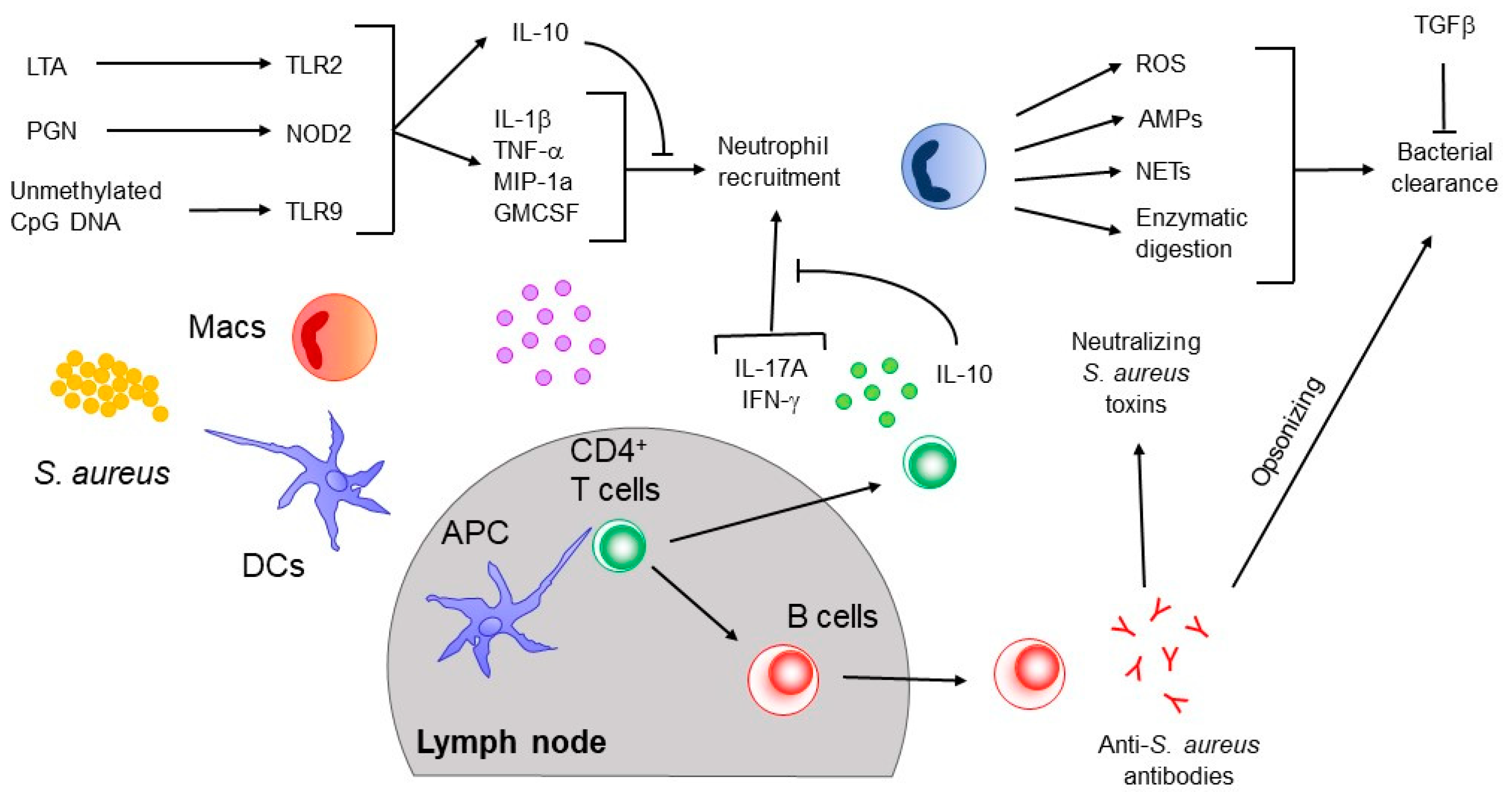
3. Innate Immune Responses to S. aureus

The innate immune responses to S. aureus and the pathogen’s strategies to counteract these responses have been the topic of many other recent and comprehensive reviews [36,37]. Here, we briefly summarize the innate response to S. aureus and focus mainly on the adaptive immune response as we discuss the new findings that can potentially inform the development of new vaccines. The innate immune system broadly recognizes invading pathogens through receptors that ligate pathogen-related molecular patterns (PAMPs), including single stranded RNA, lipopolysaccharide (LPS), and peptidoglycan (PGN). PAMPs are bound via pattern recognition receptors (PRRs), i.e., Toll-like receptors (TLRs), NOD-like receptors (NLRs), and/or C-type lectin receptors (CLRs). PRRs are mainly expressed by innate immune cells such as macrophages, monocytes, dendritic cells (DCs), and neutrophils. The sensing of PAMPs by TLRs initiates a signaling cascade that involves adaptor proteins myeloid differentiation primary response 88 (Myd88) or Toll/interleukin-1 receptor-domain-containing adapter-inducing interferon-β (TRIF). This is followed by interleukin-1 receptor-associated kinases (IRAKs), and nuclear factor kappa-light-chain-enhancer of activated B cells (NF-kB) or mitogen-activated protein kinase (MAPK) with a subsequent governance of pro-inflammatory gene expression. Alternatively, NF-kB activation by NOD2 signaling is modulated by receptor-interacting serine/threonine protein kinase 2 (RICK or RIP2).
The major S. aureus-associated PAMP sensed by the innate immune system appears to be LTA in addition to other lipoproteins, PGN, cell wall glycopolymers, RNA, and unmethylated CpG DNA. The recognition of S. aureus-LTA relies on the heterodimer, TLR2-TLR6, which senses diacylated lipoproteins such as LTA. Other receptors that recognize S. aureus PAMPs include NOD2, mannose-binding lectin, L-ficolin, surfactant protein A, TLR8, TLR9, and heterodimer TLR2-TLR1. The importance of TLR2, Myd88, IL-1 signaling, and NOD2 is underlined by several knockout mice studies. Knockout of TLR2, Myd88, IL-1R, or NOD2 increases the bacterial burden during infection compared to wild-type controls [38,39,40]. The importance of TLR, Myd88, and IL-1R signaling is mirrored in humans. Patients with defects in TLR, Myd88, IRAK4, and IL-1R signaling have higher incidence of S. aureus infections [41,42,43,44].
The binding of LTA by TLR2-TLR6 occurs on macrophages/monocytes as well as DCs. This triggers the expression/release of pro-inflammatory cytokines, such as IL-1b, IL-8, IL-10, IL-12, TNF-α and chemoattractants, MIP-1α, MCP-1, granulocyte colony-stimulating factor (GM-CSF), leukotriene B4, and complement factor 5a [45]. Chemoattractants recruit neutrophils to the site of infection, which clear the invading bacteria, while the pro-inflammatory cytokines act as chemoattractants as well as help shape the immune response [45] (Figure 1). After encountering S. aureus, DCs and macrophages travel from the site of infection to the draining lymph node where they can present S. aureus antigens to T cells. At least one study showed that that deletion of DCs by S. aureus α-toxin prevented the formation of a protective immune memory in a skin infection model [46], suggesting that DC-mediated antigen presentation is crucial for the development of successful adaptive immune responses against S. aureus (see below). The production of cytokines, as well as expression of co-stimulatory molecules by DCs help to initiate and direct the functional differentiation of T cell subsets that will ultimately shape the adaptive response to the infection.
In addition to DCs and macrophages, neutrophils have also been shown to play crucial roles in the immune response against S. aureus. Patients with neutropenia or defects in neutrophil function have higher incidences of S. aureus infections [47,48,49,50]. In animal models, it has been demonstrated that the recruitment of neutrophils leads to a decrease in bacterial burden, thus identifying neutrophils as a main player in clearing bacteria during infection. Neutrophils kill bacteria in several ways, such as via the creation of reactive oxygen species (ROS), production of antimicrobial peptides (AMPs), release of neutrophil extracellular traps (NETs), and degradation of bacteria through proteinases and hydrolases [51,52,53]. As we discuss below, although the innate immune response to S. aureus is important for controlling bacterial burden during infection, innate signaling upon natural infection appears inadequate to instruct the development of fully protective adaptive T and B cell responses against the bacteria. Work from our group has shown that manipulation of peptidoglycan O-acetylation leads to cell-wall resistance to lysozyme degradation thus shielding many staphylococcal cell-wall associated PAMPs from host detection. This results in the impaired production of various pro-inflammatory cytokines including IL-1b, IL-6, IL-23, and suboptimal development of protective Th1 and Th17 responses [54].

4. Adaptive Immune Response to S. aureus

4.1. B Cell Responses

Infection or colonization with S. aureus leads to the production of antibodies against both surface-bound proteins [55] and staphylococcal toxins [56,57]. These anti-staphylococcal antibodies are of various subclasses, i.e., IgM, IgA, and IgG. However, similarly to other successful pathogens, S. aureus has acquired strategies to evade host antibody responses. Two staphylococcal toxins, Surface Protein A (SpA) and second immunoglobulin binding protein (Sbi) can bind the Fc region of antibodies, which can prevent opsonization-mediated phagocytosis [58]. Additionally, both proteins interfere with complement activation to prevent opsonization [58]. SpA can act also as a B cell superantigen by binding the F(ab)2 portion of the B cell receptor, which leads to the inhibition of antibody production by inducing cell death [59].
A few studies have noted that antibody titers against S. aureus toxins correlate with better disease outcome. One study showed that the risk of sepsis was lower in individuals who had higher levels of IgG against α-hemolysin (Hla), δ-hemolysin (Hld), Panton Valentine leukocidin (PVL), staphylococcal enterotoxin C-1 (SEC-1), and phenol-soluble modulin α3 (PSM-α3) [60]. Another study looked at acute and convalescent antibodies levels in individuals with different types of infections, including first time skin and soft-tissue (SSTI), recurrent SSTI, and invasive infections, such as bacteremia, osteomyelitis, septic arthritis, bursitis, pyomyositis, empyema, endocarditis, or septic thrombophlebitis [61]. The patient group that had the highest convalescent titers was the invasive infection cohort. This group had an increase in production of α-Hla antibody titers, which correlated with protection against subsequent infection within 12 months. In contrast, SSTI did not induce long-lasting antibody production. These observations suggest that the route of infection can affect the type of humoral immune response mounted against S. aureus. Additionally, our recent study showed that the adoptive transfer of sera from children with invasive staphylococcal diseases protected naïve mice from systemic staphylococcal infection [62]. However, protection conferred by the sera was only observed with some of the convalescent serum samples and sera collected at 6 months post-infection were not protective, albeit the size of the study was small. These antibodies most likely neutralized S. aureus toxins, which in addition to preventing damage caused by the toxin also prevented progression of infection. Another study in mice found that serum transfer after vaccination against ClfA, ClfB, Hla, IsdA, IsdB, and SdrD prevented bacteremia and reduced dermonecrosis but did not reduce the bacterial burden in abscesses or GI colonization [63].
Although the studies discussed above indicate that B cells are capable of eliciting anti-staphylococcal responses, one of our most recent studies showed that antibodies were dispensable for protective immunity against S. aureus in mice vaccinated with four different S. aureus recombinant proteins (ClfA, IsdA, MntC, and SdrE) encapsulated in β-glucan particles [64]. Importantly, B cell-deficient mice and patients with X-linked agammaglobulinemia, a genetic disorder that results in severe B cell deficiency, do not have increased susceptibility to S. aureus infection [65,66]. Despite these observations, the majority of vaccine studies, including several clinical trials (Table 2) have focused on antibody production as a measure of induction of protective immunity. As we discuss below, there is strong evidence that T cell-mediated immunity is required for long-lasting protection against S. aureus infection. Thus, the data so far support the notion that the engagement of certain T helper cell subsets, which are usually suppressed by natural infection, is a key component for the development of protective immunity to re-infection, and therefore should be the basis of novel approaches for vaccine development.

4.2. T Cell Responses

T cells make up the other branch of the adaptive immune response and are defined by the expression of the T cell receptor (TCR) which can either be comprised of gamma and delta chains (γδTCR) or alpha and beta chains (αβTCR). One difference between these cell subsets is that γδ-T cells do not have a broad clonal diversity as αβ-T cells, and thus epitope coverage by αβ-T cells is wider compared to γδ-T cells.

4.2.1. Roles of γδ T Cells

Several studies have indicated that γδ-T cells play important roles in the control of S. aureus infection. One study described more severe skin pathology in γδ-T cell knockout mice, which was attributed to decreased IL-17 production [67]. Another study reported the expansion of certain γδ-T cell clones after infection. Furthermore, the adoptive transfer of γδ-T cells from mice previously infected with S. aureus protected naïve mice from infection, suggesting that γδ-T cells could provide some sort of protective memory to S. aureus infection [68]. However, in that study, the γδ-T cells were primed in the absence of IL-1β, which differs from what happens during natural S. aureus infection. Moreover, as measured by lesion size and bacterial burden, protection induced by the adoptively transferred γδ-T cells was modest and only apparent during early time points after infection. Nonetheless, there is precedence for a role of “memory” γδ-T cells in other bacterial infection models. For example, “memory” Vg4+ γδ-T cells have been shown to provide IL-17-dependent protection against Listeria monocytogenes oral infection [69]. Thus, it is conceivable that γδ-T cells could contribute to memory responses to S. aureus infection in specific circumstances, but this remains to be validated.

4.2.2. Roles of αβ T Cells

αβ-T cells make up the majority of T cells and their functions can be defined on the basis of their expression of the surface molecules CD4 and CD8. CD4+ T cells, also called helper T (Th) cells, contribute to immune responses against infections by providing help to B cells and CD8+ T cells. In contrast, CD8+ T cells, also called cytotoxic T cells, lyse cells that are infected by pathogens. CD4+ T cells have critical roles in the defense against extracellular pathogens, whereas CD8+ T cells preferentially recognize and kill intracellular/viral pathogens and tumor cells. CD4+ helper T cells can differentiate into different subsets, including Th1, Th2, and Th17, among others [70]. These Th subsets are mainly defined by the cytokines they produce and their role in certain immune responses. Th1 cells depend on the transcription factor, T-bet, for differentiation. They preferentially produce IL-2 and IFNγ and have been shown to help protect against intracellular pathogens [70]. The development of Th2 cells requires the transcription factor GATA-3, which is important to mediate the expression of the majority of Th2-associated cytokines, including IL-4, IL-5, and IL-13 [70]. Th2 cells are associated with humoral immunity and parasitic infections. Th17 cells require the expression of RORγt and several other transcription factors [70] and produce primarily IL-17 family cytokines (IL-17A, F, C, and E) in addition to IL-22 and, in some cases, GM-CSF. Th17 cells have been shown to mediate protection against extracellular pathogens such as bacteria and fungi [70]. In addition, Th cells can also originate T follicular helper cells (TFH), which require the transcription factor BCL-6. TFH produce cytokines, such as IL-21, which promote B cell responses [70]. The differentiation of Th cells can also result in the generation of Foxp3+ regulatory T cells (termed inducible TREG cells), which have similar functions as thymic derived (t) TREG [70].
Evidence to date suggests that both CD4+ and CD8+ T cells are important for immunity against S. aureus infection. Investigations have focused on CD4+ T cells because S. aureus is traditionally considered an extracellular pathogen. However, there is data indicating that once internalized by cells such as macrophages, S. aureus escapes phagosomes to enter the cytoplasm, and therefore becomes an intracellular pathogen [71] that requires control by CD8+ T cells or NK cells. In contrast, studies in murine models indicate that CD8+ αβ-T cells are dispensable during S. aureus infection [72,73], whereas there is an abundance of data, in empyema and skin infection models, supporting a central role of CD4+ T cells. Of note, S. aureus has developed several virulence factors to counteract CD4+ αβ-T cell responses including the superantigens staphylococcal enterotoxin A (SEA), enterotoxin B (SEB), and toxic shock syndrome toxin (TSST-1), which bind major histocompatibility complexes (MHC) II and T cell receptors (TCR). This binding of MHC class II and TCR activates the uncontrolled release of inflammatory cytokines from T cells, followed by the induction of T cell anergy or deletion. Additional staphylococcal strategies consist of the expression of an MHC class II analog protein (Map), which binds directly to TCR to attenuate T cell responses [74], and the expression of the LukED toxin that lyses T cells following ligation to the CCR5 receptor [75]. The importance of CD4+ T cells in the immune response against S. aureus in humans is supported by the observation that HIV patients with decreased CD4+ T cells have increased prevalence of S. aureus infections [76,77,78].
Studies of the mechanisms underlying CD4+ T cells roles in S. aureus infection indicate that IFNγ may be dispensable during primary infection, whereas the requirement for IL-17 varies depending on the infection model. One study found that IFNγ knockout mice had higher survival rates and lower bacterial burdens during intravenous infection compared to wild type controls [79]. Similarly, another study found that there was no effect on primary skin S. aureus infection in IFNγ-receptor knockout mice, suggesting that IFNγ signaling is not required for bacterial clearance in the skin [67]. In contrast, primary cutaneous infection in IL-17-receptor knockout mice resulted in larger lesions and bigger bacterial burdens compared to wild type mice, whereas IL-17-deficient mice had mortality rates comparable to that observed in wild type mice during intravenous infection [80,81]. Thus, during primary S. aureus infection, IFNγ seems to be dispensable while IL-17 is only conditionally required.
In contrast to the observed in primary infection, it is well established that both Th1 and Th17 CD4+ T cells are required to mediate protection against S. aureus in different models of re-infection in mice. One study found that the transfer of IFNγ producing αβ-CD4+ T cells protected against S. aureus peritoneal infection by a mechanism that involved the activation of peritoneal macrophages [82]. Likewise, protection against S. aureus peritoneal infection was conferred by an IL-17-dependent mechanism in another vaccination study [83]. Lin et al. also showed that vaccination with recombinant N-terminus of the Candida albicans adhesin protein Als3p, which is structurally similar to S. aureus ClfA, induced IFNγ and IL-17A single- and double-producing CD4+ T cells that protected against S. aureus bloodstream infection. In this model, protection was associated with increased neutrophil influx, which reduced bacterial burden [84].
In a different study, vaccination with a multiple antigen presenting system with six staphylococcal proteins led to the differentiation of CD4+ T cells that produced both IFNγ and IL-17A, which mediated protection against both bloodstream and skin infections [63]. Additionally, in a study showing that immunization intravenously could protect mice against subcutaneous infection, this protection was mediated by CD4+ T cells and was associated with an increase in IFNγ production [46]. In one of our recent studies, we found that the vaccination of mice with β-glucan particles loaded with four different proteins from S. aureus also generated an innate immune response that fostered the development of Th1 and Th17 cells that could transfer protection to naïve mice [64].
Although the requirement for IL-17 in the immune response to S. aureus is clearly illustrated by the studies mentioned above, the cellular source of this cytokine during S. aureus infection is less clear with conflicting reports showing a role for both αβ and γδ T cells. Cho et al. suggested that IL-17 was produced by γδ-T cells, which recruited neutrophils for the control of S. aureus primary skin infection [67]. In contrast, Montgomery et al. found that IL-17A and CD4+ T cells were the key to controlling S. aureus infection in the skin [73]. Additionally, Marchitto et al. showed that upon skin infection, both CD4+ and γδ-T cells produced IL-17A/F at the site of infection as well as in the draining lymph node [68]. However, they concluded that the expression of IL-17A was higher in γδ-T cells. Thus, both αβ-CD4+ T cells as well as γδ-T cells can produce IL-17 and be required to control S. aureus infection. The importance of IFNγ and IL-17 in the response to S. aureus infection in humans is supported by several studies [79,80,81,85]. The role of IL-17 is further indicated by the finding that individuals with genetic defects that interfere with the differentiation of Th17 cells, such as STAT3 loss-of-function mutations or hyper-IgE syndrome, have a higher incidence of S. aureus infection [86,87,88].
In addition to S. aureus, Th17 cells and IL-17 are crucial in the response to several other extracellular bacterial infections. Likewise, Th1 cells and IFNγ play important roles in the response to many intracellular pathogens. Although advantageous in the context of these infections, both IL-17 and IFNγ are highly inflammatory cytokines and can cause unwanted harm to host tissues. Consequently, Th1 and Th17 responses must be tightly regulated. The immune system has evolved to deploy several different mechanisms to control Th1 and Th17 responses. One important mechanism used to control IL-17 and IFNγ actions is the expression of the regulatory cytokines TGFβ and IL-10, which can be produced by many different immune cell types. The role of TGFβ in the immune response to S. aureus has not been extensively evaluated, but at least one study showed that culture of bovine mammary epithelial cells with heat-killed S. aureus leads to the expression of TGFβ [89]. Another study showed that partial genetic deletion of TGFβ Receptor 2 (Tgfbr2) or the administration of a TGFβ receptor inhibitor in mice was associated with improved bacterial clearance in the skin [90]. These data suggest that S. aureus-induced TGFβ could play a role in damping the responses against the infection in the skin, but this remains to be further studied.
The role of IL-10 in the immune response to S. aureus infection is not fully understood. In studies looking at systemic infection, IL-10 appears to be protective; IL-10 deficient mice had higher bacterial burdens compared to wild type controls [91,92]. Additionally, one study found that the incidence of S. aureus-induced arthritis was higher in IL-10-deficient mice [92]. In contrast, during skin infection, IL-10-deficient mice had smaller lesion sizes and lower bacterial burdens compared to wild type counterparts [91]. These data suggest the complex role of IL-10 during S. aureus infection may depend on the route of infection.
Importantly, when naïve human T cells were cultured with S. aureus, they produced IL-10 in addition to IL-17 [93]. However, if the T cells were cultured with S. aureus along with IL-1β, the cells secreted IL-17 and IFNγ, but not IL-10. This was in stark contrast to what was observed with C. albicans, which caused human T cells to produce IL-17 and IFNγ and very little IL-10 without any manipulation to the culture conditions. An additional study in human samples observed that higher IL-10 serum levels correlate with higher rates of mortality in patients with bacteremia caused by S. aureus [94], suggesting that it can play an inhibitory role in human immunity. Together, these observations suggest that S. aureus can somehow manipulate the immune response to prevent the differentiation of IL-17 and potentially IFNγ-producing cells and/or undermine the actions of these cells. In line with that, our recent study showed that O-acetylation of peptidoglycan by S. aureus leads to a preferential expression of IL-10 instead of IL-17 or IFNγ. Furthermore, deletion of O-acetyl transferase in S. aureus increased the expression of cytokines associated with Th17 polarization and contributes to protection from intraperitoneal reinfection via an IL-17-dependent mechanism [54]. We also found that immunized IL-10-deficient mice had improved bacterial clearance upon intraperitoneal infection with S. aureus, and importantly, protection induced by genetic deficiency of IL-10 could be transferred to naïve mice by the adoptive transfer of CD4+ T cells. Thus, the expression of IL-10 by CD4+ T cells upon primary infection with S. aureus is likely one of the factors contributing to the lack of robust long-lasting protective CD4+ T cell memory responses.
The cellular source of IL-10 and the exact mechanisms by which IL-10 shapes effector and potentially memory CD4+ T cells responses to S. aureus infection remain to be elucidated. Considering the evidence above, it is tempting to speculate that the use of adjuvants that could counteract the induction of IL-10 by S. aureus could be a promising approach for the development of new vaccines to favor the induction of robust Th17 and Th1 responses and potentially generate long-lasting protective immunity against S. aureus.

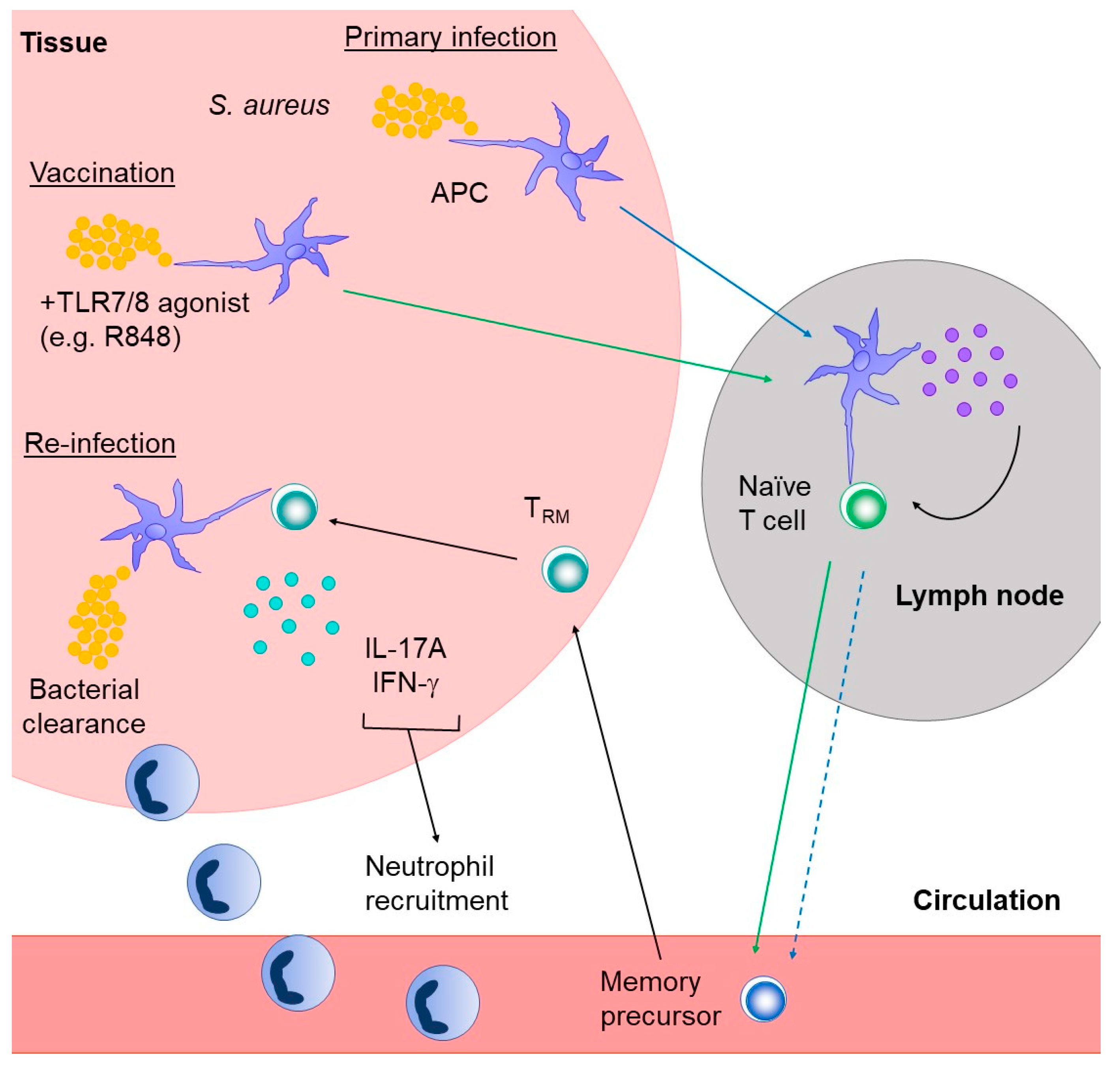
4.2.3. Tissue Resident Memory T Cells

Tissue-resident memory T cells (TRM) are a recently described memory T cell subset that is thought to reside in tissues long-term. Similar to other T cell memory subsets, TRM respond rapidly and specifically upon re-infection [95], but have the added advantage to be present at the site of injury and therefore could provide a fast and efficient response against insults. The exact mechanisms underlying the differentiation of TRM are not fully understood. Studies using different infection models [95,96,97], indicate that TRM cells develop from memory precursors that enter the tissue during the effector phase of infection and remain in the tissue afterwards [98]. However, the exact regulation of this process is unknown.
One mechanism by which tissue retention of TRM is thought to occur is through the antagonism of sphingosine 1-phosphate receptor-1 (S1PR1) by the surface molecule CD69, also called early T cell-activation antigen P60 [99]. S1PR1 regulates the egress of T cells from tissues and CD69 likely binds S1PR1 directly, causing a conformational change that interferes with its function [100]. Of note, the expression of S1PR1 along with other genes involved in secondary lymphoid organ (SLO) recirculation are regulated by the transcription factor Krupple-like factor 2 (KLF2), which is downregulated in TRM cells [101]. Additionally, if the expression of S1PR1 is induced outside of its typical regulation, it reduces the frequency of cells localizing to non-lymphoid tissues. CD103 (integrin αE) and CD49a (integrin α1) are other surface markers associated with the TRM phenotype that might aid in the retention of these cells in the tissues. CD103 binds E-cadherin with integrin beta 7 and is thought to enable the cells to enter and remain in the tissue. CD49a binds collagen along with CD29 (integrin beta 1) to help retain cells in collagen-rich tissues [102]. Additionally, TRM lack the expression of the chemokine receptors CCR7 and CD62L, which are required for homing to lymph nodes.
Several studies have detailed the importance of TRM during re-infection at specific sites such as, the lungs, skin, and female reproductive tract. Our current knowledge of TRM biology is largely based on studies focusing on CD8+ TRM cells residing in the tissue long-term, with many fewer studies detailing the characteristics of CD4+ TRM. Therefore, the bulk of the data discussed below is based on CD8+ studies. Similarities and differences with CD4+ TRM are highlighted/discussed when appropriate.
As alluded to above, the mechanisms underlying TRM cell differentiation and function are poorly defined. In addition to KLF2, mentioned above, there are several other transcription factors that may play a role in the establishment of TRM tissue residency, including Blimp-1, Hobit, T-bet, Eomes, and Runx3. Mackay et al. found that when the transcription factors Hobit and Blimp-1 were knocked out, significantly fewer TRM cells were found in the skin, gut, liver, and kidney in herpes simplex virus (HSV) and lymphocytic choriomeningitis virus (LCMV) experimental infection models [103]. This study indicated that Blimp-1 and Hobit work together to modify transcriptional profiles that are required for TRM differentiation and potentially, for function. Similarly, deletion of the transcription factor Runx3 prevented the formation of TRM cells, especially CD69+ CD103+ TRM cells localized to the gut after LCMV infection [104]. This study concluded that Runx3 was a transcriptional regulator that upregulated core genes responsible for tissue residency and downregulated genes important for circulating properties. Additionally, the downregulation of two other transcription factors, T-bet and Eomes, caused by TGF-β signaling, was seen in TRM cells migrating to the skin after HSV infection [105]. However, a complete ablation of T-bet resulted in the loss of these cells over time. This was likely because of the low expression of the IL-15 Receptor (IL15R) β chain in these cells. IL-15 signaling is necessary for the long-term survival of TRM cells in the skin.
How cytokine signaling determines the fate of tissue-resident memory T cells is not well understood however, several studies have suggested that TGFβ plays a role in programming T cells to home to and reside in peripheral tissues. TGFβ stimulation of activated CD8+ T cells in vitro causes a gene regulation signature that closely resembles that seen in TRM, which suggests that the TRM genetic signature could be regulated in part by TGFβ signaling [106]. One study demonstrated that TGFβ released from DCs in the lymph nodes before infection predisposed CD8+ T cells to home to the tissues after antigen stimulation [107]. Similarly, Thompson et al. found that TGFβ released by monocytes caused the upregulation of CD103 on naïve T cells, which helped the T cells to migrate to peripheral tissues, such as the lung [108]. Additionally, TGFβ was required for maintaining TRM within the intestines, which was linked to the induction of CD69 [109].
Although the vast majority of TRM studies in murine models focus on CD8+ T cells, at least three different studies indicate that CD4+ TRM cells are important for protection against bacterial infection. Wilk et al. showed that CD4+ TRM (CD44hi CD62Llo CD69+ CD103+/−) cells could be found in the lungs of mice after Bordetella pertussis infection and that these cells conferred protection against re-infection [110]. Another group demonstrated that CD4+ TRM found in the lungs can protect against Klebsiella pneumoniae infection without the help of circulating CD4+ T cells [111]. Importantly, a 2015 study by Stary et al. found that vaccination-induced protection against Chlamydia trachomatis infection was mediated at least in part by CD4+ TRM cells induced in the female reproductive tract of mice [112]. This result suggests that vaccines can be designed to manipulate the immune response to optimize the induction of TRM cells that can protect against bacterial infection.
TRM surface markers identified in humans correspond with surface markers found in murine TRM, including CD69, CD103, and CD49a [113]. However, a direct examination of the function of these cells in patients has not been performed and thus, the requirement of TRM for protection against bacterial infection in humans remains to be established. Nonetheless, at least a few studies indicate that in humans, both CD8+ and CD4+ TRM are required for vaccine-induced protection against viruses. Studies performed in human tissues, including the lung and skin, identified the presence of CD4+ and CD8+ T cells with specificity to ubiquitous human pathogens such as influenza [114,115], herpes simplex virus (HSV) [116], cytomegalovirus (CMV) [117], and respiratory syncytial virus (RSV) [118]. Additionally, one study looking at RSV-specific TRM cells correlated an increase in TRM cells with lower viral titers [118], suggesting that TRM cells help mediate the immune response upon re-infection, thereby providing protection.
The induction and functional requirement of TRM for long-term protective immunity against S. aureus remains to be investigated. However, if the generation of TRM cells upon primary infection with other bacteria is a requirement for protective immunity, as suggested by the data discussed above, then one could speculate that primary responses to S. aureus infection might not result in TRM cell generation (Figure 2). The generation of skin-specific TRM cells has been observed in several other infectious models, including Leishmania major [119], vaccinia virus (VACV) [120], LCMV [121], and HSV [122]. In these studies, TRM were shown to provide protection against re-infection. Furthermore, Linehan et al. found that S. epidermidis-specific CD8+ TRM cells were created after an application of a strain from a particular clade (NIHLM087 from the A20 clade) [123]. This study is of particular interest because S. epidermidis is closely related to S. aureus however, S. epidermidis is exclusively part of the microbiome, whereas S. aureus can cause pathogenic infections. Therefore, it is unlikely that the same vaccination strategy would work to induce the development of anti-S. aureus specific protective TRM cells, but that remains to be tested. Nonetheless, the fact that protective TRM responses can be generated against S. epidermidis is encouraging and might help to inform the criteria for the development of novel vaccination approaches against S. aureus. Future studies should focus on what aids in creating S. aureus-specific TRM cells and the possible hurdles that need to be overcome to create S. aureus-specific TRM cells.

5. Concluding Remarks and Future Directions

Despite promising pre-clinical studies, every anti-staphylococcal vaccine developed to date has failed in clinical trials. It is still unclear why these experimental vaccines have not translated into successful vaccines and what a successful vaccine would look like, but there have been notable advances in our understanding of how long-lasting protective immunity to pathogens develop. Moving forward, the field will benefit from this new understanding as well as of a more comprehensive characterization of T cell-mediated immunity to S. aureus, especially after secondary infections. This knowledge should provide a framework for the development of strategies to mitigate the manipulation of adaptive immunity by S. aureus, and thus inform the development of novel and efficacious vaccines. Clarifying if natural infection with S. aureus leads to the differentiation of antigen-specific TRM and their potential function (or disfunction) in both experimental models and in patients is just one of the many aspects that will contribute to the development of a protective vaccine against S. aureus. The development of new tools that facilitate the characterization of the S. aureus-specific T cell responses, such as S. aureus-specific TCR transgenic mice, will also greatly benefit the field.

Author Contributions

Writing—original draft preparation and editing, E.I.A.; writing—review and editing, G.Y.L.; writing—review and editing, G.A.M. All authors have read and agreed to the published version of the manuscript.

Funding

This research was funded by NIH research grant 2R01AI103542-06 and 1R21AI151987 (to G.A.M.), R01AI144694-01 (to G.Y.L.), R01 AI127406 (to G.Y.L. and G.A.M.) and by internal funds from the Biological Sciences Dept. CSMC (to G.A.M.).

Acknowledgments

We are grateful to the members of the Martins and Liu labs for discussions.

Conflicts of Interest

The authors declare no conflict of interest.

References

  1. Anderson, D.J.; Sexton, D.J.; Kanafani, Z.A.; Auten, G.; Kaye, K.S. Severe surgical site infection in community hospitals: Epidemiology, key procedures, and the changing prevalence of methicillin-resistant Staphylococcus aureus. Infect. Control. Hosp. Epidemiol. 2007, 28, 1047–1053. [Google Scholar] [CrossRef]
  2. Berríos-Torres, S.I.; Umscheid, C.A.; Bratzler, D.W.; Leas, B.; Stone, E.C.; Kelz, R.R.; Reinke, C.E.; Morgan, S.; Solomkin, J.S.; Mazuki, J.E.; et al. Centers for Disease Control and Prevention Guideline for the Prevention of Surgical Site Infection. JAMA Surg. 2017, 152, 784–791. [Google Scholar] [CrossRef]
  3. Peng, H.; Liu, D.; Ma, Y.; Gao, W. Comparison of community- and healthcare-associated methicillin-resistant Staphylococcus aureus isolates at a Chinese tertiary hospital, 2012–2017. Sci. Rep. 2018, 8, 17916. [Google Scholar] [CrossRef] [PubMed]
  4. Lee, B.Y.; Singh, A.; David, M.Z.; Bartsch, S.M.; Slayton, R.B.; Huang, S.S.; Zimmer, S.M.; Potter, M.A.; Macal, C.M.; Lauderdale, D.S.; et al. The economic burden of community-associated methicillin-resistant Staphylococcus aureus (CA-MRSA). Clin. Microbiol. Infect. 2013, 19, 528–536. [Google Scholar] [CrossRef] [PubMed] [Green Version]
  5. Acton, D.S.; Plat-Sinnige, M.J.; van Wamel, W.; de Groot, N.; van Belkum, A. Intestinal carriage of Staphylococcus aureus: How does its frequency compare with that of nasal carriage and what is its clinical impact? Eur. J. Clin. Microbiol. Infect. Dis. 2009, 28, 115–127. [Google Scholar] [CrossRef] [PubMed] [Green Version]
  6. van Belkum, A.; Verkaik, N.J.; de Vogel, C.P.; Boelens, H.A.; Verveer, J.; Nouwen, J.L.; Verbrugh, H.A.; Wertheim, H.F. Reclassification of Staphylococcus aureus nasal carriage types. J. Infect. Dis. 2009, 199, 1820–1826. [Google Scholar] [CrossRef] [PubMed] [Green Version]
  7. Jimenez-Truque, N.; Tedeschi, S.; Saye, E.J.; McKenna, B.D.; Langdon, W.; Wright, J.P.; Alsentzer, A.; Arnold, S.; Saville, B.R.; Wang, W.; et al. Relationship between maternal and neonatal Staphylococcus aureus colonization. Pediatrics 2012, 129, e1252-9. [Google Scholar] [CrossRef] [Green Version]
  8. Miller, L.G.; Diep, B.A. Clinical practice: Colonization, fomites, and virulence: Rethinking the pathogenesis of community-associated methicillin-resistant Staphylococcus aureus infection. Clin. Infect. Dis. 2008, 46, 752–760. [Google Scholar] [CrossRef]
  9. Skinner, D.; Keefer, S.C. Significance of bacteremia caused by staphylococcus aureus: A study of one hundred and twenty-two cases and a review of the literature concerned with experimental infection in animals. Arch. Intern. Med. 1941, 68, 851–875. [Google Scholar] [CrossRef]
  10. van Hal, S.J.; Jensen, S.O.; Vaska, V.L.; Espedido, B.A.; Paterson, D.L.; Gosbell, I.B. Predictors of mortality in Staphylococcus aureus Bacteremia. Clin. Microbiol. Rev. 2012, 25, 362–386. [Google Scholar] [CrossRef] [Green Version]
  11. Kopp, B.J.; Nix, D.E.; Armstrong, E.P. Clinical and economic analysis of methicillin-susceptible and -resistant Staphylococcus aureus infections. Ann. Pharm. 2004, 38, 1377–1382. [Google Scholar] [CrossRef] [PubMed]
  12. Giersing, B.K.; Dastgheyb, S.S.; Modjarrad, K.; Moorthy, V. Status of vaccine research and development of vaccines for Staphylococcus aureus. Vaccine 2016, 34, 2962–2966. [Google Scholar] [CrossRef] [PubMed] [Green Version]
  13. Redi, D.; Raffaelli, C.S.; Rossetti, B.; De Luca, A.; Montagnani, F. Staphylococcus aureus vaccine preclinical and clinical development: Current state of the art. N. Microbiol. 2018, 41, 208–213. [Google Scholar]
  14. Fowler, V.G., Jr.; Proctor, R.A. Where does a Staphylococcus aureus vaccine stand? Clin. Microbiol. Infect. 2014, 20 (Suppl. S5), 66–75. [Google Scholar] [CrossRef] [Green Version]
  15. Anderson, A.S.; Miller, A.A.; Donald, R.G.; Scully, I.L.; Nanra, J.S.; Cooper, D.; Jansen, K.U. Development of a multicomponent Staphylococcus aureus vaccine designed to counter multiple bacterial virulence factors. Hum. Vaccines Immunother. 2012, 8, 1585–1594. [Google Scholar] [CrossRef] [PubMed] [Green Version]
  16. Bagnoli, F.; Bertholet, S.; Grandi, G. Inferring reasons for the failure of Staphylococcus aureus vaccines in clinical trials. Front. Cell Infect. Microbiol. 2012, 2, 16. [Google Scholar] [CrossRef] [PubMed] [Green Version]
  17. Schulz, D.; Grumann, D.; Trübe, P.; Pritchett-Corning, K.; Johnson, S.; Reppschläger, K.; Gumz, J.; Sundaramoorthy, N.; Michalik, S.; Berg, S.; et al. Laboratory Mice Are Frequently Colonized with Staphylococcus aureus and Mount a Systemic Immune Response-Note of Caution for In vivo Infection Experiments. Front. Cell Infect. Microbiol. 2017, 7, 152. [Google Scholar] [CrossRef] [PubMed] [Green Version]
  18. Pritchett-Corning, K.R.; Cosentino, J.; Clifford, C.B. Contemporary prevalence of infectious agents in laboratory mice and rats. Lab. Anim. 2009, 43, 165–173. [Google Scholar] [CrossRef]
  19. Benjamin, D.K.; Schelonka, R.; White, R.; Holley, H.P.; Bifano, E.; Cummings, J.; Adcock, K.; Kaufman, D.; Puppala, B.; Riedel, P.; et al. A blinded, randomized, multicenter study of an intravenous Staphylococcus aureus immune globulin. J. Perinatol. 2006, 26, 290–295. [Google Scholar] [CrossRef] [Green Version]
  20. Rupp, M.E.; Holley, H.P., Jr.; Lutz, J.; Dicpinigaitis, P.V.; Woods, C.W.; Levine, D.P.; Veney, N.; Fowler, V.G., Jr. Phase II, randomized, multicenter, double-blind, placebo-controlled trial of a polyclonal anti-Staphylococcus aureus capsular polysaccharide immune globulin in treatment of Staphylococcus aureus bacteremia. Antimicrob. Agents Chemother. 2007, 51, 4249–4254. [Google Scholar] [CrossRef] [Green Version]
  21. DeJonge, M.; Burchfield, D.; Bloom, B.; Duenas, M.; Walker, W.; Polak, M.; Jung, E.; Millard, D.; Schelonka, R.; Eyal, F.; et al. Clinical trial of safety and efficacy of INH-A21 for the prevention of nosocomial staphylococcal bloodstream infection in premature infants. J. Pediatr. 2007, 151, 260–265.e1. [Google Scholar] [CrossRef] [PubMed]
  22. Weems, J.J., Jr.; Steinberg, J.P.; Filler, S.; Baddley, J.W.; Corey, G.R.; Sampathkumar, P.; Winston, L.; John, J.F.; Kubin, C.J.; Talwani, R.; et al. Phase II, randomized, double-blind, multicenter study comparing the safety and pharmacokinetics of tefibazumab to placebo for treatment of Staphylococcus aureus bacteremia. Antimicrob. Agents Chemother. 2006, 50, 2751–2755. [Google Scholar] [CrossRef] [PubMed] [Green Version]
  23. Magyarics, Z.; Leslie, F.; Bartko, J.; Rouha, H.; Luperchio, S.; Schörgenhofer, C.; Schwameis, M.; Derhaschnig, U.; Lagler, H.; Stiebellehner, L.; et al. Randomized, Double-Blind, Placebo-Controlled, Single-Ascending-Dose Study of the Penetration of a Monoclonal Antibody Combination (ASN100) Targeting Staphylococcus aureus Cytotoxins in the Lung Epithelial Lining Fluid of Healthy Volunteers. Antimicrob. Agents Chemother. 2019, 63, e00350-19. [Google Scholar] [CrossRef] [PubMed] [Green Version]
  24. Weisman, L.E.; Thackray, H.M.; Steinhorn, R.H.; Walsh, W.F.; Lassiter, H.A.; Dhanireddy, R.; Brozanski, B.S.; Palmer, K.G.; Trautman, M.S.; Escobedo, M.; et al. A randomized study of a monoclonal antibody (pagibaximab) to prevent staphylococcal sepsis. Pediatrics 2011, 128, 271–279. [Google Scholar] [CrossRef]
  25. Yu, X.Q.; Robbie, G.J.; Wu, Y.; Esse, M.T.; Jensen, K.; Schwartz, H.I.; Bellamy, T.; Hernandez-Illas, M.; Jafri, H.S. Safety, Tolerability, and Pharmacokinetics of MEDI4893, an Investigational, Extended-Half-Life, Anti-Staphylococcus aureus Alpha-Toxin Human Monoclonal Antibody, in Healthy Adults. Antimicrob. Agents Chemother. 2017, 61, e01020-16. [Google Scholar] [CrossRef] [Green Version]
  26. François, B.; Mercier, E.; Gonzalez, C.; Asehnoune, K.; Nseir, S.; Fiancette, M.; Desachy, A.; Plantefève, G.; Meziani, F.; de Lame, P.A.; et al. Safety and tolerability of a single administration of AR-301, a human monoclonal antibody, in ICU patients with severe pneumonia caused by Staphylococcus aureus: First-in-human trial. Intensive Care Med. 2018, 44, 1787–1796. [Google Scholar] [CrossRef]
  27. Fowler, V.G.; Allen, K.B.; Moreira, E.D.; Moustafa, M.; Isgro, F.; Boucher, H.W.; Corey, R.; Carmeli, C.; Betts, R.; Harzel, J.S.; et al. Effect of an investigational vaccine for preventing Staphylococcus aureus infections after cardiothoracic surgery: A randomized trial. JAMA 2013, 309, 1368–1378. [Google Scholar] [CrossRef]
  28. McNeely, T.B.; Shah, N.A.; Fridman, A.; Joshi, A.; Hartzel, J.S.; Keshari, R.S.; Lupu, F.; DiNubile, M.J. Mortality among recipients of the Merck V710 Staphylococcus aureus vaccine after postoperative S. aureus infections: An analysis of possible contributing host factors. Hum. Vaccines Immunother. 2014, 10, 3513–3516. [Google Scholar] [CrossRef] [Green Version]
  29. Aman, M.J. Integrated BioTherapeutics. Hum. Vaccines Immunother. 2018, 14, 1308–1310. [Google Scholar] [CrossRef] [Green Version]
  30. Landrum, M.L.; Lalani, T.; Niknian, M.; Maguire, J.D.; Hospenthal, D.R.; Fattom, A.; Taylor, K.; Fraser, J.; Wilkins, K.; Ellis, M.W.; et al. Safety and immunogenicity of a recombinant Staphylococcus aureus α-toxoid and a recombinant Panton-Valentine leukocidin subunit, in healthy adults. Hum. Vaccines Immunother. 2017, 13, 791–801. [Google Scholar] [CrossRef] [Green Version]
  31. Chen, W.H.; Pasetti, M.F.; Adhikari, R.P.; Baughman, H.; Douglas, R.; El-Khorazaty, J.; Greenberg, N.; Holtsberg, F.W.; Liao, G.C.; Reymann, M.K.; et al. Safety and Immunogenicity of a Parenterally Administered, Structure-Based Rationally Modified Recombinant Staphylococcal Enterotoxin B Protein Vaccine, STEBVax. Clin. Vaccines Immunol. 2016, 23, 918–925. [Google Scholar] [CrossRef] [PubMed] [Green Version]
  32. Schmidt, C.S.; White, C.J.; Ibrahim, A.S.; Filler, S.G.; Fu, Y.; Yeaman, M.R.; Edwards, J.E., Jr.; Hennessey, J.P., Jr. NDV-3, a recombinant alum-adjuvanted vaccine for Candida and Staphylococcus aureus, is safe and immunogenic in healthy adults. Vaccine 2012, 30, 7594–7600. [Google Scholar] [CrossRef] [PubMed] [Green Version]
  33. Gurtman, A.; Begier, E.; Mohamed, N.; Baber, J.; Sabharwal, C.; Haupt, R.M.; Edwards, H.; Cooper, D.; Jansen, K.U.; Anderson, A.S. The development of a staphylococcus aureus four antigen vaccine for use prior to elective orthopedic surgery. Hum. Vaccines Immunother. 2019, 15, 358–370. [Google Scholar] [CrossRef] [PubMed]
  34. Fattom, A.; Matalon, A.; Buerkert, J.; Taylor, K.; Damaso, S.; Boutriau, D. Efficacy profile of a bivalent Staphylococcus aureus glycoconjugated vaccine in adults on hemodialysis: Phase III randomized study. Hum. Vaccines Immunother. 2015, 11, 632–641. [Google Scholar] [CrossRef] [Green Version]
  35. Fattom, A.I.; Horwith, G.; Fuller, S.; Propst, M.; Naso, R. Development of StaphVAX, a polysaccharide conjugate vaccine against S. aureus infection: From the lab bench to phase III clinical trials. Vaccine 2004, 22, 880–887. [Google Scholar] [CrossRef]
  36. Spaan, A.N.; Surewaard, B.G.; Nijland, R.; van Strijp, J.A. Neutrophils versus Staphylococcus aureus: A biological tug of war. Annu. Rev. Microbiol. 2013, 67, 629–650. [Google Scholar] [CrossRef] [Green Version]
  37. Thomsen, I.P.; Liu, G.Y. Targeting fundamental pathways to disrupt Staphylococcus aureus survival: Clinical implications of recent discoveries. JCI Insight 2018, 3, e98216. [Google Scholar] [CrossRef] [Green Version]
  38. Deshmukh, H.S.; Hamburger, J.B.; Ahn, S.H.; McCafferty, D.G.; Yang, S.R.; Fowler, V.G., Jr. Critical role of NOD2 in regulating the immune response to Staphylococcus aureus. Infect. Immun. 2009, 77, 1376–1382. [Google Scholar] [CrossRef] [Green Version]
  39. Miller, L.S.; O’Connell, R.M.; Gutierrez, M.A.; Pietras, E.M.; Shahangian, A.; Gross, C.E.; Thirumala, A.; Cheung, A.L.; Cheng, G.; Modlin, R.L. MyD88 mediates neutrophil recruitment initiated by IL-1R but not TLR2 activation in immunity against Staphylococcus aureus. Immunity 2006, 24, 79–91. [Google Scholar] [CrossRef] [Green Version]
  40. Takeuchi, O.; Hoshino, K.; Akira, S. Cutting edge: TLR2-deficient and MyD88-deficient mice are highly susceptible to Staphylococcus aureus infection. J. Immunol. 2000, 165, 5392–5396. [Google Scholar] [CrossRef] [Green Version]
  41. Ku, C.L.; von Bernuth, H.; Picard, C.; Zhang, S.Y.; Chang, H.H.; Yang, K.; Chrabieh, M.; Issekutz, A.C.; Cunningham, C.K.; Gallin, J.; et al. Selective predisposition to bacterial infections in IRAK-4-deficient children: IRAK-4-dependent TLRs are otherwise redundant in protective immunity. J. Exp. Med. 2007, 204, 2407–2422. [Google Scholar] [CrossRef] [PubMed]
  42. Picard, C.; Puel, A.; Bonnet, M.; Ku, C.L.; Bustamante, J.; Yang, K.; Soudais, C.; Dupuis, S.; Feinberg, J.; Fieschi, C.; et al. Pyogenic bacterial infections in humans with IRAK-4 deficiency. Science 2003, 299, 2076–2079. [Google Scholar] [CrossRef] [PubMed]
  43. Picard, C.; von Bernuth, H.; Ghandil, P.; Chrabieh, M.; Levy, O.; Arkwright, P.D.; McDonald, D.; Geha, R.S.; Takada, H.; Krause, J.C.; et al. Clinical features and outcome of patients with IRAK-4 and MyD88 deficiency. Medicine 2010, 89, 403–425. [Google Scholar] [CrossRef] [PubMed] [Green Version]
  44. von Bernuth, H.; Picard, C.; Jin, Z.; Pankla, R.; Xiao, H.; Ku, C.L.; Chrabieh, M.; Mustapha, I.B.; Ghandil, P.; Camcioglu, Y.; et al. Pyogenic bacterial infections in humans with MyD88 deficiency. Science 2008, 321, 691–696. [Google Scholar] [CrossRef] [PubMed] [Green Version]
  45. Fournier, B.; Philpott, D.J. Recognition of Staphylococcus aureus by the innate immune system. Clin. Microbiol. Rev. 2005, 18, 521–540. [Google Scholar] [CrossRef] [Green Version]
  46. Lee, B.; Olaniyi, R.; Kwiecinski, J.M.; Wardenburg, J.B. Staphylococcus aureus toxin suppresses antigen-specific T cell responses. J. Clin. Investig. 2020, 130, 1122–1127. [Google Scholar] [CrossRef] [Green Version]
  47. Andrews, T.; Sullivan, K.E. Infections in patients with inherited defects in phagocytic function. Clin. Microbiol. Rev. 2003, 16, 597–621. [Google Scholar] [CrossRef] [Green Version]
  48. Bouma, G.; Ancliff, P.J.; Thrasher, A.J.; Burns, S.O. Recent advances in the understanding of genetic defects of neutrophil number and function. Br. J. Haematol. 2010, 151, 312–326. [Google Scholar] [CrossRef]
  49. González-Barca, E.; Carratalà, J.; Mykietiuk, A.; Fernández-Sevilla, A.; Gudiol, F. Predisposing factors and outcome of Staphylococcus aureus bacteremia in neutropenic patients with cancer. Eur. J. Clin. Microbiol. Infect. Dis. 2001, 20, 117–119. [Google Scholar] [CrossRef]
  50. Lakshman, R.; Finn, A. Neutrophil disorders and their management. J. Clin. Pathol. 2001, 54, 7–19. [Google Scholar] [CrossRef]
  51. Collins, L.V.; Kristian, S.A.; Weidenmaier, C.; Faigle, M.; Van Kessel, K.P.; Van Strijp, J.A.; Götz, F.; Neumeister, B.; Peschel, A. Staphylococcus aureus strains lacking D-alanine modifications of teichoic acids are highly susceptible to human neutrophil killing and are virulence attenuated in mice. J. Infect. Dis. 2002, 186, 214–219. [Google Scholar] [CrossRef] [PubMed] [Green Version]
  52. Liu, G.Y.; Essex, A.; Buchanan, J.T.; Datta, V.; Hoffman, H.M.; Bastian, J.F.; Fierer, J.; Nizet, V. Staphylococcus aureus golden pigment impairs neutrophil killing and promotes virulence through its antioxidant activity. J. Exp. Med. 2005, 202, 209–215. [Google Scholar] [CrossRef] [PubMed]
  53. Thammavongsa, V.; Missiakas, D.M.; Schneewind, O. Staphylococcus aureus degrades neutrophil extracellular traps to promote immune cell death. Science 2013, 342, 863–866. [Google Scholar] [CrossRef] [PubMed] [Green Version]
  54. Sanchez, M.; Kolar, S.L.; Müller, S.; Reyes, C.N.; Wolf, A.J.; Ogawa, C.; Singhania, R.; De Carvalho, D.D.; Arditi, M.; Underhill, D.M.; et al. O-Acetylation of Peptidoglycan Limits Helper T Cell Priming and Permits Staphylococcus aureus Reinfection. Cell Host Microbe 2017, 22, 543–551.e4. [Google Scholar] [CrossRef] [PubMed] [Green Version]
  55. Romero Pastrana, F.; Neef, J.; Koedijk, D.; de Graaf, D.; Duipmans, J.; Jonkman, M.F.; Engelmann, S.; van Dijl, J.M.; Buist, G. Human antibody responses against non-covalently cell wall-bound Staphylococcus aureus proteins. Sci. Rep. 2018, 8, 3234. [Google Scholar] [CrossRef]
  56. Stolz, S.J.; Davis, J.P.; Vergeront, J.M.; Crass, B.A.; Chesney, P.J.; Wand, P.J.; Bergdoll, M.S. Development of serum antibody to toxic shock toxin among individuals with toxic shock syndrome in Wisconsin. J. Infect. Dis. 1985, 151, 883–889. [Google Scholar] [CrossRef]
  57. Wu, Y.; Liu, X.; Akhgar, A.; Li, J.J.; Mok, H.; Sellman, B.R.; Yu, L.; Roskos, L.K.; Esser, M.T.; Ruzin, A. Prevalence of IgG and Neutralizing Antibodies against Staphylococcus aureus Alpha-Toxin in Healthy Human Subjects and Diverse Patient Populations. Infect. Immun. 2018, 86, e00671-17. [Google Scholar]
  58. Thammavongsa, V.; Kim, H.K.; Missiakas, D.; Schneewind, O. Staphylococcal manipulation of host immune responses. Nat. Rev. Microbiol. 2015, 13, 529–543. [Google Scholar] [CrossRef] [Green Version]
  59. Goodyear, C.S.; Silverman, G.J. Death by a B cell superantigen: In vivo VH-targeted apoptotic supraclonal B cell deletion by a Staphylococcal Toxin. J. Exp. Med. 2003, 197, 1125–1139. [Google Scholar] [CrossRef]
  60. Adhikari, R.P.; Ajao, A.O.; Aman, M.J.; Karauzum, H.; Sarwar, J.; Lydecker, A.D.; Johnson, J.K.; Nguyen, C.; Chen, W.H.; Roghmann, M.C. Lower antibody levels to Staphylococcus aureus exotoxins are associated with sepsis in hospitalized adults with invasive S. aureus infections. J. Infect. Dis. 2012, 206, 915–923. [Google Scholar] [CrossRef]
  61. Fritz, S.A.; Tiemann, K.M.; Hogan, P.G.; Epplin, E.K.; Rodriguez, M.; Al-Zubeidi, D.N.; Bubeck Wardenburg, J.; Hunstad, D.A. A serologic correlate of protective immunity against community-onset Staphylococcus aureus infection. Clin. Infect. Dis. 2013, 56, 1554–1561. [Google Scholar] [CrossRef] [PubMed] [Green Version]
  62. Tsai, C.M.; Soper, N.; Bennett, M.; Fallon, J.K.; Michell, A.R.; Alter, G.; Liu, G.Y.; Thomsen, I. Adoptive Transfer of Serum Samples from Children with Invasive Staphylococcal Infection and Protection Against Staphylococcus aureus Sepsis. J. Infect. Dis. 2020, jiaa482. [Google Scholar] [CrossRef] [PubMed]
  63. Zhang, F.; Ledue, O.; Jun, M.; Goulart, C.; Malley, R.; Lu, Y.J. Protection against Staphylococcus aureus Colonization and Infection by B- and T-Cell-Mediated Mechanisms. mBio 2018, 9, e01949-18. [Google Scholar] [CrossRef] [PubMed] [Green Version]
  64. Paterson, M.J.; Caldera, J.R.; Nguyen, C.; Sharma, P.; Castro, A.M.; Kolar, S.L.; Tsai, C.M.; Limon, J.J.; Becker, C.A.; Martins, G.A.; et al. Harnessing antifungal immunity in pursuit of a Staphylococcus aureus vaccine strategy. PLoS Pathog. 2020, 16, e1008733. [Google Scholar] [CrossRef]
  65. Dhalla, F.; Misbah, S.A. Secondary antibody deficiencies. Curr. Opin Allergy Clin. Immunol. 2015, 15, 505–513. [Google Scholar] [CrossRef]
  66. Hoernes, M.; Seger, R.; Reichenbach, J. Modern management of primary B-cell immunodeficiencies. Pediatr. Allergy Immunol. 2011, 22, 758–769. [Google Scholar] [CrossRef]
  67. Cho, J.S.; Pietras, E.M.; Garcia, N.C.; Ramos, R.I.; Farzam, D.M.; Monroe, H.R.; Magorien, J.E.; Blauvelt, A.; Kolls, J.K.; Cheung, A.L.; et al. IL-17 is essential for host defense against cutaneous Staphylococcus aureus infection in mice. J. Clin. Investif. 2010, 120, 1762–1773. [Google Scholar] [CrossRef] [Green Version]
  68. Marchitto, M.C.; Dillen, C.A.; Liu, H.; Miller, R.J.; Archer, N.K.; Ortines, R.V.; Alphonse, M.P.; Marusina, A.I.; Merleev, A.A.; Wang, Y.; et al. Clonal Vγ6(+)Vδ4(+) T cells promote IL-17-mediated immunity against Staphylococcus aureus skin infection. Proc. Natl. Acad. Sci. USA 2019, 116, 10917–10926. [Google Scholar] [CrossRef] [Green Version]
  69. Sheridan, B.S.; Romagnoli, P.A.; Pham, Q.M.; Fu, H.H.; Alonzo, F., 3rd; Schubert, W.D.; Freitag, N.E.; Lefrançois, L. γδ T cells exhibit multifunctional and protective memory in intestinal tissues. Immunity 2013, 39, 184–195. [Google Scholar] [CrossRef] [Green Version]
  70. Luckheeram, R.V.; Zhou, R.; Verma, A.D.; Xia, B. CD4⁺T cells: Differentiation and functions. Clin. Dev. Immunol. 2012, 2012, 925135. [Google Scholar] [CrossRef] [Green Version]
  71. Lowy, F.D. Is Staphylococcus aureus an intracellular pathogen? Trends Microbiol. 2000, 8, 341–343. [Google Scholar] [CrossRef]
  72. Joshi, A.; Pancari, G.; Cope, L.; Bowman, E.P.; Cua, D.; Proctor, R.A.; McNeely, T. Immunization with Staphylococcus aureus iron regulated surface determinant B (IsdB) confers protection via Th17/IL17 pathway in a murine sepsis model. Hum. Vaccines Immunother. 2012, 8, 336–346. [Google Scholar] [CrossRef] [PubMed] [Green Version]
  73. Montgomery, C.P.; Daniels, M.; Zhao, F.; Alegre, M.L.; Chong, A.S.; Daum, R.S. Protective immunity against recurrent Staphylococcus aureus skin infection requires antibody and interleukin-17A. Infect. Immun. 2014, 82, 2125–2134. [Google Scholar] [CrossRef] [PubMed] [Green Version]
  74. Lee, L.Y.; Leech, J.M.; Rogers, T.R.; McLoughlin, R.M. The Staphylococcus aureus Map protein is an immunomodulator that interferes with T cell-mediated responses. J. Clin. Investig. 2002, 110, 1461–1471. [Google Scholar] [CrossRef] [PubMed]
  75. Alonzo, F., 3rd; Kozhaya, L.; Rawlings, S.A.; Reyes-Robles, T.; DuMont, A.L.; Myszka, D.G.; Landau, N.R.; Unutmaz, D.; Torres, V.J. CCR5 is a receptor for Staphylococcus aureus leukotoxin ED. Nature 2013, 493, 51–55. [Google Scholar] [CrossRef] [PubMed] [Green Version]
  76. Crum-Cianflone, N.F.; Grandits, G.; Echols, S.; Ganesan, A.; Landrum, M.; Weintrob, A.; Barthel, R.; Agan, B.; Infectious Disease Clinical Research Program. Trends and causes of hospitalizations among HIV-infected persons during the late HAART era: What is the impact of CD4 counts and HAART use? J. Acquir Immun. Defic. Syndr. 2010, 54, 248–257. [Google Scholar] [CrossRef] [Green Version]
  77. Manfredi, R.; Calza, L.; Chiodo, F. Epidemiology and microbiology of cellulitis and bacterial soft tissue infection during HIV disease: A 10-year survey. J. Cutan. Pathol. 2002, 29, 168–172. [Google Scholar] [CrossRef]
  78. Manfredi, R.; Costigliola, P.; Ricchi, E.; Chiodo, F. Sepsis-bacteraemia and other infections due to non-opportunistic bacterial pathogens in a consecutive series of 788 patients hospitalized for HIV infection. Clin. Ter. 1993, 143, 279–290. [Google Scholar]
  79. Beekhuizen, H.; van de Gevel, J.S. Gamma Interferon Confers Resistance to Infection with Staphylococcus aureus in Human Vascular Endothelial Cells by Cooperative Proinflammatory and Enhanced Intrinsic Antibacterial Activities. Infect. Immun. 2007, 75, 5615–5626. [Google Scholar] [CrossRef] [Green Version]
  80. Cooper, A.J.R.; Lalor, S.J.; McLoughlin, R.M. Activation of Human Vδ2+ γδ T Cells by Staphylococcus aureus Promotes Enhanced Anti-Staphylococcal Adaptive Immunity. J. Immunol. 2020, 205, 1039–1049. [Google Scholar] [CrossRef]
  81. De Forge, L.E.; Billeci, K.L.; Kramer, S.M. Effect of IFN-gamma on the killing of S. aureus in human whole blood. Assessment of bacterial viability by CFU determination and by a new method using alamarBlue. J. Immunol. Methods 2000, 245, 79–89. [Google Scholar]
  82. Brown, A.F.; Murphy, A.G.; Lalor, S.J.; Leech, J.M.; O’Keeffe, K.M.; Mac Aogáin, M.; O’Halloran, D.P.; Lacey, K.A.; Tavakol, M.; Hearnden, C.H.; et al. Memory Th1 Cells Are Protective in Invasive Staphylococcus aureus Infection. PLoS Pathog. 2015, 11, e1005226. [Google Scholar] [CrossRef] [PubMed]
  83. Narita, K.; Hu, D.L.; Mori, F.; Wakabayashi, K.; Iwakura, Y.; Nakane, A. Role of interleukin-17A in cell-mediated protection against Staphylococcus aureus infection in mice immunized with the fibrinogen-binding domain of clumping factor A. Infect. Immun. 2010, 78, 4234–4242. [Google Scholar] [CrossRef] [PubMed] [Green Version]
  84. Lin, L.; Ibrahim, A.S.; Xu, X.; Farber, J.M.; Avanesian, V.; Baquir, B.; Fu, Y.; French, S.W.; Edwards, J.E., Jr.; Spellberg, B. Th1-Th17 cells mediate protective adaptive immunity against Staphylococcus aureus and Candida albicans infection in mice. PLoS Pathog. 2009, 5, e1000703. [Google Scholar] [CrossRef]
  85. Nurjadi, D.; Kain, M.; Marcinek, P.; Gaile, M.; Heeg, K.; Zanger, P. Ratio of T-Helper Type 1 (Th1) to Th17 Cytokines in Whole Blood Is Associated with Human β-Defensin 3 Expression in Skin and Persistent Staphylococcus aureus Nasal Carriage. J. Infect. Dis. 2016, 214, 1744–1751. [Google Scholar] [CrossRef] [Green Version]
  86. Ma, C.S.; Chew, G.Y.; Simpson, N.; Priyadarshi, A.; Wong, M.; Grimbacher, B.; Fulcher, D.A.; Tangye, S.G.; Cook, M.C. Deficiency of Th17 cells in hyper IgE syndrome due to mutations in STAT3. J. Exp. Med. 2008, 205, 1551–1557. [Google Scholar] [CrossRef] [Green Version]
  87. Milner, J.D.; Brenchley, J.M.; Laurence, A.; Freeman, A.F.; Hill, B.J.; Elias, K.M.; Kanno, Y.; Spalding, C.; Elloumi, H.Z.; Paulson, M.L.; et al. Impaired T(H)17 cell differentiation in subjects with autosomal dominant hyper-IgE syndrome. Nature 2008, 452, 773–776. [Google Scholar] [CrossRef] [Green Version]
  88. Renner, E.D.; Rylaarsdam, S.; Anover-Sombke, S.; Rack, A.L.; Reichenbach, J.; Carey, J.C.; Zhu, Q.; Jansson, A.F.; Barboza, J.; Schimke, L.F.; et al. Novel signal transducer and activator of transcription 3 (STAT3) mutations, reduced T(H)17 cell numbers, and variably defective STAT3 phosphorylation in hyper-IgE syndrome. J. Allergy Clin. Immunol. 2008, 122, 181–187. [Google Scholar] [CrossRef]
  89. Wu, J.; Ding, Y.; Wang, J.; Wang, F. Staphylococcus aureus induces TGF-β(1) and bFGF expression through the activation of AP-1 and NF-κB transcription factors in bovine mammary epithelial cells. Microb. Pathog. 2018, 117, 276–284. [Google Scholar] [CrossRef]
  90. Zhang, L.J.; Chen, S.X.; Guerrero-Juarez, C.F.; Li, F.; Tong, Y.; Liang, Y.; Liggins, M.; Chen, X.; Chen, H.; Li, M.; et al. Age-Related Loss of Innate Immune Antimicrobial Function of Dermal Fat Is Mediated by Transforming Growth Factor Beta. Immunity 2019, 50, 121–136.e5. [Google Scholar] [CrossRef] [Green Version]
  91. Leech, J.M.; Lacey, K.A.; Mulcahy, M.E.; Medina, E.; McLoughlin, R.M. IL-10 Plays Opposing Roles during Staphylococcus aureus Systemic and Localized Infections. J. Immunol. 2017, 198, 2352–2365. [Google Scholar] [CrossRef] [PubMed] [Green Version]
  92. Gjertsson, I.; Hultgren, O.H.; Tarkowski, A. Interleukin-10 ameliorates the outcome of Staphylococcus aureus arthritis by promoting bacterial clearance. Clin. Exp. Immunol. 2002, 130, 409–414. [Google Scholar] [CrossRef] [PubMed]
  93. Zielinski, C.E.; Mele, F.; Aschenbrenner, D.; Jarrossay, D.; Ronchi, F.; Gattorno, M.; Monticelli, S.; Lanzavecchia, A.; Sallusto, F. Pathogen-induced human TH17 cells produce IFN-γ or IL-10 and are regulated by IL-1β. Nature 2012, 484, 514–518. [Google Scholar] [CrossRef] [PubMed]
  94. Rose, W.E.; Shukla, S.K.; Berti, A.D.; Hayney, M.S.; Henriquez, K.M.; Ranzoni, A.; Cooper, M.A.; Proctor, R.A.; Nizet, V.; et al. Increased Endovascular Staphylococcus aureus Inoculum Is the Link Between Elevated Serum Interleukin 10 Concentrations and Mortality in Patients With Bacteremia. Clin. Infect. Dis. 2017, 64, 1406–1412. [Google Scholar] [CrossRef] [Green Version]
  95. Gebhardt, T.; Wakim, L.M.; Eidsmo, L.; Reading, P.C.; Heath, W.R.; Carbone, F.R. Memory T cells in nonlymphoid tissue that provide enhanced local immunity during infection with herpes simplex virus. Nat. Immunol. 2009, 10, 524–530. [Google Scholar] [CrossRef]
  96. Jiang, X.; Clark, R.A.; Liu, L.; Wagers, A.J.; Fuhlbrigge, R.C.; Kupper, T.S. Skin infection generates non-migratory memory CD8+ T(RM) cells providing global skin immunity. Nature 2012, 483, 227–231. [Google Scholar] [CrossRef]
  97. Masopust, D.; Vezys, V.; Marzo, A.L.; Lefrançois, L. Preferential localization of effector memory cells in nonlymphoid tissue. Science 2001, 291, 2413–2417. [Google Scholar] [CrossRef] [Green Version]
  98. Schenkel, J.M.; Masopust, D. Tissue-resident memory T cells. Immunity 2014, 41, 886–897. [Google Scholar] [CrossRef] [Green Version]
  99. Mackay, L.K.; Braun, A.; Macleod, B.L.; Collins, N.; Tebartz, C.; Bedoui, S.; Carbone, F.R.; Gebhardt, T. Cutting edge: CD69 interference with sphingosine-1-phosphate receptor function regulates peripheral T cell retention. J. Immunol. 2015, 194, 2059–2063. [Google Scholar] [CrossRef] [Green Version]
  100. Bankovich, A.J.; Shiow, L.R.; Cyster, J.G. CD69 suppresses sphingosine 1-phosophate receptor-1 (S1P1) function through interaction with membrane helix 4. J. Biol. Chem. 2010, 285, 22328–22337. [Google Scholar] [CrossRef] [Green Version]
  101. Skon, C.N.; Lee, J.Y.; Anderson, K.G.; Masopust, D.; Hogquist, K.A.; Jameson, S.C. Transcriptional downregulation of S1pr1 is required for the establishment of resident memory CD8+ T cells. Nat. Immunol. 2013, 14, 1285–1293. [Google Scholar] [CrossRef] [PubMed] [Green Version]
  102. Roberts, A.I.; Brolin, R.E.; Ebert, E.C. Integrin alpha1beta1 (VLA-1) mediates adhesion of activated intraepithelial lymphocytes to collagen. Immunology 1999, 97, 679–685. [Google Scholar] [CrossRef] [PubMed]
  103. Mackay, L.K.; Minnich, M.; Kragten, N.A.; Liao, Y.; Nota, B.; Seillet, C.; Zaid, A.; Man, K.; Preston, S.; Freestone, D.; et al. Hobit and Blimp1 instruct a universal transcriptional program of tissue residency in lymphocytes. Science 2016, 352, 459–463. [Google Scholar] [CrossRef] [PubMed] [Green Version]
  104. Milner, J.J.; Toma, C.; Yu, B.; Zhang, K.; Omilusik, K.; Phan, A.T.; Wang, D.; Getzler, A.J.; Nguyen, T.; Crotty, S.; et al. Runx3 programs CD8(+) T cell residency in non-lymphoid tissues and tumours. Nature 2017, 552, 253–257. [Google Scholar] [CrossRef] [PubMed]
  105. Mackay, L.K.; Wynne-Jones, E.; Freestone, D.; Pellicci, D.G.; Mielke, L.A.; Newman, D.M.; Braun, A.; Masson, F.; Kallies, A.; Belz, G.T.; et al. T-box Transcription Factors Combine with the Cytokines TGF-β and IL-15 to Control Tissue-Resident Memory T Cell Fate. Immunity 2015, 43, 1101–1111. [Google Scholar] [CrossRef] [Green Version]
  106. Nath, A.P.; Braun, A.; Ritchie, S.C.; Carbone, F.R.; Mackay, L.K.; Gebhardt, T.; Inouye, M. Comparative analysis reveals a role for TGF-β in shaping the residency-related transcriptional signature in tissue-resident memory CD8+ T cells. PLoS ONE 2019, 14, e0210495. [Google Scholar] [CrossRef]
  107. Mani, V.; Bromley, S.K.; Äijö, T.; Mora-Buch, R.; Carrizosa, E.; Warner, R.D.; Hamze, M.; Sen, D.R.; Chasse, A.Y.; Lorant, A.; et al. Migratory DCs activate TGF-β to precondition naïve CD8+ T cells for tissue-resident memory fate. Science 2019, 366, eaav5728. [Google Scholar] [CrossRef]
  108. Thompson, E.A.; Thompson, E.A.; Darrah, P.A.; Foulds, K.E.; Hoffer, E.; Caffrey-Carr, A.; Norenstedt, S.; Perbeck, L.; Seder, R.A.; Kedl, R.M.; et al. Monocytes Acquire the Ability to Prime Tissue-Resident T Cells via IL-10-Mediated TGF-β Release. Cell Rep. 2019, 28, 1127–1135.e4. [Google Scholar] [CrossRef] [Green Version]
  109. Zhang, N.; Bevan, M.J. Transforming Growth Factor-β Signaling Controls the Formation and Maintenance of Gut-Resident Memory T Cells by Regulating Migration and Retention. Immunity 2013, 39, 687–696. [Google Scholar] [CrossRef] [Green Version]
  110. Wilk, M.M.; Misiak, A.; McManus, R.M.; Allen, A.C.; Lynch, M.A.; Mills, K. Lung CD4 Tissue-Resident Memory T Cells Mediate Adaptive Immunity Induced by Previous Infection of Mice with Bordetella pertussis. J. Immunol. 2017, 199, 233–243. [Google Scholar] [CrossRef] [Green Version]
  111. Iwanaga, N.; Sandquist, I.; Chen, K.; Norton, E.B.; Rangel-Moreno, J.; Kolls, J.K. Klebsiella Pneumoniae Mucosal Vaccination Elicits Lung CD4+ TRM cells that are resistant to CD4 depleting antibodies. J. Immunol. 2020, 204 (Suppl. S1), 217–234. [Google Scholar]
  112. Stary, G.; Olive, A.; Radovic-Moreno, A.F.; Gondek, D.; Alvarez, D.; Basto, P.A.; Perro, M.; Vrbanac, V.D.; Tager, A.M.; Shi, J.; et al. VACCINES. A mucosal vaccine against Chlamydia trachomatis generates two waves of protective memory T cells. Science 2015, 348, aaa8205. [Google Scholar] [CrossRef] [PubMed] [Green Version]
  113. Zens, K.D.; Chen, J.K.; Farber, D.L. Vaccine-generated lung tissue-resident memory T cells provide heterosubtypic protection to influenza infection. JCI Insight 2016, 1, e85832. [Google Scholar] [CrossRef] [PubMed]
  114. Piet, B.; de Bree, G.J.; Smids-Dierdorp, B.S.; van der Loos, C.M.; Remmerswaal, E.B.; von der Thüsen, J.H.; van Haarst, J.M.; Eerenberg, J.P.; ten Brinke, A.; van der Bij, W.; et al. CD8⁺ T cells with an intraepithelial phenotype upregulate cytotoxic function upon influenza infection in human lung. J. Clin. Investig. 2011, 121, 2254–2263. [Google Scholar] [CrossRef] [PubMed] [Green Version]
  115. Pizzolla, A.; Nguyen, T.H.; Sant, S.; Jaffar, J.; Loudovaris, T.; Mannering, S.I.; Thomas, P.G.; Westall, G.P.; Kedzierska, K.; Wakim, L.M. Influenza-specific lung-resident memory T cells are proliferative and polyfunctional and maintain diverse TCR profiles. J. Clin. Investig. 2018, 128, 721–733. [Google Scholar] [CrossRef] [Green Version]
  116. Posavad, C.M.; Zhao, L.; Dong, L.; Jin, L.; Stevens, C.E.; Magaret, A.S.; Johnston, C.; Wald, A.; Zhu, J.; Corey, L.; et al. Enrichment of herpes simplex virus type 2 (HSV-2) reactive mucosal T cells in the human female genital tract. Mucosal. Immunol. 2017, 10, 1259–1269. [Google Scholar] [CrossRef] [Green Version]
  117. Gordon, C.L.; Miron, M.; Thome, J.J.; Matsuoka, N.; Weiner, J.; Rak, M.A.; Igarashi, S.; Granot, T.; Lerner, H.; Goodrum, F.; et al. Tissue reservoirs of antiviral T cell immunity in persistent human CMV infection. J. Exp. Med. 2017, 214, 651–667. [Google Scholar] [CrossRef]
  118. Jozwik, A.; Habibi, M.S.; Paras, A.; Zhu, J.; Guvenel, A.; Dhariwal, J.; Almond, M.; Wong, E.; Sykes, A.; et al. RSV-specific airway resident memory CD8+ T cells and differential disease severity after experimental human infection. Nat. Commun. 2015, 6, 10224. [Google Scholar] [CrossRef]
  119. Glennie, N.D.; Yeramilli, V.A.; Beiting, D.P.; Volk, S.W.; Weaver, C.T.; Scott, P. Skin-resident memory CD4+ T cells enhance protection against Leishmania major infection. J. Exp. Med. 2015, 212, 1405–1414. [Google Scholar] [CrossRef]
  120. Hobbs, S.J.; Osborn, J.F.; Nolz, J.C. Activation and trafficking of CD8(+) T cells during viral skin infection: Immunological lessons learned from vaccinia virus. Curr. Opin. Virol. 2018, 28, 12–19. [Google Scholar] [CrossRef]
  121. Mackay, L.K.; Rahimpour, A.; Ma, J.Z.; Collins, N.; Stock, A.T.; Hafon, M.L.; Vega-Ramos, J.; Lauzurica, P.; Mueller, S.N.; Stefanovic, T.; et al. The developmental pathway for CD103(+)CD8+ tissue-resident memory T cells of skin. Nat. Immunol. 2013, 14, 1294–1301. [Google Scholar] [CrossRef] [PubMed]
  122. Roychoudhury, P.; Swan, D.A.; Duke, E.; Corey, L.; Zhu, J.; Davé, V.; Spuhler, L.R.; Lund, J.M.; Prlic, M.; Schiffer, J.T. Tissue-resident T cell-derived cytokines eliminate herpes simplex virus-2-infected cells. J. Clin. Investig. 2020, 130, 2903–2919. [Google Scholar] [CrossRef] [PubMed] [Green Version]
  123. Linehan, J.L.; Harrison, O.J.; Han, S.J.; Byrd, A.L.; Vujkovic-Cvijin, I.; Villarino, A.V.; Sen, S.K.; Shaik, J.; Smelkinson, M.; Tamoutounour, S.; et al. Non-classical Immunity Controls Microbiota Impact on Skin Immunity and Tissue Repair. Cell 2018, 172, 784–796.e18. [Google Scholar] [CrossRef] [PubMed] [Green Version]
Figure 1. The immune response against S. aureus. Pattern recognition receptors (TLR2/NOD2/TLR9) expressed on innate immune cells such as macrophages (Mac) and dendritic cells (DC) at the infection site respond to S. aureus pathogen-related molecular patterns (LTA, peptidoglycan (PGN), and unmethylated CpG DNA). This interaction leads to the release of cytokines and chemokines (IL-1β, TNF-α, MIP-1α, and GM-CSF), which attracts neutrophils to the site of infection. Neutrophils mediate bacteria killing and clearance via reactive oxygen species (ROS), anti-microbial peptides (AMPs), neutrophil extracellular traps (NETs), and enzymatic digestion by hydrolases and proteinases. After interacting with S. aureus, Mac and DC can migrate to draining lymph nodes to present the antigen to naïve T cells. This interaction causes T cells to undergo functional differentiation, which culminates with the generation of IFNγ and IL-17-producing cells. IFNγ can activate Mac and DC to enhance antigen presentation (among other inflammatory functions). The expression of IL-17 facilitates neutrophil recruitment. The anti-inflammatory cytokines TGFβ and IL-10 can also be induced upon infection and could play both protective (limiting damage) or inhibitory (limiting immune response) roles, depending on the route of infection. Additionally, TFH cells could differentiate in response to the initial infection and could interact with B cells so as to foster the production of anti-staphylococcal antibodies that enable the opsonization of bacteria or neutralize staphylococcal toxins.
Microorganisms 08 01936 g001
Figure 2. A model for potential roles of TRM in protecting against S. aureus infection. Given the poor induction of long-lasting protective immunity to S. aureus infections, it is possible that natural S. aureus infection (broken blue arrows) fails to lead to the development of TRM cells. Vaccination strategies (green arrows) proven to induce protective TRM responses against another bacterial infection [112] could be employed to promote the induction of protective TRM cells. If generated upon vaccination, S. aureus-specific TRM cells could produce IL-17A and IFNγ leading to neutrophil recruitment and subsequent bacterial clearance upon re-infection of sites such as the skin and soft tissues.
Microorganisms 08 01936 g002
Table 1. Clinical trials of passive S. aureus immunization.
AntigenCompany/ NamePhaseTarget GroupResultStudy DatesNotesCitation
CP5/CP8Nabi/ AltastaphIITreatment of BacteriemiaCompleted2002–2004Shorter length of stay for treatment group. No further development.Rupp et al., 2007 [20]
CP5/CP8Nabi/ AltastaphIIPrevention of invasive infection in very low birth weight neonatesFailed2003–2004No difference between placebo and treatment groups.Benjamin et al., 2006 [19]
ClfABristol-Myers Squibb/ Aurexis TefibazumabIITreatment of bacteremiaFailed2005No differences between groups.Weems et al., 2006 [22]
ATP-binding cassette (ABC) transporter GrfA Neu Tec Pharma/AurograbIITreatment of deep-seated infection Failed2004–2006No difference between groupsFowler and Proctor 2015 [14]
HlaAstraZeneca/ Suvratoxumab/ MEDI4893IIMechanically ventilated adultsFailed2014–2018No significant differences between groups. Yu et al., 2017 [25]
Hla, PVL, LukED, g-hemolysin AB and CB (HlgAB, HlgCB)Arsanis Inc/ ASN100IIMechanically ventilated subjectsFailed2016–2018Did not achieve endpoint. Ended in futility.Magyarics et al., 2019 [23]
ClfA (S. aureus) and SdrG (S. epidermidis)Bristol–Myers Squibb/ Veronate/ InhibitexIIISepsis in premature infantsFailed2004–2006No difference between placebo and treated group.DeJonge et al., 2007 [21]
LTABiosynexus incorporate/ PagibmaximabIIPrevention of Sepsis in very low birth weight neonatesFailed2009–2011No significant difference between groups.Weisman et al., 2011 [24]
HlaAridis Pharmaceuticals/ AR-301IIITreatment of bacterial pneumonia or ventilator-associated pneumoniaCompleted2019–2020Promising phase II trials (Francois et al., 2018) [26]unpublished
Table 2. Clinical trials of active S. aureus immunization.
AntigenCompany/ NamePhaseTarget GroupResultStudy DatesNotesCitation
Enterotoxin A and C1, TSST, alpha-hemolysin, LukS, LukFBioTherapeutics/
IBT-V02
Pre-clinical No dataNAScheduled to enter phase I clinical trials in 2020Aman 2018 [29]
rAT/rLukS-PVNabi/
N.A.
I Completed2009–2011No further dataLandrum et al., 2017 [30]
Recombinant Enterotoxin B (rSEB)BioTherapeutics/
STEBVax
I Completed2011–2015No further dataChen et al., 2016 [31]
Unspecified Recombinant protein—bioconjugated—adjuvantedGSK/
GSK3878858A
I Ongoing2020–2020No data Unpublished
Als3pNovaDigm Therapeutics/
NDV-3A
IINasal colonization (and SSTI) in military personnelCompleted2018–2019Well tolerated in phase I trials and
provided protection against S. aureus and C. albicans
Schmidt et al., 2012 [32]
CflA/MntC/CP5/CP8Pfizer/
SA-4Ag
IIb/IIIElective orthopedic surgeryFailed2015–2019Stopped after sub-par resultsGurtman et al., 2019 [33]
CP5/CP8Nabi/
StaphVax
IIIRenal disease or orthopedic surgeryFailed2005–2006No difference between placebo and vaccinated groupsFattom et al., 2004; Fattom et al., 2015 [34,35]
IsdBMerck/
V710
IIIPrevention of infection after cardiothoracic surgeryFailed2007–2011Adverse outcomes in vaccinated groupFowler et al., 2013; McNeely et al., 2014 [26,27]
Publisher’s Note: MDPI stays neutral with regard to jurisdictional claims in published maps and institutional affiliations.

