Article

Strategies to Prevent Biofilm Infections on Biomaterials: Effect of Novel Naturally-Derived Biofilm Inhibitors on a Competitive Colonization Model of Titanium by Staphylococcus aureus and SaOS-2 Cells

1
Drug Research Program, Division of Pharmaceutical Biosciences, Faculty of Pharmacy, University of Helsinki, 00790 Helsinki, Finland
2
Department of Otorhinolaryngology-Head and Neck Surgery, Helsinki University Hospital, University of Helsinki, Kasarmikatu 11-13, 00029 HUS, 00130 Helsinki, Finland
3
Department of Clinical Microbiology, Hospital Universitario Príncipe de Asturias, 28805 Alcalá de Henares, Madrid, Spain
4
Drug Research Program, Division of Pharmaceutical Chemistry and Technology, Faculty of Pharmacy, University of Helsinki, 00790 Helsinki, Finland
*
Author to whom correspondence should be addressed.
Microorganisms 2020, 8(3), 345; https://doi.org/10.3390/microorganisms8030345
Submission received: 18 February 2020 / Revised: 26 February 2020 / Accepted: 27 February 2020 / Published: 29 February 2020
(This article belongs to the Special Issue Biology and Pathogenesis of Staphylococcus Infection)

Abstract

:
Biofilm-mediated infection is a major cause of bone prosthesis failure. The lack of molecules able to act in biofilms has driven research aimed at identifying new anti-biofilm agents via chemical screens. However, to be able to accommodate a large number of compounds, the testing conditions of these screenings end up being typically far from the clinical scenario. In this study, we assess the potential applicability of three previously discovered anti-biofilm compounds to be part of implanted medical devices by testing them on in vitro systems that more closely resemble the clinical scenario. To that end, we used a competition model based on the co-culture of SaOS-2 mammalian cells and Staphylococcus aureus (collection and clinical strains) on a titanium surface, as well as titanium pre-conditioned with high serum protein concentration. Additionally, we studied whether these compounds enhance the previously proven protective effect of pre-incubating titanium with SaOS-2 cells. Out of the three, DHA1 was the one with the highest potential, showing a preventive effect on bacterial adherence in all tested conditions, making it the most promising agent for incorporation into bone implants. This study emphasizes and demonstrates the importance of using meaningful experimental models, where potential antimicrobials ought to be tested for the protection of biomaterials in translational applications.

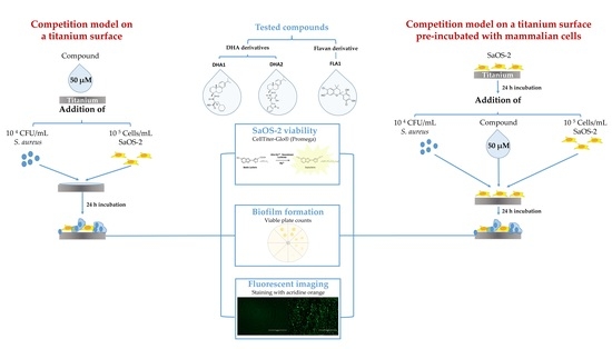
Graphical Abstract

1. Introduction

Antimicrobial resistance is one of the major healthcare challenges that is currently faced by humanity. By switching into the biofilm state, bacteria can withstand antibiotic chemotherapy, and this is increasingly regarded as the most important nonspecific mechanism of antimicrobial resistance [1,2]. Biofilms are defined as a community of cells encased within a self-produced matrix that adheres to biological or non-biological surfaces [3,4]. Because implanted medical devices can be ideal substrates for bacteria to attach, biofilm-mediated infections are one of the leading causes of prosthesis implantation failures. Biomaterial-associated infections (BAIs) represent a great clinical concern because they cause increased morbidity and distress in patients, along with high economic costs due to increased hospitalizations [5].
When implanting a biomaterial, the desired outcome is the correct integration of such material with the host tissue. However, this ideal outcome is often impacted by the presence of bacterial cells at the moment of implantation. According to the concept of “race for the surface” [6], if host cells are able to colonize the surface of the device first, the chances of bacterial cells to adhere to such surface are lower, therefore lowering the risk of implant infection [7]. A frequent route of infection for implants occurs during surgery [8], as microorganisms can be introduced on the implant surface, providing them with an advantage to colonize the unprotected surface and create a biofilm [9]. Taking this into consideration, a reasonable approach would be to design an antimicrobial material or coating that promotes tissue integration. In that direction, based on the earlier results by Perez-Tanoira et al. [10], it seems that the pre-conditioning of the material with host cells is advantageous to protect against bacterial colonization. Staphylococcus aureus is found asymptomatically on the skin [11] and its presence there enhances the risk of infection in the surgical site, which is why it is regarded as a frequent causative agent of implant-related infections, especially in orthopedics [12].
Thus far, there is a limited repertoire of compounds that are able to act on biofilms at sufficiently low concentrations, especially in the case of S. aureus [13]. In order to tackle this problem, several strategies have been proposed, which include, among others, (i) the screening of compounds libraries to identify new small molecules able to inhibit or disassemble biofilms [14], (ii) medicinal chemistry-driven approaches directed towards synthetic modifications of known antibiotics to increase their effectivity on biofilms [15], and (iii) development of novel nano-formulations to improve antibiotic penetration on biofilms [16]. Our laboratory has embraced the exploration of natural compound sources and those studies have resulted in the identification of three promising anti-biofilm leads [17,18,19], which are shown in Figure 1.
The first two compounds are dehydroabietic acid (DHA) derivatives, N-(abiet-8,11,13-trien-18-oyl) cyclohexyl-L-alanine and N-(abiet-8,11,13-trien-18-oyl)-D-tryptophan, coded DHA1 (shown in Figure 1a) and DHA2 (shown in Figure 1b), respectively. These two compounds were synthetically developed in [20] (coded 11 and 9b, respectively, in [20]), by combining the abietane moiety with amino acids, which had separately been shown to display anti-biofilm properties [17,21,22,23]. Compounds DHA1 and DHA2 were both demonstrated to prevent biofilm formation as well as to effectively disassemble pre-formed S. aureus biofilms [20], and they represent the most potent abietane-type anti-biofilm agents that have been reported thus far. The third antimicrobial candidate (show in Figure 1c) is a flavan derivative: 6-chloro-4-(6-chloro-7-hydroxy-2,4,4-trimethylchroman-2-yl)benzene-1,3-diol, coded FLA1, that has also been earlier shown to prevent bacterial colonization and decrease the viability of existing S. aureus biofilms [18] (coded 291 in [18]).
These three lead compounds are good candidates to protect medical devices from biofilm infections. Among the strategies used to protect implants are the modification of the material surfaces to inhibit bacterial adhesion [24,25] and the application of passive coatings to enhance tissue integration or compatibility. Altogether, these strategies aim at diminishing the rate of implant infection [26], which is essential not only to prevent biofilm formation per se but also to avoid aseptic loosening, which is also an important cause of prosthesis failure [27]. It is conceivable that these three lead compounds could be used to develop new materials loaded with them, or that they could be incorporated into antimicrobial coating solutions.
However, moving from in vitro to translational studies can be highly challenging. Often, antimicrobial screening is carried out on materials like polystyrene, which might provide dramatically different results from those obtained in more clinically-relevant surfaces, such as titanium. Similarly, in antibacterial screens, collection strains are often used, whose virulence and adherence capability might differ from those found in wild-type strains isolated from implants [28,29]. In addition, the promotion of tissue integration is not often tested on antimicrobial screens, and, if it happens to be included, the testing is done separately from the antibacterial properties [30,31,32,33,34]. This is less than ideal as anti-biofilm/antibacterial capability and tissue integration should be preferably tested together. In vitro co-culture, models (involving both bacterial and mammalian cells) have been applied to better mimic the clinical situation. These models do not only assess the effects of the leads or materials on bacterial and mammalian cells at the same time but they also provide information on their effect in the interaction between these two cell types [9,35].
Our goal in this investigation is to study the applicability of these naturally compound-derived anti-biofilm leads (DHA1, DHA2, and FLA1) on the antimicrobial protection of biomaterials. Biofilm inhibiting effects of these leads were first studied using the collection and clinical bacterial strains. The anti-biofilm effects were then measured on a competitive colonization model of titanium surfaces, based on the protocol developed by Perez-Tanoira et al. [36], exposed simultaneously to Staphylococcus aureus strains and human osteosarcoma cells (SaOS-2), as well as with titanium pre-conditioned with media supplemented with serum proteins. Furthermore, as it has been previously shown that the incubation of biomaterials with SaOS-2 cells before implantation can be an effective strategy to prevent bacterial adhesion and biofilm formation [10], we additionally tested if the naturally-derived anti-biofilm leads would enhance this preventive effect given by cellular pre-exposure.

2. Materials and Methods

2.1. Compounds

Two dehydroabietic acid derivatives (DHA2 and DHA1) were synthesized according to [20]. Their spectral data were identical to those reported in [23]. The flavan derivative coded FLA1, 6-chloro-4-(6-chloro-7-hydroxy-2,4,4-trimethylchroman-2-yl)benzene-1,3-diol, was purchased from TimTec (product code: ST075672, www.timtec.net). Control antibiotics were purchased from Sigma-Aldrich: rifampicin (13292-46-1) and penicillin G (69-57-8) (coded RIF and PEN, respectively).

2.2. Bacterial Strains

Bacterial studies were performed with the collection strain S. aureus ATCC 25923 (American Type Culture Collection, Manassas, Virginia) and five clinical strains isolated from hip prostheses and osteosynthesis implants at the Hospital Fundación Jiménez Díaz (Madrid, Spain) [37] (S. aureus P1, P2, P4, P18, and P61).

2.3. Effects of Compounds on Biofilm Viability in 96-Wells Microplates

Bacteria (S. aureus ATCC 25923 and the five clinical strains) were cultured in 30 g/L tryptic soy broth (TSB, Neogen®, Lansing, Michigan, USA) under aerobic conditions at 37 °C, 220 rpm for 4 h to reach exponential phase. For forming biofilms, these exponentially grown cultures (106 CFU/mL) were added into flat bottomed 96-well microplates (Nunclon Δ surface, Nunc, Roskilde, Denmark). The anti-biofilm effects of the compounds were assessed prior to biofilm formation and post-biofilm formation. This was carried out as described earlier [17]. Briefly, compounds (at a concentration of 50 µM, 0.25% DMSO) were added simultaneously with the bacterial suspension and effects were examined after incubation at 37 °C, 200 rpm for 18 h, to assess the prevention of biofilm formation. For the post-biofilm formation testing, biofilms were first formed during 18 h (37 °C, 200 rpm), compounds were added, and plates were incubated for 24 h at 37 °C, 200 rpm. Untreated biofilms (only exposed to culture media and 0.25% DMSO), cell-free wells containing only TSB and wells containing biofilms, and 0.25% of DMSO were included as solvent controls. Antibiotics RIF and PEN were used as positive controls, at the same concentration of the studied compounds. The effects on the biofilm viability were assessed following the protocol of [38] by resazurin staining. Briefly, the biofilms were washed twice (200 µL per well) with phosphate buffered saline (PBS) and stained with 20 µM resazurin for 20 min at room temperature (RT), 200 rpm. The top fluorescence of the reduced resazurin was measured at λexcitation = 560 nm and λemission = 590 nm using a Thermo Scientific Varioskan LUX Multimode microplate reader.

2.4. Selection of The Best Biomass-Producer from the Clinical Strains

In order to assess the differences in biomass production between the clinical strains and select the best strain for the co-culture studies, the biofilm biomass was quantified by crystal violet staining. To do so, after the resazurin measurement, biofilms were fixed with 99.8% MeOH (200 µL per well) for 15 min. After removal of MeOH, wells were allowed to dry and 190 µL of crystal violet (0.0023%, v/v) was added and cells were stained for 5 min at room temperature (RT), followed by 2 cycles of manual washing (200 µL per well) with milliQ-water. Wells were let dry for 15 min at RT. After this, the crystal violet-bound stain was solubilized in 96% ethanol (200 μL per well). The plates were incubated for 1 h at RT, and the stained biofilm biomass was then quantitated by measuring the absorbance values at 595 nm using a Thermo Scientific Multiskan Sky microplate spectrophotometer.

2.5. Competition Model on A Titanium Surface

2.5.1. Culture of Human Cells

Human osteosarcoma SaOS-2 cells (89050205, European Collection of Authenticated Cell cultures, ECACC) were grown in minimal essential medium (MEM) (Sigma Aldrich, St. Louis, Missouri, USA) supplemented with 10% heat-inactivated fetal bovine serum (FBS) (Sigma Aldrich, St. Louis, Missouri, USA) containing 500 UI/mL penicillin and 0.1 mg/mL streptomycin (Sigma Aldrich, St. Louis, Missouri, USA). Cells were maintained at 37 °C in 5% CO2 in a humidified incubator.

2.5.2. Cytotoxicity of Compounds in 96-Well Plates

Before proceeding with the co-culture studies, the possible cytotoxicity of the compounds to SaOS-2 cells was assessed in opaque-walled well plates, using the Promega CellTiter-Glo® luminescent cell viability assay. This assay is based on the quantification of the ATP present, which signals the presence of metabolically active cells, and therefore allows the calculation of the number of viable cells. To do this, 50,000 cells per well were seeded. They were exposed to a concentration of 50 µM of each compound (0.25% DMSO); cells without treatment were used as control, and effects of DMSO (0.25%) were also measured. Media alone wells (with MEM) were used as blanks, and in the case of RIF, a blank containing only this compound was also included, to exclude possible interference related to its red color. After 24 h incubation, Promega CellTiter-Glo® assay was carried out, and the luminescence was measured using a Thermo Scientific Varioskan LUX Multimode microplate reader.

2.5.3. Culture of Staphylococci and Human Cells

The bacteria S. aureus (25923 or the clinical strains) was pre-cultured overnight at 37 °C in 5 mL of TSB medium [39]. It was later centrifuged at 4500× g for 10 min, the supernatant was discarded, and the pellet washed three times with PBS. The optical density of the bacterial suspension was measured at λ of 600 nm with a Thermo Scientific Multiskan Sky microplate spectrophotometer according to the McFarland standard. The final concentration of the S. aureus suspension was 104 CFU/mL. This suspension was then added to titanium coupons (0.4 cm height, 1.27 cm diameter, BioSurface Technologies Corp., Bozeman, MT, USA), onto which the different tested compounds or the control antibiotics had been added at a concentration of 50 µM and inserted in the different wells of a 24-well plate (Nunclon Δ surface, Nunc, Roskilde, Denmark).
On the other hand, for the SaOS-2 cells, the media was refreshed 24 h before the experiment started with MEM 10% FBS (without antibiotics), in order to clear out possible traces of antibiotics on the maintenance media. On the day of the experiment the cells were detached with a Trypsin:EDTA solution and re-suspended in MEM 10% FBS; afterwards, they were seeded at a concentration of 105 cells/mL on the titanium coupons, onto which the bacterial suspension had been added as well as the studied compounds and control antibiotics (as described in the previous paragraph). The well plates containing the coupons were maintained in co-culture with a solution containing 104 CFU of S. aureus and 105 SaOS-2 cells in a total volume of 1 mL of MEM:PBS 5% FBS for 24 h, as originally described in [36]. Titanium with added RIF or PEN (both, 50 μM) were used as positive (antibiotic) controls. As co-culture controls, titanium coupons without the addition of the tested compounds or the control antibiotics were exposed to both cellular systems (S. aureus and SaOS-2 cells) at the concentrations previously described. In addition, bacterial controls (exposed or not to the compounds, without SaOS-2 cells), SaOS-2 cells controls (exposed or not to the compounds, without bacterial cells), and a DMSO control (0.25 % DMSO coating titanium coupon in 105 SaOS-2 cells/mL), were also included. Furthermore, an additional bacterial control (104 CFU/mL of S. aureus on 1 mL of MEM:PBS 5% FBS) was set in order to assess the possible background caused by S.aureus in the viability measurement of SaOS-2 cells by luminescence.

2.6. Competition Model on a Titanium Surface Pre-Incubated with SaOS-2 Cells

Following the protocol of Perez-Tanoira et al. [10], a concentration of 105 SaOS-2 cells/mL was seeded on titanium coupons and incubated for 24 h. After the end of the incubation period, the cell medium was removed and samples were washed three times with sterile PBS to remove any non-adherent human cells. The different tested compounds (DHA1, DHA2, FLA1, RIF, and PEN) were added to the coupons at a concentration of 50 µM. A suspension containing 10 4 CFU/mL of S. aureus and 105 SaOS-2 cells/mL on 1 mL of MEM:PBS 5% FBS was added to each coupon and incubated for 24 h. As a co-culture control, a titanium coupon that had been pre-incubated with the SaOS-2 cells and later added bacterial and SaOS-2 cells (as described before) but not exposed to compounds, was used. As a bacterial control, a suspension of 104 CFU/mL of S. aureus on 1 mL of MEM:PBS 5% FBS was added to a titanium coupon without pre-incubation with SaOS-2 cells. As a cellular control, titanium was incubated with 105 SaOS-2 cells/mL on 1 mL of MEM:PBS 5% FBS, without the addition of bacterial cells. Additionally, another bacterial control (104 CFU/mL of S. aureus on 1 mL of MEM:PBS 5% FBS ) was set in order to assess the possible background caused by S.aureus in the measurement of SaOS-2 cellular viability by luminescence.

2.7. Competition Model on a Titanium Surface Pre-Conditioned with Serum-Containing Media

Following the protocol of Campoccia et al. [40], the titanium coupons were exposed to MEM containing 10% of FBS for 30 min. After the incubation period, the medium was removed and samples were washed once with serum-free MEM. Compound DHA1 was added to the coupons at a concentration of 50 µM. A suspension containing 104 CFU/mL of S. aureus and 105 SaOS-2 cells/mL on 1 mL of MEM:PBS 5% FBS was added to each coupon and incubated for 24 h. As a co-culture control, a titanium coupon that had been pre-conditioned with the 10% FBS MEM and added bacterial cells (as described before) but not exposed to compounds, was used. In addition, a co-culture control with titanium without pre-conditioning with 10% FBS MEM was included. As bacterial controls, a suspension of 104 CFU/mL of S. aureus on 1 mL of MEM:PBS 5% FBS was added to a titanium coupon, both non-preconditioned and pre-conditioned with 10% FBS MEM. As cellular controls, titanium was incubated with 105 SaOS-2 cells/mL on 1 mL of MEM:PBS 5% FBS, without the addition of bacterial cells, both non-preconditioned and pre-conditioned with 10% FBS MEM, as in the case of the bacterial controls. Additionally, another bacterial control (104 CFU/mL of S. aureus on 1 mL of MEM:PBS 5% FBS) was set in order to assess the possible background caused by S. aureus in the measurement of SaOS-2 viability by luminescence.

2.8. Measurement of SaOS-2 Cells Viability

Coupons were washed once with PBS and cells detached with 500 µL of a 1:10 trypsin:EDTA solution. Then, 500 µL of MEM (containing 10% FBS) was added to neutralize the trypsin:EDTA solution. The resulting (1 mL) suspension was centrifuged at 150× g. The supernatant was discarded and 100 µL of MEM containing the cellular pellet was transferred to an opaque-walled well plate and the Promega CellTiter-Glo® luminescent cell viability assay was carried out. The possible background signal corresponding to S. aureus was subtracted from the co-culture wells

2.9. Bacterial Adherence and Biofilm Formation

Titanium coupons were washed with TBS to remove remaining planktonic cells and then they were transferred into Falcon tubes containing 1 mL of 0.5% (w/v) Tween® 20-TSB solution. After that, the tubes were sonicated in an Ultrasonic Cleaner 3800 water sonicator (Branson Ultrasonics, Danbury, CT, USA) at 25 °C, for 5 min at 35 kHz. The tubes were mixed vigorously for 20 s prior to and after the sonication step. Serial dilutions were performed from the resulting bacterial suspensions and plated on Tryptic Soy Agar (TSA, Neogen®, Lansing, MI, USA) plates.

2.10. Fluorescence Imaging

Titanium coupons were incubated in the same exact conditions as described in Section 2.5.3 and Section 2.6. After incubation with the different compounds, coupons were manually washed three times with PBS. The dried coupons were stained for 2 min with a rapid fluorescence staining method using acridine orange (BD Diagnostics, Sparks, MD, USA) at a concentration of 0.1 mg/mL. After the staining, coupons were rinsed with sterile water to remove the excess of dye. Images were taken with an Invitrogen EVOS M5000 imaging system (Thermo Scientific, Waltham, Massachusetts, USA). On each coupon, 8–10 fields were viewed and photographed at magnifications of 10x and 20x. Representative images are shown in the results. All imaging experiments were performed in duplicates and experiments were repeated three times.

2.11. Statistical Analysis

The quantitative data are reported as mean of at least 3 technical replicates ± SEM, and all experiments were repeated 3 times. Data were analyzed using GraphPad Prism 8 for Windows. Non-parametric tests were used. For statistical comparisons, Welch’s unpaired t-test and one-way ANOVA with Bonferroni correction were applied, and p < 0.05 was always considered as statistically significant.

3. Results

3.1. Effect on the Prevention and Killing of Biofilms Formed by S. aureus Clinical Strains in 96-well microplates

The three anti-biofilm leads studied here had been earlier tested only using collection strains [18,20]. Thus, prior to starting their effects on biomaterials, we set out to test their anti-biofilm efficacy using five different clinical S. aureus strains, isolated from hip prostheses and osteosynthesis implants [37]. These results were compared to the effect of the compounds on S. aureus ATCC 25923, the collection strain previously used as a reference. The effects on the biofilm viability were measured using a redox staining assay (resazurin-based) on biofilms grown on the polystyrene surface of 96-well microplates.
As can be seen in Figure 2a, all three compounds caused over 90% prevention of biofilm formation by all clinical strains, as well as the previously tested reference strain, with an effect fairly similar to RIF, one of the used control antibiotics. In contrast, PEN (the other control antibiotic tested here) was only efficient against the collection strain and one of the clinical strains (P61). When the effect was measured on pre-formed biofilms of the clinical strains (Figure 2b), the inhibitory effects of the three compounds was reduced (compared to Figure 2a), but, still, a significant inhibitory effect was measured in all cases and it was higher than the one displayed by PEN. The second control antibiotic, RIF, remained effective and able to cause over 60% inhibition of the viability of the clinical pre-formed biofilms (Figure 2b).
For the rest of the studies, one of the S. aureus clinical strains, P2, was selected and compared to the effect on the reference collection strain (S. aureus ATCC 25923). This decision was based on the fact that this clinical S. aureus strain P2 was the one with the highest biomass-containing biofilm when compared to the collection strain S. aureus 25923 (Figure 3).

3.2. Effect on the prevention of S. aureus Adhesion in a Competitive Colonization Model on Titanium Coupons

Prior to testing the capacity of the compounds on the competitive colonization model, their possible cytotoxic effects towards SaOS-2 cells were assessed on polystyrene 96-well microplates (Figure S1). None of the compounds showed cytotoxicity at a concentration of 50 µM on either of the materials.
Next, the effects of the compounds on SaOS-2 viability, as well as on the S. aureus 25923 and P2 viable attached cells were measured, when bacteria and human cells were simultaneously co-cultured (Figure 4). The first part of Figure 4a (to the left) shows how none of the compounds caused any inhibition of the SaOS-2 adherence to titanium, in the absence of bacterial cells. The rest of Figure 4a (middle and right panels) show the effects of the compounds on SaOS-2 viability when bacteria and human cells are simultaneously co-cultured. The presence of either the collection (SaOS-2 + S. aureus 25923, blue bars) or the clinical (SaOS-2 + S. aureus P2, red bars) S. aureus strain induced a slight proliferative effect on the SaOS-2 cells, but with no statistical relevance in any of the cases. Only compound FLA1 significantly diminished SaOS-2 viability in the presence of the clinical strain P2 (Figure 4a; SaOS-2 + S. aureus P2).
On the other hand, the presence of SaOS-2 cells caused a significant reduction of the attached viable S. aureus ATCC 25923 when compared with the material incubated only with bacteria (blue control bar versus black control bar in Figure 4b, p = 0.007). In addition, all three compounds reduced the number of viable S. aureus ATCC 25923 cells in co-cultured with SaOS-2 cells (blue bars), compared to the bacterial control (Figure 4b). Compound DHA1 managed to cause a 4-log reduction when compared to the bacteria grown in monoculture, and more than a 2-log reduction when compared to the corresponding co-culture control (Figure 4b). The reduction of bacterial adhesion obtained after treatment with compound DHA2 is slightly smaller but yet significant (p = 0.0247), being the number of S. aureus ATCC 25923 attached in DHA2-treated samples about 1-log less than in the corresponding co-culture control. Compound FLA1 caused a reduction of the viable S. aureus ATCC 25923 comparable to the one obtained with DHA1 (p < 0.001). Both controls antibiotics caused a very significant reduction in the number of viable S. aureus ATCC 25923 cells. In fact, RIF completely prevented the adherence of this collection strain.
With respect to the clinical S. aureus strain P2, the mere presence of mammalian cells did not cause a reduction of the attached viable bacteria when compared to the bacterial monoculture controls (red control bar versus black control bar in Figure 4c). Compound DHA1 significantly reduced (p < 0.001) the viable attached S. aureus P2 by almost 2-log when compared to corresponding bacterial (monoculture and co-culture) controls. Compounds DHA2 and FLA1 did not cause any significant reduction of the viable clinical bacteria in this competitive colonization assay. Antibiotics RIF and PEN significantly reduced the viable attached clinical bacterial strain when compared to both bacterial controls, but PEN lost activity, as it was the case with compound DHA1.
Figure 5 shows fluorescence microscope images of the titanium coupons in the competitive colonization model, using the reference collection strain (S. aureus ATCC 25923). Figure 5 confirms that the presence of S. aureus ATCC 25923 does not cause a reduction in the number of SaOS-2 cells. A slight change in SaOS-2 cell morphology can be noticed when comparing the SaOS-2 monoculture control (Figure 5a) with the co-culture control (Figure 5c). The presence of bacteria seems to cause a slight reduction in the size of the cells and a loosening on its adhesive shape. This change in morphology is not observed in the samples treated with the control antibiotics, given its high antibacterial activity, but it can be noticed in the samples treated with DHA1, DHA2, and FLA1 as they do not completely prevent the adherence of bacterial cells. The S. aureus ATCC 25923 remaining on those samples might be responsible for these changes observed in SaOS-2 cell morphology.

3.3. Effect on the Prevention of S. aureus Adhesion in a Competitive Colonization Model on Titanium Coupons Pre-Incubated with SaOS-2 Cells

Figure 6 shows the effects of the compounds in coupons pre-incubated with SaOS-2 cells when using both the reference S. aureus collection (ATCC 25923, blue bars) or the clinical strain (P2, red bars).
With respect to the effect on the SaOS-2 cells, only when S. aureus ATCC 25923 and SaOS-2 cells were simultaneously incubated with compounds DHA1 or DHA2, a proliferative effect was seen (p = 0.004 and 0.037, respectively) compared to the corresponding control (Figure 6a, middle panel, SaOS-2 + S. aureus 25923). In contrast, co-culture of the clinical isolate P2 caused a highly significant decrease (p < 0.001) on mammalian cell viability (Figure 6a, SaOS-2 + S. aureus P2). In the controls, mammalian cells did not survive when co-cultured in the presence of S. aureus P2 strain, neither did the SaOS-2 cells treated with the compounds DHA2 and FLA1. However, a significant proliferative effect of SaOS-2 cells was found in the presence of compound DHA1. Exposure to both control antibiotics also prevented the bacterial-induced cytotoxicity and resulted in the protection of SaOS-2 cells, similarly to compound DHA1 (Figure 6b, red bars, SaOS-2 + S. aureus P2).
Figure 6b,c shows the effects of the compounds on the viable attached bacteria when using coupons pre-incubated with SaOS-2 cells. The pre-incubation of titanium with SaOS-2 cells significantly reduced the number of attached ATCC 25923 S. aureus strain (blue control bar versus gray control bar in Figure 6b, p < 0.001). Exposure to compound DHA1 as well as the control antibiotics caused a total reduction of S. aureus ATCC 25923 attachment (Figure 6b, p < 0.001 in all cases). Compounds DHA2 and FLA1 caused a reduction of the viable attached bacteria, when compared to the bacterial (ATCC 25923) control (p < 0.001, in both cases), and the reduction was also significant when compared to the cellular pre-coated coupons (control of S. aureus 25923 + SaOS-2, Figure 6b) (p < 0.001 and p = 0.035, respectively).
In contrast, the results of Figure 6c show how the positive impact of cellular pre-conditioning was not detected for the clinical S. aureus strain P2. Interestingly, exposure to compound DHA1 significantly reduced 1-log the viable attached P2 strain when compared to both the bacteria control and the control corresponding to the pre-coating of titanium with SaOS-2 cells (SaOS-2 + S. aureus P2 strain) (p < 0.001). The activity of RIF was preserved against the clinical strain, while PEN, despite losing some activity when compared to its effect against the collection strain, also significantly reduced the viable bacteria (P2 strain) attached (p < 0.001 in both cases).
Figure 7 shows fluorescence microscope images of the titanium coupons in the competitive colonization model using titanium coupons that had been pre-coated with SaOS-2 cells. The protective effect of the pre-coating with SaOS-2 cells is visible, as the cells do not show the morphology change that was observed when the SaOS-2 cells were directly co-cultured with S. aureus ATCC 25923 (Figure 5c versus Figure 7c). When compared to the cellular monoculture control (Figure 7a), the different treatments (Figure 7d–h) do not appear to affect the morphology or the number of SaOS-2 cells.

3.4. Effect on the Prevention of S. aureus Adhesion in a Competitive Colonization Model on Titanium Coupons Pre-Conditioned with Media Containing High Serum Concentration

Figure 8 shows the effects of DHA1 on SaOS-2 viability, as well as on the attachment of S. aureus ATCC 25923 and P2 viable cells when bacteria and human cells are simultaneously co-cultured in titanium that has been pre-conditioned with media containing 10% FBS. In parts (a) and (b) of Figure 8, it can be seen how SaOS-2 adheres better to the titanium surface when it is pre-conditioned with media containing 10% FBS (p = 0.0387). However, this enhanced adherence is not observed when SaOS-2 cells are co-cultured with bacteria, nor with the collection strain (Figure 8a) or the clinical strain (Figure 8b). In turn, in Figure 8c,d, it can also be observed how both bacterial of strains adhere significantly more to the titanium surface pre-conditioned with media containing 10% FBS. (Figure 8c, p = 0.0063; Figure 8d, p = 0.0313).
In the case of the collection strain (Figure 8c), it can be again observed how the presence of SaOS-2 cells decreased the attachment of S. aureus (p < 0.001). The pre-conditioning with FBS does not make a difference in bacterial adherence when bacterial cells are co-cultured with SaOS-2 cells (Figure 8c). Exposure to the compound DHA1 significantly reduced the attached S. aureus ATCC 25923 when compared to the corresponding controls of the subgroups: S. aureus 25923 non-preconditioned FBS; S. aureus 25923 preconditioned FBS; S. aureus 25923 + SaOS-2 non-preconditioned FBS, and S. aureus 25923 + SaOS-2 preconditioned FBS (p < 0.001; p < 0.001; p = 0.026, and p = 0.019, respectively). As for the clinical P2 strain (Figure 8d), the effect observed in Figure 4c is repeated, the presence of SaOS-2 cells does not decrease the adherence of the S. aureus P2 strain. The pre-conditioning with media containing 10% FBS does not influence the adherence of S. aureus P2 when co-cultured with SaOS-2 cells (Figure 8d). Once more, the compound DHA1 significantly reduced the attached S. aureus P2 strain when compared to the corresponding controls of the different subgroups: S. aureus P2 non-preconditioned FBS; S. aureus P2 preconditioned FBS; S. aureus P2 + SaOS-2 non-preconditioned FBS, and S. aureus P2 + SaOS-2 preconditioned FBS (p = 0.017; p = 0.0011; p = 0.027, and p = 0.025, respectively).

4. Discussion

In this study, the applicability of three previously discovered antimicrobial compounds to be incorporated into implanted medical devices that would prevent the formation of S. aureus biofilms was studied. To examine such effects, a competitive model was utilized, allowing us to investigate, in a more realistic scenario, the implantation of a biomaterial, which provides a substratum to host either tissue–cell integration or bacterial colonization. These two phenomena are in conflict because, after the adherence of either one, the surface is less prone to colonization by the other. Tissue integration and bacterial contamination of medical devices have been extensively studied as independent phenomena [41]. However, only recently, experimental assays have been established in which fair competition can be investigated during the development of new biomaterial-coating strategies [36,42]. Using such tools, it has been recently shown that the incubation of typical implant materials with human SaOS-2 cells before implantation represents an innovative and effective way to reduce the bacterial living space available and prevent S. aureus adhesion, thus protecting biomaterials against biofilm formation [10]. Such an approach would offer an attractive concept that could be further enhanced by the presence of antimicrobial compounds.
In the current study, we hypothesized that two DHA derivatives (DHA1 and DHA2) and a flavan derivative (FLA1), which our group had previously reported as promising anti-biofilm leads [18,20], could find applicability in the protection of medical devices against infections. Here, we started by evaluating their antimicrobial efficacy against clinical bacterial strains [37]. In the case of S. aureus, its origin is particularly relevant. Besides being a causative agent of implant infections, S. aureus is part of the normal bacterial flora of human skin and mucosal surfaces. One of the most relevant virulence factors expressed by S. aureus is the cell wall-anchored (CWA) protein, surface proteins covalently attached to peptidoglycan that significantly influence the colonization ability and posterior survival of the bacteria in the host, which is particularly relevant in medical devices infections [43]. Among these, the microbial surface component recognizing adhesive matrix molecules (MSCRAMM) facilitates the adherence of the bacteria to the host tissue; in the case of bone implants, the capacity of S. aureus to interact with collagenous proteins might be important in the pathogenesis of possible osteomyelitis [44]. This makes it obvious that the inter-strain virulence, and as a consequence, the efficacy of the tested compounds, may drastically change [28]. Our results show how that, despite being very efficient at both preventing and killing biofilms formed by the reference S. aureus ATCC 25923 strain, the antibiotic PEN loses a great part of its activity when tested against most of the clinical strains (all except P61, Figure 2a,b). On the contrary, compounds DHA1, DHA2, FLA1, as well as the control RIF, kept their preventive activity against all the clinical strains (Figure 2a). All of the tested compounds were also able to inhibit at least 30% of the viability of already formed biofilms by both the reference collection and the clinical strains. It was also observed differences in the biomass quantity produced by different clinical strains, which could represent an additional virulence factor that varies depending on the strain. In Figure 3, it can be seen how S. aureus P2 produces more biomass than the collection strain. This can influence the treatment and clearance of these clinical biofilms, in comparison with the collection strain.
Moreover, efficacy in preventing biofilm formation of the different compounds was tested on titanium, given its relevance as biomaterial used for orthopedic implants. In Figure 4, it can be seen that compound DHA1 preserved its antimicrobial activity when tested on titanium. However, compound DHA2 was shown to lose its prevention capability on this material. This change is even more evident with the clinical strain, where neither the compound DHA2 nor FLA1 managed to prevent biofilm formation (Figure 4c).
Titanium treated with DHA1, DHA2, or FLA1 was studied in a competitive colonization model with both bacteria and human cells. We used an S. aureus concentration of 104 CFU/mL as more than 102 CFU of S. aureus is necessary to establish a prosthesis infection and 104 CFU/mL in surgery without an implant. On the other hand, it has been proposed that using concentrations higher than 106 CFU/mL might be questionable and not relevant from a clinical perspective [45,46]. According to present results, and in concordance with the ones reported by [36], the presence of S. aureus did not significantly affect the viability of SaOS-2 cells (Figure 4a). Previously, Yue et al. [47] have shown that low bacterial concentrations increase cellular adhesion, likely as a result of the stress response caused by bacteria on the mammalian cells, which are then forced to compete more effectively and withstand cellular detachment. This effect was observed in both the collection strain and the clinical strain. None of the compounds produced a negative effect on SaOS-2 cellular viability, but surprisingly, the cells treated with the compound FLA1 died when exposed to the clinical S. aureus P2 strain (Figure 4a). From fluorescence imaging (Figure 5), it can be seen how the presence of bacteria had an effect on the morphology of SaOS-2, despite not affecting the number of attached cells. It can be observed that this change in morphology is reduced in presence of RIF (Figure 5g), likely due to the fact that this antibiotic reduces practically to zero the presence of S. aureus ATCC 25923 viable cells.
As mentioned before, the success of an anti-infective prosthesis relies not only on the efficacy to eradicate bacteria but also in its ability to promote bone-implant osseointegration. Titanium is an inert material that does not accelerate this process, which is why many approaches towards implant development aim at bio-functionalizing titanium to improve its bioactivity. The difficulty remains in modifying the titanium in such a way that it promotes osseointegration while being antimicrobial, as on many occasions, the antimicrobial agents incorporated are cytotoxic to the osteoblasts. Nie et al. [48] developed a biofunctionalized titanium with bacitracin that proved to be antimicrobial and cytocompatible in vitro. The efficacy of such titanium on preventing infection and improving osteoinductivity was later proven in vivo [49]. This is a good example of how an antimicrobial that is intended to form part of a prosthesis has not only to be proven in vitro as antimicrobial but also cytocompatible, as both qualities are essential for correct integration of the prosthesis. The in vitro system described in this study not only assess both qualities in the same assay, but it also gives information on the effect that the antimicrobial has on the adherence of the material of each cell type, mammalian and bacterial, when they are present together.
The second part of this study aimed at assessing the utility of the compounds in enhancing the positive effect of pre-incubating materials with human SaOS-2 cells [10]. As previously demonstrated, the development of an infection would highly depend on what type of cells colonize the surface of an implant first, the cells of the host or the invading bacteria cells. Unfortunately, during surgery, bacterial cells frequently are in advantage as they can be introduced onto an implant before integration with host tissue even starts, and they have a faster replication rate. Giving advantage to the host cells would facilitate tissue integration and diminish the risk of bacterial infection [9]. By combining the protective effect of pre-conditioning titanium with mammalian cells with the previously reported anti-biofilm capability of the studied compounds, it was expected to accomplish a drastic reduction of the biofilm formation.
The pre-exposure of SaOS-2 to S. aureus 25923 generates the same slight proliferation effect on the mammalian cells at 24 h (Figure 6 in comparison to Figure 4). This proliferation is less acute in the case of RIF and PEN, probably due to their high efficacy of killing the bacterial cells. None of the compounds appeared to affect the morphology of the cells. Perez-Tanoira et al. [10] reported a drop in the proliferation of SaOS-2 cells at 48 h exposed to the same concentration of the clinical isolate S. aureus 15981. In our study, the presence of the clinical S. aureus strain P2 dramatically decreases SaOS-2 cell viability in co-culture, which may be explained by a higher virulence of this strain. This drop on the cell viability can also be observed in the groups treated with DHA2 and FLA1, probably due to the lack of antibacterial activity (Figure 6a).
It has to be taken into account, that when implanting a medical device, the first event occurring, before the race for the surface starts, is the interaction of the implant’s surface with physiological fluids and the subsequent adsorption of host proteins. The adhesion of contaminating bacteria usually occurs once the implant is protein-coated. The adhesive glycoprotein fibrinogen is a major plasma glycoprotein. At sites of trauma and injury, fibrinogen serves as a major host substrate for S. aureus adhesion, as this bacterium expresses fibrinogen-biding proteins [44]. For this reason, we tested the pre-conditioning of titanium with fibrinogen at a physiological concentration (1g/L) [50], following the protocol of Opperman et al. [51]. There were no differences in the adherence of S. aureus to a titanium surface pre-conditioned with fibrinogen when compared to the non-conditioned surface (Figure S2). These results are in line with the ones obtained by Rao et al. [52] that showed how the pre-conditioning of titanium with fibrinogen and fibrinogen clotted with thrombin promoted osseointegration, but the biofilm formation by Enterococcus faecalis in these bio-functionalized materials did not show significant differences with the titanium control.
Next, we also tested the influence of pre-conditioning the titanium surface with media supplemented with a higher concentration of FBS, in the same competition model, as it contains other major proteins present in the blood, such as albumin, which can also be adsorbed by titanium [53,54]. We demonstrated that the adherence of bacteria is higher when the surface is pre-conditioned in this manner, and this bacterial enhancement occurs in both bacterial strains (Figure 8c,d). Nevertheless, mammalian cell adhesion to the preconditioned surface seemed also to increase, which is the reason why the results obtained in this part of the study are very similar to the ones obtained in the competition model without pre-conditioning. The compound DHA1 prevented bacterial adherence of both the collection and clinical S. aureus strains also on pre-conditioned titanium (Figure 8c,d).
In this study, we analyzed three compounds that, based on our earlier results, were regarded as promising candidates to prevent S. aureus biofilms related infections. However, out of the three, we demonstrated here that only compound DHA1 remains effective in conditions that are more likely to be encountered during an in vivo bacterial infection in an orthopedic implant. In the case of the compound DHA2, its lack of activity might be given by the fact that the concentration used here is slightly below its MIC [20]. As with most antimicrobials, a common concern is unspecific cytotoxicity. Both DHA derivatives had already been tested on mammalian cells, specifically HL cells (originating from the human respiratory tract). Compound DHA1 did not cause any reduction in the viability of this cell line, but with a concentration of 100 µM of DHA2, only 23% of the cells remained viable. This is why a concentration of 50 µM was chosen, as in a 96-well microplate system, it did not show cytotoxic effect but kept a considerably strong anti-biofilm capability. An increase of the concentration on the co-culture system would have probably shown a higher antimicrobial effect, but also a cytotoxic one, so it would have anyway limited the efficacy of DHA2 as a possible implant coating. On the contrary, the compound FLA1 was used in a dosage that was expected to be effective [18], and despite the effectiveness shown in 96-well microplates, the lack of activity in the competitive colonization system indicates that it is most likely not suitable for protection of implants.
In this study, the compound DHA1 was shown to be a promising candidate to form part of a bone implant, based on the results of the competitive colonization model. However, this co-culture system used here is not meant to replace in vivo experimentation, and it can still be further improved, for instance, by utilizing primary osteoblast cells. It has been shown how the results obtained in a co-culture system utilizing osteoblast cells differ from the ones obtained with SaOS-2 cells [36]. The co-culture using SaOS-2 is very convenient (methodologically-wise), but it is to be kept in mind that because SaOS-2 is a sarcoma cell line, it has a faster division rate, and can be in general more resilient to adverse conditions. Utilizing autologous osteoblasts of the patient would be the ideal scenario to avoid possible rejection of the prosthesis. The co-culture model could also be further enriched by the addition of immune cells, such as neutrophils, which are also present at the moment of implantation. In any case, as it stands, the co-culture model of S. aureus and SaOS-2 cells does offer a deeper insight into the protective capacity of investigational antimicrobial compounds, particularly those intended for the protection of bone implants.
These results highlight the importance of developing new protocols in vitro that would more effectively select the best antimicrobial candidates for biomedical applications, and that would minimize the translational gap between in vitro screening data and the in vivo clinical scenario.

5. Conclusions

We concluded that one DHA derivative, DHA1, would be a promising candidate for the coating of biomaterials in order to prevent biofilm formation. This compound shows a preventive activity of S. aureus biofilm formation by both collection and clinical strains, and it prevents S. aureus adhesion to titanium surfaces, while also favoring the adhesion of mammalian cells to titanium.

Supplementary Materials

The following are available online at https://www.mdpi.com/2076-2607/8/3/345/s1, Figure S1: Effect of the two DHA derivatives (DHA1 and DHA2), the flavonoid derivative (FLA1) and two control antibiotics (RIF and PEN) on SaOS-2 viability when cultured in 96 wells polystyrene plates. Figure S2. Viable counts of S. aureus (ATCC 25923 and clinical strain P2) after 24 h incubation on titanium coupons and titanium coupons pre-conditioned with 1 g/L of fibrinogen.

Author Contributions

Conceptualization, A.F.; methodology, J.Z.P., R.P.-T. and I.R.; software, I.R.; validation, I.R. and A.F.; formal analysis, I.R.; investigation, I.R.; resources, J.Y.-K., J.Z.P., K.S., A.F. and T.J.K.; data curation, I.R.; writing—original draft preparation, I.R.; writing—review and editing, A.F., T.J.K., R.P.-T. and J.Y.-K.; visualization, I.R., and A.F.; supervision, A.F., K.S.; project administration, A.F.; funding acquisition, A.F. and J.Y.-K. All authors have read and agreed to the published version of the manuscript.

Funding

This project has received funding from the European Union’s Horizon 2020 research and innovation program under the Marie Sklodowska—Curie grant agreement No 722467 (PRINT-AID consortium). We also acknowledge the funding received from the Jane and Aatos Erkko Foundation.

Acknowledgments

We thank Pablo Reigada for his help in graphic design.

Conflicts of Interest

The authors declare no conflict of interest. The information and views set out in this article are those of the authors and do not necessarily reflect the official opinion of the European Union. Neither the European Union institutions and bodies nor any person acting on their behalf may be held responsible for the use which may be made of the information contained therein.

References

  1. Kumar, A.; Alam, A.; Rani, M.; Ehtesham, N.Z.; Hasnain, S.E. Biofilms: Survival and defense strategy for pathogens. Int. J. Med Microbiol. Ijmm 2017, 307, 481–489. [Google Scholar] [CrossRef] [PubMed]
  2. Singh, S.; Singh, S.K.; Chowdhury, I.; Singh, R. Understanding the Mechanism of Bacterial Biofilms Resistance to Antimicrobial Agents. Open Microbiol. J. 2017, 11, 53–62. [Google Scholar] [CrossRef] [PubMed] [Green Version]
  3. Paharik, A.E.; Horswill, A.R. The Staphylococcal Biofilm: Adhesins, Regulation, and Host Response. Microbiol. Spectr. 2016, 4. [Google Scholar] [CrossRef] [Green Version]
  4. Hall-Stoodley, L.; Costerton, J.W.; Stoodley, P. Bacterial biofilms: From the Natural environment to infectious diseases. Nat. Rev. Microbiol. 2004, 2, 95. [Google Scholar] [CrossRef] [PubMed]
  5. Jamsen, E.; Furnes, O.; Engesaeter, L.B.; Konttinen, Y.T.; Odgaard, A.; Stefansdottir, A.; Lidgren, L. Prevention of deep infection in joint replacement surgery A review. Acta Orthop. 2010, 81, 660–666. [Google Scholar] [CrossRef] [PubMed]
  6. Gristina, A.G.; Naylor, P.T.; Myrvik, Q. The Race for the Surface: Microbes, Tissue Cells, and Biomaterials. In Molecular Mechanisms of Microbial Adhesion; Springer: New York, NY, USA, 1989; pp. 177–211. [Google Scholar]
  7. Gristina, A.G. Biomaterial-centered infection: Microbial adhesion versus tissue integration. Science 1987, 237, 1588–1595. [Google Scholar] [CrossRef] [PubMed]
  8. Lidwell, O.M.; Lowbury, E.J.L.; Whyte, W.; Blowers, R.; Stanley, S.J.; Lowe, D. Airborne contamination of wounds in joint replacement operations: The relationship to sepsis rates. J. Hosp. Infect. 1983, 4, 111–131. [Google Scholar] [CrossRef]
  9. Subbiahdoss, G.; Kuijer, R.; Grijpma, D.W.; van der Mei, H.C.; Busscher, H.J. Microbial biofilm growth vs. tissue integration: “The race for the surface” experimentally studied. Acta Biomater. 2009, 5, 1399–1404. [Google Scholar] [CrossRef]
  10. Perez-Tanoira, R.; Aarnisalo, A.A.; Eklund, K.K.; Han, X.; Soininen, A.; Tiainen, V.M.; Esteban, J.; Kinnari, T.J. Prevention of Biomaterial Infection by Pre-Operative Incubation with Human Cells. Surg Infect (Larchmt) 2017, 18, 336–344. [Google Scholar] [CrossRef]
  11. Grice, E.A.; Segre, J.A. The skin microbiome. Nat. Rev. Microbiol. 2011, 9, 244–253. [Google Scholar] [CrossRef]
  12. Campoccia, D.; Baldassarri, L.; Pirini, V.; Ravaioli, S.; Montanaro, L.; Arciola, C.R. Molecular epidemiology of Staphylococcus aureus from implant orthopaedic infections: Ribotypes, agr polymorphism, leukocidal toxins and antibiotic resistance. Biomaterials 2008, 29, 4108–4116. [Google Scholar] [CrossRef]
  13. Jaskiewicz, M.; Janczura, A.; Nowicka, J.; Kamysz, W. Methods Used for the Eradication of Staphylococcal Biofilms. Antibiotics 2019, 8, 174. [Google Scholar] [CrossRef] [PubMed] [Green Version]
  14. Worthington, R.J.; Richards, J.J.; Melander, C. Small molecule control of bacterial biofilms. Org. Biomol. Chem. 2012, 10, 7457–7474. [Google Scholar] [CrossRef] [PubMed] [Green Version]
  15. Huigens, R.W., 3rd. The Path to New Halogenated Quinolines With Enhanced Activities Against Staphylococcus epidermidis. Microbiol. Insights 2018, 11, 1178636118808532. [Google Scholar] [CrossRef] [PubMed]
  16. Kłodzińska, S.N.; Wan, F.; Jumaa, H.; Sternberg, C.; Rades, T.; Nielsen, H.M. Utilizing nanoparticles for improving anti-biofilm effects of azithromycin: A head-to-head comparison of modified hyaluronic acid nanogels and coated poly (lactic-co-glycolic acid) nanoparticles. J. Colloid Interface Sci. 2019, 555, 595–606. [Google Scholar] [CrossRef]
  17. Fallarero, A.; Skogman, M.; Kujala, J.; Rajaratnam, M.; Moreira, V.M.; Yli-Kauhaluoma, J.; Vuorela, P. (+)-Dehydroabietic acid, an abietane-type diterpene, inhibits Staphylococcus aureus biofilms in vitro. Int. J. Mol. Sci. 2013, 14, 12054–12072. [Google Scholar] [CrossRef] [Green Version]
  18. Manner, S.; Skogman, M.; Goeres, D.; Vuorela, P.; Fallarero, A. Systematic Exploration of Natural and Synthetic Flavonoids for the Inhibition of Staphylococcus aureus Biofilms. Int. J. Mol. Sci. 2013, 14, 19434–19451. [Google Scholar] [CrossRef] [Green Version]
  19. Manner, S.; Fallarero, A. Screening of Natural Product Derivatives Identifies Two Structurally Related Flavonoids as Potent Quorum Sensing Inhibitors against Gram-Negative Bacteria. Int. J. Mol. Sci. 2018, 19, 1346. [Google Scholar] [CrossRef] [Green Version]
  20. Manner, S.; Vahermo, M.; Skogman, M.E.; Krogerus, S.; Vuorela, P.M.; Yli-Kauhaluoma, J.; Fallarero, A.; Moreira, V.M. New derivatives of dehydroabietic acid target planktonic and biofilm bacteria in Staphylococcus aureus and effectively disrupt bacterial membrane integrity. Eur. J. Med. Chem. 2015, 102, 68–79. [Google Scholar] [CrossRef]
  21. Kolodkin-Gal, I.; Romero, D.; Cao, S.; Clardy, J.; Kolter, R.; Losick, R. D-amino acids trigger biofilm disassembly. Science 2010, 328, 627–629. [Google Scholar] [CrossRef] [Green Version]
  22. Sarkar, S.; Pires, M.M. d-Amino acids do not inhibit biofilm formation in Staphylococcus aureus. PLoS ONE 2015, 10, e0117613. [Google Scholar] [CrossRef]
  23. Hochbaum, A.I.; Kolodkin-Gal, I.; Foulston, L.; Kolter, R.; Aizenberg, J.; Losick, R. Inhibitory effects of D-amino acids on Staphylococcus aureus biofilm development. J. Bacteriol. 2011, 193, 5616–5622. [Google Scholar] [CrossRef] [PubMed] [Green Version]
  24. Boelens, J.J.; Dankert, J.; Murk, J.L.; Weening, J.J.; van der Poll, T.; Dingemans, K.P.; Koole, L.; Laman, J.D.; Zaat, S.A. Biomaterial-associated persistence of Staphylococcus epidermidis in pericatheter macrophages. J. Infect. Dis. 2000, 181, 1337–1349. [Google Scholar] [CrossRef] [PubMed] [Green Version]
  25. Szymczyk, P.; Junka, A.; Ziolkowski, G.; Smutnicka, D.; Bartoszewicz, M.; Chlebus, E. The ability of S.aureus to form biofilm on the Ti-6Al-7Nb scaffolds produced by Selective Laser Melting and subjected to the different types of surface modifications. Acta Bioeng. Biomech. 2013, 15, 69–76. [Google Scholar]
  26. Goodman, S.B.; Yao, Z.Y.; Keeney, M.; Yang, F. The future of biologic coatings for orthopaedic implants. Biomaterials 2013, 34, 3174–3183. [Google Scholar] [CrossRef] [Green Version]
  27. Raphel, J.; Holodniy, M.; Goodman, S.B.; Heilshorn, S.C. Multifunctional coatings to simultaneously promote osseointegration and prevent infection of orthopaedic implants. Biomaterials 2016, 84, 301–314. [Google Scholar] [CrossRef] [Green Version]
  28. Chessa, D.; Ganau, G.; Spiga, L.; Bulla, A.; Mazzarello, V.; Campus, G.V.; Rubino, S. Staphylococcus aureus and Staphylococcus epidermidis Virulence Strains as Causative Agents of Persistent Infections in Breast Implants. PLoS ONE 2016, 11, e0146668. [Google Scholar] [CrossRef]
  29. Sabouni, F.; Mahmoudi, S.; Bahador, A.; Pourakbari, B.; Sadeghi, R.H.; Ashtiani, M.T.H.; Nikmanesh, B.; Mamishi, S. Virulence Factors of Staphylococcus aureus Isolates in an Iranian Referral Children’s Hospital. Osong Public Health Res Perspect 2014, 5, 96–100. [Google Scholar] [CrossRef] [Green Version]
  30. Barkarmo, S.; Longhorn, D.; Leer, K.; Johansson, C.B.; Stenport, V.; Franco-Tabares, S.; Kuehne, S.A.; Sammons, R. Biofilm formation on polyetheretherketone and titanium surfaces. Clin. Exp. Dent. Res. 2019, 5, 427–437. [Google Scholar] [CrossRef] [PubMed]
  31. Huang, T.B.; Li, Y.Z.; Yu, K.; Yu, Z.; Wang, Y.; Jiang, Z.W.; Wang, H.M.; Yang, G.L. Effect of the Wnt signal-RANKL/OPG axis on the enhanced osteogenic integration of a lithium incorporated surface. Biomater. Sci. 2019, 7, 1101–1116. [Google Scholar] [CrossRef]
  32. Johnson, C.T.; Wroe, J.A.; Agarwal, R.; Martin, K.E.; Guldberg, R.E.; Donlan, R.M.; Westblade, L.F.; García, A.J. Hydrogel delivery of lysostaphin eliminates orthopedic implant infection by Staphylococcus aureus and supports fracture healing. Proc. Natl. Acad. Sci. USA 2018, 115, E4960–E4969. [Google Scholar] [CrossRef] [Green Version]
  33. Tran, P.A.; O’Brien-Simpson, N.; Palmer, J.A.; Bock, N.; Reynolds, E.C.; Webster, T.J.; Deva, A.; Morrison, W.A.; O’Connor, A.J. Selenium nanoparticles as anti-infective implant coatings for trauma orthopedics against methicillin-resistant Staphylococcus aureus and epidermidis: In vitro and in vivo assessment. Int. J. Nanomed. 2019, 14, 4613–4624. [Google Scholar] [CrossRef] [Green Version]
  34. Van Hengel, I.A.J.; Riool, M.; Fratila-Apachitei, L.E.; Witte-Bouma, J.; Farrell, E.; Zadpoor, A.A.; Zaat, S.A.J.; Apachitei, I. Selective laser melting porous metallic implants with immobilized silver nanoparticles kill and prevent biofilm formation by methicillin-resistant Staphylococcus aureus. Biomaterials 2017, 140, 1–15. [Google Scholar] [CrossRef]
  35. Zhao, B.; van der Mei, H.C.; Rustema-Abbing, M.; Busscher, H.J.; Ren, Y. Osteoblast integration of dental implant materials after challenge by sub-gingival pathogens: A co-culture study in vitro. Int. J. Oral Sci. 2015, 7, 250. [Google Scholar] [CrossRef] [PubMed]
  36. Perez-Tanoira, R.; Han, X.; Soininen, A.; Aarnisalo, A.A.; Tiainen, V.M.; Eklund, K.K.; Esteban, J.; Kinnari, T.J. Competitive colonization of prosthetic surfaces by Staphylococcus aureus and human cells. J. Biomed. Mater. Res. Part A 2017, 105, 62–72. [Google Scholar] [CrossRef] [PubMed]
  37. Esteban, J.; Gomez-Barrena, E.; Cordero, J.; Martin-de-Hijas, N.Z.; Kinnari, T.J.; Fernandez-Roblas, R. Evaluation of quantitative analysis of cultures from sonicated retrieved orthopedic implants in diagnosis of orthopedic infection. J. Clin. Microbiol. 2008, 46, 488–492. [Google Scholar] [CrossRef] [PubMed] [Green Version]
  38. Skogman, M.E.; Vuorela, P.M.; Fallarero, A. Combining biofilm matrix measurements with biomass and viability assays in susceptibility assessments of antimicrobials against Staphylococcus aureus biofilms. J. Antibiot. 2012, 65, 453–459. [Google Scholar] [CrossRef] [Green Version]
  39. Kinnari, T.J.; Soininen, A.; Esteban, J.; Zamora, N.; Alakoski, E.; Kouri, V.P.; Lappalainen, R.; Konttinen, Y.T.; Gomez-Barrena, E.; Tiainen, V.M. Adhesion of staphylococcal and Caco-2 cells on diamond-like carbon polymer hybrid coating. J. Biomed. Mater. Res. Part A 2008, 86A, 760–768. [Google Scholar] [CrossRef]
  40. Campoccia, D.; Montanaro, L.; Agheli, H.; Sutherland, D.S.; Pirini, V.; Donati, M.E.; Arciola, C.R. Study of Staphylococcus aureus adhesion on a novel nanostructured surface by chemiluminometry. Int. J. Artif. Organs 2006, 29, 622–629. [Google Scholar] [CrossRef]
  41. Sun, J.; Zhu, Y.; Meng, L.; Chen, P.; Shi, T.; Liu, X.; Zheng, Y. Electrophoretic deposition of colloidal particles on Mg with cytocompatibility, antibacterial performance, and corrosion resistance. Acta Biomater. 2016, 45, 387–398. [Google Scholar] [CrossRef]
  42. Chu, L.; Yang, Y.; Yang, S.; Fan, Q.; Yu, Z.; Hu, X.L.; James, T.D.; He, X.P.; Tang, T. Preferential Colonization of Osteoblasts Over Co-cultured Bacteria on a Bifunctional Biomaterial Surface. Front. Microbiol. 2018, 9, 2219. [Google Scholar] [CrossRef]
  43. Foster, T.J.; Geoghegan, J.A.; Ganesh, V.K.; Hook, M. Adhesion, invasion and evasion: The many functions of the surface proteins of Staphylococcus aureus. Nat. Rev. Microbiol. 2014, 12, 49–62. [Google Scholar] [CrossRef] [PubMed] [Green Version]
  44. Hammerschmidt, S.; Rohde, M.; Preissner, K.T. Extracellular Matrix Interactions with Gram-Positive Pathogens. Microbiol. Spectr. 2019, 7. [Google Scholar] [CrossRef]
  45. Southwood, R.T.; Rice, J.L.; McDonald, P.J.; Hakendorf, P.H.; Rozenbilds, M.A. Infection in experimental hip arthroplasties. J. Bone Jt. Surg. Br. Vol. 1985, 67, 229–231. [Google Scholar] [CrossRef]
  46. Tande, A.J.; Patel, R. Prosthetic joint infection. Clin. Microbiol. Rev. 2014, 27, 302–345. [Google Scholar] [CrossRef] [PubMed] [Green Version]
  47. Yue, C.; van der Mei, H.C.; Kuijer, R.; Busscher, H.J.; Rochford, E.T. Mechanism of cell integration on biomaterial implant surfaces in the presence of bacterial contamination. J. Biomed. Mater. Res. Part A 2015, 103, 3590–3598. [Google Scholar] [CrossRef] [PubMed]
  48. Nie, B.e.; Ao, H.; Zhou, J.; Tang, T.; Yue, B. Biofunctionalization of titanium with bacitracin immobilization shows potential for anti-bacteria, osteogenesis and reduction of macrophage inflammation. Colloids Surf. B Biointerfaces 2016, 145, 728–739. [Google Scholar] [CrossRef]
  49. Nie, B.; Ao, H.; Long, T.; Zhou, J.; Tang, T.; Yue, B. Immobilizing bacitracin on titanium for prophylaxis of infections and for improving osteoinductivity: An in vivo study. Colloids Surf. BBiointerfaces 2017, 150, 183–191. [Google Scholar] [CrossRef]
  50. Acharya, S.S.; Dimichele, D.M. Rare inherited disorders of fibrinogen. Haemophilia 2008, 14, 1151–1158. [Google Scholar] [CrossRef]
  51. Opperman, T.J.; Kwasny, S.M.; Williams, J.D.; Khan, A.R.; Peet, N.P.; Moir, D.T.; Bowlin, T.L. Aryl rhodanines specifically inhibit staphylococcal and enterococcal biofilm formation. Antimicrob. Agents Chemother. 2009, 53, 4357–4367. [Google Scholar] [CrossRef] [Green Version]
  52. Rao, S.; Hashemi Astaneh, S.; Villanueva, J.; Silva, F.; Takoudis, C.; Bijukumar, D.; Souza, J.C.M.; Mathew, M.T. Physicochemical and in-vitro biological analysis of bio-functionalized titanium samples in a protein-rich medium. J. Mech. Behav. Biomed. Mater. 2019, 96, 152–164. [Google Scholar] [CrossRef]
  53. Feng, B.; Weng, J.; Yang, B.C.; Chen, J.Y.; Zhao, J.Z.; He, L.; Qi, S.K.; Zhang, X.D. Surface characterization of titanium and adsorption of bovine serum albumin. Mater. Charact. 2002, 49, 129–137. [Google Scholar] [CrossRef]
  54. Anderson, L.; Anderson, N.G. High resolution two-dimensional electrophoresis of human plasma proteins. Proc. Natl. Acad. Sci. USA 1977, 74, 5421–5425. [Google Scholar] [CrossRef] [PubMed] [Green Version]
Figure 1. Chemical structures of the two dehydroabietic acid (DHA) derivatives, (a) N-(abiet-8,11,13-trien-18-oyl)cyclohexyl-L-alanine and (b) N-(abiet-8,11,13-trien-18-oyl)-D-tryptophan, coded DHA1 and DHA2, as well as the flavan derivative (c), 6-chloro-4-(6-chloro-7-hydroxy-2,4,4-trimethylchroman-2-yl)benzene-1,3-diol, coded FLA1.
Microorganisms 08 00345 g001
Figure 2. Effect of the two dehydroabietic acid (DHA) derivatives (DHA1 and DHA2), the flavonoid-derivative (FLA1), and two control antibiotics (RIF and PEN), at a concentration of 50 µM, when exposed before biofilm are formed (a) or on pre-formed S. aureus biofilms (b). Biofilms formed by one collection strain (ATCC 25923) and 5 different clinical isolates were tested. Statistical differences are indicated in comparison to the collection strain, *** p < 0.001. Results are expressed as mean ± SEM of three technical replicates in experiments repeated three times.
Microorganisms 08 00345 g002
Figure 3. Biofilm biomass produced by S. aureus ATCC 25923 in comparison with five clinical strains resulting after 18 h incubation (a) and 42 h incubation (b). Statistical differences are indicated in comparison to the collection strain, ** p < 0.01. Results are expressed as mean ± SEM of three technical replicates in experiments repeated three times.
Microorganisms 08 00345 g003
Figure 4. Effect of the two DHA derivatives (DHA1 and DHA2), the flavonoid-derivative (FLA1), and two control antibiotics (RIF and PEN) on the competitive colonization assay performed in titanium coupons. (a) Results corresponding to the viability of SaOS-2 cells when cultured on titanium coupons alone (SaOS-2, grey bars), or co-cultured with S. aureus ATCC 25923 (SaOS-2 + S. aureus 25923, blue bars), or P2 clinical S. aureus strain (SaOS-2 + S. aureus P2, red bars); (b,c) Results corresponding to the effects on attached viable S. aureus measured when the ATCC 25923 (b) or P2 clinical S. aureus strain (c) was used. Percentage of viability of SaOS-2 cells was calculated with respect to untreated controls after 24-h incubation on titanium coupons, using glow luminescence signal resulting from ATP production by viable SaOS-2 cells. Viable counts (log of CFU/mL) of S. aureus ATCC 25923 or the clinical strain P2 were also measured after 24-h incubation on titanium coupons when co-cultured with SaOS-2 cells. “*” represents differences with the corresponding monoculture control and “#” represents differences with the corresponding co-culture controls (* p < 0.05; ** p < 0.01; *** p < 0.001)/(# p < 0.05; ## p < 0.01; ### p < 0.001). Results are expressed as mean ± SEM of three technical replicates in experiments repeated three times.
Microorganisms 08 00345 g004
Figure 5. Representative fluorescence microscope images of titanium coupons treated under different conditions in the competitive colonization model. Upper row of the images (Figure 5a–c) correspond to the controls: (a) titanium covered by 105 human cells/mL (cell control); (b) titanium covered by 104 CFU/mL of S. aureus ATCC 25923 (bacterial control), and (c) 104 CFU/mL of S. aureus ATCC 25923 and 105 human SaOS-2 cells/mL (co-culture control). Middle row of images (Figure 5d–f) correspond to titanium coupons with the two DHA derivatives (d) DHA1 and (e) DHA2, and the flavonoid-derivative (f) FLA1, all added at a concentration of 50 µM and co-cultured with S. aureus ATCC 25923 and 105 human SaOS-2 cells/mL. The bottom row of images (Figure 5g,h) are representative images of titanium coupons with the two control antibiotics (g) RIF and (h) PEN, all added at a concentration of 50 µM and co-cultured with 104 CFU/mL of S. aureus ATCC 25923 and 105 human SaOS-2 cells/mL. The samples were stained with acridine orange (BD Diagnostics, Sparks, MD, USA).
Microorganisms 08 00345 g005
Figure 6. Effect of the two DHA derivatives (DHA1 and DHA2), the flavonoid derivative (FLA1) and two control antibiotics (RIF and PEN) on competitive colonization on titanium coupons pre-incubated for 24 h with SaOS-2 cells. (a) Results corresponding to the viability of SaOS-2 cells when cultured on titanium coupons alone (SaOS-2, grey bars), or co-cultured with S. aureus ATCC 25923 (SaOS-2 + S. aureus 25923, blue bars), or P2 clinical S. aureus strain (SaOS-2 + S. aureus P2, red bars); (b,c) Results corresponding to the effects on attached viable S. aureus measured when the ATCC 25923 (b) or P2 clinical S. aureus strain (c) was used. Percentage of viability of SaOS-2 cells was calculated with respect to untreated controls after 24-h incubation on titanium coupons, using glow luminescence signal resulting from ATP production by viable SaOS-2 cells. Viable counts (log of CFU/mL) of S. aureus ATCC 25923 or the clinical strain P2 were also measured after 24-h incubation on titanium coupons when co-cultured with SaOS-2 cells. “*” represents differences with the corresponding monoculture control and “#” represents differences with the corresponding co-culture controls (* p < 0.05; ** p < 0.01; *** p < 0.001)/(# p < 0.05; ## p < 0.01; ### p < 0.001). Results are expressed as mean ± SEM of three technical replicates in experiments repeated three times.
Microorganisms 08 00345 g006
Figure 7. Representative fluorescence microscope images of titanium coupons treated under different conditions in a competitive colonization model with cellular pre-coating. Upper row of the images (Figure 7a–c) correspond to the controls: (a) titanium covered by 105 human cells/mL (cell control); (b) titanium covered by 104 CFU/mL of S. aureus ATCC 25923 (bacterial control), and (c) 104 CFU/mL of S. aureus ATCC 25923 and 105 human SaOS-2 cells/mL (co-culture control). Middle row of images (Figure 7d–f) correspond to titanium coupons with the two DHA derivatives (d) DHA1 and (e) DHA2, and the flavonoid derivative (f) FLA1, all added at a concentration of 50 µM and co-cultured with S. aureus ATCC 25923 and 105 human SaOS-2 cells/mL. The bottom row of images (Figure 7g,h) are representative images of titanium coupons coated with the two control antibiotics (g) RIF and (h) PEN, all added at a concentration of 50 µM and co-cultured with 104 CFU/mL of S. aureus ATCC 25923 and 105 human SaOS-2 cells/mL. The samples were stained with acridine orange (BD Diagnostics, Sparks, MD, USA).
Microorganisms 08 00345 g007
Figure 8. Effect of the DHA derivative DHA1 on the competitive colonization assay performed in titanium coupons pre-conditioned with media supplemented with FBS. (a,b) Results corresponding to the viability of SaOS-2 cells when ATCC 25923 (a, blue bars) or P2 clinical S. aureus strain (b, red bars) were used. (c,d) Results corresponding to the effects of attached viable S. aureus measured when ATCC 25923 (c, blue bars) or P2 clinical strain (d, red bars) were used. The percentage of viability of SaOS-2 cells was calculated with respect to untreated controls after 24-h incubation on titanium coupons, using glow luminescence signal resulting from ATP production by viable SaOS-2 cells. Viable counts (log of CFU/mL) of S. aureus 25923 and the clinical strain P2, respectively, were also measured after 24-h incubation on titanium coupons when co-cultured with SaOS-2 cells. “*” represents differences with the control in mono-culture exposed to the non-preconditioned titanium coupon (SaOS-2 non- preconditioned with FBS/ S. aureus 25923 non-preconditioned/S. aureus P2 non-preconditioned). “#” represents differences with the control of the subgroups (* p < 0.05; ** p < 0.01; *** p < 0.001)/(# p < 0.05; ## p < 0.01; ### p < 0.001). Results are expressed as mean ± SEM of three technical replicates; experiments repeated three times.
Microorganisms 08 00345 g008