Article

Cryptosporidial Infection Suppresses Intestinal Epithelial Cell MAPK Signaling Impairing Host Anti-Parasitic Defense

1
Center for Emerging and Zoonotic Diseases, College of Veterinary Medicine, South China Agricultural University, Guangzhou 510642, China
2
Department of Medical Microbiology and Immunology, Creighton University School of Medicine, Omaha, NE 68198-5880, USA
3
Institute of Animal Health, Guangdong Academy of Agricultural Sciences, Guangzhou 510640, China
*
Author to whom correspondence should be addressed.
Microorganisms 2021, 9(1), 151; https://doi.org/10.3390/microorganisms9010151
Submission received: 12 December 2020 / Revised: 7 January 2021 / Accepted: 8 January 2021 / Published: 12 January 2021
(This article belongs to the Section Parasitology)

Abstract

:
Cryptosporidium is a genus of protozoan parasites that infect the gastrointestinal epithelium of a variety of vertebrate hosts. Intestinal epithelial cells are the first line of defense and play a critical role in orchestrating host immunity against Cryptosporidium infection. To counteract host defense response, Cryptosporidium has developed strategies of immune evasion to promote parasitic replication and survival within epithelial cells, but the underlying mechanisms are largely unclear. Using various models of intestinal cryptosporidiosis, we found that Cryptosporidium infection caused suppression of mitogen-activated protein kinase (MAPK) signaling in infected murine intestinal epithelial cells. Whereas expression levels of most genes encoding the key components of the MAPK signaling pathway were not changed in infected intestinal epithelial cells, we detected a significant downregulation of p38/Mapk, MAP kinase-activated protein kinase 2 (Mk2), and Mk3 genes in infected host cells. Suppression of MAPK signaling was associated with an impaired intestinal epithelial defense against C. parvum infection. Our data suggest that cryptosporidial infection may suppress intestinal epithelial cell MAPK signaling associated with the evasion of host antimicrobial defense.

1. Introduction

Cryptosporidium, a protozoan parasite that infects a variety of vertebrate hosts [1,2], can cause a life-threatening infection in the gastrointestinal tract and other mucosal surfaces in AIDS patients [3,4]. More recent epidemiological studies have revealed that Cryptosporidium is one of the most common pathogens responsible for moderate-to-severe diarrhea in children younger than two years old [5]. Human infections are mainly caused by two species: C. parvum and C. hominis [6]. Cryptosporidium oocysts undergo excystation and release infective sporozoites; the released sporozoite then attach to the apical membrane of intestinal epithelial cells and forms an intracellular vacuole in which the parasite develops [7]. The internalized sporozoite then matures and undergoes asexual reproduction (merogony) to produce merozoites and release into the lumen. Merozoites can then either infect other epithelial cells or mature into gametocytes (sexual reproduction). After fertilization in the intestinal tract, oocysts are generated and shed in the feces of an infected host.
Epithelial cells are an important component of gastrointestinal mucosal immunity [8]. They establish various types of barriers to protect the intestinal mucosa from commensal microbes or invasion of pathogenic organisms. Due to the intracellular but extracellular nature of the parasitophorous vacuole established by Cryptosporidium in infected host cells, epithelial cells play a critical role in the initiation, regulation, and resolution of both innate and adaptive immune reactions against Cryptosporidium infection [9]. Following Cryptosporidium parvum infection, intestinal epithelial cells display a series of early innate immune reactions, including expression of adhesion molecules, production and release of antimicrobial peptides and inflammatory chemokines and cytokines [10,11,12,13,14]. Production of antimicrobial peptides (e.g., β-defensin 2) and nitric oxide can kill C. parvum or inhibit parasite growth [15]. The release of chemokines and cytokines from infected epithelial cells can also mobilize and activate immune effector cells to the infection sites [9]. Activation of the TLR/MyD88/NF-ĸB signaling pathway appears to be essential for these epithelial responses [16].
To enable the completion of its life cycle in the host, Cryptosporidium has developed strategies of immune evasion within infected epithelial cells, particularly during the early infection stages [17,18]. However, the underlying mechanisms of immune evasion are largely unclear. Cryptosporidium infection can activate NF-κB signaling to activate anti-apoptotic cell death signaling in infected cells, which may benefit the parasite survival within infected epithelial cells [17]. The infection can cause the depletion of signal transducer and activator of transcription 1α (STAT1α), a critical transcription factor in IFN-γ signaling, resulting in the suppression of IFN-γ-dependent gene transactivation in intestinal epithelium [19]. The infection of host epithelial cells suppresses the expression of the C-C motif chemokine ligand 20 (CCL20), a cytokine with anti-parasitic capacity, which is detrimental to parasite clearance [20].
In this study, we present data by demonstrating suppression of the mitogen-activated protein kinase (MAPK) signaling in murine intestinal epithelium following C. parvum infection. Whereas expression levels of most genes coding the key components of the MAPK signaling pathway were not changed in infected intestinal epithelial cells, we detected significant downregulation of p38/Mapk, MAP kinase-activated protein kinase 2 (Mk2), and Mk3 genes in infected host cells. Suppression of MAPK signaling was associated with an impaired intestinal epithelial defense against C. parvum infection. Our data suggest that cryptosporidial infection may suppress intestinal epithelial cell MAPK signaling to counteract host antimicrobial defense.

2. Materials and Methods

2.1. C. parvum and Cell Lines

C. parvum oocysts were purchased from a commercial source (Iowa strain, Bunch Grass Farm, Deary, ID, USA). The IEC4.1 cell line, transformed but non-tumorigenic intestinal epithelial cells from neonatal mice [21], was received as a kind gift from Dr. Pingchang Yang (McMaster University, Hamilton, ON, Canada). The muINTEPI, a murine intestinal epithelial cell line [22], was purchased from InSCREENeX Cellular Screening Technologies (Lower Saxony, Germany). Culture media were supplied with 10% FBS (Ambion, MA, USA) and antibiotics (100 IU/mL of penicillin and 100 µg/mL of streptomycin).

2.2. Infection Models and Infection Measurements

Models of intestinal cryptosporidiosis using intestinal epithelial cell lines were employed, as previously described [23,24]. The neonatal murine infection model of intestinal cryptosporidiosis was used for in vivo experiments [12,25]. Neonates (5 days after birth) received C. parvum oocysts by oral gavage (105 oocysts per mice) to develop intestinal cryptosporidiosis. Mice received phosphate buffered saline (PBS) by oral gavage were used as control. At 24, 48, and 72 h after C. parvum oocysts or PBS administration, animals were sacrificed, and ileum intestine tissues were collected. At least five animals from each group were sacrificed and ileum epithelium tissues were obtained for biochemical analyses. Real-time PCR, immunofluorescence microscopy, and immunohistochemistry were used to assess C. parvum infection, as previously reported [24,26]. Anti-PCNA (Proliferating cell nuclear antigen, Abcam, MA, USA) was used to stain proliferating cells.

2.3. Agilent Microarray Analysis

The Agilent SurePrint G3 Human Gene Expression Microarray and service to process the samples were applied to genome-wide analysis, as previously described [15]. Briefly, cells were collected after exposure to C. parvum infection for 24 h. Total RNA was isolated using the RNeasy Mini kit (Qiagen, Hilden, Germany). A mixture of equal amounts of total RNAs from each group was used as the control. RNA (2 µg RNA) from each sample was labeled with the Agilent Gene Expression Hybridization Kit (Agilent, CA, USA). Hybridization and quantification of the labeled signals were preformed and the LC Sciences were carried out in accordance with MIAME guidelines.

2.4. Quantitative Real-Time PCR and Western Blot

For quantitative analysis of mRNA and C. parvum RNA expression, comparative real-time PCR was performed, as previous reported [15,23,24], using the SYBR Green PCR Master Mix (Applied Biosystems, Carlsbad, CA, USA). Briefly, total RNA was isolated and possible remaining DNA was removed using TRI-reagent, treated with DNA-free Kit (Ambion, MA, USA). Real-time PCR was then performed using 25 ng of template cDNA from reverse transcription for each RNA gene of interest. The expression level of each RNA was calculated using the ΔΔCt method and normalized to glyceraldehyde-3-phosphate dehydrogenase (Gapdh). The sequence for all the PCR primers were listed in Table S1. For Western blotting, whole cell extracts were prepared using the Mammalian Protein Extraction Reagent (Fisher) with cocktail protease inhibitors. Cell pellet was incubated in the Mammalian Protein Extraction Reagent, centrifuged at 16,100× g for 20 min and the supernatants were collected. The following antibodies to Phospho-p38/Mapk (Cell Signaling Technology), p38/Mapk (Cell Signaling Technology) and Gapdh (Sigma-Aldrich, MO, USA, 0.2 µg/mL) were used. Details for Western blot were as described in our previous studies [15,23,24].

2.5. Statistical Analysis

All values are presented as mean ±S.E. Means of each group were from at least three independent experiments and compared with Student’s t test (unpaired) or the ANOVA test when appropriate. p values < 0.05 were treated as statistically significant.

3. Results

3.1. Suppression of MAPK Signaling in Intestinal Epithelium Following C. parvum Infection

We recently performed a genome-wide transcriptome analysis of IEC4.1 cells following C. parvum infection for 24 h [24]. Infected IEC4.1 cells demonstrated a significant alteration in gene expression profile (GEO database: GSE112247) [24]. Intriguingly, expression levels of MAPK-controlled genes, such as Mef2a/C/D, Znhit1, Bmi-1, Usf1, Creb, Pla2, and Mnk1/2 [27,28,29,30,31,32,33], were generally either not changed or decreased in infected cells, with a 24.6% to 54.1% decrease compared to that in the non-infected control cells (Figure 1A).Consistent with results from previous studies [9,15,23,24], many other inflammatory genes not directly related to the MAPK signaling were upregulated in the infected cells, such as Ifnb1, IL20, Ligp1, Ido2, and Celf1 (Figure 1A). Therefore, we speculated that C. parvum infection may suppress p38/MAPK signaling activity in infected intestinal epithelial cells. To address this possibility, we infected IEC4.1 cells with C. parvum for 24 h and then measured the expression levels of MAPK-controlled genes in infected cells in response to a MAPK activator (anisomycin, AN) [34]. Expression levels of Il-6 and Tnf-α, both of which are representative genes induced through the MAPK signaling upon AN stimulation [35,36], were significantly lower in infected cells than that in AN-treated non-infected cells (Figure 1B). Accordingly, activation of MAPK signaling in response to anisomycin stimulation, reflected by the phosphorylation of p38/Mapk [35,36], was partially inhibited in infected cells compared to that in the noninfected control cells (Figure 1B). Therefore, C. parvum infection results in the suppression of MAPK signaling activity in infected intestinal epithelial cells.

3.2. Expression Profile of Genes Encoding the Key Elements of the MAPK Signaling Pathway in Intestinal Epithelial Cells Following Infection

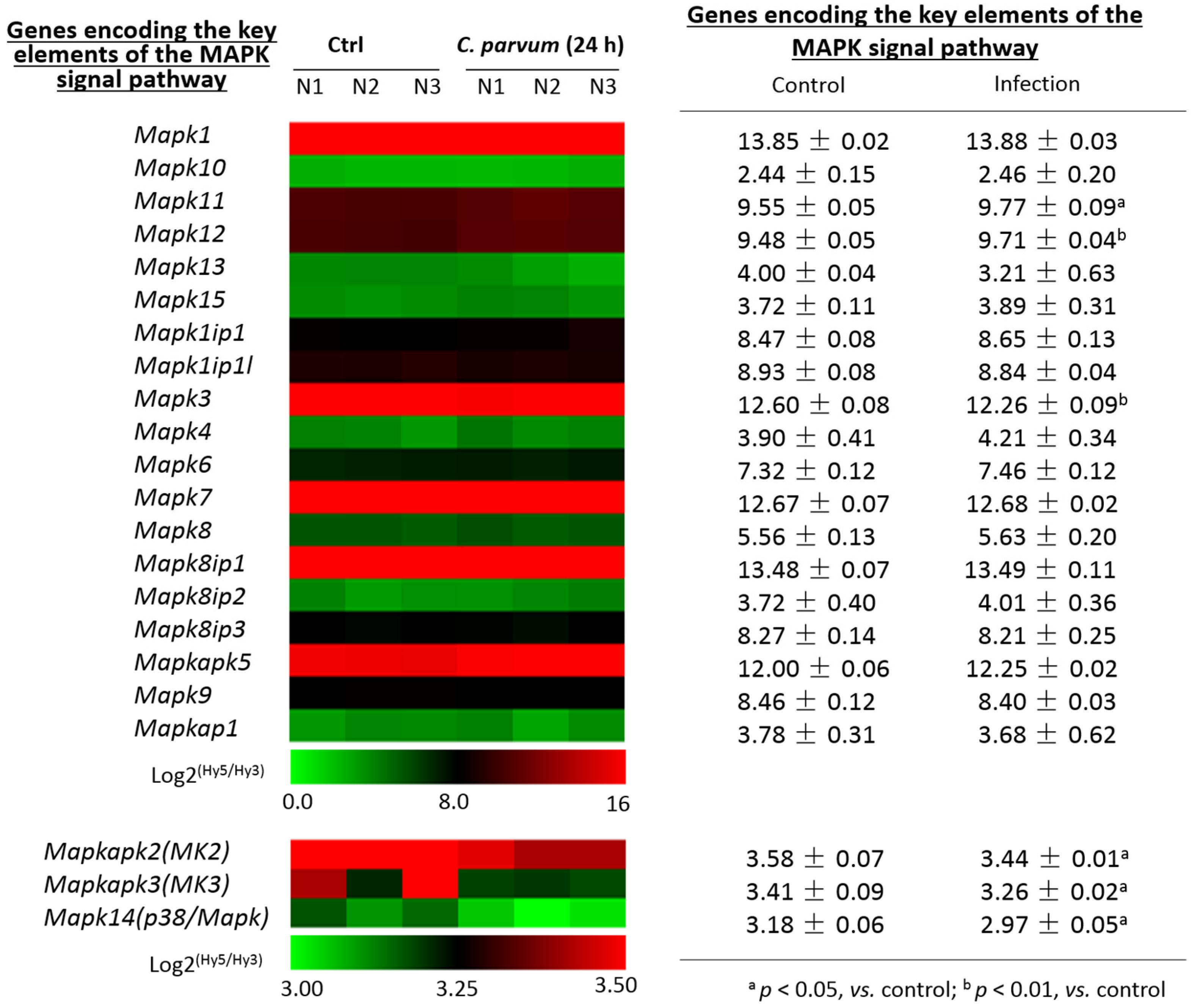
Using the same dataset from our previous transcriptome analysis of IEC4.1 cells following infection by C. parvum for 24 h, as described above [24], we looked at the expression levels for these genes encoding the key elements of the MAPK signal pathway. Expression levels of most genes coding the key elements of the MAPK signaling pathway were not altered IEC4.1 in cells following C. parvum infection for 24 h (Figure 2). However, the expression levels of p38/Mapk, MAP kinase-activated protein kinase 2 (Mk2), and Mk3 genes were significantly decreased in infected cells, compared to that in the non-infected control (Figure 2).

3.3. Downregulation of p38MAPK, MK2 and MK3 Genes in Infected Host Cells of Various Models of C. parvum Infection

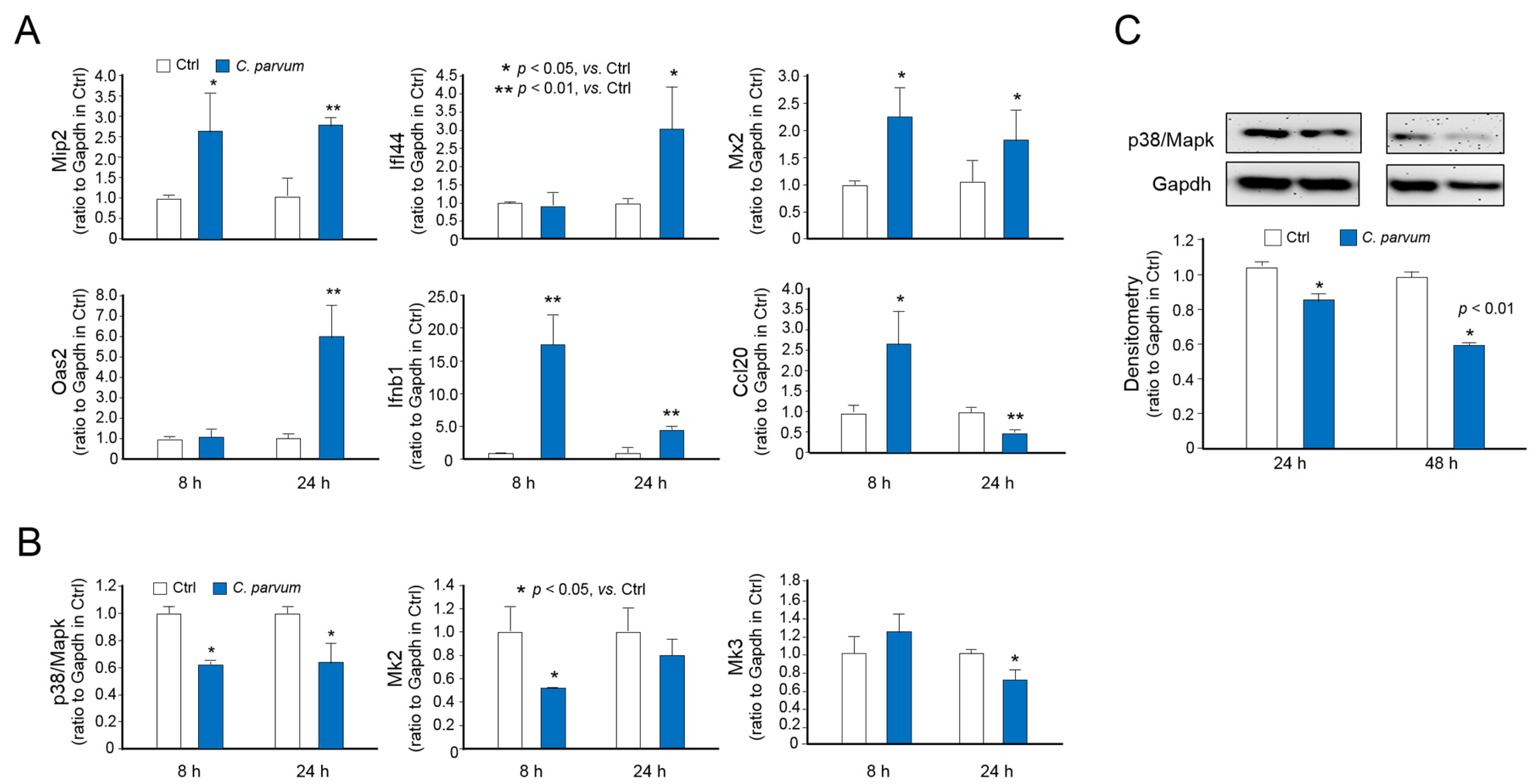
Consistent with results from previous studies [24,37], we detected the upregulation of several inflammatory and defense genes using real-time PCR in IEC4.1 cells following C. parvum infection, including Mip2, Ifi44, Mx2, Oas2, and Ifnb1 (Figure 3A). Whereas the induction of Ccl20 was observed in IEC4.1 cells following C. parvum infection for 8 h, a significant decrease in the Ccl20 expression level was detected in cells following infection for 24 h (Figure 3A), confirming the suppression of Ccl20 in host cells following C. parvum infection in a previous report [20]. We further confirmed the decreased expression of p38/Mapk, Mk2, and Mk3 in infected cells (Figure 3B). Moreover, Western blotting further confirmed decreased p38/Mapk protein content in infected IEC4.1 cells (Figure 3C).
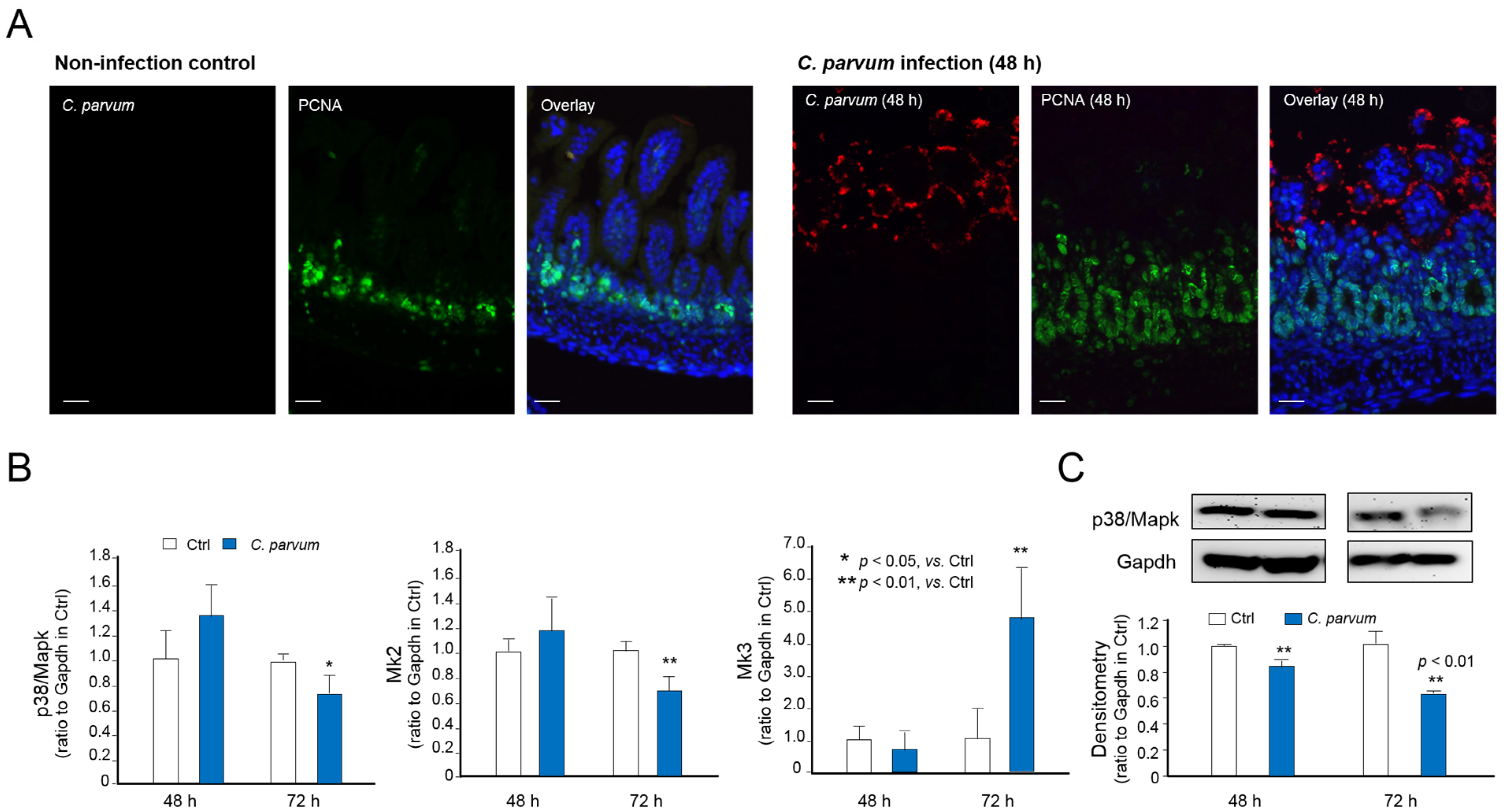
Using a well-documented model of intestinal cryptosporidiosis in neonatal mice through the oral administration of the parasite [12,25], we detected the infection of C. parvum to the intestinal epithelium at the villus region and increased cell proliferation at the crypt region, as previously reported [12,25], by immunofluorescent staining (Figure 4A). We also detected the decreased expression of p38/Mapk, Mk2, and Mk3 in isolated intestinal epithelium from infected neonatal mice (Figure 4B). Western blotting further confirmed a decrease of p38/Mapk protein content in the intestinal epithelium from infected neonatal mice (Figure 4C). In addition, decreased expression of p38/Mapk, Mk2, and Mk3 genes in infected cells was detected in another murine intestinal epithelial cell line (mulINTEP1 cells) following C. parvum infection [22] (Figure 5).

3.4. Suppression of MAPK Signaling Impairs Intestinal Epithelial Innate Defense against C. parvum

We then investigated the possible impact of the suppression of MAPK signaling on host epithelial antimicrobial defense. When cells were exposed to C. parvum oocysts for infection in the presence of a MAPK activator anisomycin, a significant decrease of infection burden was detected (Figure 6A). Accordingly, an increase of infection burden was observed in cells treated with a MAPK inhibitor, SP600125 [38] (Figure 6A). The impact of MAPK signaling on the infection burden in IEC4.1 cells was not due to the effects on parasite attachment or invasion, as a similar attachment/invasion pattern was observed in cells treated with the MAPK inhibitor SP600125 (Figure 6B). Interestingly, a slight increase of the parasite attachment/invasion was measured in cells treated with the MAPK activator anisomycin (Figure 6B), suggesting that the alterations in the infection burden (after exposure to parasites for 24 h as measured in Figure 6A) in cells treated with the MAPK activator or inhibitor was due to MAPK-mediated epithelial anti-parasite defense, rather than their impact on the parasite attachment or invasion.

4. Discussion

Several immune-evasive strategies have been proposed to address how C. parvum evades host innate antimicrobial defense [39]. As one of the ancient host antimicrobial strategies to intracellular pathogens [40], the anti-apoptotic mechanism in infected host cells during early C. parvum infection stage may facilitate parasite propagation and survival [17,41]. Whereas C. parvum infection eventually causes host cell death, infection inhibits apoptotic cell death of directly infected host cells during the early stage of infection [41]. Moreover, it has been demonstrated that infection induces depletion of STAT1α and suppresses expression of CCL20 in intestinal epithelial cells, resulting in the suppression of epithelial antimicrobial defense [13,20]. In this study, we observed the suppression of p38/MAPK signaling in intestinal epithelium following C. parvum infection. We detected significant downregulation of p38/Mapk, Mk2, and Mk3 genes in infected intestinal epithelial cells. Furthermore, suppression of p38/MAPK signaling was associated with an impaired intestinal epithelial defense against C. parvum infection. Therefore, our data suggest a new strategy by which C. parvum impedes host antimicrobial signaling to evade host immune defense.
Intestinal epithelial cells express several pathogen pattern recognition receptors, such as Toll-like receptors (TLRs), C-type lectin receptors, NOD-like receptors and RIG-I-like receptors. While their receptor-proximal signaling mechanisms vary, these pattern recognition receptors can activate both MAPK and NF-κB pathways, which are crucial to generating immune responses [42]. We and other laboratories previously demonstrated activation of the TLR/MyD88/NF-κB signal pathway in intestinal epithelium following Cryptosporidium infection [16,17,41]. Nevertheless, here we observed that the vast majority of MAPK-controlled genes show either no change or are suppressed in intestinal epithelial cells following C. parvum infection. Suppression of MAPK signaling in infected cells was evident by a significant inhibition of cellular responses to anisomycin stimulation. How Cryptosporidium infection impedes MAPK signaling activation is still unclear. Nevertheless, we detected significant downregulation of p38/Mapk, Mk2 and Mk3 in infected cells. Consistent with results from a previous report [20], we also detected downregulation of Ccl20 in host cells following C. parvum infection for 24 h. Of note, Ccl20 has been demonstrated as one of the defense genes that are controlled by MAPK and NF-κB signaling in intestinal epithelial cells [35,36,43,44].
The expression of proteases that can degrade specific components of MAPK cascades is one mechanism used by pathogens to modulate MAPK signaling in infected host cells. For examples, the leishmanolysin (also known as GP63) protease of Leishmania major can degrade TGF-beta activated kinase 1/MAP3K7 binding protein 1, resulting in the inhibition of MAPK7-mediated p38α activation in infected host cells [45]. The lethal toxin from Bacillus anthracis contains a protease, which can cleave the MAPK-docking domain of MKKs, and thus, inhibits MAPK activation and suppresses inflammatory cytokine production in macrophages [46]. Similarly, several members of the Yersinia bacterial genus use type III secretion systems to inject proteins into host cells to interfere with host intracellular signaling [47]. YopJ is one of such proteins that can inhibit both MAPK and NF-κB signaling in host cells (40). Interestingly, Cryptosporidium species carry Cryptosporidium parvum virus 1 (CSpV1) virus, a virus of the family Partitiviridae, genus Cryspovirus that infects Cryptosporidium (41). The CSpV1 genome comprises two distinct dsRNAs, sized 1786 bp (CSpV1-dsRdRp) and 1374 bp (CSpV1-dsCA) (42). The predicted protein sequence from the CSpV1-dsCA has a limited similarity with mitogen-activated c-June NH2 terminal protein kinases (JNK/p38/MAPK) from mammalian cells (42). It merits further investigation to clarify whether CSpV1 dsRNAs from Cryptosporidium can target MAPK signaling in infected host cells. RNA transcripts with very low protein-coding potential from Cryptosporidium have been demonstrated to be delivered into infected host epithelial cells and modulate host cell gene transcription [26].
Pathogens have often evolved effective mechanisms of inhibiting immune responses and a common strategy is targeting the host intracellular signaling networks, including the NF-κB and interferon signal pathways [48]. Given the importance of MAPK signaling in regulating the immune response, it is not surprising that many pathogens can modulate MAPK activation to evade host immune defense, such as Bacillus anthracis, Leishmania major, and M. tuberculosis [45,46,47,49]. Coupled with the fact that the MAPK signaling pathway usually cross-talks with many other signaling cascades important to epithelial innate defense [42], we would speculate that the suppression of MAPK signaling is associated with an impaired intestinal epithelial immunity against Cryptosporidium infection. Our findings also suggest the potential of the pharmacological targeting of MAPK signaling to control Cryptosporidium infection.

Supplementary Materials

The following are available online at https://www.mdpi.com/2076-2607/9/1/151/s1, Table S1: List of primers used for RT-PCR.

Author Contributions

X.-M.C., W.H. conceived and designed the experiments. W.H., J.L., A.-Y.G., S.D., M.L., Y.W. and N.W.M. performed the experiments. X.-M.C., W.H., A.-Y.G. analyzed the data. L.X. and Y.F. provided some suggestions on experimental design and writing. W.H. and X.-M.C. wrote the manuscript. All authors have read and agreed to the published version of the manuscript.

Funding

This study was supported by funding from the National Institutes of Health (AI116323, AI136877, and AI141325) and the Nebraska Cancer and Smoking Disease Research Program (LB595) to XMC. Wei He was a visiting scholar supported by the China Scholarship Council, 111 Project (D20008), and Innovation Team Project of Guangdong Universities (2019KCXTD001). The project described was also supported by Grant Number G20RR024001 from the National Center for Research Resources.

Institutional Review Board Statement

This study was conducted in strict accordance with the recommendations in the Guide for the Care and Use of Laboratory Animals of the National Institutes of Health under the Assurance of Compliance Number A3348-01. All animal experiments were done in accordance with procedures (protocol number #0959) approved by the Institutional Animal Care and Use Committee of Creighton University.

Informed Consent Statement

Not applicable.

Data Availability Statement

Publicly available datasets were analyzed in this study. This data can be found here: https://www.ncbi.nlm.nih.gov/geo/query/acc.cgi?acc=GSE112247.

Acknowledgments

We thank B.L.B. (Creighton) for her assistance in writing the manuscript.

Conflicts of Interest

The authors declare that the research was conducted in the absence of any commercial or financial relationships that could be construed as a potential conflict of interest.

References

  1. Chalmers, R.M.; Katzer, F. Looking for cryptosporidium: The application of advances in detection and diagnosis. Trends Parasitol. 2013, 5, 237–251. [Google Scholar] [CrossRef] [PubMed]
  2. Ryan, U.; Fayer, R.; Xiao, L. Cryptosporidium species in humans and animals: Current understanding and research needs. Parasitology 2014, 13, 1667–1685. [Google Scholar] [CrossRef] [PubMed] [Green Version]
  3. Striepen, B. Parasitic infections: Time to tackle cryptosporidiosis. Nature 2013, 7475, 189–191. [Google Scholar] [CrossRef] [PubMed] [Green Version]
  4. Checkley, W.; White, A.C., Jr.; Jaganath, D.; Arrowood, M.J.; Chalmers, R.M.; Chen, X.M.; Fayer, R.; Griffiths, J.K.; Guerrant, R.L.; Hedstrom, L.; et al. A review of the global burden, novel diagnostics, therapeutics, and vaccine targets for cryptosporidium. Lancet Infect. Dis. 2015, 1, 85–94. [Google Scholar] [CrossRef] [Green Version]
  5. Kotloff, K.L.; Nataro, J.P.; Blackwelder, W.C.; Nasrin, D.; Farag, T.H.; Panchalingam, S.; Wu, Y.; Sow, S.O.; Sur, D.; Breiman, R.F.; et al. Burden and aetiology of diarrhoeal disease in infants and young children in developing countries (the global enteric multicenter study, gems): A prospective, case-control study. Lancet 2013, 9888, 209–222. [Google Scholar] [CrossRef]
  6. Putignani, L.; Menichella, D. Global distribution, public health and clinical impact of the protozoan pathogen cryptosporidium. Interdiscip. Perspect Infect. Dis. 2010, 2010, 753512. [Google Scholar] [CrossRef] [Green Version]
  7. Chen, X.M.; Keithly, J.S.; Paya, C.V.; LaRusso, N.F. Cryptosporidiosis. N. Engl. J. Med. 2002, 22, 1723–1731. [Google Scholar] [CrossRef]
  8. Peterson, L.W.; Artis, D. Intestinal epithelial cells: Regulators of barrier function and immune homeostasis. Nat. Rev. Immunol. 2014, 3, 141–153. [Google Scholar] [CrossRef]
  9. McDonald, V.; Korbel, D.S.; Barakat, F.M.; Choudhry, N.; Petry, F. Innate immune responses against cryptosporidium parvum infection. Parasite Immunol. 2013, 2, 55–64. [Google Scholar] [CrossRef]
  10. Laurent, F.; Kagnoff, M.F.; Savidge, T.C.; Naciri, M.; Eckmann, L. Human intestinal epithelial cells respond to cryptosporidium parvum infection with increased prostaglandin h synthase 2 expression and prostaglandin e2 and f2alpha production. Infect. Immun. 1998, 4, 1787–1790. [Google Scholar] [CrossRef] [Green Version]
  11. Pollok, R.C.; Farthing, M.J.; Bajaj-Elliott, M.; Sanderson, I.R.; McDonald, V. Interferon gamma induces enterocyte resistance against infection by the intracellular pathogen cryptosporidium parvum. Gastroenterology 2001, 1, 99–107. [Google Scholar] [CrossRef] [PubMed]
  12. Barakat, F.M.; McDonald, V.; Di Santo, J.P.; Korbel, D.S. Roles for nk cells and an nk cell-independent source of intestinal gamma interferon for innate immunity to cryptosporidium parvum infection. Infect. Immun. 2009, 11, 5044–5049. [Google Scholar] [CrossRef] [PubMed] [Green Version]
  13. Choudhry, N.; Petry, F.; van Rooijen, N.; McDonald, V. A protective role for interleukin 18 in interferon gamma-mediated innate immunity to cryptosporidium parvum that is independent of natural killer cells. J. Infect. Dis. 2012, 1, 117–124. [Google Scholar] [CrossRef] [PubMed] [Green Version]
  14. Bedi, B.; McNair, N.N.; Forster, I.; Mead, J.R. Il-18 cytokine levels modulate innate immune responses and cryptosporidiosis in mice. J. Eukaryot. Microbiol. 2015, 1, 44–50. [Google Scholar] [CrossRef] [PubMed]
  15. Zhou, R.; Gong, A.Y.; Eischeid, A.N.; Chen, X.M. Mir-27b targets ksrp to coordinate tlr4-mediated epithelial defense against cryptosporidium parvum infection. PLoS Pathog. 2012, 5, e1002702. [Google Scholar] [CrossRef] [PubMed] [Green Version]
  16. Chen, X.M.; O’Hara, S.P.; Nelson, J.B.; Splinter, P.L.; Small, A.J.; Tietz, P.S.; Limper, A.H.; LaRusso, N.F. Multiple tlrs are expressed in human cholangiocytes and mediate host epithelial defense responses to cryptosporidium parvum via activation of nf-kappab. J. Immunol. 2005, 11, 7447–7456. [Google Scholar] [CrossRef] [Green Version]
  17. Chen, X.M.; Levine, S.A.; Splinter, P.L.; Tietz, P.S.; Ganong, A.L.; Jobin, C.; Gores, G.J.; Paya, C.V.; LaRusso, N.F. Cryptosporidium parvum activates nuclear factor kappab in biliary epithelia preventing epithelial cell apoptosis. Gastroenterology 2001, 7, 1774–1783. [Google Scholar] [CrossRef]
  18. Liu, J.; Deng, M.; Lancto, C.A.; Abrahamsen, M.S.; Rutherford, M.S.; Enomoto, S. Biphasic modulation of apoptotic pathways in cryptosporidium parvum-infected human intestinal epithelial cells. Infect. Immun. 2009, 2, 837–849. [Google Scholar] [CrossRef] [Green Version]
  19. Choudhry, N.; Korbel, D.S.; Edwards, L.A.; Bajaj-Elliott, M.; McDonald, V. Dysregulation of interferon-gamma-mediated signalling pathway in intestinal epithelial cells by cryptosporidium parvum infection. Cell Microbiol. 2009, 9, 1354–1364. [Google Scholar] [CrossRef]
  20. Guesdon, W.; Auray, G.; Pezier, T.; Bussiere, F.I.; Drouet, F.; Le Vern, Y.; Marquis, M.; Potiron, L.; Rabot, S.; Bruneau, A.; et al. Ccl20 displays antimicrobial activity against cryptosporidium parvum, but its expression is reduced during infection in the intestine of neonatal mice. J. Infect. Dis. 2015, 8, 1332–1340. [Google Scholar] [CrossRef] [Green Version]
  21. Li, X.C.; Jevnikar, A.M.; Grant, D.R. Expression of functional icam-1 and vcam-1 adhesion molecules by an immortalized epithelial cell clone derived from the small intestine. Cell Immunol. 1997, 1, 58–66. [Google Scholar] [CrossRef] [PubMed]
  22. Selvakumar, T.A.; Bhushal, S.; Kalinke, U.; Wirth, D.; Hauser, H.; Koster, M.; Hornef, M.W. Identification of a predominantly interferon-lambda-induced transcriptional profile in murine intestinal epithelial cells. Front. Immunol. 2017, 8, 1302. [Google Scholar] [CrossRef] [PubMed] [Green Version]
  23. Wang, Y.; Gong, A.Y.; Ma, S.; Chen, X. Delivery of parasite cdg7_flc_0990 rna transcript into intestinal epithelial cells during cryptosporidium parvum infection suppresses host cell gene transcription through epigenetic mechanisms. Cell. Microbiol. 2017, 11, 1462–5814. [Google Scholar]
  24. Li, M.; Gong, A.Y.; Zhang, X.T.; Wang, Y.; Mathy, N.W.; Martins, G.A.; Strauss-Soukup, J.K.; Chen, X.M. Induction of a long noncoding rna transcript, nr_045064, promotes defense gene transcription and facilitates intestinal epithelial cell responses against cryptosporidium infection. J. Immunol. 2018, 12, 3630–3640. [Google Scholar] [CrossRef]
  25. Lantier, L.; Lacroix-Lamande, S.; Potiron, L.; Metton, C.; Drouet, F.; Guesdon, W.; Gnahoui-David, A.; Le Vern, Y.; Deriaud, E.; Fenis, A.; et al. Intestinal cd103+ dendritic cells are key players in the innate immune control of cryptosporidium parvum infection in neonatal mice. PLoS Pathog. 2013, 12, e1003801. [Google Scholar] [CrossRef] [Green Version]
  26. Wang, Y.; Gong, A.Y.; Ma, S.; Chen, X.; Li, Y.; Su, C.J.; Norall, D.; Chen, J.; Strauss-Soukup, J.K.; Chen, X.M. Delivery of parasite RNA transcripts into infected epithelial cells during cryptosporidium infection and its potential impact on host gene transcription. J. Infect. Dis. 2017, 4, 636–643. [Google Scholar] [CrossRef] [Green Version]
  27. Cuadrado, A.; Corrado, N.; Perdiguero, E.; Lafarga, V.; Munoz-Canoves, P.; Nebreda, A.R. Essential role of p18hamlet/srcap-mediated histone h2a.Z chromatin incorporation in muscle differentiation. EMBO J. 2010, 12, 2014–2025. [Google Scholar] [CrossRef] [Green Version]
  28. Kim, J.; Hwangbo, J.; Wong, P.K. P38 mapk-mediated bmi-1 down-regulation and defective proliferation in atm-deficient neural stem cells can be restored by akt activation. PLoS ONE 2011, 1, e16615. [Google Scholar] [CrossRef] [Green Version]
  29. Malcolm, T.; Chen, J.; Chang, C.; Sadowski, I. Induction of chromosomally integrated hiv-1 ltr requires rbf-2 (usf/tfii-i) and ras/mapk signaling. Virus Genes 2007, 2, 215–223. [Google Scholar] [CrossRef]
  30. Hwang, Y.S.; Oh, S.W.; Park, S.H.; Lee, J.; Yoo, J.A.; Kwon, K.; Park, S.J.; Kim, J.; Yu, E.; Cho, J.Y.; et al. Melanogenic effects of maclurin are mediated through the activation of camp/pka/creb and p38 mapk/creb signaling pathways. Oxid. Med. Cell Longev. 2019, 2019, 9827519. [Google Scholar] [CrossRef] [Green Version]
  31. Hu, S.B.; Zou, Q.; Lv, X.; Zhou, R.L.; Niu, X.; Weng, C.; Chen, F.; Fan, Y.W.; Deng, Z.Y.; Li, J. 9t18:1 and 11t18:1 activate the mapk pathway to regulate the expression of pla2 and cause inflammation in huvecs. Food Funct. 2020, 1, 649–661. [Google Scholar] [CrossRef]
  32. Dreas, A.; Mikulski, M.; Milik, M.; Fabritius, C.H.; Brzozka, K.; Rzymski, T. Mitogen-activated protein kinase (mapk) interacting kinases 1 and 2 (mnk1 and mnk2) as targets for cancer therapy: Recent progress in the development of mnk inhibitors. Curr. Med. Chem. 2017, 28, 3025–3053. [Google Scholar] [CrossRef] [PubMed]
  33. Bardwell, A.J.; Frankson, E.; Bardwell, L. Selectivity of docking sites in mapk kinases. J. Biol. Chem. 2009, 19, 13165–13173. [Google Scholar] [CrossRef] [PubMed] [Green Version]
  34. Xiong, W.; Kojic, L.Z.; Zhang, L.; Prasad, S.S.; Douglas, R.; Wang, Y.; Cynader, M.S. Anisomycin activates p38 map kinase to induce ltd in mouse primary visual cortex. Brain Res. 2006, 1, 68–76. [Google Scholar] [CrossRef] [PubMed]
  35. Zanello, G.; Berri, M.; Dupont, J.; Sizaret, P.Y.; D’Inca, R.; Salmon, H.; Meurens, F. Saccharomyces cerevisiae modulates immune gene expressions and inhibits etec-mediated erk1/2 and p38 signaling pathways in intestinal epithelial cells. PLoS ONE 2011, 4, e18573. [Google Scholar] [CrossRef] [PubMed]
  36. Chatterjee, D.; Chaudhuri, K. Vibrio cholerae o395 outer membrane vesicles modulate intestinal epithelial cells in a nod1 protein-dependent manner and induce dendritic cell-mediated th2/th17 cell responses. J. Biol. Chem. 2013, 6, 4299–4309. [Google Scholar] [CrossRef] [PubMed] [Green Version]
  37. Heo, I.; Dutta, D.; Schaefer, D.A.; Iakobachvili, N.; Artegiani, B.; Sachs, N.; Boonekamp, K.E.; Bowden, G.; Hendrickx, A.P.A.; Willems, R.J.L.; et al. Modelling cryptosporidium infection in human small intestinal and lung organoids. Nat. Microbiol. 2018, 7, 814–823. [Google Scholar] [CrossRef]
  38. Bennett, B.L.; Sasaki, D.T.; Murray, B.W.; O’Leary, E.C.; Sakata, S.T.; Xu, W.; Leisten, J.C.; Motiwala, A.; Pierce, S.; Satoh, Y.; et al. Sp600125, an anthrapyrazolone inhibitor of jun n-terminal kinase. Proc. Natl. Acad. Sci. USA 2001, 24, 13681–13686. [Google Scholar] [CrossRef] [Green Version]
  39. Xie, H.; Lei, N.; Gong, A.Y.; Chen, X.M.; Hu, G. Cryptosporidium parvum induces sirt1 expression in host epithelial cells through downregulating let-7i. Hum. Immunol. 2014, 8, 760–765. [Google Scholar] [CrossRef] [Green Version]
  40. Barber, G.N. Host defense, viruses and apoptosis. Cell Death Differ. 2001, 2, 113–126. [Google Scholar] [CrossRef] [Green Version]
  41. Liu, J.; Drescher, K.M.; Chen, X.M. Micrornas and epithelial immunity. Int. Rev. Immunol. 2009, 3–4, 139–154. [Google Scholar] [CrossRef] [PubMed] [Green Version]
  42. Arthur, J.S.; Ley, S.C. Mitogen-activated protein kinases in innate immunity. Nat. Rev. Immunol. 2013, 9, 679–692. [Google Scholar] [CrossRef] [PubMed]
  43. Sandri, S.; Hatanaka, E.; Franco, A.G.; Pedrosa, A.M.; Monteiro, H.P.; Campa, A. Serum amyloid a induces ccl20 secretion in mononuclear cells through mapk (p38 and erk1/2) signaling pathways. Immunol. Lett. 2008, 1, 22–26. [Google Scholar] [CrossRef] [PubMed]
  44. Dommisch, H.; Chung, W.O.; Jepsen, S.; Hacker, B.M.; Dale, B.A. Phospholipase c, p38/mapk, and nf-kappab-mediated induction of mip-3alpha/ccl20 by porphyromonas gingivalis. Innate Immun. 2010, 4, 226–234. [Google Scholar] [CrossRef] [Green Version]
  45. Halle, M.; Gomez, M.A.; Stuible, M.; Shimizu, H.; McMaster, W.R.; Olivier, M.; Tremblay, M.L. The leishmania surface protease gp63 cleaves multiple intracellular proteins and actively participates in p38 mitogen-activated protein kinase inactivation. J. Biol. Chem. 2009, 11, 6893–6908. [Google Scholar] [CrossRef] [Green Version]
  46. Turk, B.E. Manipulation of host signalling pathways by anthrax toxins. Biochem. J. 2007, 3, 405–417. [Google Scholar] [CrossRef] [Green Version]
  47. Trosky, J.E.; Liverman, A.D.; Orth, K. Yersinia outer proteins: Yops. Cell Microbiol. 2008, 3, 557–565. [Google Scholar] [CrossRef]
  48. Roy, C.R.; Mocarski, E.S. Pathogen subversion of cell-intrinsic innate immunity. Nat. Immunol. 2007, 11, 1179–1187. [Google Scholar] [CrossRef]
  49. Mukherjee, S.; Keitany, G.; Li, Y.; Wang, Y.; Ball, H.L.; Goldsmith, E.J.; Orth, K. Yersinia yopj acetylates and inhibits kinase activation by blocking phosphorylation. Science 2006, 5777, 1211–1214. [Google Scholar] [CrossRef] [Green Version]
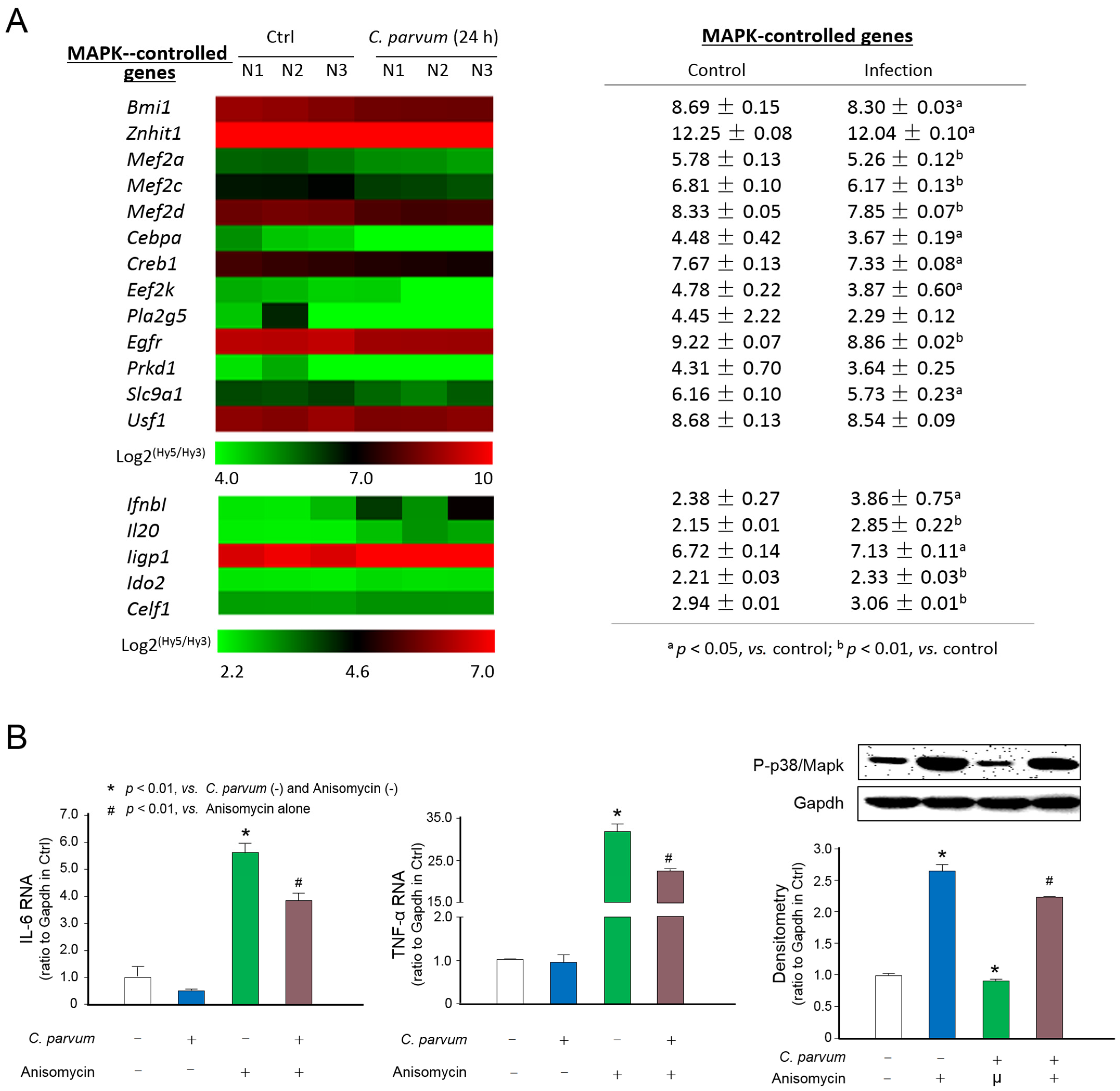
Figure 1. Suppression of MAPK signaling in intestinal epithelium following C. parvum infection. (A) Heatmap of expression levels of MAPK-controlled genes in host cells following C. parvum infection. IEC4.1 cells were exposed to C. parvum infection for 24 h. Total RNA was isolated for genome-wide transcriptome analysis via microarray. Expression levels of MAPK-controlled genes and selected inflammatory genes not directly related to the MAPK signaling are presented as the log2 (Hy5/Hy3), which passed the filtering criteria variation across the samples (n = 3). a p < 0.05, vs., control; b p < 0.01, vs. control. (B) Suppression of Il-6 and Tnf-α expression and inhibition of phosphorylation of p38/Mapk in C. parvum-infected intestinal epithelial cells in response to MAPK activator stimulation. IEC4.1 cells were exposed to C. parvum infection for 24 h and then treated with the MAPK activator anisomycin for up to 4 h. Anisomycin-mediated expression levels of IL-6 and TNF-α were measured. Phosphorylation of p38/Mapk was assessed using Western blot. Gapdh was also blotted for control. “+” and “−” represent cells treated with and without the according reagents, respectively. Representative gel images were shown. Data represent three independent experiments.
Microorganisms 09 00151 g001
Figure 2. Expression profile of genes key to the MAPK signaling pathway in intestinal epithelial cells following C. parvum infection. Heatmap of expression levels of genes key to the MAPK signal pathway in host cells following C. parvum infection, presented as the log2 (Hy5/Hy3) ratios, which passed the filtering criteria variation across the samples (n = 3). IEC4.1 cells were exposed to C. parvum infection for 24 h and RNA was isolated for genome-wide transcriptome analysis via microarray. Expression levels of genes key to the MAPK signal pathway are shown. a p < 0.05, vs. control; b p < 0.01, vs. control.
Microorganisms 09 00151 g002
Figure 3. Downregulation of p38/Mapk, Mk2 and Mk3 genes in intestinal epithelial cells following C. parvum infection. (A) RNA levels of a panel of inflammatory genes in IEC4.1 cells following C. parvum infection. Cells were exposed to C. parvum infection for 8 and 24 h. RNA levels of these genes were measured by using real-time quantitative PCR. (B) RNA levels of p38/Mapk, Mk2 and Mk3 genes in IEC4.1 cells following C. parvum infection. Cells were exposed to C. parvum infection for 8 and 24 h. RNA levels of p38/Mapk, Mk2 and Mk3 genes were measured. (C) Protein level of p38/Mapk in IEC4.1 cells following C. parvum infection. Cells were exposed to C. parvum infection for 24 h and 48 h. Protein level of p38/Mapk was assessed by using Western blot. Gapdh was also blotted for control. Data represent three independent experiments.
Microorganisms 09 00151 g003
Figure 4. Downregulation of p38/Mapk, Mk2 and Mk3 genes in intestinal epithelium of neonatal mice following C. parvum infection in vivo. (A) Immunofluorescent staining of ileum from neonatal mice with and with C. parvum infection. Mice at the age of 5 days after birth received C. parvum oocysts by oral gavage (105 oocysts each mouse). Mice which received phosphate buffered saline by oral gavage were used as control. Tissue sections were triple stained with anti-C. parvum (showing in red), anti-PCNA (showing proliferating cells in green) and DAPI (showing nuclei in blue). (B) RNA levels of p38/Mapk, Mk2 and Mk3 genes in isolated intestinal epithelium from infected neonatal animals. RNA levels of p38/Mapk, Mk2 and Mk3 genes were measured by using real-time PCR. (C) Protein level of p38/Mapk in isolated intestinal epithelium from infected neonatal animals. Protein level of p38/Mapk was assessed by using Western blot. Gapdh was also blotted for control. DAPI = 4′,6-diamidino-2-phenylindole; PCNA = Proliferating cell nuclear antigen.
Microorganisms 09 00151 g004
Figure 5. Downregulation of p38/Mapk, Mk2 and Mk3 genes in mulINTEP1 cells following C. parvum infection. RNA levels of p38/Mapk, Mk2 and Mk3 genes in mulINTEP1 cells following C. parvum infection. Cells were exposed to C. parvum infection for 48 and 72 h. RNA levels of p38/Mapk, Mk2 and Mk3 genes were measured by using real-time PCR. Data represent three independent experiments.
Microorganisms 09 00151 g005
Figure 6. Suppression of MAPK signaling impairs intestinal epithelial defense against C. parvum infection. (A) Activation of MAPK signaling decreased the infection burden of C. parvum in host cells. IEC4.1 cells were exposed to C. parvum infection for 24 h in the presence or absence of the MAPK activator anisomycin or inhibitor SP600125. Infection burden of C. parvum was quantified. (B) Suppression of MAPK signaling in intestinal epithelial cells on the attachment and invasion of C. parvum to host cells. Cells were exposed to C. parvum infection for 2 h (for attachment and invasion) in the presence or absence of the MAPK activator anisomycin or inhibitor SP600125. Infection burden of C. parvum was quantified. The expression levels of IL-6 and TNF- were measured in cells treated with anisomycin or SP600125 to confirm their effects on MAPK signaling. “+” and “−” represent cells treated with and without the according reagents, respectively. Data represent three independent experiments.
Microorganisms 09 00151 g006
Publisher’s Note: MDPI stays neutral with regard to jurisdictional claims in published maps and institutional affiliations.