Article

Identification of Potential Citrate Metabolism Pathways in Carnobacterium maltaromaticum

by
Heng Li
1,2,
Nancy E. Ramia
3,
Frédéric Borges
3,
Anne-Marie Revol-Junelles
3,
Finn Kvist Vogensen
4 and
Jørgen J. Leisner
2,*
1
Pasteurien College, Soochow University, Suzhou 215123, China
2
Department of Veterinary and Animal Sciences, Faculty of Health and Medical Sciences, University of Copenhagen, DK-1870 Frederiksberg, Denmark
3
Laboratoire d’Ingénierie des Biomolécules (LIBio), Ecole Nationale Supérieure d’Agronomie et des Industries Alimentaires (ENSAIA), Université de Lorraine, LIBio, F-54000 Nancy, France
4
Department of Food Science, Faculty of Science, University of Copenhagen, DK-1958 Frederiksberg, Denmark
*
Author to whom correspondence should be addressed.
Microorganisms 2021, 9(10), 2169; https://doi.org/10.3390/microorganisms9102169
Submission received: 25 September 2021 / Revised: 13 October 2021 / Accepted: 15 October 2021 / Published: 18 October 2021
(This article belongs to the Special Issue Functional Characterization of Lactic Acid Bacteria)

Abstract

:
In the present study, we describe the identification of potential citrate metabolism pathways for the lactic acid bacterium (LAB) Carnobacterium maltaromaticum. A phenotypic assay indicated that four of six C. maltaromaticum strains showed weak (Cm 6-1 and ATCC 35586) or even delayed (Cm 3-1 and Cm 5-1) citrate utilization activity. The remaining two strains, Cm 4-1 and Cm 1-2 gave negative results. Additional analysis showed no or very limited utilization of citrate in media containing 1% glucose and 22 or 30 mM citrate and inoculated with Cm 6-1 or ATCC 35586. Two potential pathways of citrate metabolism were identified by bioinformatics analyses in C. maltaromaticum including either oxaloacetate (pathway 1) or tricarboxylic compounds such as isocitrate and α-ketoglutarate (pathway 2) as intermediates. Genes encoding pathway 1 were present in two out of six strains while pathway 2 included genes present in all six strains. The two potential citrate metabolism pathways in C. maltaromaticum may potentially affect the sensory profiles of milk and soft cheeses subjected to growth with this species.

1. Introduction

The main metabolic activity of lactic acid bacteria (LAB) in milk fermentation is lactic acid production. Various species and strains of LAB, including Carnobacterium maltaromaticum isolated from Mozzarella cheese, are also able to metabolize citrate, which leads to the production of aroma compounds [1,2,3,4] . C. maltaromaticum is common in milk and soft cheeses as well as in meat and fish products. It is also isolated from a range of cold and temperate environments. Some strains of C. maltaromaticum (previously named Carnobacterium piscicola) are fish pathogens [5,6,7,8].
The presence of C. maltaromaticum in milk and soft cheeses may affect the sensory profiles of such products [9]. In this context, citrate is of interest as LAB produces flavor compounds from this compound, including acetate, diacetyl, acetoin, and 2,3-butanediol [1,3,4,10,11,12].
To extend our knowledge on potential citrate metabolism pathways in C. maltaromaticum, we here compare phenotypic tests for citrate metabolism with analysis of whole-genome sequences to identify genes predicted to be involved in citrate metabolism. The main aim was to supply information on the potential sensory applicability of using C. maltaromaticum as adjunct cultures in soft cheeses.

2. Materials and Methods

2.1. Strains Collection, Phenotypic Tests, and Bioinformatic Analyses

Table 1 lists C. maltaromaticum strains used in phenotypic tests and bioinformatics and their sources.

2.2. Phenotypic Tests for Citrate Metabolism and Test for Growth on a Citrate-Containing Medium

An agar medium for the detection of citrate utilizing LAB was modified from the procedure described by Kempler and McKay [14]. The basal medium contained 0.25% milk protein hydrolysate peptone (Bacto, Mt Pritchard, Australia) and 1.5% agar, autoclaved at 121 °C for 15 min, after which sterile 0.4% nonfat milk (Arla Foods, Aarhus, Denmark) and 0.5% w/v glucose were added, and pH was adjusted to 6.6. Finally, two solutions, one containing 10% potassium ferricyanide and one containing 1 g ferric citrate and 1 g sodium citrate in 40 mL water, were heat-treated at 100 °C for 30 min and 10 mL of each solution was added to 1 L agar medium. Plates were dried in the dark overnight at 30 °C and bacterial cultures were streaked, either directly from frozen culture vials or an overnight culture propagated in All Purpose Tween broth (APT) (Difco, Sparks, MD, USA). Incubation was performed under aerobic or microaerophilic conditions (plates were incubated in a jar with air exhausted by a tea light) in the dark at 25 °C. Cultures were scored positive if they developed a blue color after 3 or 5 days.
A determination of citrate metabolism by chemical analysis was carried out by sub-culturing the Cm 6-1 and ATCC 35586 strains in APT broth (Difco, Beckton, Dickinson and Company, Sparks, MD, USA) with 1% glucose and 22 or 30 mM citrate. Strains were sub-cultured twice o/n in APT broth at 25 °C before adding 10 µl of 22 h cultures to 10 mL of APT broth. The cultures were incubated anaerobically (Anaerogen, Oxoid, Basingstoke, UK) for 9 days at 25 °C before sampling. Viable counts were determined by the transfer of appropriate dilutions of sterile peptone saline to APT agar plates that were incubated for three days anaerobically at 25 °C before enumeration. Citric acid was determined by MS-Omics (Vedbæk, Denmark) using a gas chromatography-mass spectrometry (GC-MS) method. Samples were derivatized as described by Smart et al. [15] and the derivatization reaction was stopped by the addition of chloroform. Subsequently, the chloroform phase with derivatives was injected into the GC. All samples were analyzed in a randomized order. Analysis was performed using gas chromatography (7890B, Agilent, Santa Clara, CA, USA) coupled with a quadrupole mass spectrometry detector (5977B, Agilent). The system was controlled by ChemStation (Agilent). Raw data were converted to netCDF format using Chemstation. Then, the data were imported and processed in MATLAB R2018b (Mathworks, Inc., Natick, MA, USA) using the PARADISe software described by Johnsen et al. [16].
Sample analysis for contents of short-chain fatty acids was carried out by MS-Omics as follows. Samples were acidified using hydrochloride acid, and deuterium-labeled internal standards were added. All samples were analyzed in a randomized order. Analysis was performed using a high polarity column (Zebron™ ZB-FFAP, GC Cap. Column 30 m × 0.25 mm × 0.25 µm) installed in a GC coupled with a quadropole detector and controlled by ChemStation, similar to the analysis for citric acid. Raw data was converted to netCDF format using Chemstation before the data was imported and processed in Matlab R2014b (Mathworks, Inc.) using the PARADISe software [16].

2.3. Whole-Genome Sequencing

Isolates were grown on APT agar for 24 h at 30 °C and genomic DNA was extracted and purified using the DNeasy Blood & Tissue Kit, according to the manufacturer’s instructions (Qiagen GmbH, Hilden, Germany). The quality of the extracted DNA was assessed using a Nanodrop ND-1000 spectrophotometer (Nanodrop Technologies, Wilmington, DE, USA) and electrophoresis on a 1.0% (w/v) agarose gel. Sequencing was performed using MiSeq (Illumina) at a 300 bp paired-end read format. Sequencing reads were de novo assembled with CLC Genomics Workbench 10.1.1 (Qiagen, Aarhus, Denmark).

2.4. Bioinformatic Analyses for Genome Annotation

The presence of genes included in citrate metabolism pathways 1 and 2 were examined for a number of Carnobacterium spp. whole-genome sequences. A single nucleotide polymorphism (SNP) cladogram was constructed using Carnobacterium spp. sequences downloaded from NCBI GenBank (n = 21), RAST annotation server (n = 1), and the C. maltaromaticum isolates from the present study (n = 6). The annotations of the pathways were predicted via the RAST annotation server [17,18,19]. The SNP tree was constructed by CSI Phylogeny 1.4 (https://cge.cbs.dtu.dk, accessed on 17 June 2021) with ATCC 35586 as the reference genome. The select minimum depth at SNP positions was set at 10x. The select minimum relative depth at SNP positions was 10%. The select minimum distance between SNPs (prune) was 10 bp, while the select minimum SNP quality was 30. Finally, the select minimum read mapping quality was 25 and the select minimum Z-score was set at 1.96. Then, the phylogenetic tree was imported to iTOL (Interactive Tree of Life) for visualization (https://itol.embl.de, accessed on 17 June 2021) [20]. Leaf sorting was set as the default and the tree was re-rooted at midpoint with ATCC 35586 as the reference genome.
Detailed genome annotations were carried out using a combined bioinformatical approach including the RAST annotation server [17,18,19], SMART [21], Pfam 32.0 [22], Uniprot database [23], KEGG database [24], and local Blast within the CLC Genomics Workbench 10.1.1. Briefly, whole-genome assemblies were uploaded to the seed-based RAST annotation server for genome annotation and in MicroScope [25]. To improve the annotation accuracy for genes found to be associated with citrate metabolism, based on the KEGG pathway website, sequence alignments for specific genes were downloaded from the Pfam and Uniprot databases. The hits with an e-score of 1e−20 or lower were considered as indicative of putative functional proteins or enzymes [6].
Moreover, the contigs containing the cit operon were retrieved from the whole-genome sequence and blasted against all the strains. The results were visualized in GView (Figure 1 and Figure 2 ) [26]. The presence of transmembrane helices was predicted by using the TMHMM server [27] at DTU Bioinformatics at the Department of Bio and Health Informatics of the Technical University of Denmark. Protein domain predictions were obtained from SMART (http://smart.embl-heidelberg.de, accessed on 17 June 2021). Figure 1 and Figure 2 list the annotated gene products encoding the functions of citrate uptake or citrate metabolism with oxaloacetate as an intermediate (pathway 1) or the truncated TCA cycle (pathway 2).

3. Results and Discussion

C. maltaromaticum citrate metabolism was tested on a citrate-containing agar medium [2,14]. The positive controls, Enterococcus faecalis ATCC 29212, Lactiplantibacillus pentosus DSMZ 20314, and Lactiplantibacillus plantarum LMG 11405, all showed strong activity, whereas Pediococcus acidilactici PAH and Lactococcus lactis ATTC 11454 showed no activity, which was also the case for Carnobacterium gallinarum LMG 9841 and Carnobacterium divergens LMG 9199. Four C. maltaromaticum strains (Cm 6-1, Cm 3-1, Cm 5-1, and ATCC 35586) showed weak activity, among which the Cm 3-1 and Cm 5-1 strains had delayed positive phenotypes during aerobic incubation at 25 °C (Table 1). The remaining strains, Cm 4-1 and Cm 1-2, gave negative results under these conditions. Incubation under microaerophilic conditions gave similar results.
An additional experiment examined citrate utilization of the two strains Cm 6-1 and ATCC 35586 during growth in APT broth with 22 or 30 mM citrate. Bacterial concentrations increased, irrespectively of initial citrate concentration, from log 6.1 CFU/mL (Cm 6-1) or log 6.3 CFU/mL (ATCC 35586) to log 8.7 CFU/mL (Cm 6-1) or log 9.1 CFU/mL (ATCC 35586) after 9 days of incubation at 25 °C. The medium with the lowest initial concentration of citrate (22 mM) contained either 18.7 (ATCC 35586; an average of three replica cultures) or 19.5 (Cm 6-1; an average of four replica cultures) mM citrate at end of incubation. One ATCC 35586 sample that only contained 12 mM was noted as an outlier and was not included in the average. Similarly, the medium with the highest initial concentration of citrate (30 mM) contained 28.25 mM at the end of incubation irrespective of strain (average of four replica cultures in each case, see Table S2 for details). Taking into consideration that the analysis had a relative standard deviation of 3%, the results indicate that the two strains did not—or only to a limited extent—utilize citrate when present in relatively high amounts. Production of short-chain fatty acids was also examined in this experiment. Both strains showed limited production of acetic acid: 1.8 mM or 1.2 mM for the 3Ba-II-6 strain and 2.5 or 1.5 mM for the ATCC 35586 strain in APT broth with 22 or 30 mM citrate, respectively. Production of formic acid depended on strain: 11.1 or 9.1 mM for the 3Ba-II-6 strain and 2.4 or 0.6 mM for the ATCC 35586 strain in APT broth with 22 or 30 mM citrate, respectively. These values were calculated by subtraction of concentrations found in sterile media: 5.5 mM or 7.6 mM acetic acid and 2.5 or 4.7 mM formic acid in APT broth with 22 or 30 mM citrate, respectively.
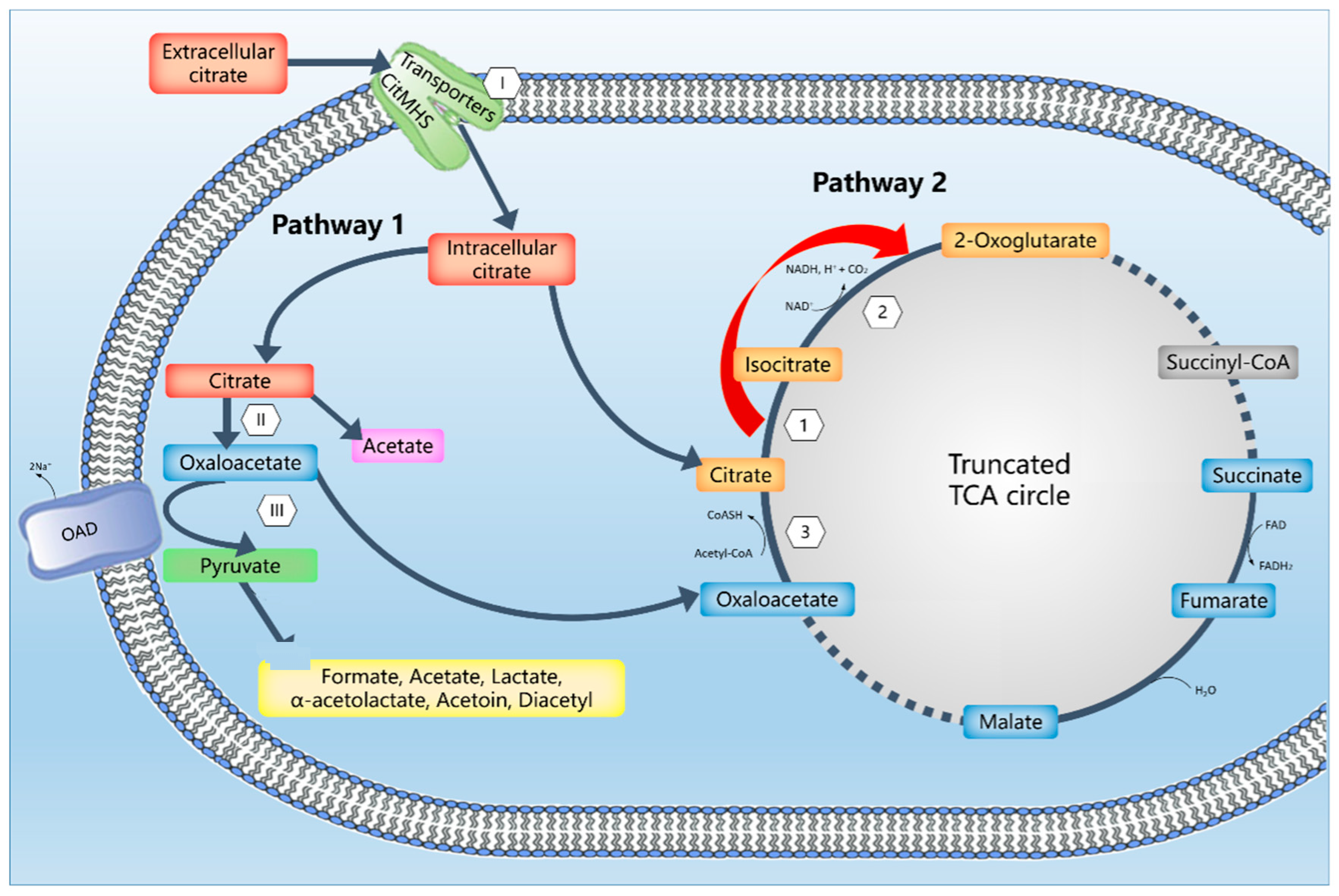
Several genes, identified by genome annotations, were associated with two different pathways for citrate metabolism in C. maltaromaticum ( Figure 1Figure 2 and Figure 3, Table S1). Pathway 1 constitutes the typical LAB citrate metabolism pathway and includes oxaloacetate as an intermediate, and pathway 2 includes TCA compounds as intermediates ( Figure 1Figure 2 and Figure 3). All examined strains encoded the pathway 2 genes whereas only two strains encoded the pathway 1 genes (Table 1). A screening of whole-genome sequences of 14 additional C. maltaromaticum strains showed the presence of genes associated with pathway 1 in three other strains and the presence of genes associated with pathway 2 in all 14 strains (Figure S1). SNP analysis indicated that the C. maltaromaticum strains possessing pathway 1 genes are not necessarily closely related (Figure S1).
None of these pathways were detected in five strains of Carnobacterium divergens or one strain each for Carnobacterium funditum and Carnobacterium viridans, whereas pathway 1 was observed in one strain of Carnobacterium pleistocenium. The presence of pathway 1 genes in only some C. maltaromaticum strains indicate that their presence could either be due to a transfer event from another species or they have been lost from several lineages of C. maltaromaticum. In relation to the first possibility, we have previously discussed the capability of genome expansion in C. maltaromaticum compared to other Carnobacterium spp. [28]. BLAST analyses, as well as the alignment and construction of phylogenetic trees of predicted protein domains of C. maltaromaticum Cm 6-1 pathway 1 gene products, indicated that the most similar gene sequences were generally found in the related Isobaculum melis, with Vagococcus salmoninarum possessing the second-closest related set of genes (results not shown). It will be of interest to conduct further studies on whether citrate pathway 1 genes are localized on plasmids in order to evaluate the stability of their presence in individual strains.
The overall organization of the pathway 1 gene cluster is similar in the four strains Cm 4-1, Cm 6-1, JIP 2891, and 10040100629 (Figure 1 and Figure S2A). Following the citM-CDEFGT cluster nomenclature [29] and with the Cm 6-1 sequence as an example, the locus tag 3134 encodes a GntR family transcriptional regulator CitO that may regulate the activity of the cit promoters [30,31]. Locus tags 3135 and 3136 are hypothetical proteins probably associated with the active citrate lyase complex and similar to CitG and CitT [29]. Locus tag 3141 was predicted as a CitMHS transporter for citrate/H+ (CitM), a metal–citrate complex protein that transports the citrate2-–Mg2+ complex [29,32,33]. Locus tags 3142, 3143, and 3144 encoded CitDEF: citrate lyase alpha, beta, and gamma chains, respectively. Locus tag 3145 encoded CitC: citrate lyase ligase. The locus tags 3137 and 3140 encoded oxaloacetate decarboxylase (OAD) beta and alpha chains, respectively.
In the L. lactis biovar. diacetylactis CRL264 strain, citrate transport, and metabolism are composed of the chromosomal citM(ψcitO)-I-CDEFXG cluster [34]. However, in the present study, we only observed the cluster of citM(H)-CDEFGT(X) without citI gene.
The gene products associated with locus tags 3138 and 3139 are related to transmembrane function; region and transmembrane helices were predicted for the 3139 gene product by use of the TMHMM server. These proteins may be associated with the functionality of the OAD-beta chain, and the latter was also predicted to contain transmembrane helices. Transmembrane OAD has previously been identified in LAB and has been associated with the type II citrate pathway in E. faecalis [12,35].
Soluble OAD are classified into three groups (EC 1.1.1.38, EC1.1.1.39, EC1.1.1.40) based on the malic enzymes (MEs). Notably, the soluble OAD (EC1.1.1.40) was identified in the Cm 6-1 strain (locus tag 1547), indicating an additional potential ability to convert oxaloacetate to pyruvate [36,37].
Pathway 2 includes the conversion of citrate into isocitrate and α-ketoglutarate by a truncated tricarboxylic acid (TCA) cycle containing the genes encoding aconitate hydratase and isocitrate dehydrogenase but lacking the genes encoding the reactions from α-ketoglutarate to succinate (Figure 2, Figure 3, and Figure S2B). This pathway is described for other LAB [38,39,40]. The pathway 2 genes clustered together with a gene encoding citrate synthase that synthesizes citrate from oxaloacetate and acetyl-CoA (Figure 2, Figure 3, and Figure S2B). Thus, oxaloacetate or citrate, perhaps imported by a presumptive transporter (L-malate or Citrate/H+ symporter, CimH, locus tag 1548 in Cm 6-1 sequence) encoded by a gene present in all six strains (Table 1) are presumable substrates.
The experimental conditions employed in the initial phenotypic screening of Carnobacterium maltaromaticum citrate degradation did not induce pathway 1, as indicated by the negative result for strain Cm 4-1 that contains the genes for pathway 1 but showed a negative phenotype (Table 1). Thus, the observed phenotype on citrate utilizing media appeared not to be associated with the presence of pathway 1 genes.
Furthermore, APT broth with 1% glucose and 22 or 30 mM citrate did not appear to support citrate utilization by C. maltaromaticum ATCC 35586 (with genes encoding pathway 2) or Cm 6-1 (with genes encoding pathways 1 and 2) or to affect the production of short-chain fatty acids to any significant extent. The presence of glucose may suppress the conversion of citrate in E. faecalis but not in L. lactis, Lacticaseibacillus casei, or L. plantarum [3,41,42]. As the medium employed in the initial phenotypical screening—which showed some citrate utilization as described above—also contains glucose, it is currently not clear what effect glucose might have on C. maltaromaticum citrate catabolism. Among other carbohydrates, galactose may exert an enhancing effect for citrate metabolism in E. faecalis, L. casei, and L. plantarum. Suppression of citrate metabolism by lactose was not observed in L. casei, L. plantarum, or Leuconostoc mesenteroides [3,43] and depended on the medium in E. faecalis [44].
pH also affects citrate catabolism, with the highest activity observed between pH 5.5 and 6.0 in Lc. lactis and Leuconostoc spp. [45], whereas other studies show that low pH values increased activity in Lc. lactis, L. casei, and L. plantarum [3,46]. Citrate may also be an inducer, as shown for E. faecalis and L. mesenteroides [31,47,48,49], although this compound does not appear as a satisfactory condition for C. maltaromaticum during growth in APT broth with citrate.
Succinate is a product of the reductive TCA cycle for some LAB, such as strains belonging to L. pentosus and L. plantarum [50,51]. Implicated enzymes include malate dehydrogenase, fumarase, and fumarate reductase [50,51]. Using RAST, we found a potential fumarate hydratase gene (=fumarase) (locus tag 2880 in the Cm 6-1 sequence) and a gene encoding a redox protein related to succinate dehydrogenase/fumarate reductase (locus tag 3513 in the Cm 6-1 sequence) in all genomes for strains included in Table 1, whereas there was no evidence for a malate dehydrogenase gene. This potential lack of one of the genes necessary for encoding such a pathway agrees with the lack of reports on the production of succinate by C. maltaromaticum.
Pyruvate serves as a substrate for C. maltaromaticum for the production of lactic and acetic acid, formic acid, CO2, acetoin, diacetyl, and 2-3-butanediol [5]. Oxaloacetate and pyruvate may be converted into α-ketoglutarate by the aspartate aminotransferase and alanine aminotransferase enzymes, respectively [49]. These two enzymes are widely detected in various LABs. Isocitrate dehydrogenase in pathway 2 also converts isocitrate into α-ketoglutarate (Figure 3). The α-ketoglutarate compound may then undergo a transamination reaction with leucine, resulting in the formation of 3-methyl butanal and 3-methyl butanol. These two compounds are also produced by C. maltaromaticum [49,52,53,54]. The overall effects of citrate on pyruvate and α-ketoglutarate catabolism in C. maltaromaticum are not known. It will be of interest to examine this further, especially with regard to the synthesis of the flavor compounds diacetyl and 3-methyl-butanal.
In conclusion, this study shows that some strains of C. maltaromaticum appear to encode two different pathways for metabolizing citrate. To our knowledge, this is the first genetic analysis on potential citrate metabolism pathways for the Carnobacterium genus. Currently, the conditions that activate the citrate pathways are not known and further studies are required to illuminate them. As this species has potential as an adjunct culture in soft cheese products, it would also be of interest to investigate whether citrate catabolism in C. maltaromaticum by pathway 1 and/or pathway 2 may result in the production of flavor compounds under conditions prevailing in such products. We are currently researching that direction by examining the external regulation of citrate catabolism in Carnobacterium maltaromaticum.

Supplementary Materials

The following are available online at https://www.mdpi.com/article/10.3390/microorganisms9102169/s1, Table S1. Compilation of genes related to citrate metabolism in Carnobacterium maltaromaticum 3Ba-6-II. Table S2. The citrate utilization of the two Carnobacterium maltaromaticum strains Cm 6-1 and ATCC 5586. Figure S1. Comparison of SNP cladogram with the absence or presence of citrate pathway 1 and 2 genes in published genomes of C. maltaromaticum as well as other Carnobacterium spp. The filled squares indicate the confirmed presence while empty squares indicate the absence of pathway genes. The assembly accession includes IDs/numbers from NCBI bioproject, GenBank accession numbers, and RAST annotation server IDs. Figure S2. (A) Organization of pathway 1 gene cluster in the strains Cm 4-1, Cm 6-1, JIP 2891, and 10040100629. Mobile elements were situated next to the gene encoding the citrate lyase ligase in the 10040100629 strain. (B) Organization of truncated pathway 2 gene cluster in the nine strains Cm 5-1, Cm 3-1, Cm 6-1, Cm 1-2, Cm 4-1, JIP 2891, 10040100629, LMA28, and ATCC 35586.

Author Contributions

Conceptualization, H.L., F.B., F.K.V. and J.J.L.; methodology, H.L. and J.J.L.; software, H.L., N.E.R. and F.B.; validation, H.L., N.E.R., F.B., A.-M.R.-J. and J.J.L.; formal analysis, H.L., F.B. and J.J.L.; investigation, H.L., and J.J.L.; resources, H.L, N.E.R., F.B., A.-M.R.-J., F.K.V. and J.J.L.; data curation, H.L.; writing—original draft preparation, H.L. and J.J.L.; writing—review and editing, H.L., N.E.R., F.B., A.-M.R.-J., F.K.V. and J.J.L.; visualization, H.L.; supervision, J.J.L.; project administration, J.J.L.; funding acquisition, H.L., F.B., A.-M.R.-J., F.K.V. and J.J.L. All authors have read and agreed to the published version of the manuscript.

Funding

This work was supported by a young scholar grant to Heng Li from Soochow University (GJ12100120).

Institutional Review Board Statement

Not applicable.

Informed Consent Statement

Not applicable.

Data Availability Statement

The genome sequences of Cm 4-1, Cm 6-1, Cm 1-2, Cm 3-1, and Cm 5-1 strains (BioProject ID: PRJEB40278) and of JIP 2891 and 10040100629 strains (BioProject ID: PRJEB40429) were uploaded to GenBank. The Carnobacterium divergens Cd 1-1 sequence was from the RAST annotation server (job ID #692064). Sequences of ATCC 35586, LMA 28, and the remaining strains were downloaded from GenBank. Accession numbers are listed in Figure S1.

Acknowledgments

We thank Vi Phuong Thi Nguyen and Gitte Petersen for their excellent technical support regarding the phenotypic tests.

Conflicts of Interest

The authors declare no conflict of interest. The funders had no role in the design of the study; in the collection, analyses, or interpretation of data; in the writing of the manuscript, or in the decision to publish the results.

References

  1. Hugenholtz, J. Citrate metabolism in lactic acid bacteria. FEMS Microbiol. Rev. 1993, 12, 165–178. [Google Scholar] [CrossRef]
  2. Morea, M.; Baruzzi, F.; Cocconcelli, P. Molecular and physiological characterization of dominant bacterial populations in traditional Mozzarella cheese processing. J. Appl. Microbiol. 1999, 87, 574–582. [Google Scholar] [CrossRef]
  3. Palles, T.; Beresford, T.; Condon, S.; Cogan, T.M. Citrate metabolism in Lactobacillus casei and Lactobacillus plantarum. J. Appl. Microbiol. 1998, 85, 147–154. [Google Scholar] [CrossRef]
  4. Bintsis, T. Lactic acid Bacteria as starter cultures: An update in their metabolism and genetics. AIMS Microbiol. 2018, 4, 665–684. [Google Scholar] [CrossRef] [PubMed]
  5. Leisner, J.J.; Laursen, B.G.; Prévost, H.; Drider, D.; Dalgaard, P. Carnobacterium: Positive and negative effects in the environment and in foods. FEMS Microbiol. Rev. 2007, 31, 592–613. [Google Scholar] [CrossRef] [Green Version]
  6. Leisner, J.J.; Hansen, M.A.; Larsen, M.H.; Hansen, L.; Ingmer, H.; Sørensen, S.J. The genome sequence of the lactic acid bacterium, Carnobacterium maltaromaticum ATCC 35586 encodes potential virulence factors. Int. J. Food Microbiol. 2012, 152, 107–115. [Google Scholar] [CrossRef]
  7. Toranzo, A.; Romalde, J.; Nunez, S.; Figueras Huerta, A.; Barja, J. An epizootic in farmed, market-sized rainbow trout in Spain caused by a strain of Carnobactenum piscicola of unusual virulence. Dis. Aquat. Org. 1993, 121, 6107–6108. [Google Scholar] [CrossRef]
  8. Roh, H.; Kim, B.S.; Lee, M.K.; Park, C.-I.; Kim, D.-H. Genome-wide comparison of Carnobacterium maltaromaticum derived from diseased fish harbouring important virulence-related genes. J. Fish Dis. 2020, 43, 1029–1037. [Google Scholar] [CrossRef]
  9. Afzal, M.I.; Ariceaga, C.C.G.; Lhomme, E.; Ali, N.K.; Payot, S.; Burgain, J.; Gaiani, C.; Borges, F.; Revol-Junelles, A.-M.; Delaunay, S.; et al. Characterization of Carnobacterium maltaromaticum LMA 28 for its positive technological role in soft cheese making. Food Microbiol. 2013, 36, 223–230. [Google Scholar] [CrossRef] [PubMed]
  10. Østlie, H.M.; Treimo, J.; Narvhus, J.A. Effect of temperature on growth and metabolism of probiotic bacteria in milk. Int. Dairy J. 2005, 15, 989–997. [Google Scholar] [CrossRef]
  11. Singh, T.; Drake, M.A.; Cadwallader, K.R. Flavor of Cheddar cheese: A chemical and sensory perspective. Compr. Rev. Food Sci. Food Saf. 2003, 2, 166–189. [Google Scholar] [CrossRef]
  12. García-Quintáns, N.; Blancato, V.S.; Repizo, G.D.; Magni, C.; López, P. Citrate metabolism and aroma compound production in lactic acid bacteria. In Molecular Aspects of Lactic Acid Bacteria for Traditional and New Applications; Research Signpost: Trivandrum, India, 2008. [Google Scholar]
  13. Leisner, J.J.; Vogensen, F.K.; Kollmann, J.; Aideh, B.; Vandamme, P.; Vancanneyt, M.; Ingmer, H. α-Chitinase activity among lactic acid bacteria. Syst. Appl. Microbiol. 2008, 31, 151–156. [Google Scholar] [CrossRef]
  14. Kempler, G.M.; McKay, L.L. Improved medium for detection of citrate-fermenting Streptococcus lactis subsp. diacetylactis. Appl. Environ. Microbiol. 1980, 39, 926–927. [Google Scholar] [CrossRef] [Green Version]
  15. Smart, K.F.; Aggio, R.B.M.; Van Houtte, J.R.; Villas-Bôas, S.G. Analytical platform for metabolome analysis of microbial cells using methyl chloroformate derivatization followed by gas chromatography-mass spectrometry. Nat. Protoc. 2010, 5, 1709–1729. [Google Scholar] [CrossRef]
  16. Johnsen, L.G.; Skou, P.B.; Khakimov, B.; Bro, R. Gas chromatography–mass spectrometry data processing made easy. J. Chromatogr. A 2017, 1503, 57–64. [Google Scholar] [CrossRef] [PubMed]
  17. Aziz, R.K.; Bartels, D.; Best, A.A.; DeJongh, M.; Disz, T.; Edwards, R.A.; Formsma, K.; Gerdes, S.; Glass, E.M.; Kubal, M. The RAST Server: Rapid annotations using subsystems technology. BMC Genom. 2008, 9, 75–content. [Google Scholar] [CrossRef] [Green Version]
  18. Overbeek, R.; Olson, R.; Pusch, G.D.; Olsen, G.J.; Davis, J.J.; Disz, T.; Edwards, R.A.; Gerdes, S.; Parrello, B.; Shukla, M.; et al. The SEED and the Rapid Annotation of microbial genomes using Subsystems Technology (RAST). Nucleic Acids Res. 2014, 42, 206–214. [Google Scholar] [CrossRef] [PubMed]
  19. Brettin, T.; Davis, J.J.; Disz, T.; Edwards, R.A.; Gerdes, S.; Olsen, G.J.; Olson, R.; Overbeek, R.; Parrello, B.; Pusch, G.D.; et al. RASTtk: A modular and extensible implementation of the RAST algorithm for building custom annotation pipelines and annotating batches of genomes. Sci. Rep. 2015, 5, 8365. [Google Scholar] [CrossRef] [Green Version]
  20. Letunic, I.; Bork, P. Interactive tree of life (iTOL) v3: An online tool for the display and annotation of phylogenetic and other trees. Nucleic Acids Res. 2016, 44, 242–245. [Google Scholar] [CrossRef] [PubMed]
  21. Schultz, J.; Copley, R.R.; Doerks, T.; Ponting, C.P.; Bork, P. SMART: A web-based tool for the study of genetically mobile domains. Nucleic Acids Res. 2000, 28, 231–234. [Google Scholar] [CrossRef] [PubMed]
  22. El-Gebali, S.; Mistry, J.; Bateman, A.; Eddy, S.R.; Luciani, A.; Potter, S.C.; Qureshi, M.; Richardson, L.J.; Salazar, G.A.; Smart, A. The Pfam protein families database in 2019. Nucleic Acids Res. 2019, 47, 427–432. [Google Scholar] [CrossRef]
  23. Consortium, U. UniProt: A hub for protein information. Nucleic Acids Res. 2015, 43, 204–212. [Google Scholar] [CrossRef] [PubMed]
  24. Kanehisa, M. The KEGG Database Novartis Foundation Symposium. 2002, 247, 91–100.
  25. Vallenet, D.; Belda, E.; Calteau, A.; Cruveiller, S.; Engelen, S.; Lajus, A.; Le Fèvre, F.; Longin, C.; Mornico, D.; Roche, D.; et al. MicroScope—An integrated microbial resource for the curation and comparative analysis of genomic and metabolic data. Nucleic Acids Res. 2013, 41, 636–647. [Google Scholar] [CrossRef]
  26. Petkau, A.; Stuart-Edwards, M.; Stothard, P.; Van Domselaar, G. Interactive microbial genome visualization with GView. Bioinformatics 2010, 26, 3125–3126. [Google Scholar] [CrossRef] [PubMed]
  27. Krogh, A.; Larsson, B.; Von Heijne, G.; Sonnhammer, E.L. Predicting transmembrane protein topology with a hidden Markov model: Application to complete genomes. J. Mol. Biol. 2001, 305, 567–580. [Google Scholar] [CrossRef] [PubMed] [Green Version]
  28. Iskandar, C.F.; Borges, F.; Taminiau, B.; Daube, G.; Zagorec, M.; Remenant, B.; Leisner, J.J.; Hansen, M.A.; Sørensen, S.J.; Mangavel, C.; et al. Comparative genomic analysis reveals ecological differentiation in the genus Carnobacterium. Front. Microbiol. 2017, 8, 357. [Google Scholar] [CrossRef] [Green Version]
  29. Eisenbach, L.; Geissler, A.J.; Ehrmann, M.A.; Vogel, R.F. Comparative genomics of Lactobacillus sakei supports the development of starter strain combinations. Microbiol. Res. 2019, 221, 1–9. [Google Scholar] [CrossRef]
  30. Blancato, V.S.; Pagliai, F.A.; Magni, C.; Gonzalez, C.F.; Lorca, G.L. Functional analysis of the citrate activator CitO from Enterococcus faecalis implicates a divalent metal in ligand binding. Front. Microbiol. 2016, 7, 101. [Google Scholar] [CrossRef]
  31. Blancato, V.S.; Repizo, G.D.; Suárez, C.A.; Magni, C. Transcriptional regulation of the citrate gene cluster of Enterococcus faecalis Involves the GntR family transcriptional activator CitO. J. Bacteriol. 2008, 190, 7419–7430. [Google Scholar] [CrossRef] [Green Version]
  32. Lensbouer, J.J.; Doyle, R.P. Secondary transport of metal–citrate complexes: The CitMHS family. Crit. Rev. Biochem. Mol. Biol. 2010, 45, 453–462. [Google Scholar] [CrossRef]
  33. Blancato, V.S.; Magni, C.; Lolkema, J.S. Functional characterization and Me2+ ion specificity of a Ca2+-citrate transporter from Enterococcus faecalis. FEBS J. 2006, 273, 5121–5130. [Google Scholar] [CrossRef] [PubMed] [Green Version]
  34. Manno, M.T.; Zuljan, F.; Alarcón, S.; Esteban, L.; Blancato, V.; Espariz, M.; Magni, C. Genetic and phenotypic features defining industrial relevant Lactococcus lactis, L. cremoris and L. lactis biovar. diacetylactis strains. J. Biotechnol. 2018, 282, 25–31. [Google Scholar] [CrossRef] [PubMed] [Green Version]
  35. Repizo, G.D.; Blancato, V.S.; Mortera, P.; Lolkema, J.S.; Magni, C. Biochemical and genetic characterization of the Enterococcus faecalis oxaloacetate decarboxylase complex. Appl. Environ. Microbiol. 2013, 79, 2882–2890. [Google Scholar] [CrossRef] [PubMed] [Green Version]
  36. Sender, P.D.; Martín, M.G.; Peirú, S.; Magni, C. Characterization of an oxaloacetate decarboxylase that belongs to the malic enzyme family. FEBS Lett. 2004, 570, 217–222. [Google Scholar] [CrossRef] [PubMed] [Green Version]
  37. Espariz, M.; Repizo, G.; Blancato, V.; Mortera, P.; Alarcón, S.; Magni, C. Identification of malic and soluble oxaloacetate decarboxylase enzymes in Enterococcus faecalis. FEBS J. 2011, 278, 2140–2151. [Google Scholar] [CrossRef] [PubMed]
  38. Morishita, T.; Yajima, M. Incomplete operation of biosynthetic and bioenergetic functions of the citric acid cycle in multiple auxotrophic lactobacilli. Biosci. Biotechnol. Biochem. 1995, 59, 251–255. [Google Scholar] [CrossRef] [Green Version]
  39. Lapujade, P.; Cocaign-Bousquet, M.; Loubiere, P. Glutamate biosynthesis in Lactococcus lactis subsp. lactis NCDO 2118. Appl. Environ. Microbiol. 1998, 64, 2485–2489. [Google Scholar] [CrossRef] [Green Version]
  40. Wang, H.; Baldwin, K.A.; O’Sullivan, D.J.; McKay, L.L. Identification of a gene cluster encoding Krebs cycle oxidative enzymes linked to the pyruvate carboxylase gene in Lactococcus lactis ssp. lactis C2. J. Dairy Sci. 2000, 83, 1912–1918. [Google Scholar] [CrossRef]
  41. Rea, M.C.; Cogan, T.M. Glucose prevents citrate metabolism by enterococci. Int. J. Food Microbiol. 2003, 88, 201–206. [Google Scholar] [CrossRef]
  42. Pudlik, A.M.; Lolkema, J.S. Citrate uptake in exchange with Intermediates in the citrate metabolic pathway in Lactococcus lactis IL1403. J. Bacteriol. 2011, 193, 706–714. [Google Scholar] [CrossRef] [Green Version]
  43. Schmitt, P.; Diviès, C.; Merlot, C. Utilization of citrate by Leuconostoc mesenteroides subsp. cremoris in continuous culture. Biotechnol. Lett. 1990, 12, 127–130. [Google Scholar] [CrossRef]
  44. Sarantinopoulos, P.; Kalantzopoulos, G.; Tsakalidou, E. Citrate metabolism by Enterococcus faecalis FAIR-E 229. Appl. Environ. Microbiol. 2001, 67, 5482–5487. [Google Scholar] [CrossRef] [PubMed] [Green Version]
  45. Starrenburg, M.J.; Hugenholtz, J. Citrate fermentation by Lactococcus and Leuconostoc spp. Appl. Environ. Microbiol. 1991, 57, 3535–3540. [Google Scholar] [CrossRef] [Green Version]
  46. García-Quintáns, N.; Magni, C.; de Mendoza, D.; López, P. The citrate transport system of Lactococcus lactis subsp. lactis biovar diacetylactis is induced by acid stress. Appl. Environ. Microbiol. 1998, 64, 850–857. [Google Scholar] [CrossRef] [Green Version]
  47. Marty-Teysset, C.; Posthuma, C.; Lolkema, J.S.; Schmitt, P.; Divies, C.; Konings, W.N. Proton motive force generation by citrolactic fermentation in Leuconostoc mesenteroides. J. Bacteriol. 1996, 178, 2178–2185. [Google Scholar] [CrossRef] [PubMed] [Green Version]
  48. Martino, G.P.; Quintana, I.M.; Espariz, M.; Blancato, V.S.; Nizo, G.G.; Esteban, L.; Magni, C. Draft Genome sequences of four Enterococcus faecium strains isolated from Argentine cheese. Genome Announc. 2016, 4, 1515–1576. [Google Scholar] [CrossRef] [Green Version]
  49. D’Angelo, M.; Martino, G.P.; Blancato, V.S.; Espariz, M.; Hartke, A.; Sauvageot, N.; Benachour, A.; Alarcón, S.H.; Magni, C. Diversity of volatile organic compound production from leucine and citrate in Enterococcus faecium. Appl. Microbiol. Biotechnol. 2020, 104, 1175–1186. [Google Scholar] [CrossRef]
  50. Cselovszky, J.; Wolf, G.; Hammes, W.P. Production of formate, acetate, and succinate by anaerobic fermentation of Lactobacillus pentosus in the presence of citrate. Appl. Microbiol. Biotechnol. 1992, 37, 94–97. [Google Scholar] [CrossRef]
  51. Dudley, E.G.; Steele, J.L. Succinate production and citrate catabolism by Cheddar cheese nonstarter lactobacilli. J. Appl. Microbiol. 2005, 98, 14–23. [Google Scholar] [CrossRef]
  52. Larrouture-Thiveyrat, C.; Montel, M.-C. Effects of environmental factors on leucine catabolism by Carnobacterium piscicola. Int. J. Food Microbiol. 2003, 81, 177–184. [Google Scholar] [CrossRef]
  53. Afzal, M.I.; Delaunay, S.; Paris, C.; Borges, F.; Revol-Junelles, A.-M.; Cailliez-Grimal, C. Identification of metabolic pathways involved in the biosynthesis of flavor compound 3-methylbutanal from leucine catabolism by Carnobacterium maltaromaticum LMA 28. Int. J. Food Microbiol. 2012, 157, 332–339. [Google Scholar] [CrossRef] [PubMed]
  54. Afzal, M.I.; Boulahya, K.-A.; Paris, C.; Delaunay, S.; Cailliez-Grimal, C. Effect of oxygen on the biosynthesis of flavor compound 3-methylbutanal from leucine catabolism during batch culture in Carnobacterium maltaromaticum LMA 28. J. Dairy Sci. 2013, 96, 352–359. [Google Scholar] [CrossRef] [PubMed]
Figure 1. (A) Comparison of strains with contigs including genes encoding pathway 1. For comparison, whole-genome sequences of three strains included in the initial Single Nucleotide Polymorphism analysis (SNP) (Figure S1) were added: JIP 2891 and 10040100629 are sequences for isolates from an adult, diseased pike, and LMA28 is a sequence of an isolate from soft, ripened cheese. (B) Example of the genes predicted to be included in pathway 1 in the C. maltaromaticum Cm 6-1 strain.
Microorganisms 09 02169 g001
Figure 2. (A) Comparison of strains with contigs including genes encoding pathway 2. Three additional strains were included for comparison, see legend to Figure 1A. (B) Example of the genes predicted to be included in pathway 2 in the C. maltaromaticum Cm 6-1 strain.
Microorganisms 09 02169 g002
Figure 3. The prediction of citrate metabolism pathways in the C. maltaromaticum Cm 6-1 strain (see Table 1, Figure 1 and Figure 2 for a complete list of genes). Pathway 1 constitutes the typical citrate catabolism pathway with oxaloacetate as an intermediate. I: Citrate MHS transporter; II: citrate lyase complex; III: oxaloacetate decarboxylase complex. Pathway 2 includes isocitrate and α-ketoglutarate as intermediates. 1: Aconitate hydratase; 2: isocitrate dehydrogenase, 3: citrate synthase. A potentially associated citrate transporter is not shown in the figure.
Microorganisms 09 02169 g003
Table 1. Overview of phenotypical and bioinformatical analyses for selected strains of Carnobacterium maltaromaticum.
StrainIsolated fromCitrate Metabolic Phenotype aGenes Encoding Specific PathwayRef.
Pathway 1 bPathway 2 c
Cm 4-1dSphagnum pond++[13]
Cm 6-1Sphagnum pond(+)++[13]
Cm 1-2Sphagnum pond+[13]
Cm 3-1Sphagnum pond(+)(d)+[13]
Cm 5-1Sphagnum pond(+)(d)+[13]
ATCC35586Diseased salmon(+)+[6]
a indicates a negative phenotype, (+) indicates a weak positive phenotype, (d) = delayed reaction. b See Figure 1B and Table S1 for a list of genes. c See Figure 2B and Table S1 for a list of genes. d Numbers for Sphagnum pond isolates as given in Leisner et al. [13]. The first number indicates in each case a specific rep-PCR cluster.
Publisher’s Note: MDPI stays neutral with regard to jurisdictional claims in published maps and institutional affiliations.