Article

Development of High-Throughput Multiplex Serology to Detect Serum Antibodies against Coxiella burnetii

1
Division of Infections and Cancer Epidemiology, German Cancer Research Center (DKFZ), 69120 Heidelberg, Germany
2
Faculty of Biosciences, Heidelberg University, 69120 Heidelberg, Germany
3
German National Consiliary Laboratory of Coxiella burnetii, 70191 Stuttgart, Germany
4
State Health Office Baden-Württemberg, 70565 Stuttgart, Germany
5
Bundeswehr Institute of Microbiology, 80937 Munich, Germany
6
Bundeswehr Medical Service Headquarters VI-2, Medical Intelligence & Information (MI2), 80637 Munich, Germany
7
Department of Population Science, American Cancer Society, Atlanta, GA 30303-1002, USA
*
Author to whom correspondence should be addressed.
Microorganisms 2021, 9(11), 2373; https://doi.org/10.3390/microorganisms9112373
Submission received: 12 October 2021 / Revised: 10 November 2021 / Accepted: 11 November 2021 / Published: 17 November 2021
(This article belongs to the Special Issue Intracellular Bacteria: From Basic Research to Clinics)

Abstract

:
The causative agent of Q fever, the bacterium Coxiella burnetii (C. burnetii), has gained increasing interest due to outbreak events and reports about it being a potential risk factor for the development of lymphomas. In order to conduct large-scale studies for population monitoring and to investigate possible associations more closely, accurate and cost-effective high-throughput assays are highly desired. To address this need, nine C. burnetii proteins were expressed as recombinant antigens for multiplex serology. This technique enables the quantitative high-throughput detection of antibodies to multiple antigens simultaneously in a single reaction. Based on a reference group of 76 seropositive and 91 seronegative sera, three antigens were able to detect C. burnetii infections. Com1, GroEL, and DnaK achieved specificities of 93%, 69%, and 77% and sensitivities of 64%, 72%, and 47%, respectively. Double positivity to Com1 and GroEL led to a combined specificity of 90% and a sensitivity of 71%. In a subgroup of seropositives with an increased risk for chronic Q fever, the double positivity to these markers reached a specificity of 90% and a sensitivity of 86%. Multiplex serology enables the detection of antibodies against C. burnetii and appears well-suited to investigate associations between C. burnetii infections and the clinical manifestations in large-scale studies.

1. Introduction

The intracellular bacterium Coxiella burnetii (C. burnetii) is the causative agent of the zoonotic disease Q fever, which mainly affects domestic ruminants. In sheep, goats, and cattle, the infection can cause reproductive failures or late-term abortions. The pathogen can be transmitted by infected ticks or through contact with manure or the birth products of infected animals. Humans in close proximity can also acquire a C. burnetii infection, usually by the inhalation of contaminated dust particles [1,2]. Hence, people with occupation-based exposures to infected animals, e.g., veterinarians or farmers, are at risk of developing Q fever. Seroprevalences among these high-risk populations range from 3% to 84%, while seroprevalences among general populations range from 1.3% to 13.6% [2,3,4]. In hyperendemic regions, seroprevalence rates of up to 48.6% were reported [5].
Recently, Q fever has been observed with increasing attention due to several major outbreaks. The largest occurred in the Netherlands between 2007 and 2009 and affected over 4000 people [6,7,8,9]. Reports about a potentially causative role in the development of lymphomas have increased the significance of C. burnetii as a public health issue even further [10,11,12,13]. There is particular interest in large-scale studies to examine the role of C. burnetii. For this purpose, accurate and cost-effective high-throughput methods are desirable [10,14].
Approximately 50% of the individuals who acquire C. burnetii infections develop ‘acute Q fever’ which is accompanied by unspecific, flu-like symptoms and hence often remains misdiagnosed or undiagnosed [1,15,16]. Others do not experience any symptoms at all, which contributes to the underestimation of the actual C. burnetii prevalence [17]. In 1–5% of symptomatic and asymptomatic cases, the infection proceeds to a persistent state with severe clinical manifestations, referred to as ‘chronic Q fever’ [18]. Confusingly, this term has been used synonymously with ‘Q fever endocarditis’, which is by far the most frequent clinical manifestation of persistent C. burnetii infections. Recent studies have also found C. burnetii to play a role in chronic infections of the intima of large arteries [18,19]. Further clinical manifestations include arthritis, osteomyelitis, and hepatitis [1,20,21].
The gold standard for diagnosing Q fever, acute or chronic, is an indirect immunofluorescence assay (IFA). An IFA is a serological detection method based on fixated whole cell C. burnetii cultures, either obtained from the spleen of infected mice (Phase I), or after several passages in eggs or cell cultures, causing changes in surface lipopolysaccharides (Phase II).
While acute infections are associated with high anti-Phase II IgM and IgG antibody titers, high levels of anti-Phase I IgG antibodies accompany persistent infections [22]. Anti-Phase I IgG endpoint titers of ≥1:1024 are of special interest in the diagnosis of chronic Q fever. Patients meeting this criterion can be characterized as having ‘possible chronic Q fever’ as proposed by the Dutch consensus guideline on chronic Q fever diagnostics [18,21]. Although there are controversies about the exact cutoff and additional diagnostic criteria, the IFA remains fundamentally important for the diagnosis of Q fever [21,23].
The generation of IFAs, however, requires biosafety level 3 conditions, making it laborious and cost-intensive [22]. Furthermore, the assay conduct is non-automated and, therefore, not suitable for large-scale studies [14]. Hence, multiple approaches to substitute IFAs have been conducted. Antigen ELISAs omit the need to culture C. burnetii by using recombinant proteins instead of whole cell lysates and allow the simultaneous analysis of many serum samples.
Here, we present the integration of C. burnetii antigens into our multiplex serology platform [24]. This bead-based technique follows the principle of an antigen ELISA, while further enabling the simultaneous measurement of antibodies against multiple antigens in a single reaction. Up to 2000 samples per day can be processed cost effectively, as it has been demonstrated in multiple studies [25,26,27,28]. By including C. burnetii antigens, we enable powerful large-scale studies which will help to better understand the role of C. burnetii in potential clinical manifestations, e.g., by investigating respective case-control studies. Here, we expressed nine different C. burnetii proteins, previously described as immunogenic, for multiplex serology, and tested their ability to discriminate between the sera of C. burnetii infected and uninfected individuals.

2. Materials and Methods

2.1. Reference Sera

Human sera, previously tested for C. burnetii infections, were obtained from the German National Consiliary Laboratory of Coxiella burnetii in Stuttgart, Germany. The reference serostatus was determined by a semi-quantitative Q fever immunofluorescence assay (IFA), IgG (Focus Diagnostics, Cypress, CA, USA). For each serum, the endpoint titers of C. burnetii Phase I and Phase II were measured.
Patients without any detectable antibodies against C. burnetii in Phase I or Phase II were considered seronegative. Seropositive patients exhibited endpoint titers against C. burnetii Phase I ranging from 1:32 to 1:8192, with a median of 1:512. Endpoints titers against Phase II ranged from 1:64 to 1:65,536, with a median of 1:4096. In total, the reference panel was comprised of 76 sera with positive and 91 sera with negative reference statuses. Of the 76 seropositive references, 28 had a Phase I titer of ≥1:1024.
Since no further information about the sera was available, the seroresponses to five control antigens from four ubiquitous human pathogens were determined (as described below) to compare the two reference groups. These control antigens were the envelope glycoprotein G (gG) from the herpes simplex virus type 1, the DNA polymerase processivity factor BMRF1 (EA-D), the trans-activator protein BZLF1 (ZEBRA) from the Epstein-Barr virus, and the major capsid protein VP1 from the human polyomavirus JC and human polyomavirus 6 [29,30]. Both qualitative (i.e., seroprevalence) and quantitative responses (i.e., antibody titers) for these control antigens were as expected, and no significant differences between the two reference groups were detected in serum dilutions of 1:100 or 1:1000.

2.2. The Generation of Recombinant C. burnetii Antigens

Nine C. burnetii proteins were selected as antigens for multiplex serology, based on previously reported seroreactivity or expert opinion (Table 1). The respective protein sequences were obtained from the NCBI and were codon-optimized for expression in Escherichia coli. Genes were synthesized by Eurofins genomics (Ebersberg, Germany) and cloned into a modified pGEX4T3 vector. Hence, each antigen sequence was expressed as a double fusion protein with an N-terminal GST-tag and a C-terminal peptide tag derived from the SV40 large T-antigen [31]. The successful expression of the full-length recombinant proteins was verified by an anti-tag ELISA, as well as anti-GST and anti-tag Western blot.

2.3. Multiplex Serology

Antibody responses to the C. burnetii antigens, as well as the control antigens, were determined for each reference serum using multiplex serology, as previously described [20]. Briefly, each GST-fusion protein was coupled to a set of fluorescence-labelled glutathione casein-coated polystyrene beads (SeroMap, Luminex Corp., Austin, TX, USA). The different bead sets were spectrally distinguishable and thereby enabled the simultaneous testing of serum antibodies to different antigens within a single reaction. Serum antibodies, which were bound to the respective antigens, were subsequently quantified using a biotinylated anti-human IgG secondary antibody (1:1000, #109-065-064, Jackson Immunoresearch, West Grove, PA, USA) and streptavidin-R-phycoerythrin (MossBio, Pasadena, MD, USA). The signal of the fluorescent reporter conjugate, as well as the signal assigning the respective antigens, were measured on a Luminex 200 instrument (Luminex Corp., Austin, TX, USA) and were expressed as median fluorescence intensity (MFI).
The reactivity to each antigen was determined in serum dilutions of 1:100 (primary analysis) and 1:1000 (shown in Tables S1–S4 and Figures S1 and S2). As expected, MFI values were lower in the 1:1000 serum dilution, with many measurements falling below the technical minimum cutoff of 30 MFI (lower limit of quantitation). Nevertheless, the results generated for the 1:100 serum dilution were generally reproduced in the 1:1000 dilution.

2.4. Data Processing and Statistical Analysis

Raw MFI values were corrected by subtracting the unspecific background determined in empty controls, as well as the serum-specific anti-GST background. Negative net values were set to 1 MFI. Continuous values were characterized by means and standard deviations. Reference samples with a positive reference status were compared to those with a negative reference status using a nonparametric Mann–Whitney U test. p-values below 0.05 were considered statistically significant.
Continuous multiplex serology results were dichotomized using antigen-specific cutoffs obtained by the receiver operating characteristic (ROC) analysis, either by maximizing Youden’s index or by setting a minimum value for the specificity measure. Test agreement was determined by dividing concordant outcomes by the total number of tests.
Cohen’s kappa coefficient was calculated to assess assay agreement beyond chance. Kappa results were interpreted as follows: values <0: less-than-chance agreement; 0–0.2: slight agreement; 0.2–0.4: fair agreement; 0.4–0.6: moderate agreement; 0.6–0.8: substantial agreement; 0.8–1.0: almost perfect agreement.
Analyses were conducted with R 3.6.0 using the package pROC (version 1.16.2) [39].

3. Results

3.1. Seroresponses to C. burnetii Antigens and Concordance with the Reference Assay

To validate the C. burnetii multiplex serology assay, antibody responses to nine selected C. burnetii proteins (Table 2) were measured in 167 reference sera, 76 of which were seropositive. The quantitative measurements were visualized according to the predefined serostatus (Figure 1).
MFI values were compared between the two groups (Table 2). Antibody responses against the proteins Com1, CBU_0370, CBU_0937, and GroEL showed a significant difference between the two reference groups. However, only Com1 and GroEL were positively correlated with a positive reference assay serostatus and were, therefore, suitable to detect C. burnetii infections.
Sensitivity, specificity, and agreement beyond chance (Cohen’s kappa) were determined and are summarized in Table 3. Moderate agreement with the reference assay was observed for Com1 and GroEL, with kappa values of 0.59 and 0.41, respectively. The achieved specificities and sensitivities were 93% and 64% for Com1, and 69% and 72% for GroEL, respectively. A fair agreement was found for DnaK with a kappa value of 0.25, a specificity of 77%, and a sensitivity of 47%.

3.2. Assay Performance in Patients with High C. burnetii Phase I Endpoint Titers

Considering that especially persistent infections play a role in clinical manifestations, the assay performance is particularly important in samples displaying high levels of anti-Phase I IgG antibodies.
For this purpose, we performed a subgroup analysis of the samples with a positive reference status for C. burnetii. Out of the 76 seropositive samples, 28 had an endpoint titer to C. burnetii Phase I IgG above the IFA reference assay cutoff of 1:1024. This subgroup was compared to the reference group of 91 seronegative sera without any detectable C. burnetii Phase I titers.
In seropositives with high Phase I titers, the performances of Com1, GroEL, and DnaK substantially improved with kappa values of 0.78, 0.56, and 0.40, respectively. The sensitivities were 93%, 84%, and 79% while the specificities reached 86%, 79%, and 68%, respectively (Table 4).

3.3. Optimizing Assay Parameters for Seroepidemiological Case-Control Studies

Sensitivity and specificity are statistical measures of test performance which are inversely correlated. Modifying cutoffs increases one of these measures while decreasing the other. Hence, assay cutoffs need to be determined in accordance with the study goals.
Commonly, case-control setups are used to investigate risk factors for clinical outcomes. In these studies, type II errors (false negatives) are preferred over type I errors (false positives) to minimize the risk of chance findings.
Therefore, the cutoffs of the discriminative antigens were pre-specified to yield a minimum specificity of 90% (Table 5). This resulted in a sensitivity of 66% for Com1 and 34% for GroEL. Due to the required minimal cutoff of 30 MFI, a specificity of 90% was not achievable for the antigen DnaK.
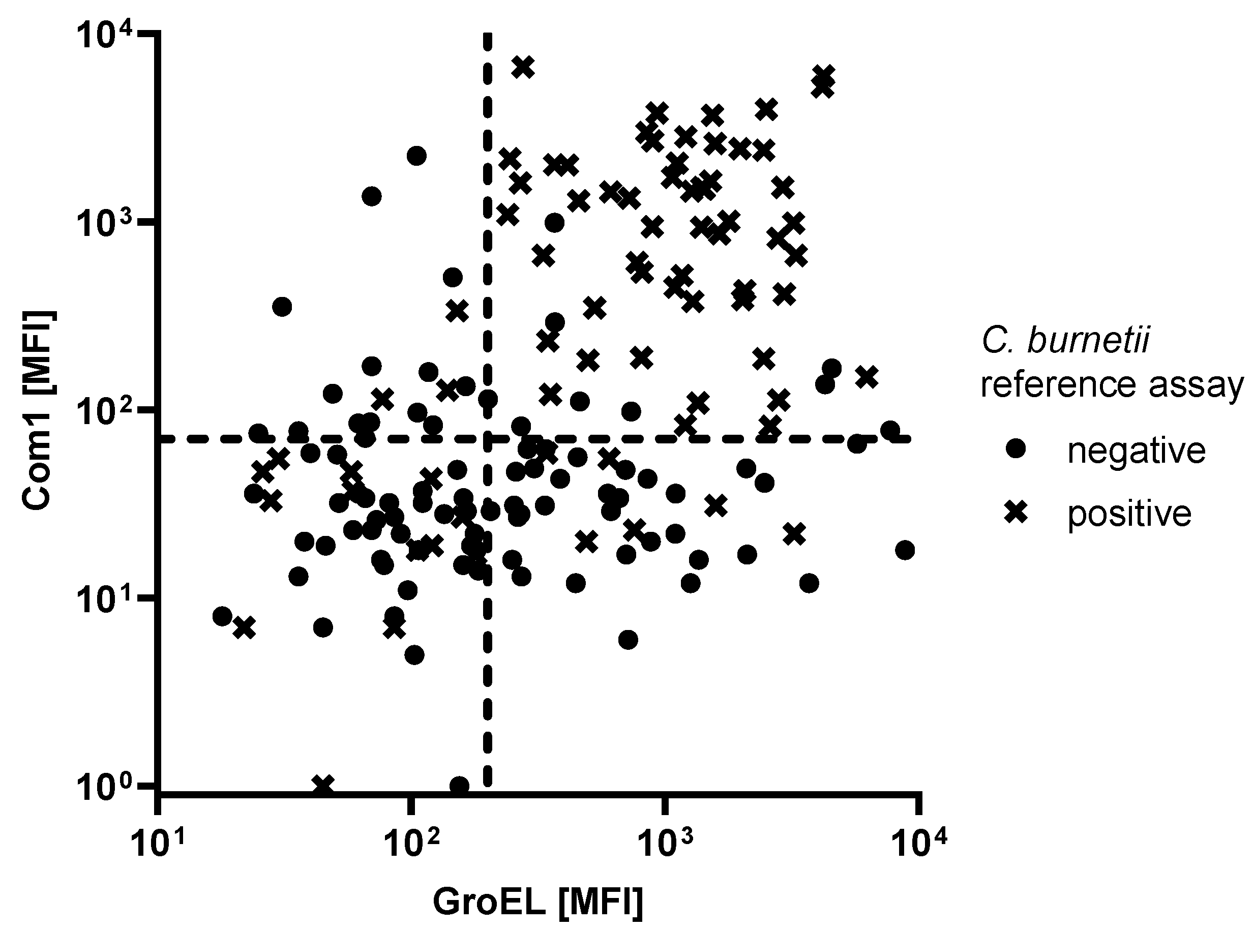
A major advantage of multiplexed assays is the possibility to combine antigens to boost assay performance (Figure 2). Considering all the seropositive samples, double positivity to Com1 and GroEL yielded a sensitivity of 71%, which outperformed Com1 as a single marker (Table 5). Combinations that included DnaK were not able to increase assay performance.
In the subgroup of patients with a Phase I endpoint titer of ≥1:1024, Com1, GroEL, and the combination of both achieved sensitivities of 86%, 50%, and 86%, respectively.
By increasing the cutoffs of Com1 and GroEL from 100 and 110 MFI, respectively, to 180 and 400 MFI, respectively, a specificity of 100% was achieved at a marginal loss in sensitivity (from 86% to 79%).

4. Discussion

We demonstrated that multiplex serology is a valuable tool for large-scale studies addressing C. burnetii infections. Analyzing up to 2000 serum samples per day, this technique has been used in diverse seroepidemiological studies to detect antibodies against a variety of different pathogens, e.g., Helicobacter pylori, human papillomaviruses, and herpes viruses [25,26,27,28]. The inclusion of C. burnetii broadens the spectrum of measurable pathogens and enables powerful studies to investigate the association between C. burnetii infections and potential clinical outcomes.
The best discrimination between seropositive and seronegative C. burnetii reference samples was achieved by the outer membrane-associated protein Com1 [40]. Its immunogenicity was initially described in 1990 and has been reproduced in several studies. Sensitivities range from 47% to 94% and specificities range from 68% to 90%, varying by the immunoassay method, host species, and the investigated population [36,38,41,42]. To the best of our knowledge, the sensitivity and specificity of our combined antigen assay is the highest reported in humans so far.
Especially in chronic Q fever patients, Com1 achieved a good performance [33,43,44]. Vranakis et al. reported a specificity and sensitivity above 90%. Due to a lack of clinical or follow-up data from our reference groups, we were not able to diagnose and analyze chronic Q fever cases separately. Instead, we performed a subgroup analysis based on the endpoint titer against C. burnetii Phase I, which is strongly associated with chronic Q fever [45].
As proposed by the Dutch Q fever consensus group, patients with an IFA Phase I endpoint titer of ≥1:1024 were considered ‘possible chronic Q fever’ cases [18,21]. This definition has been controversially discussed in a diagnostic context [23]. Epidemiologically, it is, however, suited to subdividing patients by their risk of developing chronic Q fever. In this subgroup, Com1 achieved a sensitivity of 86% and specificity of 93%. We thereby demonstrate that multiplex serology performs particularly well in patients with a higher risk of chronic Q fever and the respective clinical manifestations.
Com1 was described as a good serological marker by Xiong et al. [32]. Their study showed that among seven potential molecular markers, Com1 antibodies were particularly persistent, and were detected in 52% of acute late Q fever patients and 50% of convalescent Q fever patients. This finding strongly supports the role of Com1 as a serological biomarker, as clinical manifestations, e.g., endocarditis or hepatitis, might occur years after the primary infection. Especially in large-scale settings, biomarkers which are detectable within a large time frame are of special advantage, as large heterogeneity between patient samples might exist, e.g., due to different time spans between the blood draw and the primary infection.
GroEL and DnaK were also persistent and accurate molecular markers described by Xiong et al. [32,37,44,46,47,48]. Using multiplex serology, they achieved specificities of 84% and 79%, respectively, and sensitivities of 79% and 68%, respectively.
GroEL is a heat shock protein utilized as serological marker in a broad range of different organisms, e.g., Burkholderia pseudomallei, and Helicobacter pylori [49,50]. It does not come as a surprise that cross-reactivities with C. burnetii GroEL have been reported, especially in cases of rickettsial spotted fever and Legionella pneumonia infections [32]. This might explain the relatively high proportion of false positives when GroEL was used as a sole marker. The same principle applies to DnaK, which is also a chaperone used as serological marker for multiple organisms [51,52]. The reference sera tested here derived from individuals exhibiting unspecific flu-like symptoms, which were forwarded to the reference laboratory to be tested for C. burnetii infections. In the individuals who already showed signs of infection, the false positive rates might, therefore, be elevated compared to an asymptomatic population.
A main advantage of multiplex serology is, however, that unspecific reactivity can be addressed by considering multiple antigens. Here, GroEL can be combined with Com1 measurements to increase the specificity. When optimizing assay parameters for case-control studies, we pre-specified specificity at 90%. Among all 76 seropositive samples, sensitivity achieved 71%, which outperformed Com1 as a sole marker. Considering the patients with the high Phase I titer only, the sensitivity reached 86%. Alternatively, the specificity can be set to 100% by increasing the cutoffs accordingly, which can be of special interest to investigate risk factors to rare outcomes, e.g., lymphomas. The assay performance in these kind of studies, case-control or population-based, needs to be validated further in the future.
Overall, the integration of C. burnetii into the multiplex serology platform enables large-scale seroepidemiological studies to investigate possible associations with this infection. Multiplexing C. burnetii antigens with antigens derived from other pathogens will enable the assessment of the seroreactivity to multiple infectious agents simultaneously, providing a broader view on different infections and co-infections.

5. Summary

This study presents the development of a multiplex serology assay for the high-throughput detection of C. burnetii antibodies in human sera. Nine selected antigens (YbgF, CBU_937, CBU_370, AdaA, Com1, GroEL, DnaK, Mip, and CBU_1425) were expressed as recombinant GST-fusion proteins and were used as targets in this bead-based suspension array. Validation was performed in a panel of 167 reference sera from whom 91 were pre-classified as seronegative and 76 as seropositive. Com1, GroEL, and DnaK were able to discriminate between these two groups with specificities of 93%, 69%, and 77%, respectively, and sensitivities of 64%, 72%, and 47%, respectively. A combination of the markers Com1 and GroEL outperformed single antigens with a combined specificity and sensitivity of 90% and 71%, respectively. By restricting the analysis to a subgroup of seropositives exhibiting an anti-Phase I endpoint titer of ≥ 1:1024, the specificities of Com1 and GroEL increased to 93% and 84%, respectively, and the sensitivities to 86% and 79%, respectively. Combining both markers resulted in a specificity of 90% and sensitivity of 86%.
The multiplex serology assay presented here enables large-scale seroepidemiological studies to investigate the associations between C. burnetii infections and the clinical manifestations.

Supplementary Materials

Supplemental tables and figures show results of the assay measured in a serum dilution of 1:1000. The following are available online at: https://www.mdpi.com/article/10.3390/microorganisms9112373/s1. Table S1: Quantitative seroresponses to nine C. burnetii antigens; Table S2: Performance of C. burnetii antigens; Table S3: Performance of C. burnetii antigens measured in 28 C. burnetii patients with elevated Phase I endpoint titers; Table S4: multiplex serology assay characteristics and performance of the antigens GroEL and Com1; Figure S1: Antibody levels in the reference population; Figure S2: Antibody responses against Com1 and GroEL.

Author Contributions

Conceptualization: T.W., D.F., L.R.T., S.F.F.; Methodology: R.J., T.W., L.S.; Formal Analysis: R.J., L.S.; Investigation: R.J.; Resources: L.D., D.F., S.F.F., L.R.T.; Writing—Original Draft Preparation: R.J.; Writing—Review and Editing: L.D., L.S., D.F., L.R.T., S.F.F., T.W.; Visualization: R.J., L.S.; Supervision: T.W., S.F.F. All authors have read and agreed to the published version of the manuscript.

Funding

This research received no external funding.

Institutional Review Board Statement

Ethical review and approval were waived for this study, as all samples were completely anonymized before shipping and sampling. It is not possible to re-identify human subjects.

Data Availability Statement

Data is available upon request.

Conflicts of Interest

The authors declare no conflict of interest.

References

  1. Angelakis, E.; Raoult, D. Q fever. Vet. Microbiol. 2010, 140, 297–309. [Google Scholar] [CrossRef] [Green Version]
  2. Maurin, M.; Raoult, D. Q fever. Clin. Microbiol. Rev. 1999, 12, 518–553. [Google Scholar] [CrossRef] [Green Version]
  3. European Centre for Disease Prevention and Control. Technical Report: Risk Assessment on Q Fever; ECDC: Stockholm, Sweden, 2010; p. 40. [Google Scholar]
  4. Schimmer, B.; Notermans, D.W.; Harms, M.G.; Reimerink, J.H.; Bakker, J.; Schneeberger, P.; Mollema, L.; Teunis, P.; van Pelt, W.; van Duynhoven, Y. Low seroprevalence of Q fever in The Netherlands prior to a series of large outbreaks. Epidemiol. Infect. 2012, 140, 27–35. [Google Scholar] [CrossRef]
  5. Pascual-Velasco, F.; Montes, M.; Marimon, J.M.; Cilla, G. High seroprevalence of Coxiella burnetii infection in Eastern Cantabria (Spain). Int. J. Epidemiol. 1998, 27, 142–145. [Google Scholar] [CrossRef] [Green Version]
  6. Delsing, C.E.; Kullberg, B.J.; Bleeker-Rovers, C.P. Q fever in The Netherlands from 2007 to 2010. Neth. J. Med. 2010, 68, 382–387. [Google Scholar]
  7. Amitai, Z.; Bromberg, M.; Bernstein, M.; Raveh, D.; Keysary, A.; David, D.; Pitlik, S.; Swerdlow, D.; Massung, R.; Rzotkiewicz, S.; et al. A large Q fever outbreak in an urban school in central Israel. Clin. Infect. Dis. 2010, 50, 1433–1438. [Google Scholar] [CrossRef] [Green Version]
  8. Gilsdorf, A.; Kroh, C.; Grimm, S.; Jensen, E.; Wagner-Wiening, C.; Alpers, K. Large Q fever outbreak due to sheep farming near residential areas, Germany, 2005. Epidemiol. Infect. 2008, 136, 1084–1087. [Google Scholar] [CrossRef]
  9. Porten, K.; Rissland, J.; Tigges, A.; Broll, S.; Hopp, W.; Lunemann, M.; van Treeck, U.; Kimmig, P.; Brockmann, S.O.; Wagner-Wiening, C.; et al. A super-spreading ewe infects hundreds with Q fever at a farmers′ market in Germany. BMC Infect. Dis. 2006, 6, 147. [Google Scholar] [CrossRef] [Green Version]
  10. Melenotte, C.; Million, M.; Audoly, G.; Gorse, A.; Dutronc, H.; Roland, G.; Dekel, M.; Moreno, A.; Cammilleri, S.; Carrieri, M.P.; et al. B-cell non-Hodgkin lymphoma linked to Coxiella burnetii. Blood 2016, 127, 113–121. [Google Scholar] [CrossRef] [Green Version]
  11. van Roeden, S.E.; van Houwelingen, F.; Donkers, C.M.J.; Hogewoning, S.J.; de Lange, M.M.A.; van der Hoek, W.; Kampschreur, L.M.; Bonten, M.J.M.; Hoepelman, A.I.M.; Bleeker-Rovers, C.P.; et al. Exposure to Coxiella burnetii and risk of non-Hodgkin lymphoma: A retrospective population-based analysis in The Netherlands. Lancet Haematol. 2018, 5, e211–e219. [Google Scholar] [CrossRef]
  12. Melenotte, C.; Mezouar, S.; Ben Amara, A.; Benatti, S.; Chiaroni, J.; Devaux, C.; Costello, R.; Kroemer, G.; Mege, J.L.; Raoult, D. A transcriptional signature associated with non-Hodgkin lymphoma in the blood of patients with Q fever. PLoS ONE 2019, 14, e0217542. [Google Scholar] [CrossRef]
  13. Lee, W.Y. Hairy cell transformation of human peripheral blood lymphocytes by Coxiella burnetii. Yonsei Med. J. 1993, 34, 11–21. [Google Scholar] [CrossRef]
  14. de Boer, P.T.; de Lange, M.M.A.; Wielders, C.C.H.; Dijkstra, F.; van Roeden, S.E.; Bleeker-Rovers, C.P.; Oosterheert, J.J.; Schneeberger, P.M.; van der Hoek, W. Cost-effectiveness of screening program for chronic Q fever, The Netherlands. Emerg. Infect. Dis. 2020, 26, 238–246. [Google Scholar] [CrossRef]
  15. Eldin, C.; Melenotte, C.; Mediannikov, O.; Ghigo, E.; Million, M.; Edouard, S.; Mege, J.L.; Maurin, M.; Raoult, D. From Q fever to coxiella burnetii infection: A paradigm change. Clin. Microbiol. Rev. 2017, 30, 115–190. [Google Scholar] [CrossRef] [Green Version]
  16. van Loenhout, J.A.; Wielders, C.C.; Morroy, G.; Cox, M.J.; van der Hoek, W.; Hautvast, J.L.; Paget, W.J.; van der Velden, J. Severely impaired health status of non-notified Q fever patients leads to an underestimation of the true burden of disease. Epidemiol. Infect. 2015, 143, 2580–2587. [Google Scholar] [CrossRef]
  17. van der Hoek, W.; Dijkstra, F.; Schimmer, B.; Schneeberger, P.M.; Vellema, P.; Wijkmans, C.; ter Schegget, R.; Hackert, V.; van Duynhoven, Y. Q fever in the Netherlands: An update on the epidemiology and control measures. Eurosurveillance 2010, 15, 19520. [Google Scholar] [CrossRef]
  18. Wegdam-Blans, M.C.; Kampschreur, L.M.; Delsing, C.E.; Bleeker-Rovers, C.P.; Sprong, T.; van Kasteren, M.E.; Notermans, D.W.; Renders, N.H.; Bijlmer, H.A.; Lestrade, P.J.; et al. Chronic Q fever: Review of the literature and a proposal of new diagnostic criteria. J. Infect. 2012, 64, 247–259. [Google Scholar] [CrossRef]
  19. Wegdam-Blans, M.C.; Vainas, T.; van Sambeek, M.R.; Cuypers, P.W.; Tjhie, H.T.; van Straten, A.H.; Teijink, J.A. Vascular complications of Q-fever infections. Eur. J. Vasc. Endovasc. Surg. 2011, 42, 384–392. [Google Scholar] [CrossRef] [Green Version]
  20. Landais, C.; Fenollar, F.; Thuny, F.; Raoult, D. From acute Q fever to endocarditis: Serological follow-up strategy. Clin. Infect. Dis. 2007, 44, 1337–1340. [Google Scholar] [CrossRef] [Green Version]
  21. Kampschreur, L.M.; Wegdam-Blans, M.C.; Wever, P.C.; Renders, N.H.; Delsing, C.E.; Sprong, T.; van Kasteren, M.E.; Bijlmer, H.; Notermans, D.; Oosterheert, J.J.; et al. Chronic Q fever diagnosis-consensus guideline versus expert opinion. Emerg. Infect. Dis. 2015, 21, 1183–1188. [Google Scholar] [CrossRef] [Green Version]
  22. Fournier, P.E.; Marrie, T.J.; Raoult, D. Diagnosis of Q fever. J. Clin. Microbiol. 1998, 36, 1823–1834. [Google Scholar] [CrossRef] [Green Version]
  23. Raoult, D. Chronic Q fever: Expert opinion versus literature analysis and consensus. J. Infect. 2012, 65, 102–108. [Google Scholar] [CrossRef]
  24. Waterboer, T.; Sehr, P.; Michael, K.M.; Franceschi, S.; Nieland, J.D.; Joos, T.O.; Templin, M.F.; Pawlita, M. Multiplex human papillomavirus serology based on in situ-purified glutathione s-transferase fusion proteins. Clin. Chem. 2005, 51, 1845–1853. [Google Scholar] [CrossRef] [Green Version]
  25. Kreimer, A.R.; Ferreiro-Iglesias, A.; Nygard, M.; Bender, N.; Schroeder, L.; Hildesheim, A.; Robbins, H.A.; Pawlita, M.; Langseth, H.; Schlecht, N.F.; et al. Timing of HPV16-E6 antibody seroconversion before OPSCC: Findings from the HPVC3 consortium. Ann. Oncol. 2019, 30, 1335–1343. [Google Scholar] [CrossRef]
  26. Butt, J.; Blot, W.J.; Teras, L.; Visvanathan, K.; Le Marchand, L.; Haiman, C.; Chen, Y.; Sesso, H.D.; Wassertheil-Smoller, S.; Ho, G.; et al. Serological responses to Helicobacter pylori proteins and risk of colorectal cancer in diverse populations across the United States. Helicobacter 2018, 23. [Google Scholar] [CrossRef]
  27. Simon, J.; Schroeder, L.; Ingarfield, K.; Diehl, S.; Werner, J.; Brenner, N.; Liu, Z.; Pawlita, M.; Pring, M.; Butt, J.; et al. Epstein-Barr virus and human papillomavirus serum antibodies define the viral status of nasopharyngeal carcinoma in a low endemic country. Int. J. Cancer 2020, 147, 461–471. [Google Scholar] [CrossRef]
  28. Butt, J.; Murugan, R.; Hippchen, T.; Olberg, S.; van Straaten, M.; Wardemann, H.; Stebbins, E.; Krausslich, H.G.; Bartenschlager, R.; Brenner, H.; et al. From multiplex serology to serolomics—A novel approach to the antibody response against the SARS-CoV-2 proteome. Viruses 2021, 13, 749. [Google Scholar] [CrossRef]
  29. Gossai, A.; Waterboer, T.; Nelson, H.H.; Michel, A.; Willhauck-Fleckenstein, M.; Farzan, S.F.; Hoen, A.G.; Christensen, B.C.; Kelsey, K.T.; Marsit, C.J.; et al. Seroepidemiology of human polyomaviruses in a US population. Am. J. Epidemiol. 2016, 183, 61–69. [Google Scholar] [CrossRef]
  30. Brenner, N.; Mentzer, A.J.; Butt, J.; Michel, A.; Prager, K.; Brozy, J.; Weissbrich, B.; Aiello, A.E.; Meier, H.C.S.; Breuer, J.; et al. Validation of multiplex serology detecting human herpesviruses 1–5. PLoS ONE 2018, 13, e0209379. [Google Scholar] [CrossRef]
  31. Sehr, P.; Zumbach, K.; Pawlita, M. A generic capture ELISA for recombinant proteins fused to glutathione S-transferase: Validation for HPV serology. J. Immunol. Methods 2001, 253, 153–162. [Google Scholar] [CrossRef]
  32. Xiong, X.; Wang, X.; Wen, B.; Graves, S.; Stenos, J. Potential serodiagnostic markers for Q fever identified in Coxiella burnetii by immunoproteomic and protein microarray approaches. BMC Microbiol. 2012, 12, 35. [Google Scholar] [CrossRef] [Green Version]
  33. Sekeyova, Z.; Kowalczewska, M.; Vincentelli, R.; Decloquement, P.; Flores-Ramirez, G.; Skultety, L.; Raoult, D. Characterization of antigens for Q fever serodiagnostics. Acta Virol. 2010, 54, 173–180. [Google Scholar] [CrossRef] [PubMed]
  34. Coleman, S.A.; Fischer, E.R.; Cockrell, D.C.; Voth, D.E.; Howe, D.; Mead, D.J.; Samuel, J.E.; Heinzen, R.A. Proteome and antigen profiling of Coxiella burnetii developmental forms. Infect. Immun. 2007, 75, 290–298. [Google Scholar] [CrossRef] [PubMed] [Green Version]
  35. Chen, C.; Bouman, T.J.; Beare, P.A.; Mertens, K.; Zhang, G.Q.; Russell-Lodrigue, K.E.; Hogaboam, J.P.; Peters, B.; Felgner, P.L.; Brown, W.C.; et al. A systematic approach to evaluate humoral and cellular immune responses to Coxiella burnetii immunoreactive antigens. Clin. Microbiol. Infect. 2009, 15 (Suppl. 2), 156–157. [Google Scholar] [CrossRef] [Green Version]
  36. Stellfeld, M.; Gerlach, C.; Richter, I.G.; Miethe, P.; Fahlbusch, D.; Polley, B.; Sting, R.; Pfeffer, M.; Neubauer, H.; Mertens-Scholz, K. Evaluation of the diagnostic potential of recombinant coxiella burnetii Com1 in an ELISA for the diagnosis of Q fever in sheep, goats and cattle. Microorganisms 2020, 8, 1235. [Google Scholar] [CrossRef]
  37. Jiao, J.; Xiong, X.; Qi, Y.; Gong, W.; Duan, C.; Yang, X.; Wen, B. Serological characterization of surface-exposed proteins of Coxiella burnetii. Microbiology 2014, 160, 2718–2731. [Google Scholar] [CrossRef]
  38. Beare, P.A.; Chen, C.; Bouman, T.; Pablo, J.; Unal, B.; Cockrell, D.C.; Brown, W.C.; Barbian, K.D.; Porcella, S.F.; Samuel, J.E.; et al. Candidate antigens for Q fever serodiagnosis revealed by immunoscreening of a Coxiella burnetii protein microarray. Clin. Vaccine Immunol. 2008, 15, 1771–1779. [Google Scholar] [CrossRef] [Green Version]
  39. Robin, X.; Turck, N.; Hainard, A.; Tiberti, N.; Lisacek, F.; Sanchez, J.C.; Muller, M. pROC: An open-source package for R and S+ to analyze and compare ROC curves. BMC Bioinform. 2011, 12, 77. [Google Scholar] [CrossRef]
  40. Hendrix, L.R.; Mallavia, L.P.; Samuel, J.E. Cloning and sequencing of Coxiella burnetii outer membrane protein gene com1. Infect. Immun. 1993, 61, 470–477. [Google Scholar] [CrossRef] [Green Version]
  41. Hendrix, L.R.; Samuel, J.E.; Mallavia, L.P. Identification and cloning of a 27-kDa Coxiella burnetii immunoreactive protein. Ann. N. Y. Acad. Sci. 1990, 590, 534–540. [Google Scholar] [CrossRef]
  42. Yadav, J.P.; Malik, S.V.S.; Dhaka, P.; Kumar, M.; Sirsant, B.; Gourkhede, D.; Barbuddhe, S.B.; Rawool, D.B. Comparison of two new in-house Latex Agglutination Tests (LATs), based on the DnaK and Com1 synthetic peptides of Coxiella burnetii, with a commercial indirect-ELISA, for sero-screening of coxiellosis in bovines. J. Microbiol. Methods 2020, 170, 105859. [Google Scholar] [CrossRef]
  43. Vranakis, I.; Mathioudaki, E.; Kokkini, S.; Psaroulaki, A. Com1 as a promising protein for the differential diagnosis of the two forms of Q fever. Pathogens 2019, 8, 242. [Google Scholar] [CrossRef] [PubMed] [Green Version]
  44. Vigil, A.; Chen, C.; Jain, A.; Nakajima-Sasaki, R.; Jasinskas, A.; Pablo, J.; Hendrix, L.R.; Samuel, J.E.; Felgner, P.L. Profiling the humoral immune response of acute and chronic Q fever by protein microarray. Mol. Cell. Proteom. 2011, 10. [Google Scholar] [CrossRef] [Green Version]
  45. Kampschreur, L.M.; Oosterheert, J.J.; Koop, A.M.; Wegdam-Blans, M.C.; Delsing, C.E.; Bleeker-Rovers, C.P.; De Jager-Leclercq, M.G.; Groot, C.A.; Sprong, T.; Nabuurs-Franssen, M.H.; et al. Microbiological challenges in the diagnosis of chronic Q fever. Clin. Vaccine Immunol. 2012, 19, 787–790. [Google Scholar] [CrossRef] [Green Version]
  46. Miller, H.K.; Kersh, G.J. Analysis of recombinant proteins for Q fever diagnostics. Sci. Rep. 2020, 10, 20934. [Google Scholar] [CrossRef] [PubMed]
  47. Psaroulaki, A.; Mathioudaki, E.; Vranakis, I.; Chochlakis, D.; Yachnakis, E.; Kokkini, S.; Xie, H.; Tsiotis, G. In the search of potential serodiagnostic proteins to discriminate between acute and chronic Q fever in humans. Some promising outcomes. Front. Cell. Infect. Microbiol. 2020, 10, 557027. [Google Scholar] [CrossRef] [PubMed]
  48. Fernandes, I.; Rousset, E.; Dufour, P.; Sidi-Boumedine, K.; Cupo, A.; Thiery, R.; Duquesne, V. Evaluation of the recombinant Heat shock protein B (HspB) of Coxiella burnetii as a potential antigen for immunodiagnostic of Q fever in goats. Vet. Microbiol. 2009, 134, 300–304. [Google Scholar] [CrossRef]
  49. Michel, A.; Waterboer, T.; Kist, M.; Pawlita, M. Helicobacter pylori multiplex serology. Helicobacter 2009, 14, 525–535. [Google Scholar] [CrossRef] [PubMed]
  50. Woo, P.C.; Leung, P.K.; Wong, S.S.; Ho, P.L.; Yuen, K.Y. groEL encodes a highly antigenic protein in Burkholderia pseudomallei. Clin. Diagn. Lab. Immunol. 2001, 8, 832–836. [Google Scholar] [CrossRef] [Green Version]
  51. Zella, D.; Curreli, S.; Benedetti, F.; Krishnan, S.; Cocchi, F.; Latinovic, O.S.; Denaro, F.; Romerio, F.; Djavani, M.; Charurat, M.E.; et al. Mycoplasma promotes malignant transformation in vivo, and its DnaK, a bacterial chaperone protein, has broad oncogenic properties. Proc. Natl. Acad. Sci. USA 2018, 115, E12005–E12014. [Google Scholar] [CrossRef] [Green Version]
  52. Lemos, J.A.; Burne, R.A.; Castro, A.C. Molecular cloning, purification and immunological responses of recombinants GroEL and DnaK from Streptococcus pyogenes. FEMS Immunol. Med. Microbiol. 2000, 28, 121–128. [Google Scholar] [CrossRef] [PubMed]
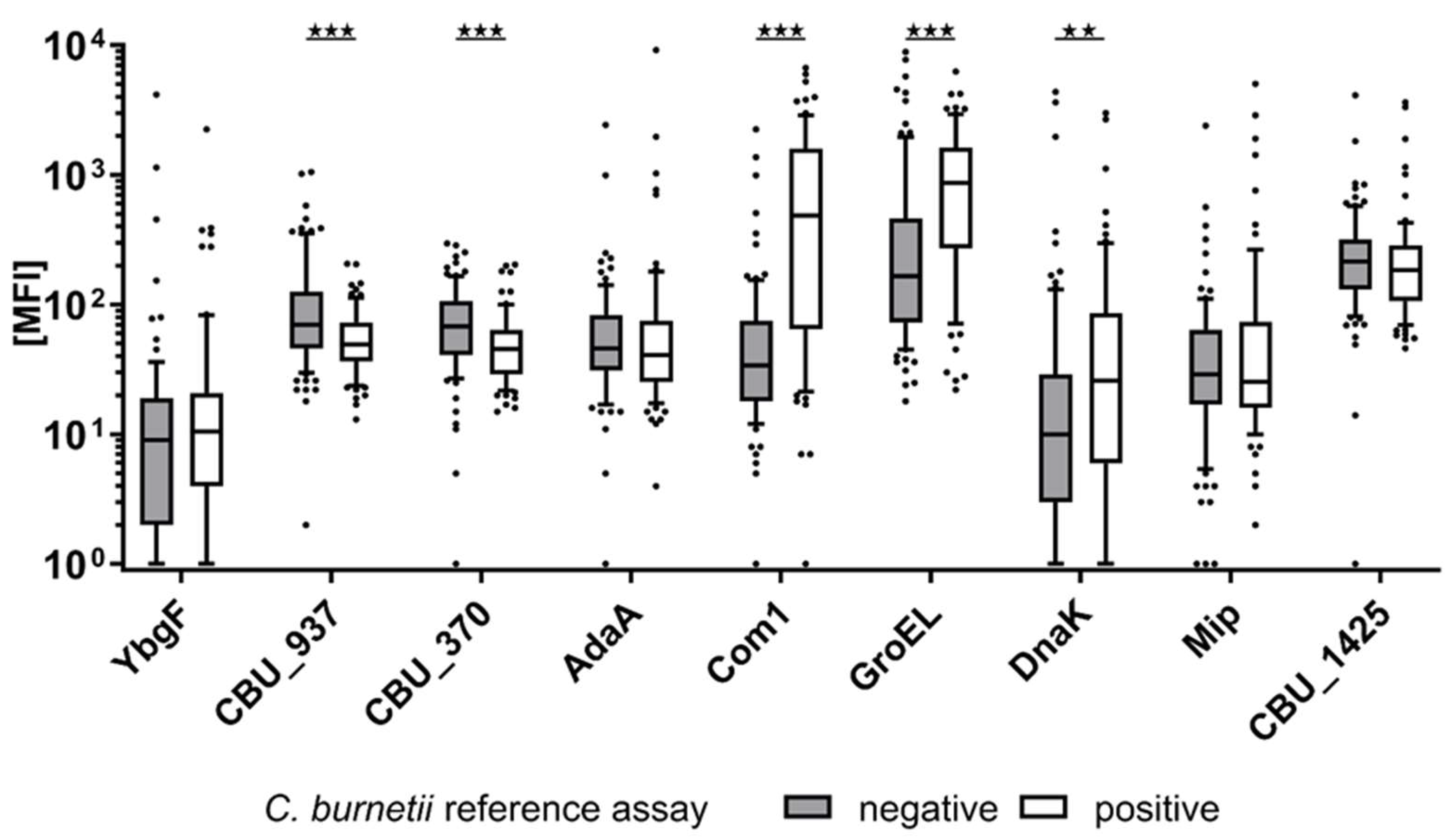
Figure 1. Antibody levels in the reference panel of 76 C. burnetii seropositive and 91 seronegative reference samples. CBU_937, CBU_370, Com1, and GroEL show a significantly different distribution (p < 0.05) between the two reference groups. Stars above boxplots indicate the level of significance, p < 0.01 (**); p < 0.01 (***).
Microorganisms 09 02373 g001
Figure 2. Antibody responses against the C. burnetii antigens Com1 and GroEL measured in 76 C. burnetii seropositive and 91 seronegative patients. Dashed lines (x = 200 MFI; y = 70 MFI) indicate the respective cutoffs to obtain a specificity of 90% and a combined sensitivity of 71%.
Microorganisms 09 02373 g002
Table 1. C. burnetii proteins selected as target antigens for the multiplex serology assay.
Antigen
Locus Tag
Antigen SymbolRefSeq
Accession
Protein DescriptionSubcellular
Location
Reference
CBU_0092YbgFNP_819144.2Cell division coordinator CpoBPeriplasmXiong et al. [32]
CBU_0937 NP_819950.2LbtU family protein
(siderophore porin)
SecretedSekeyova et al. [33]
CBU_0370 NP_819410.1Membrane-associated proteinMembrane
CBU_0952AdaANP_819961.1Acute disease antigen ASecretedColeman et al. [34]
Chen et al. [35]
CBU_1910Com1NP_820887.1Outer membrane proteinMembraneStellfeld et al. [36] Jiao et al. [37]
Xiong et al. [32]
Chen et al. [35]
Beare et al. [38]
CBU_1718GroELNP_820699.160 kDa chaperoninCytoplasmXiong et al. [32]
Coleman et al. [34]
CBU_1290DnaKNP_820282.1ChaperoneCytoplasm, Endoplasm, MembraneXiong et al. [32]
CBU_0630MipNP_819660.1Peptidyl-prolyl cis-trans isomeraseSecretedXiong et al. [32]
CBU_1425 NP_820409.117 kDa common antigenMembrane
Table 2. Quantitative seroresponses to nine selected C. burnetii antigens measured with multiplex serology in a serum dilution of 1:100. Reference samples were grouped according to a positive (pos.) or negative (neg.) C. burnetii reference assay test result. Groups were compared using Mann–Whitney U test. A p-value below 0.05 was considered statistically significant.
Antigen Locus TagAntigen Symbol C. burnetii Pos.
(MFI) (SD)
C. burnetii Neg.
(MFI) (SD)
p-Value
CBU_0092YbgF65 (266)77 (451)0.58
CBU_0937 60 (38)128 (173)<0.001
CBU_0370 55 (40)84 (60)<0.001
CBU_0952AdaA228 (1073)100 (269)0.26
CBU_1910Com11094 (1423)105 (291)<0.001
CBU_1718GroEL1209 (1205)697 (1518)<0.001
CBU_1290DnaK151 (470)138 (621)<0.01
CBU_0630Mip206 (703)81 (259)0.81
CBU_1425 324 (584)313 (469)0.13
Table 3. Performance of nine C. burnetii antigens measured in 76 reference samples with positive, and 91 with negative, C. burnetii serostatuses. Quantitative measurement values were dichotomized using the given cutoff (maximum value of the Youden’s index) to calculate assay specificity, sensitivity, and agreement (kappa).
Antigen Locus TagAntigen Symbol Cutoff (MFI)Specificity (%)Sensitivity (%)Cohen’s Kappa κ (95% CI)
CBU_0092YbgF3289210.11 (−0.05 to 0.27)
CBU_0937 675472−0.26 (−0.41 to −0.11)
CBU_0370 675380−0.32 (−0.47 to −0.18)
CBU_0952AdaA337442−0.15 (−0.29 to −0.01)
CBU_1910Com117893640.59 (0.47 to 0.72)
CBU_1718GroEL34069720.41 (0.27 to 0.55)
CBU_1290DnaK30 177470.25 (0.10 to 0.40)
CBU_0630Mip30 15147−0.02 (−0.17 to 0.13)
CBU_1425 1247938−0.15 (−0.30 to −0.01)
1 A technical minimum cutoff of 30 MFI was applied.
Table 4. Quantitative seroresponses to nine selected C. burnetii antigens measured with multiplex serology in a serum dilution of 1:100. Reference samples were grouped according to a positive (pos.) or negative (neg.) C. burnetii reference assay test result. Groups were compared using Mann–Whitney U test. A p-value below 0.05 was considered statistically significant.
Antigen Locus TagAntigen SymbolCutoff (MFI)Specificity (%) Sensitivity (%)Cohen’s Kappa κ (95% CI)
CBU_0092YbgF30 186250.12 (−0.13 to 0.37)
CBU_0937 824293−0.30 (−0.52 to −0.08)
CBU_0370 586082−0.31 (−0.48 to −0.14)
CBU_0952AdaA436350−0.08 (−0.24 to 0.08)
CBU_1910Com11799386 0.78 (0.64 to 0.91)
CBU_1718GroEL74784790.56 (0.39 to 0.73)
CBU_1290DnaK3179680.40 (0.21 to 0.59)
CBU_0630Mip30 15143−0.05 (−0.23 to 0.14)
CBU_1425 1247943−0.10 (−0.24 to 0.03)
1 A technical minimum cutoff of 30 MFI was applied.
Table 5. Final multiplex serology assay characteristics and performance of antigens Com1 and GroEL, as well as a combination of both antigens. Assay performance was determined in an overall C. burnetii positive population (n = 76) and a subgroup with high Phase I endpoint titers of ≥1:1024, as an approximation for chronic Q fever (n = 28).
Seropositive Reference PopulationAntigenCutoff (MFI)Specificity (%)Sensitivity (%)
76 C. burnetii seropositive patientsCom1 (CBU_1910)1409066
GroEL (CBU_1718)13609034
Com 1 + GroEL
(double positives)
70/2009071
28 C. burnetii seropositive patients with a Phase I titer of ≥1:1024Com1 (CBU_1910)1409086
GroEL (CBU_1718)13609050
Com 1 + GroEL
(double positives)
100/1109086
180/40010079
Publisher’s Note: MDPI stays neutral with regard to jurisdictional claims in published maps and institutional affiliations.