Article

Regulatory Control of Rishirilide(s) Biosynthesis in Streptomyces bottropensis

1
Institute for Pharmaceutical Biology and Biotechnology, Albert-Ludwigs-Universität, 79104 Freiburg, Germany
2
Faculty of Medicine, Institute for Biochemistry and Molecular Biology, ZBMZ, Albert-Ludwigs-Universität, 79104 Freiburg, Germany
3
Department of Chemistry-Biology, Institute of Organic Chemistry II, Universität Siegen, 57068 Siegen, Germany
*
Author to whom correspondence should be addressed.
Microorganisms 2021, 9(2), 374; https://doi.org/10.3390/microorganisms9020374
Submission received: 22 January 2021 / Revised: 7 February 2021 / Accepted: 9 February 2021 / Published: 12 February 2021
(This article belongs to the Section Molecular Microbiology and Immunology)

Abstract

:
Streptomycetes are well-known producers of numerous bioactive secondary metabolites widely used in medicine, agriculture, and veterinary. Usually, their genomes encode 20–30 clusters for the biosynthesis of natural products. Generally, the onset and production of these compounds are tightly coordinated at multiple regulatory levels, including cluster-situated transcriptional factors. Rishirilides are biologically active type II polyketides produced by Streptomyces bottropensis. The complex regulation of rishirilides biosynthesis includes the interplay of four regulatory proteins encoded by the rsl-gene cluster: three SARP family regulators (RslR1-R3) and one MarR-type transcriptional factor (RslR4). In this work, employing gene deletion and overexpression experiments we revealed RslR1-R3 to be positive regulators of the biosynthetic pathway. Additionally, transcriptional analysis indicated that rslR2 is regulated by RslR1 and RslR3. Furthermore, RslR3 directly activates the transcription of rslR2, which stems from binding of RslR3 to the rslR2 promoter. Genetic and biochemical analyses demonstrated that RslR4 represses the transcription of the MFS transporter rslT4 and of its own gene. Moreover, DNA-binding affinity of RslR4 is strictly controlled by specific interaction with rishirilides and some of their biosynthetic precursors. Altogether, our findings revealed the intricate regulatory network of teamworking cluster-situated regulators governing the biosynthesis of rishirilides and strain self-immunity.

1. Introduction

Streptomycetes are soil bacteria that are well-known for their complex morphological differentiation and ability to produce a plethora of natural products with pharmaceutical value [1,2,3]. To date, the genus Streptomyces produces more than half of the clinically used antibiotics and is still considered to be a promising source for novel antibiotics discovery [4].
To coordinate development and secondary metabolite production, Streptomyces developed a complex regulatory network with multiple overlapping cascade systems. This includes sigma factors, transduction systems, and transcriptional factors of different families [5,6,7].
The onset and level of natural product production are determined in accordance to environmental signals, nutrition, and the physiological state of growing cells [6,8]. In Streptomyces, specific low molecular weight signalling molecules such as gamma-butyrolactones and guanosine penta/tetraphosphate ((p)ppGpp) trigger secondary metabolism production and morphological differentiation [9,10,11,12]. Recently, intracellular second messenger cyclic di-GMP was also shown to exert a regulatory function on biosynthesis of natural products [13,14,15].
Genes involved in synthesis of natural product are clustered together. In general, Streptomyces chromosome harbors 20–30 biosynthetic gene clusters for secondary metabolites [6,16]. Typically, a biosynthetic gene cluster encodes a cluster-situated regulator, which directly controls the transcriptional levels of genes for production of the cognate natural product. Interestingly, some clusters can contain none or several regulatory genes [6,17,18,19,20]. The cluster-situated regulator in turn is controlled by higher-level regulatory elements like pleiotropic or global regulators [21]. Most of the cluster-situated regulators act as activators, and belong to the SARP (Streptomyces antibiotic regulatory proteins) family [6,21]. The most intensively studied SARPs are ActII-ORF4 and RedD, activators of actinorhodin and undecylprodigiosin production in S. coelicolor, respectively [6,22,23,24,25]. The promoter region of actII-ORF4 is a regulatory target of few superior transcriptional factors including AdpA, AbsA2, DasR, AfsQ1 and others [26,27,28,29].
Streptomyces bottropensis is a known producer of the macrocyclic peptide antibiotic bottromycin and type II polyketides—rishirilides and mensacarcin [30]. Rishirilide A and B act as inhibitors of α2-macroglobulin, whereas rishirilide B exhibits an additional inhibition activity towards glutathione-S-transferase [31,32]. Genes for rishirilides biosynthesis (rsl-genes) are clustered together and were recently functionally characterized [33].
Rishirilides are synthesized from the valine-derived isobutyl-CoA starter unit and malonyl-CoA extenders, followed by decarboxylation of one acetate unit giving a methyl group. The minimal PKS complex derives a linear tricyclic intermediate, which is further modified by cluster-encoded tailoring enzymes [33,34]. Interestingly, the rishirilide scaffold is formed in two key steps: a reductive ring opening of an epoxide moiety on polycyclic intermediate by the flavin-dependent reductase RslO5, followed by an extraordinary oxidative carbon backbone rearrangement catalyzed by flavin-dependent Baeyer-Villiger monooxygenase RslO9 [35].
In addition to biosynthetic and transporter genes, four regulators are encoded by the rsl-gene cluster. However, detailed function of these transcriptional factors in the biosynthesis of rishirilides remained unclear. In this work, we have generated rslR1, rslR2, rslR3, and rslR4 deficient mutants and analyzed their ability to produce rishirilides. Inactivation of rslR1-R3 completely abolished biosynthesis, whereas a deletion of rslR4 had no substantial effect on a production level. The overexpression of rslR1, rslR2, and rslR3 in S. bottropensis greatly increased titers of rishirilides accumulation, suggesting that RslR1, RslR2, and RslR3 are positive regulators. In order to identify putative interconnections between studied regulators, the expression of regulatory genes was analyzed in wild type and in mutant strains by RT-PCR and a GusA reporter system. RslR3 was shown to exert positive control on the transcription of rslR2. This finding was further confirmed by direct binding of RslR3 to the rslR2 promoter. Moreover, both RslR1 and RslR2 negatively control transcription of rslR2.
RslR4 is involved in the regulation of strain immunity. It modulates transcriptional levels of the MFS transporter rslT4 and its own gene. We also demonstrated that the final biosynthetic products and key pathway intermediates interact with RslR4 and release the protein from a target DNA.

2. Materials and Methods

2.1. Bacterial Strains, Plasmids, and Growth Conditions

The bacterial strains and plasmids used in this work are described in Table S1. S. bottropensis strains were grown at 28 °C in liquid YMPG medium [36] supplemented with 0.2% valine for rishirilide production (YMPGv), and on MS agar for harvesting spores and plating conjugations. Escherichia coli strains were grown in LB medium at 37 °C unless otherwise stated. Where necessary, the media were supplemented with antibiotics: apramycin (50 µg mL−1), kanamycin (50 µg mL−1), spectinomycin (100 µg mL−1), chloramphenicol (25 µg mL−1), and ampicillin (100 µg mL−1). All antibiotics were purchased from Carl Roth, Karlsruhe, Germany.

2.2. Generation of Gene Deletion Mutants

The double cross-over mutants were generated using Red-Et mediated technology [37]. Initially, an integrase gene (int) on a cosmid cos4, carrying the rsl-gene cluster, was inactivated to prevent an integration of the final construct into S. bottropensis chromosome. For this purpose, cos4 was introduced into E. coli BW25113 (pIJ790) where the integrase gene was replaced by an ampicillin resistance gene, which was amplified from pBluescript using primer pair Int-aatP::amp-f/r with int-homology extensions. The resulting cosmid cos4-int::bla was confirmed by PCR analysis and transformed into E. coli BW25113 (pIJ790).
Next, the spectinomycin resistance cassette was amplified from pCDFduet using primer pair rslR-RedET-Fw and rslR-RedET-Rv listed in Table S2. The obtained corresponding PCR products with homology extensions to rslR1, rslR2, rslR3, and rslR4 were individually electroporated into E. coli BW25113 (pIJ790) cos4-int::bla to replace the desired regulatory gene by the spectinomycin resistance cassette. The resulting constructs cos4-int::bla-rslR1::Sp, cos4-int::bla-rslR2::Sp, cos4-int::bla-rslR3::Sp, and cos4-int::bla-rslR4::Sp were confirmed by PCR and individually introduced into S. bottropensis via intergeneric conjugation with E. coli ET12567 (pUZ8002), harboring the desired construct. The double cross-over mutants were screened for resistance to spectinomycin and sensitivity to apramycin, and further confirmed by PCR analysis.

2.3. Construction of the Plasmids for Complementation and Overexpression

For complementation experiment, a regulatory gene together with a 500 bp upstream promoter region was amplified by PCR using chromosomal DNA of S. bottropensis as a template and primer pair listed in Table S2. The obtained PCR product was purified, digested with XbaI, and cloned into XbaI/EcoRV sites of the φC31-based integrative plasmid pSET152. The yielded plasmid pSET-rslR was conjugated into the respective gene deletion mutant. To complement S. bottropensis ∆R3, plasmid pTES-rslR3 was constructed. For this reason, a coding sequence of rslR3, along with the ribosome binding site (RBS), was amplified with R3-XbaI-pTESa/rslR3MuDet-Fw primers (Table S2) and cloned into XbaI/EcoRV-cleaved pTES. In resulting pTES-rslR3, the expression of rslR3 is under control of constitutive ermE-promoter.
To overexpress regulatory genes, their coding sequences, along with RBS, were amplified from S. bottropensis chromosome with primers listed in Table S2. The obtained PCR fragments were digested with respective restriction endonucleases, which recognition sites are highlighted in the primer sequence, and then ligated into the same sites of the linearized multicopy expression vector pUWL-H. In these plasmids, the expression of genes is under control of the strong constitutive promoter ermEp.

2.4. Strain Fermentation and Analysis of Rishirilides Production

Spores of the wild type and of mutant strains were inoculated into 50 mL of YMPGv in 300-mL flasks and grown for five days with rotary shaking. Then, supernatant was separated from cell pellet by centrifugation and extracted with the same volume of ethyl acetate. After evaporation, the extracts were dissolved in methanol and used for UHPLC-MS analysis as described before [35]. To evaluate the production level, total rishirilide was quantified. Total rishirilide refers to rishirilide A, B, D, and lupinacidin A.

2.5. Total RNA Isolation and RT-PCR

Total RNA was isolated from S. bottropensis strains, grown for 72 h in YMPGv medium, using RNeasy Mini kit (Qiagen, Venlo, Netherlands) according to recommendations from the supplier. To get rid of DNA contaminations, RNA samples were treated with DNaseI (Promega, Madison, Wisconsin, United States) and checked by PCR prior cDNA synthesis. Then, cDNA was synthesized from 1 µg of total RNA using Protoscript II Reverse Transcriptase (NEB, Ipswich, MA, USA) according to instructions of the manufacturer. To analyze the transcription profile of genes of interest, 200 ng of cDNA were used as a template in PCR reaction with primers listed in Table S2. Obtained PCR products were separated on a 1.5% agarose gel. Total RNA was isolated from three independent replicates.

2.6. Gus-Assay

Promoter regions of rsl-genes were amplified by PCR using S. bottropensis chromosomal DNA as a template and primer pairs listed in Table S2. The PCR-products were purified, digested with XbaI and KpnI restriction endonucleases, and cloned into pGUS. Recombinant plasmids were conjugated into S. bottropensis and in the respective mutants. β-glucuronidase activity was measured in strains lysate after 72 h (rslR1p and rslR2p) and 48 h (rslR4p and rslT4p) of growth in YMPGv medium. Measurement of β-glucuronidase activity was carried out as described before [38] in biological triplicate.

2.7. Production and Purification of RslR3DBD

To produce native or truncated versions of RslR3, the coding sequences were amplified by PCR using primer pairs listed in Table S2. The PCR products were digested with NdeI and HindIII and inserted into the respective sites of pET28a. The resulting plasmids were confirmed by sequencing and further introduced into E. coli BL21 (DE3) Star. RslR3DBD was produced with N-terminal hexahistidine tag. E. coli BL21 (DE3) Star pET28a-R3DBD was incubated in LB medium at 37 °C until the optical density at 600 nm reached 0.5, then IPTG (0.1 mM) was added and incubation continued at 18 °C overnight. The cells were harvested by centrifugation and resuspended in a binding buffer (50 mM Na2HPO4, 0.5 M NaCl, 2% glycerol, pH 7.5 supplemented with PMSF and DNaseI). After cell disruption by sonication, the cleared lysate was applied to Ni-NTA and washed with increasing concentration of imidazole (binding buffer containing 40, 80, and 100 mM imidazole). Protein was eluted with 250 mM imidazole and analyzed by SDS-PAGE.

2.8. Production and Purification of RslR4

The open reading frame of rslR4 along with 51 bp located upstream of the gene was amplified by PCR using chromosomal DNA of S. bottropensis as a template and primer pair R4-NcoI-17/R4-XhoI (Table S2, see Figure S3 for RslR4 sequence). The PCR product was digested with NcoI and XhoI and ligated into the same sites of pET28a. The resulting construct pET28-rslR4 was sequenced and introduced into E. coli BL21 (DE3) Star. To produce RslR4, the culture was grown in LB medium until optical density reached 0.4. After the addition of 0.2 mM IPTG, the culture was grown for another four hours at 22 °C. Cells were harvested by centrifugation and directly used for protein purification or frozen at −20 °C until required. The cell pellet was dissolved in a buffer (50 mM TrisHCl, 0.5 M NaCl, 20 mM imidazole, pH 8.0) supplemented with PMSF and DNaseI, and disrupted by two passages through a French pressure cell press. Lysate was separated from insoluble fraction by centrifugation and applied for protein binding with Ni-NTA resin. After that, resin was washed with the buffer containing 40 mM imidazole and protein was eluted by a linear gradient from 100 to 200 mM imidazole. Eluted fractions were analyzed for the presence of RslR4 by SDS-PAGE. Samples with RslR4 were collected and protein concentration was estimated by Bradford assay.

2.9. Electrophoretic Mobility Shift Assay

Putative promoter regions of rsl-genes were amplified by PCR using S. bottropensis chromosome as a template and primers listed in Table S2. The PCR products were labeled at 5′-end with [γ-33P]-ATP using T4 polynucleotide kinase. DNA-binding reactions were carried out at 28 °C for 15 min (or for 25 min with RslR3DBD) in a binding buffer (20 mM TrisHCl pH 7.9, 100 mM KCl, 10 mM MgCl2, 1 mM EDTA, 1 mM DTT, 12.5% glycerol) supplemented with 1 µg poly(dI-dC) and 4.5 µg BSA. DNA-protein complexes were resolved on 8% polyacrylamide native gels in TBE buffer and visualized by phosphorimaging on Typhoon FLA7000. For retardation assay, unlabeled DNA probe was added to the reaction mixture at 10- to 500- molar excess (as indicated in results section).
To test rishirilides and pathway intermediates as ligands of RslR4, compounds were incubated with the protein (100 nM) prior adding rslR4-T4 intergenic region to the reaction. The following concentrations of the ligands were used: 1, 10, 50, 100 µM and 0.25, 0.5, 1 mM. The tested ligands were purified as described before [33,35] or in Supplementary Materials (information about structure elucidation of compounds isolated from ∆rslO7 mutant).

3. Results

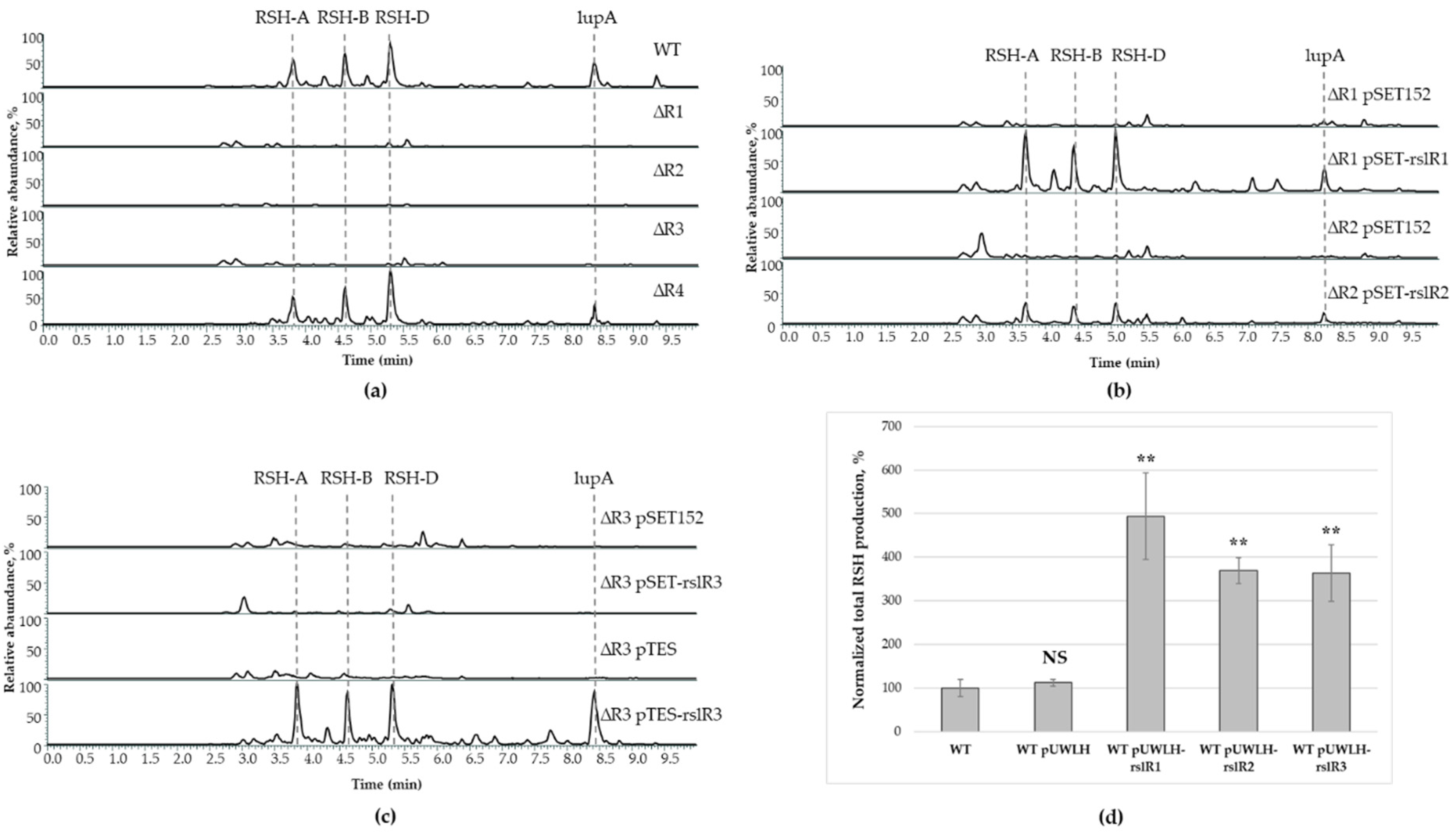
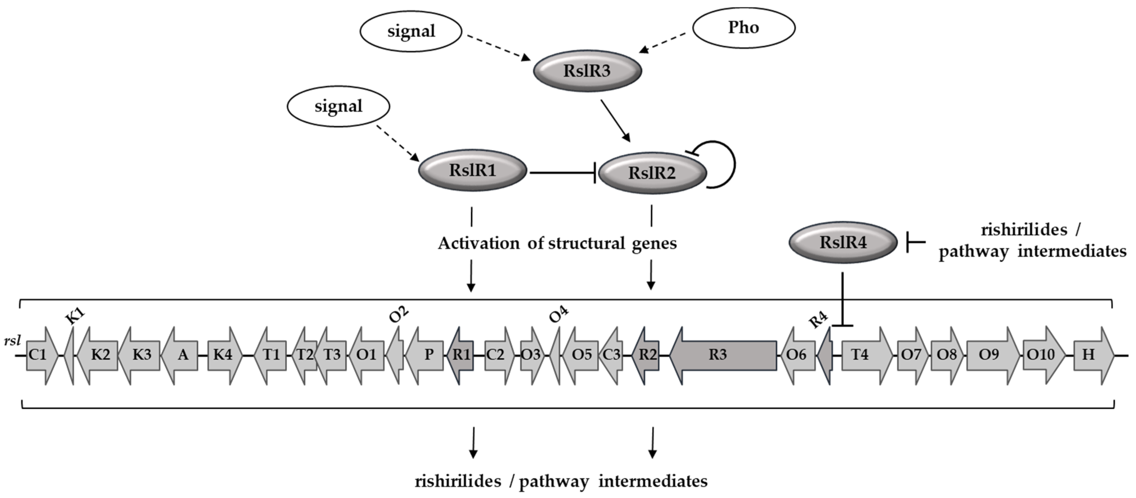
3.1. Biosynthesis of Rishirilides Is Positively Regulated by RslR1, RslR2, and RslR3 Cluster-Situated Regulators

The rsl-gene cluster encodes four regulatory proteins. Analysis of domain architecture of RslR1 and RslR2 identified an N-terminal winged helix-turn-helix (HTH) DNA-binding and Bacterial Transcriptional Activation (BTA) domains that are typical for members of the SARP-family regulatory proteins. RslR3 is a 1112-amino acid transcriptional factor which belongs to large SARPs and has multi-domain architecture similar to AfsR and PolY [19,39]. Like RslR1 and RslR2, it consists of an N-terminal SARP domain, which is followed by an additional ATPase (AAA/NB-ARC) domain and a C-terminal tetratricopeptide repeat (TPR) domain that usually mediates protein–protein interactions. RslR4 is a member of the MarR-family (Multiple Antibiotic Resistance Regulator) transcriptional factors.
To investigate the role of these regulators in rishirilide biosynthesis, we inactivated them individually in the S. bottropensis chromosome by in frame replacement of their coding sequences with the spectinomycin resistance gene. To estimate rishirilides production, the resulted mutants together with wild type strain were grown in YMPGv medium for five days and their metabolite profiles were analyzed using UHPLC-MS. Inactivation of rslR1, rslR2, and rslR3 completely abolished production of all rishirilide compounds indicating their positive role in the biosynthesis (Figure 1a). Inactivation of rslR4 did not distinctly affect rishirilides production.
To exclude the possibility of any polar effect and to confirm that changes in the production of rishirilides were solely due to the inactivation of the targeted regulatory gene, the integrative pSET152-based construct carrying a native rslR gene allele together with its promoter region was introduced into mutants. Complementation of rslR1 and rslR2 mutants restored the production of rishirilides. Complementation of S. bottropensis ∆R3 failed when rslR3 was expressed from its putative promoter region in pSET-rslR3. However, complementation with pTES-rslR3, where the regulatory gene is under control of the constitutive ermE-promoter (Figure 1b,c), restored the ability of rslR3 mutant to produce rishirilides. This result suggests that rslR3 lacks its own promoter, that was further confirmed by gene co-expression analysis (see Section 3.3). In order to overexpress rslR1-R3 in the wild type strain, replicative multi-copy plasmids expressing each regulatory gene from the ermE-promoter (pUWLH-rslR1, pUWLH-rslR2, and pUWLH-rslR3) were constructed and conjugated into S. bottropensis. Indeed, overexpression of rslR1, rslR2, and rslR3 in the wild type increased rishirilides production from 3- to 5-fold (Figure 1d). These results are in agreement with the gene deletion experiments, and indicate that the SARP regulatory proteins RslR1, RslR2, and RslR3 are positive regulators of the rsl-gene cluster. Moreover, the activity of all three proteins is required for rishirilide biosynthesis. On the other hand, RslR4 might be not involved in direct regulation of transcription of structural genes. However, it may control strain self-resistance to produced compound(s).

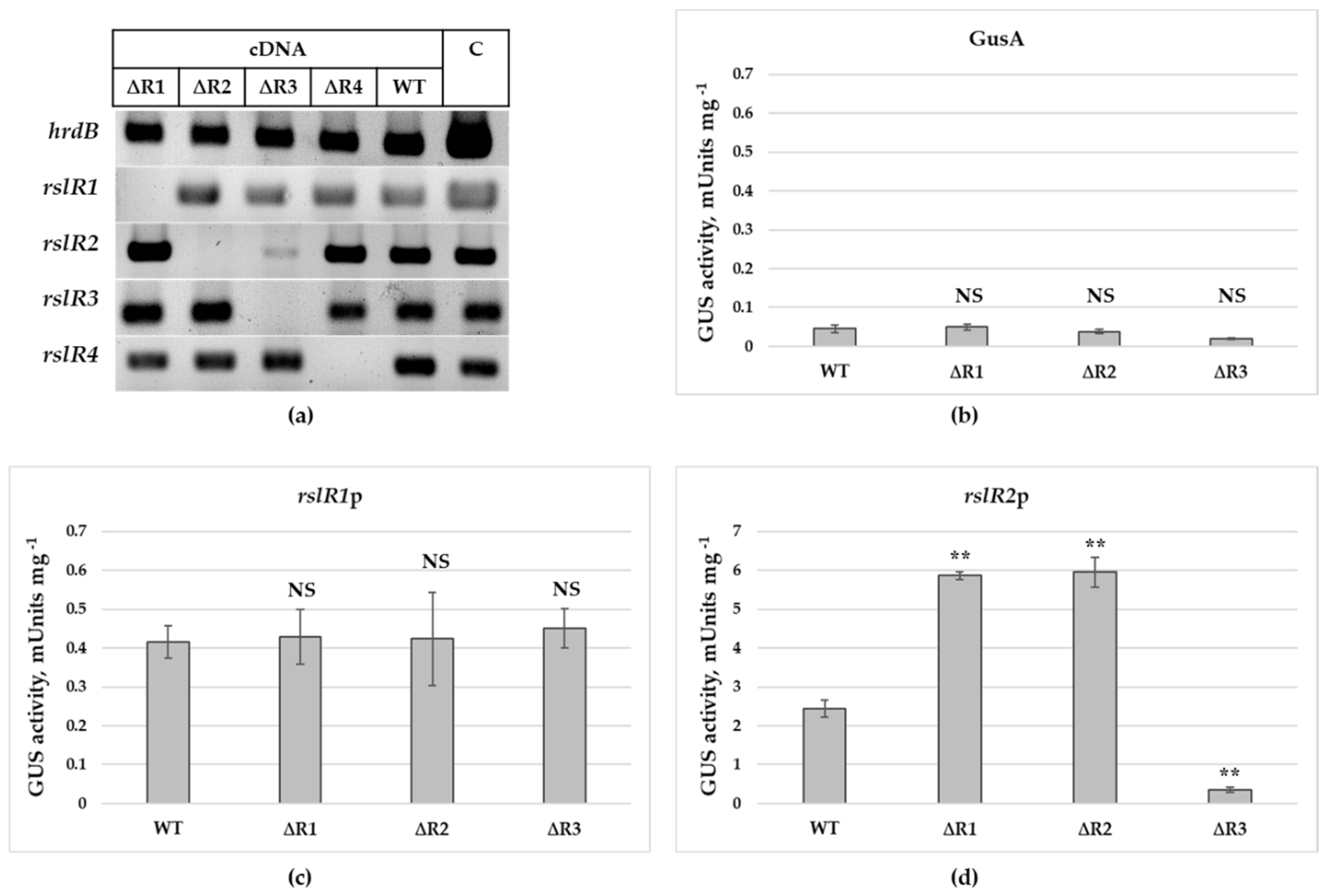
3.2. Expression Pattern of Regulatory Genes in Wild-Type and Mutant Strains

S. bottropensis initiates rishirilides production after 48 h of growth in fermentation medium. To determine putative interconnections between regulatory genes, their expression was analyzed in the wild type and in the rslR1-, rslR2-, rslR3-, and rslR4-disruption mutants. Total RNA was isolated from the strains after 72 h of growth and genes transcripts were analyzed by RT-PCR. As a control, the hrdB gene, which encodes a RNA polymerase principal sigma factor, was used.
In the semi-quantitative RT-PCR analysis, the level of transcripts of rslR4 was the same in the wild type strain and in S. bottropensis ΔR1, S. bottropensis ΔR2, and S. bottropensis ΔR3 (Figure 2a). This result indicates that rslR4 transcription is independent from a control by RslR1, RslR2 or RslR3. Transcriptional profiles of rslR3 and rslR1 were very similar in all analyzed strains as well, indicating that both genes are independently expressed. In contrast, the low level of rslR2 transcripts in S. bottropensis ΔR3 indicates that rslR2 may be positively regulated by RslR3. In addition, transcription of rslR2 might be under a negative control of RslR1 due to the increased abundance of rslR2 transcripts in S. bottropensis ∆R1 (Figure 2a).
To verify these results as well as to investigate the possibility of self-regulation, expression of rslR1 and rslR2 at transcriptional level was analyzed in the wild type and in S. bottropensis ΔR1, S. bottropensis ΔR2, and S. bottropensis ΔR3 using the GusA-reporter system [38]. Promoter regions of rslR1 and rslR2 were cloned upstream of the β-glucuronidase gene and the obtained plasmids were then conjugated into the wild type and mutant strains. Activity of GusA in strain lysates was measured after 72 h of growth. Strains carrying the empty vector pGUS were used as a control (Figure 2b). There was no significant difference in the rslR1 promoter activity in the wild type and in S. bottropensis ΔR1, S. bottropensis ΔR2, and S. bottropensis ΔR3 (Figure 2c). These data are in agreement with the results of the RT-PCR experiments. In contrast, the transcription of rslR2 was strongly downregulated in the rslR3 mutant and increased two-fold in S. bottropensis ΔR1 and S. bottropensis ΔR2 (Figure 2d). These data indicate that RslR3 and RslR1 can act as positive and negative regulators, respectively. Moreover, RslR2 acts as a negative regulator of its own transcription.
In summary, these data suggest that transcription of rslR2 is controlled by multiple regulators. It is activated by RslR3 and downregulated by RslR1 as well as by RslR2. Thus, RslR3 controls the pathway specific regulator RslR2. Finally, transcription of rslR4 is independent from the other Rsl-regulatory proteins.

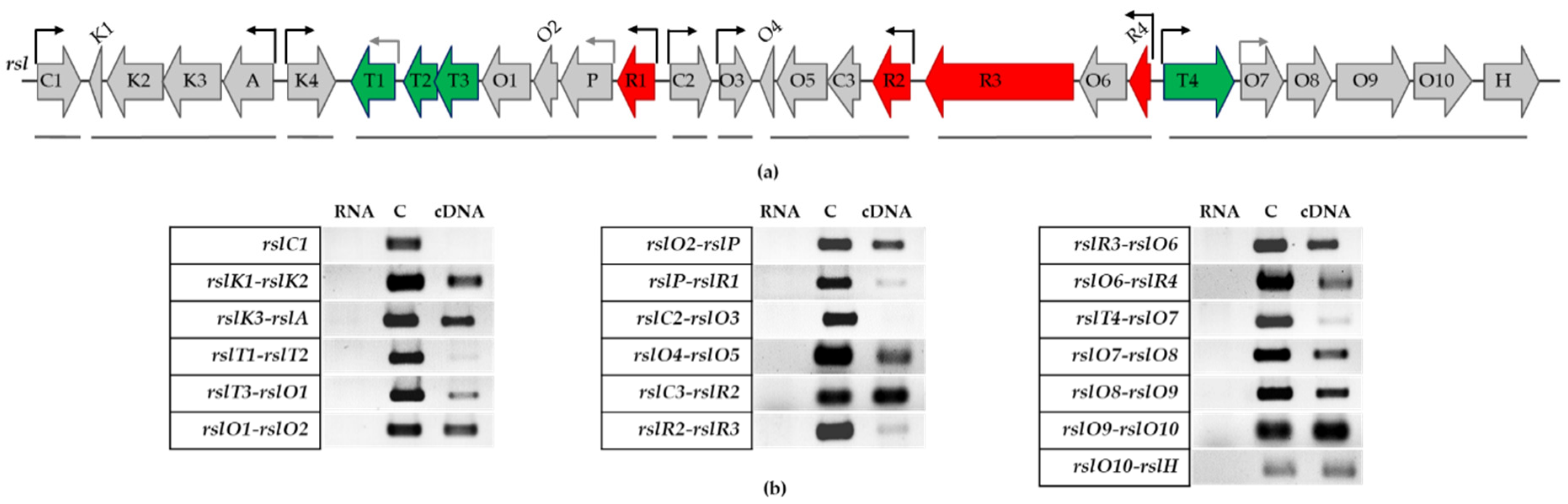
3.3. Transcriptional Organization of rsl-Genes

To identify putative promoter regions in the cluster, the transcriptional organization of the rsl-genes was determined. The co-transcribed genes were mapped by RT-PCR with primers, which anneal across gene pairs using cDNA from the wild type strain as a template. The obtained PCR products represent junction sequences between the genes (Figure 3). Amplification products between rslK1 and rslK2, as well as rslK3 and rslA, indicate that the minimal PKS genes form a single operon. The PCR products obtained for gene sets rslT1-rslT2, rslT3-rslO1, rslO1-rslO2, rslO2-rslP, and rslP-rslR1 indicate that genes between rslR1 and rslT1 are transcribed together. The faint PCR products obtained for rslT1-rslT2 and rslP-rslR1 indicate that there may be promoters located upstream of rslT1 and rslP in addition to a promoter upstream of rslR1. The PCR products were also obtained for further gene pairs (Figure 3) but not for rslC2-rslO3. These data indicate that genes between rslR2 and rslO4, rslR4 and rslR3, and rslT4 and rslH clearly form transcription units and that there are promoters located upstream of rslC2, rslO3, rslR2, rslR4, and rslT4. The weak PCR fragment for rslR3-rslR2 may suggest the co-transcription of both genes. However, successful complementation of S. bottropensis ∆R2 by pSET-rslR2 confirmed the existence of a promoter upstream of rslR2 because in our complementation construct rslR2 can only be expressed from its own promoter. Interestingly, the transcription unit rslR4-rslO6-rslR3 indicates that rslR3 lacks its own promoter region. This explains the ability to complement S. bottropensis ∆R3 only by expressing rslR3 from the constitutive ermE-promoter. Complementation failed when a fragment containing rslR3 and the region between rslO6 and rslR3 was used.

3.4. RslR3 is a Multi-Domain Protein That Regulates rslR2

To further confirm direct interaction of RslR3 with the promoter region of rslR2, a radio-labeled DNA probe was used in an electrophoretic mobility shift assay with a recombinant RslR3. Beside the rslR2p, rslR1p and promoters of rsl-transcriptional units were used. Production of the recombinant full-length RslR3 with an N-terminal hexa-histidine tag resulted in insoluble protein, which hampered the investigation of its DNA-binding ability. It has been shown that truncated versions of AfsR containing either the SARP and ATPase domains or only the SARP domain, show the same DNA interaction pattern as the full-length protein [39]. As domain organization of RslR3 is similar to AfsR, an analogous experiment was performed. Two truncated versions of RslR3, consisting of only the SARP-domain (RslR3DBD) and the SARP-domain accompanied by the ATPase domain (RslR3DBDP) were constructed. Only the RslR3DBD version could be produced as a soluble protein and was used for further studies. Increasing concentrations of purified RslR3DBD were incubated with a DNA fragment containing the promoter region of rslR2 (Figure 4a). DNA-protein complexes were formed at concentrations of 0.5–1 µM. RslR3DBD yielded a bit smeared and broad bands, which could be due to instability of the truncated protein. To confirm a specific binding of RslR3DBD to rslR2p, the protein (1 µM) was incubated with labeled DNA probe at the presence of 10 to 500-fold excess of unlabeled DNA. Unbound labeled rslR2p was detectable indicating a specific binding of RslR3DBD to the promoter region of rslR2 (Figure 4b). No binding was observed using rslR1p or any other tested promoter region (Figure 4c,d).
Altogether, these results are in a line with data of RT-PCR and in vivo reporter system experiments. This finding suggests that rslR2 is directly activated by RslR3 and is its sole target in rishirilide gene cluster.

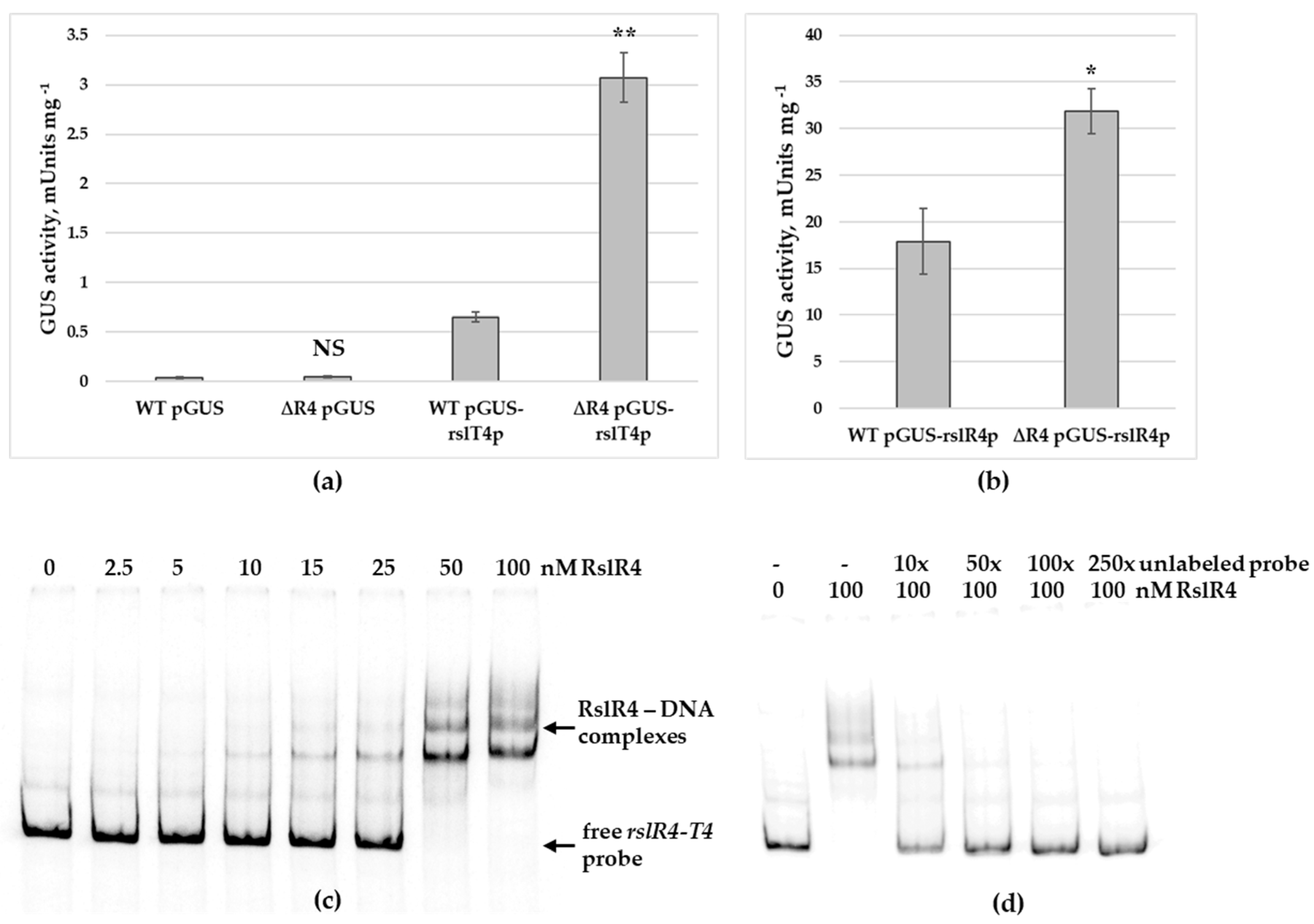
3.5. RslR4 Negatively Regulates Transcription of MFS Transporter Gene rslT4 and Its Own Expression

RslR4 is a member of the MarR family of transcriptional factors, often involved in regulating antibiotic resistance, stress responses, catabolism of aromatic compounds, and virulence regulation [40,41,42,43,44]. Most of the members are known to be transcriptional repressors of genes located next to it, including transporters of the major facilitator superfamily (MFS). Gene rslR4 is located upstream and divergently oriented to rslT4, encoding MFS membrane protein. It is predicted to be an efflux pump providing strain self-resistance to rishirilide(s) and pathway intermediates. We assumed that RslR4 negatively controls rslT4 transcription until rishirilide and/or its intermediate are accumulated.
To study the function of RslR4 on transcription of rslT4 and its own gene, two plasmids pGUS-rslT4p and pGUS-rslR4p were constructed. In these plasmids, promoters of respective genes are fused with the β-glucuronidase reporter gene gusA. Constructs were introduced into S. bottropensis and into S. bottropensis ∆R4. Both strains carrying pGUS-rslT4p were grown in YMPGv medium. After two days, β-glucuronidase activity in the lysates was measured. In comparison to the wild type strain, S. bottropensis ∆R4 showed ca. 5-fold higher β-glucuronidase activity, indicating that RslR4 represses rslT4 transcription (Figure 5a). The same experiment was carried out with the strains harboring pGUS-rslR4p. Here, a 2-fold increased β-glucuronidase activity in S. bottropensis ∆R4 in comparison to the wild type strain was observed (Figure 5b). This result concludes that RslR4 also negatively controls transcription of its own gene, acting as an autorepressor.
To confirm direct binding of RslR4 to intergenic region between rslR4 and rslT4, a recombinant C-terminal histidine-tagged protein was heterologously produced in E. coli (DE3) Star, and its DNA-binding ability was analyzed by EMSA. Figure 5c shows a formation of retarded bands with increased concentration of RslR4. MarR regulators act as dimers and binds to palindromic sequences within target DNA [45]. The existence of several protein-DNA complexes indicates the presence of multiple binding sites in a given DNA sequence. Multiple sites could facilitate differential regulation of rslT4 and rslR4 genes. Next, a competition assay was carried out to prove binding specificity. As shown in Figure 5d, an excess of non-radioactive probe specifically competed with the labeled probe for binding to RslR4.

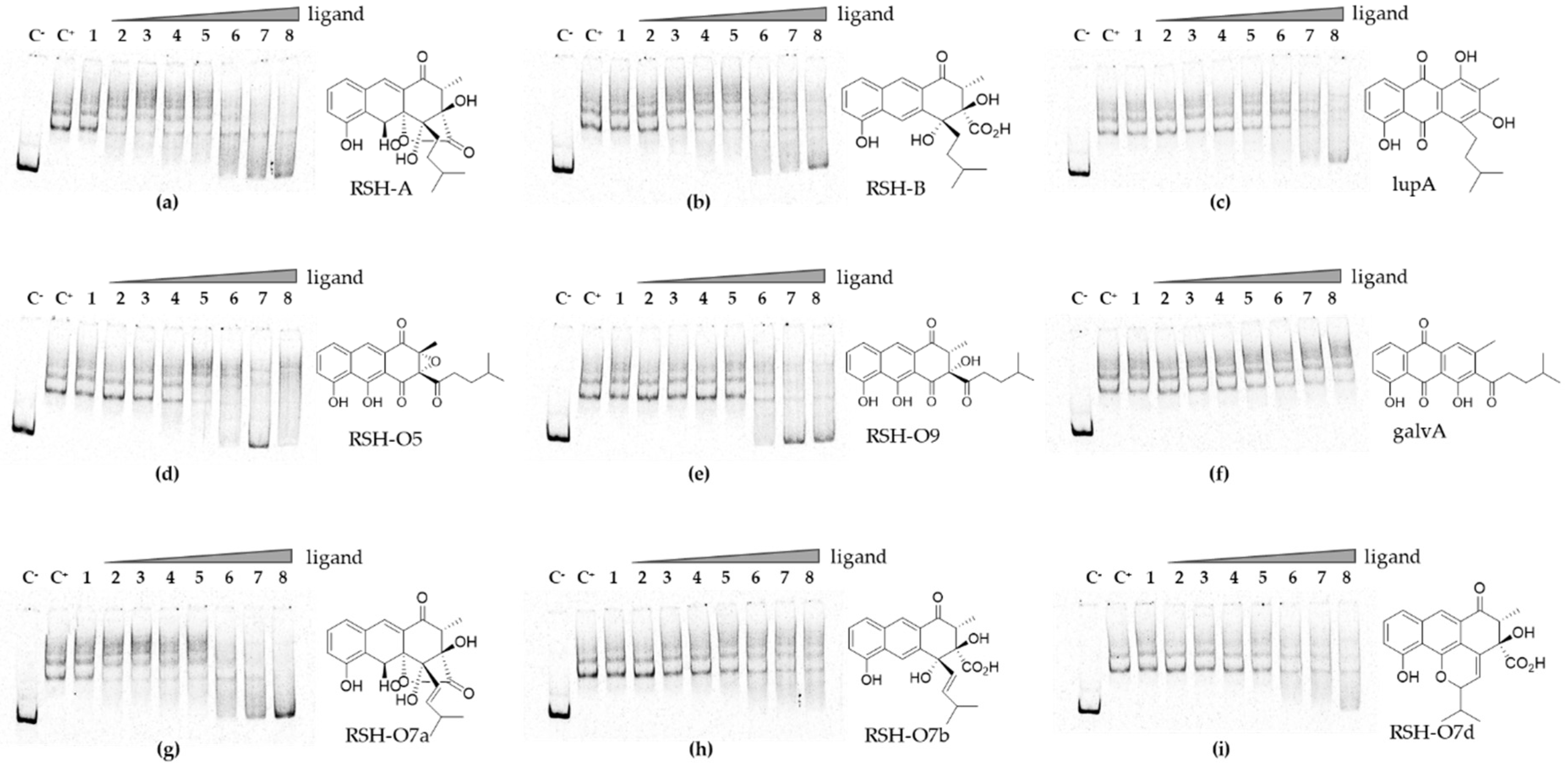
3.6. Rishirilides and Intermediates of the Pathway Interact with RslR4

The final product or pathway intermediate(s) often exert a regulatory role on strain resistance by modulating activity of cognate regulator. To assess the effect of end products and key intermediates on DNA binding by RslR4, the recombinant protein was incubated with the rslR4-T4 region in reaction mixture containing different concentrations of putative ligands. In addition, we tested the newly isolated alkene derivatives RSH-O7a-d, which are accumulated in an enoyl reductase RslO7-deficient mutant. As shown in Figure 6, the interaction of RslR4 with its ligand molecules is concentration dependent. Rishirilides, RSH-O5, RSH-O9, RSH-O7a, and lupinacidin A at high concentration could eliminate DNA–protein interaction (Figure 6a–e,g). Little interaction was observed for RSH-O7b-d (Figure 6h,i) and no interaction was observed for the shunt product galvaquinone A (accumulated in ∆rslO1 and ∆rslO4 mutants) (Figure 6f). This result demonstrated that RslR4 DNA-binding ability is regulated by the final products and some key intermediates of the rishirilide pathway.
Altogether, these findings demonstrate that RslR4 negatively controls transcription of rslR4 and rslT4 genes by direct binding to their intergenic region. The repression is eliminated upon RslR4 interaction with cognate ligand molecules. This leads to the expression of the MFS transporter gene rslT4 and provides strain with a resistance to synthesized metabolites.

4. Discussion

Streptomycetes are a rich source of natural products. Genes involved in the biosynthesis of secondary metabolite are joined together in the biosynthetic gene cluster, and usually encode one or a few regulatory proteins. Cluster-situated regulator(s) directly control transcription of biosynthetic genes, acting as activators or repressors. Moreover, expression of the regulatory genes is under control of different higher-level regulatory systems. Such cascade regulation coordinates production of secondary metabolite accordingly to strain’s physiology, developmental state, and environmental conditions.
Unlike many gene clusters that encode only one single pathway specific regulator, the rsl-gene cluster harbors four regulatory genes. Bioinformatic analysis of the proteins revealed three SARPs, RslR1, RslR2, and RslR3, and the MarR family transcriptional factor RslR4. SARP-like regulators usually activate a cognate biosynthetic gene cluster. They positively regulate transcription of biosynthetic genes and/or another SARP in the cluster.
In this study, we investigated the impact of four cluster-situated regulatory proteins on biosynthesis of rishirilides. Inactivation of each SARP completely abolished production of rishirilides and intermediates of the pathway, whereas deletion of rslR4 revealed no distinct effect. Consistently, overexpression of rslR1, rslR2, or rslR3 in S. bottropensis led to an increase of rishirilides production. These results strongly suggest that these transcriptional factors function as activators, all influencing rishirilide biosynthesis.
The presence of several SARPs in a gene cluster often goes along with a cascade of pathway-specific regulatory steps. This was shown for biosynthesis of tylosin, pristinamycin, and polymyxin [17,18,19,46,47]. Expression of rslR1, rslR3, and rslR4 remained at the same level in the wild type and mutants lacking one of the regulatory genes, indicating that their transcription is independent from the rsl-regulatory genes. In contrast, expression profile of the rslR2 gene in the wild type and in mutant strains revealed that its transcription is dependent on RslR1 and RslR3. The reduced expression of rslR2 in S. bottropensis ∆R3 suggests that rslR2 is activated by RslR3. In contrast, RslR1 likely exerts a negative control on rslR2, as rslR2 transcription is increased in S. bottropensis ∆R1. These findings are additionally confirmed by results of in vivo transcriptional analysis. Moreover, we also revealed RslR2 to be a self-repressor. The self-modulating activity of RslR2 could be explained by analogy to JadR1, a cluster-situated regulator from jadomycin biosynthesis in S. venezuelae. Upon binding of jadomycin, JadR1 activates the biosynthetic gene cluster and represses transcription of its own gene [48]. Analogous, rishirilides, or intermediates of the pathway may serve as ligands for RslR2 in a dose-dependent manner. In general, well-tuned regulation of rslR2 by RslR1 and RslR2 may help to maintain the biosynthesis of compounds at certain level, avoiding inhibition of strain growth. To further evaluate regulatory significance and investigate the exact role of RslR1 and RslR2, the recombinant proteins need to be produced and apply in experiments. Unfortunately, despite all our attempts, we failed to obtain these regulators in a soluble form.
RslR3 might be the highest-level cluster-situated regulator acting on rishirilides biosynthesis. Based on the protein structure, its activity could be modulated by ATPase domain or phosphorylation governed by particular kinase(s). ATPase activity of the structurally similar regulators PolY and AfsR triggers polymerization of the proteins and enhance their DNA binding activity [19,39,49]. Moreover, in vivo experiments revealed that endogenous ADP/ATP pool affected PolY activity, indicating a possible sensor ability of the ATPase domain and its regulatory function in modulating protein activity in response to current physiological conditions [19]. In addition, an activity of AfsR was shown to be regulated by multiple kinases [50].
Similar to PolY, RslR3 regulates transcription of another cluster-situated regulatory gene rslR2. The evidence that RslR3 did not bind to other promoter regions of rsl-transcriptional unit concludes that rslR2p is the only direct target of this regulator.
To avoid self-inhibition by produced compounds, S. bottropensis encodes the regulator RslR4 to control an expression of the cognate exporter gene rslT4. Typical for MarR-like regulators, rslR4 is divergently oriented to its target rslT4 that allows specific binding of RslR4 to intergenic region between the genes to repress transcription of both. Indeed, in vivo transcriptional levels of both genes were upregulated in S. bottropensis ∆R4 in comparison to the wild type strain. Negative autoregulation may prevent excessive accumulation of the regulator. This allows the keeping of narrow range of the protein concentration and results in more sensitive response to ligands.
RslR4 modulates the expression of rslT4 in response to rishirilides and compounds from their biosynthetic pathway. The particularly interesting result is that both, the linear tricyclic intermediates (RSH-O5 and RSH-O9) and rishirilide-scaffold compounds interact with RslR4, and act as repression-relieving ligands. The ability to respond to pathway intermediates indicates that S. bottropensis cells initiate mechanism of self-immunity before the amount of synthesized end products reaches a critical level. This also suggests that intermediates are sufficient to trigger export and couple the mechanism of strain immunity to a biosynthetic pathway.
Based on these findings, we propose the following regulatory scheme (Figure 7). Rishirilides biosynthesis requires expression of three cluster-situated regulators, RslR1, RslR2, and RslR3 since the individual gene deletions completely abolished the rishirilides production or any known pathway intermediate(s). The biosynthesis is likely triggered by RslR3, which is present in the cell pool, upon signal-induced activation of the protein. Regulatory control exerted by RslR3 is mediated through activation of rslR2 by direct binding to its promoter region. Most probably, rsl-genes are mainly regulated by the pathway specific regulator RslR2 together with RslR1. The transcription of rslR2 is negatively regulated by RslR1 and an autorepression mechanism, while the expression of rslR1 is likely activated by other regulatory elements of the cell. RslR4 is a repressor of the MFS transporter gene rslT4 and its own gene transcription. The end products and pathway intermediates release repression by RslR4 that results in production of RslT4, providing cells with resistance to the synthesized compounds.

Supplementary Materials

The following materials are available online at https://www.mdpi.com/2076-2607/9/2/374/s1. Figure S1: Transcriptional profile of regulatory genes in the wild type strain and in different mutants, Figure S2: Domain organization and sequence of RslR3, Figure S3: Sequences alignment of RslR4 versions, Figure S4: LC-MS analysis of metabolic compounds produced by S. bottropensis and S. bottropensisrslO7, Generation of S. bottropensisrslO7, isolation, purification and NMR data for compounds RSH-O7a-d, Table S1: Strains and plasmids used in this work, Table S2: Primers List.

Author Contributions

Project design and conceptualization, O.T., R.M., and A.B.; resources, A.B., H.-G.K., and T.P.; experimental procedures, O.T., R.M., and X.Y.; structure elucidation of compounds, T.P.; draft preparation, O.T.; review and editing, all authors. All authors have read and agreed to the published version of the manuscript.

Funding

This research was funded by DFG, grant number 278002225/RTG 2202.

Institutional Review Board Statement

Not applicable.

Informed Consent Statement

Not applicable.

Data Availability Statement

The data presented in this study are available in this article and Supplementary Materials.

Acknowledgments

We thank Judy Wong for proof-reading of the manuscript.

Conflicts of Interest

The authors declare no conflict of interest.

References

  1. Flärdh, K.; Buttner, M.J. Streptomyces morphogenetics: Dissecting differentiation in a filamentous bacterium. Nat. Rev. Genet. 2009, 7, 36–49. [Google Scholar] [CrossRef]
  2. Quinn, G.A.; Banat, A.M.; Abdelhameed, A.M.; Banat, I.M. Streptomyces from traditional medicine: Sources of new innovations in antibiotic discovery. J. Med. Microbiol. 2020, 69, 1040–1048. [Google Scholar] [CrossRef]
  3. Procópio, R.E.D.L.; Da Silva, I.R.; Martins, M.K.; De Azevedo, J.L.; De Araújo, J.M. Antibiotics produced by Streptomyces. Braz. J. Infect. Dis. 2012, 16, 466–471. [Google Scholar] [CrossRef] [Green Version]
  4. Sivalingam, P.; Hong, K.; Pote, J.; Prabakar, K. Extreme Environment Streptomyces: Potential Sources for New Antibacterial and Anticancer Drug Leads? Int. J. Microbiol. 2019, 2019, 5283948. [Google Scholar] [CrossRef] [PubMed] [Green Version]
  5. Tsypik, O.; Makitrynskyy, R.; Bera, A.; Song, L.; Wohlleben, W.; Fedorenko, V.; Ostash, B. Role of GntR Family Regulatory Gene SCO1678 in Gluconate Metabolism in Streptomyces coelicolor M145. BioMed Res. Int. 2017, 2017, 9529501. [Google Scholar] [CrossRef]
  6. Liu, G.; Chater, K.F.; Chandra, G.; Niu, G.; Tan, H. Molecular Regulation of Antibiotic Biosynthesis in Streptomyces. Microbiol. Mol. Biol. Rev. 2013, 77, 112–143. [Google Scholar] [CrossRef] [Green Version]
  7. Xia, H.; Li, X.; Li, Z.; Zhan, X.; Mao, X.; Li, Y. The Application of Regulatory Cascades in Streptomyces: Yield Enhancement and Metabolite Mining. Front. Microbiol. 2020, 11, 406. [Google Scholar] [CrossRef] [PubMed] [Green Version]
  8. Martín, J.F.; Liras, P. The Balance Metabolism Safety Net: Integration of Stress Signals by Interacting Transcriptional Factors in Streptomyces and Related Actinobacteria. Front. Microbiol. 2020, 10, 3120. [Google Scholar] [CrossRef]
  9. Kong, D.; Wang, X.; Nie, J.; Niu, G. Regulation of Antibiotic Production by Signaling Molecules in Streptomyces. Front. Microbiol. 2019, 10, 2927. [Google Scholar] [CrossRef] [Green Version]
  10. Hesketh, A.; Chen, W.; Ryding, J.; Chang, S.; Bibb, M. The global role of ppGpp synthesis in morphological differentiation and antibiotic production in Streptomyces coelicolor A3(2). Genome Biol. 2007, 8, R161. [Google Scholar] [CrossRef] [PubMed] [Green Version]
  11. Gomez-Escribano, J.P.; Martín, J.F.; Hesketh, A.; Bibb, M.J.; Liras, P. Streptomyces clavuligerus relA-null mutants overproduce clavulanic acid and cephamycin C: Negative regulation of secondary metabolism by (p)ppGpp. Microbiology 2008, 154, 744–755. [Google Scholar] [CrossRef] [Green Version]
  12. Hesketh, A.; Sun, J.; Bibb, M.J. Induction of ppGpp synthesis in Streptomyces coelicolor A3(2) grown under conditions of nutritional sufficiency elicits actII-ORF4 transcription and actinorhodin biosynthesis. Mol. Microbiol. 2001, 39, 136–144. [Google Scholar] [CrossRef] [Green Version]
  13. Nuzzo, D.; Makitrynskyy, R.; Tsypik, O.; Bechthold, A. Cyclic di-GMP cyclase SSFG_02181 from Streptomyces ghanaensis ATCC14672 regulates antibiotic biosynthesis and morphological differentiation in streptomycetes. Sci. Rep. 2020, 10, 12021. [Google Scholar] [CrossRef]
  14. Makitrynskyy, R.; Tsypik, O.; Nuzzo, D.; Paululat, T.; Zechel, D.L.; Bechthold, A. Secondary nucleotide messenger c-di-GMP exerts a global control on natural product biosynthesis in streptomycetes. Nucleic Acids Res. 2020, 48, 1583–1598. [Google Scholar] [CrossRef] [Green Version]
  15. Nuzzo, D.; Makitrynskyy, R.; Tsypik, O.; Bechthold, A. Identification and Characterization of Four c-di-GMP-Metabolizing Enzymes from Streptomyces ghanaensis ATCC14672 Involved in the Regulation of Morphogenesis and Moenomycin A Biosynthesis. Microorganisms 2021, 9, 284. [Google Scholar] [CrossRef]
  16. Nett, M.; Ikeda, H.; Moore, B.S. Genomic basis for natural product biosynthetic diversity in the actinomycetes. Nat. Prod. Rep. 2009, 26, 1362–1384. [Google Scholar] [CrossRef] [PubMed]
  17. Bate, N.; Stratigopoulos, G.; Cundliffe, E. Differential roles of two SARP-encoding regulatory genes during tylosin biosynthesis. Mol. Microbiol. 2002, 43, 449–458. [Google Scholar] [CrossRef]
  18. Bignell, D.R.D.; Bate, N.; Cundliffe, E. Regulation of tylosin production: Role of a TylP-interactive ligand. Mol. Microbiol. 2007, 63, 838–847. [Google Scholar] [CrossRef]
  19. Li, R.; Liu, G.; Xie, Z.; He, X.; Chen, W.; Deng, Z.; Tan, H. PolY, a transcriptional regulator with ATPase activity, directly activates transcription ofpolRin polyoxin biosynthesis inStreptomyces cacaoi. Mol. Microbiol. 2010, 75, 349–364. [Google Scholar] [CrossRef] [PubMed]
  20. Ostash, B.; Saghatelian, A.; Walker, S. A Streamlined Metabolic Pathway for the Biosynthesis of Moenomycin A. Chem. Biol. 2007, 14, 257–267. [Google Scholar] [CrossRef] [PubMed] [Green Version]
  21. Bibb, M.J. Regulation of secondary metabolism in streptomycetes. Curr. Opin. Microbiol. 2005, 8, 208–215. [Google Scholar] [CrossRef]
  22. Gramajo, H.C.; Takano, E.; Bibb, M.J. Stationary-phase production of the antibiotic actinorhodin in Streptomyces coelicolor A3(2) is transcriptionally regulated. Mol. Microbiol. 1993, 7, 837–845. [Google Scholar] [CrossRef] [Green Version]
  23. Arias, P.; Fernández-Moreno, M.A.; Malpartida, F. Characterization of the Pathway-Specific Positive Transcriptional Regulator for Actinorhodin Biosynthesis inStreptomyces coelicolor A3(2) as a DNA-Binding Protein. J. Bacteriol. 1999, 181, 6958–6968. [Google Scholar] [CrossRef] [Green Version]
  24. Fujii, T.; Gramajo, H.C.; Takano, E.; Bibb, M.J. redD and actII-ORF4, pathway-specific regulatory genes for antibiotic production in Streptomyces coelicolor A3(2), are transcribed in vitro by an RNA polymerase holoenzyme containing sigma hrdD. J. Bacteriol. 1996, 178, 3402–3405. [Google Scholar] [CrossRef] [PubMed] [Green Version]
  25. White, J.; Bibb, M. bldA dependence of undecylprodigiosin production in Streptomyces coelicolor A3(2) involves a pathway-specific regulatory cascade. J. Bacteriol. 1997, 179, 627–633. [Google Scholar] [CrossRef] [Green Version]
  26. Wolański, M.; Donczew, R.; Kois-Ostrowska, A.; Masiewicz, P.; Jakimowicz, D.; Zakrzewska-Czerwińska, J. The Level of AdpA Directly Affects Expression of Developmental Genes in Streptomyces coelicolor. J. Bacteriol. 2011, 193, 6358–6365. [Google Scholar] [CrossRef] [Green Version]
  27. Rigali, S.; Titgemeyer, F.; Barends, S.; Mulder, S.; Thomae, A.W.; Hopwood, D.A.; Van Wezel, G.P. Feast or famine: The global regulator DasR links nutrient stress to antibiotic production by Streptomyces. EMBO Rep. 2008, 9, 670–675. [Google Scholar] [CrossRef] [PubMed] [Green Version]
  28. Wang, R.; Mast, Y.; Wang, J.; Zhang, W.; Zhao, G.; Wohlleben, W.; Lu, Y.; Jiang, W.-H. Identification of two-component system AfsQ1/Q2 regulon and its cross-regulation with GlnR inStreptomyces coelicolor. Mol. Microbiol. 2012, 87, 30–48. [Google Scholar] [CrossRef] [PubMed]
  29. McKenzie, N.L.; Nodwell, J.R. Phosphorylated AbsA2 Negatively Regulates Antibiotic Production in Streptomyces coelicolor through Interactions with Pathway-Specific Regulatory Gene Promoters. J. Bacteriol. 2007, 189, 5284–5292. [Google Scholar] [CrossRef] [Green Version]
  30. Yan, X.; Probst, K.; Linnenbrink, A.; Arnold, M.; Paululat, T.; Zeeck, A.; Bechthold, A. Cloning and Heterologous Expression of Three Type II PKS Gene Clusters fromStreptomyces bottropensis. ChemBioChem 2011, 13, 224–230. [Google Scholar] [CrossRef] [PubMed]
  31. Iwaki, H.; Nakayama, Y.; Takahashi, M.; Uetsuki, S.; Kido, M.; Fukuyama, Y. Structures of rishirilides A and B, alpha 2-macroglobulin inhibitors produced by Streptomyces rishiriensis OFR-1056. J. Antibiot. 1984, 37, 1091–1093. [Google Scholar] [CrossRef] [Green Version]
  32. Komagata, D.; Sawa, R.; Kinoshita, N.; Imada, C.; Sawa, T.; Naganawa, H.; Hamada, M.; Okami, Y.; Takeuchi, T. Isolation of glutathione-S- transferase inhibitors. J. Antibiot. 1992, 45, 1681–1683. [Google Scholar] [CrossRef] [Green Version]
  33. Schwarzer, P.; Tsypik, O.; Zuo, C.; Alali, A.; Wunsch-Palasis, J.; Heitzler, T.; Derochefort, J.; Bernhardt, M.; Yan, X.; Paululat, T.; et al. Early Steps in the Biosynthetic Pathway of Rishirilide B. Molecules 2020, 25, 1955. [Google Scholar] [CrossRef] [Green Version]
  34. Schwarzer, P.; Wunsch-Palasis, J.; Bechthold, A.; Paululat, T. Biosynthesis of Rishirilide B. Antibiotics 2018, 7, 20. [Google Scholar] [CrossRef] [Green Version]
  35. Tsypik, O.; Makitrynskyy, R.; Frensch, B.; Zechel, D.L.; Paululat, T.; Teufel, R.; Bechthold, A. Oxidative Carbon Backbone Rearrangement in Rishirilide Biosynthesis. J. Am. Chem. Soc. 2020, 142, 5913–5917. [Google Scholar] [CrossRef]
  36. Ostash, B.; Rebets, Y.; Myronovskyy, M.; Tsypik, O.; Ostash, I.; Kulachkovskyy, O.; Datsyuk, Y.; Nakamura, T.; Walker, S.; Fedorenko, V. Identification and characterization of the Streptomyces globisporus 1912 regulatory gene lndYR that affects sporulation and antibiotic production. Microbiology 2011, 157, 1240–1249. [Google Scholar] [CrossRef] [Green Version]
  37. Gust, B.; Chandra, G.; Jakimowicz, D.; Yuqing, T.; Bruton, C.J.; Chater, K.F. λ Red-Mediated Genetic Manipulation of Antibiotic-Producing Streptomyces. Elsevier 2004, 54, 107–128. [Google Scholar]
  38. Myronovskyi, M.; Welle, E.; Fedorenko, V.; Luzhetskyy, A. β-Glucuronidase as a Sensitive and Versatile Reporter in Actinomycetes. Appl. Environ. Microbiol. 2011, 77, 5370–5383. [Google Scholar] [CrossRef] [PubMed] [Green Version]
  39. Tanaka, A.; Takano, Y.; Ohnishi, Y.; Horinouchi, S. AfsR Recruits RNA Polymerase to the afsS Promoter: A Model for Transcriptional Activation by SARPs. J. Mol. Biol. 2007, 369, 322–333. [Google Scholar] [CrossRef] [PubMed]
  40. Xiong, A.; Gottman, A.; Park, C.; Baetens, M.; Pandza, S.; Matin, A. The EmrR Protein Represses the Escherichia coli emrRAB Multidrug Resistance Operon by Directly Binding to Its Promoter Region. Antimicrob. Agents Chemother. 2000, 44, 2905–2907. [Google Scholar] [CrossRef] [PubMed] [Green Version]
  41. Davis, J.R.; Sello, J.K. Regulation of genes in Streptomyces bacteria required for catabolism of lignin-derived aromatic compounds. Appl. Microbiol. Biotechnol. 2009, 86, 921–929. [Google Scholar] [CrossRef] [PubMed]
  42. Chang, Y.-M.; Jeng, W.-Y.; Ko, T.-P.; Yeh, Y.-J.; Chen, C.K.-M.; Wang, A.H.-J. Structural study of TcaR and its complexes with multiple antibiotics from Staphylococcus epidermidis. Proc. Natl. Acad. Sci. USA 2010, 107, 8617–8622. [Google Scholar] [CrossRef] [PubMed] [Green Version]
  43. Fuangthong, M.; Atichartpongkul, S.; Mongkolsuk, S.; Helmann, J.D. OhrR Is a Repressor of ohrA, a Key Organic Hydroperoxide Resistance Determinant in Bacillus subtilis. J. Bacteriol. 2001, 183, 4134–4141. [Google Scholar] [CrossRef] [PubMed] [Green Version]
  44. Ellison, D.W.; Miller, V.L. Regulation of virulence by members of the MarR/SlyA family. Curr. Opin. Microbiol. 2006, 9, 153–159. [Google Scholar] [CrossRef]
  45. Perera, I.C.; Grove, A. Molecular Mechanisms of Ligand-Mediated Attenuation of DNA Binding by MarR Family Transcriptional Regulators. J. Mol. Cell Biol. 2010, 2, 243–254. [Google Scholar] [CrossRef] [PubMed] [Green Version]
  46. Mast, Y.; Guezguez, J.; Handel, F.; Schinko, E. A Complex Signaling Cascade Governs Pristinamycin Biosynthesis in Streptomyces pristinaespiralis. Appl. Environ. Microbiol. 2015, 81, 6621–6636. [Google Scholar] [CrossRef] [PubMed] [Green Version]
  47. Handel, F.; Kulik, A.; Mast, Y. Investigation of the Autoregulator-Receptor System in the Pristinamycin Producer Streptomyces pristinaespiralis. Front. Microbiol. 2020, 11, 580990. [Google Scholar] [CrossRef]
  48. Wang, L.; Tian, X.; Wang, J.; Yang, H.; Fan, K.; Xu, G.; Yang, K.; Tan, H. Autoregulation of antibiotic biosynthesis by binding of the end product to an atypical response regulator. Proc. Natl. Acad. Sci. USA 2009, 106, 8617–8622. [Google Scholar] [CrossRef] [Green Version]
  49. Lee, P.-C.; Umeyama, T.; Horinouchi, S. afsS is a target of AfsR, a transcriptional factor with ATPase activity that globally controls secondary metabolism in Streptomyces coelicolor A3(2). Mol. Microbiol. 2002, 43, 1413–1430. [Google Scholar] [CrossRef]
  50. Horinouchi, S. AfsR as an integrator of signals that are sensed by multiple serine/threonine kinases in Streptomyces coelicolor A3(2). J. Ind. Microbiol. Biotechnol. 2003, 30, 462–467. [Google Scholar] [CrossRef]
Figure 1. Rishirilides production by S. bottropensis strains. (a) UHPLC-MS analysis of culture extracts from S. bottropensis (WT) and mutant strains (S. bottropensis ∆R1, ∆R2, ∆R3, and ∆R4); (b) UHPLC-MS analysis of culture extracts from complemented rslR1, rslR2 and (c) rslR3 mutant strains. Strains carrying the empty vector pSET152 or pTES were used as a control. (d) Levels of total rishirilide (RSH) production by the wild type and strains which overexpress regulatory genes. The mean value of total rishirilide production in S. bottropensis (WT) and S. bottropensis pUWLH (WT pUWLH) was taken as 100%. Error bars represent standard deviations. Significant differences in total rishirilide production between S. bottropensis and strains overexpressing regulatory genes were calculated by a two-tailed t-test. Asterisks represent the significance value (** p < 0.01), whereas NS refers to not-significant differences.
Microorganisms 09 00374 g001
Figure 2. Transcriptional profile of regulatory genes in the wild type strain and in different mutants. (a) sqRT-PCR analysis of rslR1, rslR2, rslR3, rslR4, and hrdB transcripts in S. bottropensis strains. PCR was performed using cDNA and chromosomal DNA (C) as a template. β-glucuronidase activity from a promoterless construct (b), a construct containing the rslR1 promoter (c), a construct containing the rslR2 promoter (d) determined in the wild type strain and in S. bottropensis ΔR1, S. bottropensis ΔR2, and S. bottropensis ΔR3. Cultures and subsequent β-glucuronidase measurements were done in triplicate. Values were normalized to equal amounts of dry biomass. Error bars represent standard deviations. Significant differences in β-glucuronidase between the wild type and tested strains were calculated by a two-tailed t-test. Asterisks represent the significance value (** p < 0.01), whereas NS refers to not-significant differences.
Microorganisms 09 00374 g002
Figure 3. Transcriptional organization of rsl-genes. (a) Genetic organization of the rishirilide gene cluster. Regulatory, biosynthetic, and transporter genes are highlighted in red, grey, and green, respectively. The solid lines under the genes depict transcriptional units, black bent arrows are promoter regions, grey bent arrows are putative promoters; (b) RT-PCR analysis of rsl-genes co-transcription. RNA, genomic DNA (C), and cDNA were used as a template in PCR reactions. The functions of rsl-genes are described in [30,33,35].
Microorganisms 09 00374 g003
Figure 4. Electrophoretic mobility shift assay to study binding of RslR3DBD to promoter region of rslR1, rslR2, and structural genes. (a) EMSA containing rslR2p; (b) competition assay of RslR3DBD (1 µM) with rslR2p carrying 10, 100, 250, and 500-fold excess of unlabeled probe; (c) EMSA containing rslR1p; (d) EMSA containing promoters of structural genes.
Microorganisms 09 00374 g004
Figure 5. RslR4 negatively regulates transcription of rslT4 and rslR4 genes. (a) Transcriptional activity of rslT4 promoter in S. bottropensis and in S. bottropensis ∆R4. (b) Transcriptional activity of rslR4 promoter in S. bottropensis and in S. bottropensis ∆R4. Cultures and subsequent β-glucuronidase measurements were done in triplicate. Values were normalized to equal amounts of dry biomass. Error bars represent standard deviations. Significant differences in β-glucuronidase between S. bottropensis and S. bottropensis ∆R4 were calculated by a two-tailed t-test. Asterisks represent the significance value (* p < 0.05, ** p < 0.01), whereas NS refers to not-significant differences. (c) Electrophoretic mobility shift assay to study interaction of RslR4 with rslR4-T4 region. Increasing concentrations of RslR4 were mixed with labeled rslR4-T4 DNA probe. (d) Competition assay of RslR4 (100 nM) with rslR4-T4 region carrying 10-, 50-, 100- and 250-fold excess of unlabeled probe.
Microorganisms 09 00374 g005
Figure 6. Electrophoretic mobility shift assay used to analyze effects of ligand molecules on RslR4 binding to the rslR4-T4 region. RslR4 (100 nM) was incubated with the rslR4-T4 region and increasing concentrations of the ligands (a) rishirilide A, (b) rishirilide B, (c) lupinacidin A, (d) pathway intermediate RSH-O5, (e) pathway intermediate RSH-O9, (f) shunt product galvaquinone A, (g) RSH-O7a, (h) RSH-O7b, (i) RSH-O7d. Lanes C, negative control containing only labeled rslR4-T4, lanes C+, positive control containing RslR4 and labeled rslR4-T4, lanes 1, positive control containing DMSO, lanes 2–8, samples containing different concentrations of the ligands (1, 10, 50, 100 µM and 0.25, 0.5, 1 mM, respectively).
Microorganisms 09 00374 g006
Figure 7. Proposed regulatory scheme of rishirilides biosynthesis.
Microorganisms 09 00374 g007
Publisher’s Note: MDPI stays neutral with regard to jurisdictional claims in published maps and institutional affiliations.

