Article

Fecal Microbiome and Resistome Profiling of Healthy and Diseased Pakistani Individuals Using Next-Generation Sequencing

1
Department of Biology Education, Kyungpook National University, 80 Daehak-ro, Buk-gu, Daegu 41566, Korea
2
Center for Genome Sciences, Rehman Medical College, Phase-V, Hayatabad, Peshawar 25000, Pakistan
3
Executive Development Center, Sukkur Institute of Business Administration University, Sindh 65200, Pakistan
4
AlviArmani International, Mississauga, ON M4W-3E2, Canada
5
Department of Biotechnology, Institute of Integrative Biosciences, CECOS University of IT & Emerging Sciences, Peshawar 25000, Pakistan
*
Authors to whom correspondence should be addressed.
Microorganisms 2021, 9(3), 616; https://doi.org/10.3390/microorganisms9030616
Submission received: 27 February 2021 / Revised: 13 March 2021 / Accepted: 15 March 2021 / Published: 17 March 2021
(This article belongs to the Special Issue Gut Microbiota Dysbiosis 2.0)

Abstract

:
In this paper, we aimed to characterize the fecal microbiome and its resistomes of healthy and diseased subjects infected with multidrug-resistant Escherichia coli using next-generation sequencing (NGS). After initial screening, 26 stools samples belonging to healthy (n = 13) and diseased subjects (n = 13) were selected and subjected to NGS. A total of 23 and 42 antibiotic-resistant genes (ARGs) conferring resistance to 6 and 9 classes of antibiotics were identified in the resistomes of healthy and diseased subjects, respectively. Bacteroidetes were found to be the major phylum in both healthy and diseased subjects; however, Proteobacteria was predominantly present in the diseased subjects only. Microbial dysbiosis and predominance of various ARGs in the resistome of diseased subjects reflect the excessive usage of antibiotics in Pakistan and warrants immediate attention to regulate the use of various antimicrobials.

1. Introduction

The emergence of multidrug-resistant (MDR) bacteria is becoming a serious threat to human health. The human gut microbiome plays an important role in the emergence and transmission of antibiotic-resistant microbes [1]. Human gut microbiota essentially acts as a potential reservoir of antibiotic resistance genes (ARGs). A resistome includes all ARGs within a specific microbial niche [2,3]. Resistomes of different individuals are influenced by various factors such as geographical location, age, antibiotic usage, diet, environment, lifestyle, and socioeconomic status [4]. These factors also alter the composition of the human microbiome thereby making resistome closely correlated to the microbiome [3,4]. The advent of high-throughput sequencing technologies fast-tracked the microbiome research and characterized the gut microbiota as “superorganism” [5]. The human gut microbiota plays essential roles in metabolism, physiology, and development of immune system. Although gut microbiota serves as a “superorganism”, it is highly dynamic and is frequently altered by diet, age, antibiotics, various infections, and host genetic factors [6].
Among the various factors, antibiotic administration has been reported to profoundly affect the composition of the human microbiome and its resistome drastically [7]. Antibiotics administration alters the composition of the human microbiome leading to gut microbial dysbiosis [8]. The essential functions of the gut microbiome such as vitamin production, nutrient supply, and protection against pathogens are negatively affected by microbial dysbiosis [9]. A dysbiotic gut microbiome has been linked with various ailments such as developmental, immunological, and metabolic disorders. In addition, gut microbiome dysbiosis increases the susceptibility to develop various serious infectious diseases [9,10].
Antibiotics with broad-spectrum activity have been reported to affect the abundance of human gut microbiota by up to 30%, thereby causing a rapid and significant reduction in the taxonomic composition of gut microbes [11,12]. The effects of antibiotics administration on gut microbiota may last for a long time ranging from months to years. Furthermore, excessive antibiotic administration deteriorates the microenvironment of the human gut microbiome leading to the emergence of “pathobionts” such as Escherichia coli, Salmonella enterica, Shigella flexneri, Klebsiella, Acinetobacter, and Pseudomonas [12,13]. Consequently, the acquisition of various ARGs by these pathobionts can lead to serious health consequences [14]. Antibiotic mediated gut microbial dysbiosis has been characterized by the loss of microbial diversity, reduced abundance of specific taxa [15], increased susceptibility to various infections, escalated proliferation of various superbugs (methicillin-resistant Staphylococcus aureus (MRSA) and vancomycin-resistant Enterococcus) [16], and disruption of intestinal mucus layers [14]. Currently, a number of studies demonstrated that antibiotic mediated gut microbial dysbiosis can be effectively reversed through the incorporation of various strategies such as use of probiotics, probiotics synbiotics, fecal microbiota transplant (FMT), bacterial consortium transplant (BCT), and phage therapy [17,18].
Exploration of human resistome can help in designing effective diagnostic and therapeutic strategies that are the need of the hour owing to the pandemic status of AMR. Policies regulating antibiotic usage in humans and animals have been reported to influence the prevalence of ARGs in the resistomes of various individuals from different countries [19,20]. Metagenomics studies contributed significantly to understanding the bacterial communities associated ARGs in diverse samples such as human stools samples, animal fecal samples, ready-to-eat food [21], and urban resistome [22]. Large-scale metagenomic studies [19,20] explored the gut resistome of healthy people from different countries and studied the abundance pattern of ARGs. Increased abundance of ARGs was found in the gut resistome of individuals from the countries with higher antibiotic usage such as China and Spain than people belonging to the countries with strict AMR regulating policies such as Denmark. Currently, among low- to middle-income countries, Pakistan constitutes the third highest consumer of antibiotics after India and China [23].
Identifying the key features of country specific resistome can help understanding the abundance pattern of particular ARGs and designing interventory strategies. Large scale metagenomics studies aiming to explore the microbiome and resistome of healthy Pakistani people are scarce, and to the best of our knowledge the poultry gut microbial and abundance profile of various ARGs are only explored so far by one of our previous studies [24].
Strict national policies regulating the antimicrobial usage (AMU) in both human and veterinary medicine are lacking in Pakistan [23,25,26]. Higher AMU in Pakistan warrants investigating the gut resistomes of healthy and sick individuals infected with AMR bacteria in order to ascertain the prevalence of various ARGs; however, such metagenomics studies are needed in Pakistan. This cross-sectional metagenomic study aimed to investigate for the first time the fecal microbiome and its resistomes of healthy and individuals infected with AMR bacteria belonging to the Peshawar, Khyber Pakhtunkhwa region of Pakistan using next-generation sequencing (NGS).

2. Materials and Methods

2.1. Study Design

This cross-sectional study was carried out at the tertiary healthcare center of Peshawar, Pakistan (34°1′33.3012″ N and 71°33′36.4860″ E). Ethical approval (Ref: RMI/RMI-REC/Approval/33) for this study was obtained from the Ethics Committee of Tertiary Healthcare Center, Peshawar Pakistan.

2.2. Sample Collection

A tertiary health care center of Peshawar, Pakistan was selected for the sample collection. The study objectives were explained to the patients and informed consents were signed. Initially, we screened inpatients (n = 420) diagnosed with various bacterial infections. Inpatients with the confirmed bacterial infection and willingness to participate in this study were enrolled for further analysis. All the important clinical and demographic data were carefully recorded from the selected patients and healthy controls (Tables S1 and S2, Supplementary Materials). Stool samples were collected from all enrolled patients and screened for MDR bacterial strains. Stool samples under aseptic measures were streaked onto the MacConkey and Eosin Methylene Blue agar plates (Oxoid, Basingstoke, Hampshire, UK). The streaked plates were incubated for 18–24 h at 35–37 °C. Slow-growing strains were incubated for a longer duration extending up to 48 h. All the bacterial isolates were identified using the standard morphological and biochemical tests [27]. Antibiotic susceptibility testing (AST) was performed using the Kirby–Bauer disc diffusion method. Bacterial strains were tested against aminoglycosides, neomycin, gentamycin, streptomycin, chloramphenicol, quinolones and fluoroquinolones, ofloxacin, nalidixic acid, sulfonamides, sulfamethoxazole, tetracycline, beta-lactam, ampicillin, nitrofurans, and cephalosporins following Clinical and Laboratory Standards Institute (CLSI) guidelines [28]. Patients testing negative for MDR bacterial strains were excluded from this study. Stool samples from the 13 healthy participants (7 males, 6 females; mean age 48.6 ± 11 years) with no antibiotic usage in the last six months were used as negative controls for fecal microbiome and its resistome profiling.

2.3. Extraction, Quantification and Normalization of Genomic DNA

Genomic DNA was isolated from a 0.2 g stool sample using a commercial kit (PureLinkTM Microbiome DNA Purification Kit) following the manufacturer’s instructions (Invitrogen, Thermo Fisher Scientific, Waltham, MA, USA) with little modifications. The use of standard bead-beater recommended by the manufacturer was replaced by simple laboratory benchtop vortexer (CLASSIC Vortex Mixer product code F202A0173, VELP Scientifica, Via Stazione 16-20865-Usmate Velate (MB), Italy). Bead-tubes containing the samples were fixed horizontally on the pad of bench top vortexer with the help of scotch tape at room temperature and vortexed at 2000 rpm for 8 min. To assure the NGS quality control (QC), all the extracted DNA samples were then quantified using Qubit fluorometer following manufacturer’s instructions (Qubit™ fluorometer, Invitrogen, Carlsbad, CA, USA) [29]. The quality of DNA samples were also checked using 1.0% agarose gel. Following quality control (DNA qualitative and quantitative analysis), all the DNA samples were then normalized to 0.2 ng/µL (1 ng/5µL).

2.4. NGS Libraries Preparation

Sequencing libraries were prepared using Illumina® (San Diego, CA, USA) Nextera XT DNA Library Preparation Kit (FC-131-1096) and Nextera XT Index Kit v2 Set A (FC-131-2001) as per the manufacturer’s instructions. For library preparation, 1ng dsDNA was subjected to tagmentation. dsDNA was fragmented and adapters were added to both ends. Unique Illumina idexes (i7/i5) were added to tagmented DNA through limited cycles of PCR amplification. The PCR amplified products were purified using AMPure XP beads and washed twice with 80% freshly prepared ethanol. Then, bead-based normalization of the purified products were done by using 45µL mixture of beads Library Normalization Additives 1 (LNA1), and Library Normalization Beads 1 (LNB1), and washed by using wash solution provided in the kit. Finally, each library was eluted in 0.1N NaOH and stored in storage buffer. All the libraries were pooled by mixing 5 µL each and subsequently 24 µL from the pooled sample (pooled DNA libraries) was mixed with 576 µL HTI (Hybridization Buffer) and subjected to denaturation at 98 °C for 2 min. Furthermore, 30 µL of 12.5 pM PhiX (internal sequencing control) was added to the pooled libraries thereby making a final volume of 600 µL, which was then loaded on to Illumina MiSeq Reagent Kit v2 (300 cycles; MS-103-1002, Illumina Inc., CA, USA) for paired end sequencing (2 × 150 bp) on Illumina MiSeq sequencer (Illumina, San Diego, CA, USA).

2.5. NGS Bioinformatics Analysis

After FASTQ files (raw data) were generated through MiSeq (Illumina sequencer), 26 dual-index barcoded metagenomes were demultiplexed using CASAVA 1.8.2. The sorted FASTQ files were then subjected to NGS QC using Trimmomatic 0.36 to remove all technical biases, low-quality reads (Q > 30), and adapters. To assure shotgun metagenome sequencing QC, filtered FASTQ files were further filtered from the host DNA using a computational tool KneadData v. 0.6. The filtered, high quality metagenomics data sets (n = 26) were subjected to unique clade-specific marker genes based microbial profiling using a computational tool Metagenomic Phylogenetic Analysis (MetaPhlAn3) [30]. Bacterial taxonomic profiling was followed by resistome analysis. All the high quality filtered reads were subjected to the resistome analysis using the default settings of Bowtie2 through Antimicrobial Resistance Identification By Assembly (ARIBA) MEGAres database [31]. All ARGs were confirmed through a minimum of 16 reads (2 × 150 bp) each and a 100% identity match to the reference genome. All read alignments were manually inspected to validate the existence of various ARGs. The abundance of ARGs were estimated on the basis of relative number of reads assigned to each ARG.

3. Results

3.1. Selection of Samples for Shotgun Metagenome Analysis

Out of 420 screened patients, 13 patients (7 males, 6 females) with a mean age of 48.6 ± 11 years were found resistant to more than 3 major classes of antibiotics and were named as MDR E. coli infected patients. To explore the gut microbiome and resistome of MDR E. coli infected patients (hereafter named as diseased subjects) in addition to the healthy controls, we selected the 13 MDR infected stool samples for shotgun metagenome sequencing. A total of 26 stool samples belonging to healthy and diseased subjects (13 each) were analyzed using shotgun metagenome sequencing.

3.2. Shotgun Metagenome Sequencing

The NGS quality filtering system discarded reads quality score <Q30 and read length less than 60 nucleotides. A total of 28,842,214 filtered paired-end reads were obtained. The high-quality NGS reads (comprising of healthy and diseased samples) were processed for bacterial taxonomic and resistome profiling.

3.3. Bacterial Profiling of Fecal Microbiome of Healthy Subjects at Various Levels

The percentage abundance of different taxonomic ranks was quantified based on reads assigned to each clade. A total of 3 phyla, 6 classes, 6 orders, 6 families, and 9 genera. Diverse species were identified in the fecal microbiome of healthy subjects (Figure 1), among which 15 were dominantly found. Bacteroidetes were identified as the major representative phylum with the relative abundance of 90.8% followed by Firmicutes (9%). Proteobacteria was identified as the minor phylum with the relative abundance of <1% (Figure 1A). The major class identified was Bacteroidia (90.8%), followed by Clostridia (6.6%), and Negativicutes (1.9%). While the minor (<1%) classes identified Bacilli, Firmicutes unclassified, and Gammaproteobacteria. Among the total 6 orders identified, Bacteroidales (90.8%) were identified as the major one followed by Clostridiales (6.6%), and Veillonellales (1.9%). While the minor orders (<1%) identified were Lactobacillales, Firmicutes unclassified, and Enterobacterales. Prevotellaceae (90.3%) was identified as the dominant family followed by Lachnospiraceae (5.7%), and Veillonellaceae (1.9%). The minor (<1%) families identified were Enterobacteriaceae, Ruminococcaceae, and Bacteroidaceae. Prevotella was identified as the major genus with the relative abundance of 90.3%. Genus Prevotella was followed by Roseburia (2.2%), Butyrivibrio (2.1%), Dialister (1.8%), unclassified Lachnospiraceae (1.1%). The minor genera (<1%) were found to be Bacteroides, Faecalibacterium, Escherichia, and Klebsiella (Figure 1B). Prevotella copri was identified as the major species (74.6%). A number of other Prevotella species were also dominantly found, for instance Prevotella sp. AM42 24 (8.2%), Prevotella sp. CAG 520 (3.6%), Prevotella sp. 885 (1.7%), and Prevotella sp. CAG 279 (1.3%). Similarly, dominant species other than Prevotella spp., are Butyrivibrio crossotus (2.1%), Dialister sp. CAG 357 (1.8%), Roseburia faecis (1.3%), Eubacterium rectale (1.1%). Diverse minor species (<1%) were identified (Figure 1C), for instance, E. coli, Klebsiella pneumoniae, Prevotella stercorea, Prevotella sp. CAG 5226, and Prevotella stercorea.

3.4. Bacterial Profiling of Fecal Microbiome of Diseased Subjects at Various Levels

A total of 4 phyla, 6 classes, 6 orders, 10 families, 13 genera, and 23 species were identified in the fecal microbiome of diseased subjects (Table 1, Figure 1). Bacteroidetes (58.3%) was identified as the major phylum followed by Proteobacteria (15.8%), Actinobacteria (14.7%), and Firmicutes (11.2%) (Figure 1A). Bacteroidia (58.3%) was identified as the major class followed by Gammaproteobacteria (15.8%), Actinobacteria (14.7%), Bacilli (6.7%), and Clostridia (4.3%), while minor (<1%) class identified was Negativicutes. Bacteroidales was identified as the dominant order followed by Enterobacterales (15.8%), Bifidobacteriales (14.7%), Lactobacillales (6.7%), and Clostridiales (4.3%), while Selenomonadales was identified as the minor (<1%) order. Prevotellaceae 39.6% was identified as the major family followed by Bacteroidaceae 18.7%, Enterobacteriaceae 15.8%, Bifidobacteriaceae 14.7%, Enterococcaceae 6.3%, and Ruminococcaceae (3.8%). The minor (<1%) families identified were Lachnospiraceae, Streptococcaceae, Clostridiaceae, and Selenomonadaceae. Prevotella (39.6%) was identified as the major genus followed by Bacteroides (18.7%), Bifidobacterium (14.7%), Escherichia (9.6%), Enterococci (6.3%), Klebsiella (4.9%), and Faecalibacterium (3.8%). The minor (<1%) genera identified were Streptococcus, Clostridium, Blautia, Megamonas, Citrobacter, and Kluyvera (Figure 1B). The species identified were Prevotella copri (34.5%) Bifidobacterium longum (14.7%), Bacteroides dorei (13.3%), E. coli (9.5%), Enterococcus faecium (6.3%), Faecalibacterium prausnitzii (3.8%), Klebsiella pneumoniae (3%), Bacteroides vulgatus (2.8%), Prevotella stercorea (2.6%), Prevotella sp. CAG 5226 (1.5%), Klebsiella quasipneumoniae (1.4%), and Bacteroides plebeius (1.2%). Furthermore, 11 minor (<1%) species were also identified in the fecal microbiome of diseased subjects (Figure 1C).

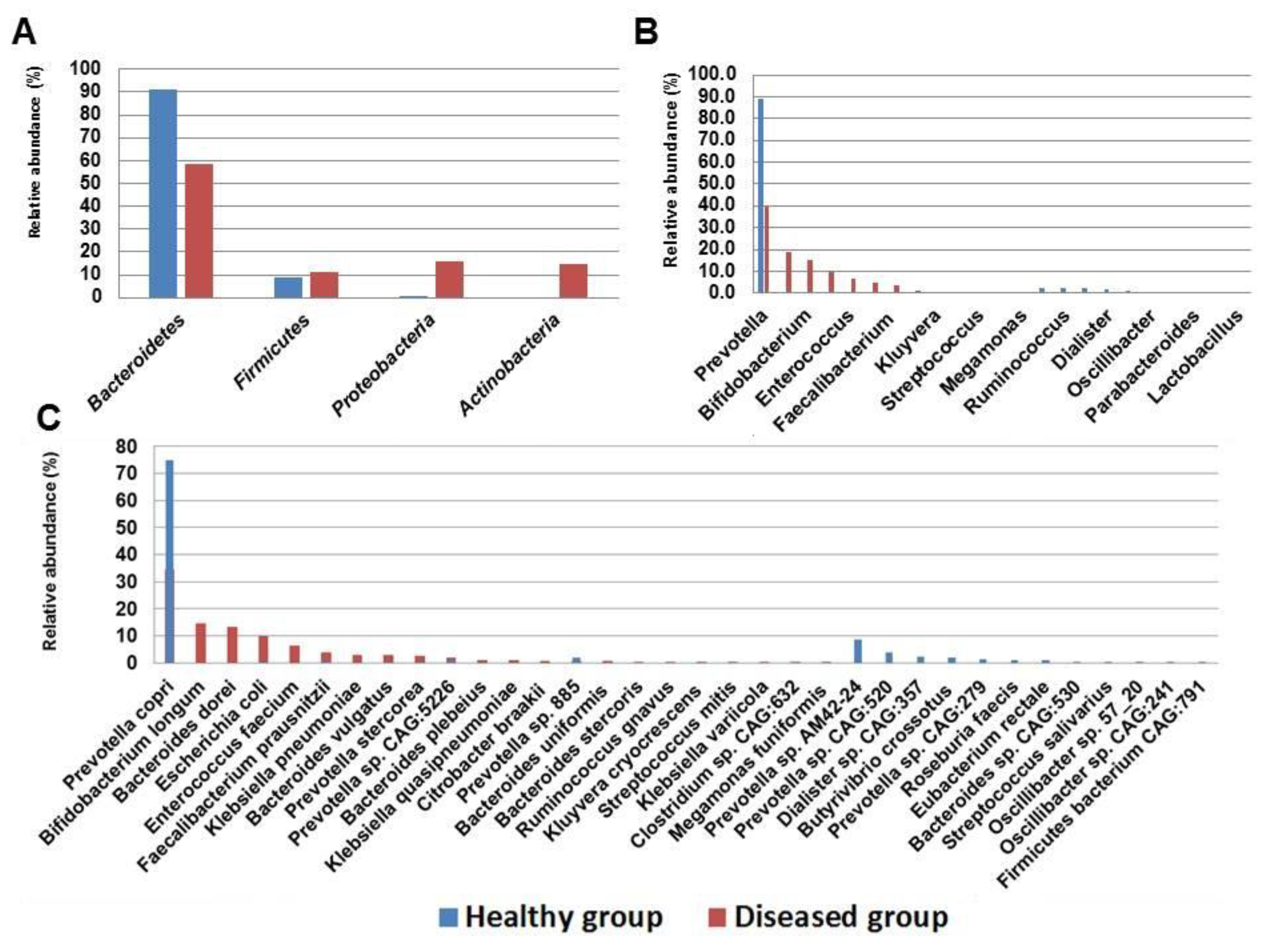
3.5. Comparative Bacterial Profiling of Healthy and Diseased Subjects

Bacteroidetes were identified as a major abundant phylum in both healthy controls (90.8%) and diseased subjects (58.3%). Albeit, Bacteroidetes constitute the major abundant phylum in both healthy and diseased subjects, however, relative percentage abundance of Bacteroidetes was found to be higher in healthy controls (Figure 1, Table 1). Firmicutes (8.9%) constitute as the second abundant phylum in healthy controls while Proteobacteria (15.8%) was the second major phylum in the diseased subjects. Conversely, Proteobacteria was found to be the least (<1%) abundant phylum in the healthy controls. The percentage abundance of microbial communities at different taxonomic ranks such as phylum, class, order, family, genus, and species of both healthy and diseased subjects are shown in Figure 1A–C. Prevotella was identified as the major genus in both healthy controls (90.3%) and diseased subjects (39.6%); however, the relative percentage abundances of both groups vary greatly (Figure 1C, Table 1). Furthermore, the various pathobionts genera such as Escherichia and Klebsiella were abundantly found in the fecal microbiome of diseased subjects, while in the healthy controls these genera constituted for <1%.

3.6. NGS-Based Resistome Analysis

Resistome analysis of healthy and diseased subjects revealed the presence of diverse ARGs (355,959 reads) conferring resistance to multiple antibiotics; namely, tetracycline, beta-lactam, macrolide-lincosamide-streptogramin (MLS), aminoglycoside, sulphonamide, multidrug efflux pump system, rifampin, quinolone, and trimethoprim.

3.7. Resistome of Healthy Subjects

The resistome of healthy controls were composed of 23 ARGs conferring resistance to various classes of antibiotics such as tetracycline, beta-lactam, MLS, sulphonamide, aminoglycoside, and multidrug efflux pumps. The abundance of various ARGs were estimated on the basis of their reads for instance, tetracycline associated ARGs (80%; n = 7, tet32, tet40, tetA) were found to be the major genes followed by beta-lactam (17.3%; n = 5; blacfxA3, blacfxA6, blaCTX), and MLS (2.5%; n = 3, MphA, ermF, ermB). Tetracycline, beta-lactam, and MLS associated ARGs were found as the dominant genes while the relative abundance of ARGs associated with efflux pump (n = 3, mdtA, emrK, mdtL), aminoglycoside (n = 3, aadA5, aph(3′′)-Ib, aacA4), sulphonamide (n = 2, sul1/sul3) were found to be < 1% (Figure 2, Table 2).

3.8. Resistome of Diseased Subjects

The resistome of diseased subjects were composed of 42 ARGs conferring resistance to 9 classes of antibiotics such as tetracycline, beta-lactam, rifampin, MLS, multidrug efflux pumps, quinolone, aminoglycoside, sulphonamide, and trimethoprim (Figure 2, Table 2). Tetracycline associated ARGs were found to be the major genes (50.4%; n = 8, tetM, tetO, tetQ), followed by beta-lactam (21.3%; n = 8, blaCTX-M, blaCMH-1, blaCMY), rifampin (10%; n = 1, rpoB), MLS (5.9%; n = 5, ermB, ermF, ermX), multidrug efflux pump (5.3%, n = 10 mdtG, mdtH, mdtN), quinolone (2.7%; n = 2, qnrB, qnrS), aminoglycoside (2.6%; n = 5, aac6, aadA5, acrE), sulphonamide (1.6%; n = 2 sul1, sul2), and trimethoprim (0.2%; n = 1, DfrA17).

3.9. Comparative Resistome Analysis

A greater number of ARGs were identified in diseased subjects (n = 42) than that of healthy controls (n = 23). Similarly, in contrast to the healthy subjects, the resistome of diseased subjects showed higher diversity in terms of ARGs to be associated with 9 classes of antibiotics namely tetracycline, beta-lactam, macrolide-lincosamide-streptogramin (MLS), aminoglycoside, sulphonamide, multidrug efflux pump system, rifampin, quinolone, and trimethoprim (Figure 2). While resistome of healthy controls showed resistance to only 6 classes of antibiotics namely tetracycline, beta-lactam, MLS, sulphonamide, aminoglycoside, and multidrug efflux pumps (Table 2). Moreover, tetracycline associated ARGs were found abundantly in the gut resistomes of both healthy (80%) and diseased (50.4%) subjects. Similarly, beta-lactam-associated ARGs were found to be the second most abundant ARGs in the gut resistomes of both healthy and diseased subjects with a relative abundance of 17.3% and 21.3%, respectively (Figure 2).

4. Discussion

Shotgun metagenome sequencing has been widely used to explore various ARGs in diverse hosts [14,19,20]. NGS-based metagenomic approach is widely adopted by the developed countries for resistome analysis for surveillance and diagnostic purposes however, being a third-world country with limited resources, Pakistan still lags behind in this race [32,33]. The present study is the first of its kind, which used an NGS-based metagenomic approach to investigate the gut resistome of healthy and diseased human subjects belonging to Peshawar, Pakistan. A number of ARGs were identified in the resistome of both healthy and diseased subjects. The presence of various ARG types in the resistome of healthy subjects is in agreement with a recent metagenomic study [34]. A metagenomic study identified various ARGs in the resistome of Yanomami Amerindian villagers whose ancestors were isolated for more than 11,000 years and have never been administered any synthetic antibiotics [35]. The presence of ARGs in these isolated villagers implies that ARGs are inherent features of the human microbiome. Although ARGs have been implicated as the inherent features of the human microbiome, albeit excessive antibiotic administration upsurges the acquisition, transmission, and dissemination of ARGs in the gut resistomes of different hosts [36]. Moreover, the antibiotic treatment causes the modification of gut resistome thereby leading to the predominance of resistant strains and their associated ARGs [37,38]. In Pakistan, antibiotics are available over the counter and self-medication is commonly practiced in the general population and also in hospital community settings [39]. A recent multicenter cross-sectional study carried out in urban areas of Punjab, Pakistan, evaluated the sale extent of non-prescribed antibiotics. Out of 353 pharmacy stores, 96.9% of medical stores and pharmacy centers were found to be dispensing antibiotics without any prescription where as 3.1% of pharmacy stores were found to be dispensing antibiotics on the basis of authentic prescription [40]. Recently, the total consumption of antibiotics in Pakistan has been increased by 65% from the year 2000 to 2010 [41]. Furthermore, a recent study also highlighted that the inappropriate use of antibiotics in both general and hospital community settings contributes to the deadly AMR situation in Pakistan [42]. We, therefore, speculate that widespread presence of diverse ARGs in the resistomes of healthy subjects is likely due to the unjustified use of antibiotics in Pakistan.
Greater diversity and predominance of ARGs were observed in the resistome of diseased subjects, which could be justified by the fact that they were already on antibiotic treatment for various bacterial infections. The association of antibiotic administration to the emergence of increased ARGs in the diseased subjects is supported by a previous study indicating that intake of antimicrobials causes potential expansion of gut resistome [43]. A number of studies indicated the widespread presence of various AMR bacterial strains and their associated ARGs, for instance, a study carried out in tertiary health care hospital of indicated that all Acinetobacter and greater than 70% P. aeruginosa isolates were found to be MDR [44,45]. Furthermore, our results are supported by a prior study that explored the publicly available datasets of 24 metagenomic samples collected from the healthy controls (n = 6) and antibiotic-treated individuals (n = 18). Their results indicate that antibiotic administration in diseased subjects for a short duration causes the diversification of gut resistome, increases the abundance of ARGs and specific pathogenic bacterial strains [46]. Moreover, using shotgun metagenomics, a group of researchers quantified alterations in the gut microbiota of hematological patients under antibiotic prophylaxis grouped in two different cohorts. Ciprofloxacin was administered in one cohort while cotrimoxazole in the other. Their results indicated reduction in gut microbial diversity of both the treated cohorts up to the similar extent, however their gut resistome differs owing to the use of a particular group of antibiotics [1]. In addition, the higher diversity of ARGs in diseased subjects is in agreement with a recent study indicating that the overall relative abundance of various ARGs was higher significantly in patients than that of healthy controls [34]. We observed a high abundance of tetracycline and beta-lactam associated ARGs in the resistomes of both healthy and diseased subjects. The prevalence of tetracycline associated ARGs in the healthy subjects is consistent with a previous study [47]. Feng et al. [47] established a catalog of resistome to study the abundance pattern of various ARG types and fecal microbial communities from healthy people (n = 180) belonging to 11 different countries. They identified 507 subtypes (from 20 types of ARGs), among which the tetracycline associated ARGs have been grouped in the top seven prevalent ARGs. Their results implies that tetracycline associated ARGs occurs commonly in human resistome [47]. Globally, tetracycline associated ARGs predominantly present in the human gut resistomes as evidenced by few studies indicating the high abundance of tetracycline associated ARGs among all the identified ARG types in the resistomes of Chinese, Danish, American, and Spanish people [20,48].
Data related to metagenomic studies are scarce in Pakistan; however, the drug resistance pattern of Shigella isolates belonging to the Faisalabad region was studied using a molecular approach. Shigella isolates exhibited resistance predominantly to ampicillin (96.84%) and tetracycline (93.68%) which is in line with our results indicating a high abundance of tetracycline and beta-lactam associated ARGs in the diseased subjects [49]. The high diversity of various ARGs conferring resistance to multiple classes of antibiotics (Table 2) in the diseased subjects can be attributed to the self-medication, over-the-counter availability of antibiotics, wrong prescription practices, and lack of legislation regulating antibiotic usage across Pakistan [50]. Furthermore, physicians in Pakistan usually prescribe antibiotics to the patients prior to the result of AST which could be another possible reason for the diversification of their gut resistomes. Prescription of antibiotics without AST has been considered as the potential cause of AMR in the developing countries such as India and Pakistan [51,52]. The widespread inappropriate use of antibiotics in Pakistan has led to the emergence of ARGs not only in human but also in food animals and their surrounding environments such as water bodies. In addition to humans, various ARGs have been detected in various environmental samples of Pakistan. Using real-time PCR, a research group detected the high abundance of sulI and dfrA1 in the river bodies of Northern Pakistan [53].
The increased abundance of Bacteroidetes in the fecal microbiota of healthy controls is supported by the fact that Bacteroidetes are the major representatives of the normal gut [54]. Bacteroidetes were identified as the most abundant phylum in both healthy and diseased subjects; however, their relative abundances varied greatly (Figure 1, Table 1). Moreover, Proteobacteria was identified as the second major phyla in the diseased subjects while in the healthy controls it accounted for <1% which implies that the lower diversity of microbial communities was likely caused by microbial dysbiosis in diseased subjects. The predominance of Proteobacteria in the diseased subjects is in agreement with previous studies indicating its association with different diseases and microbial disruption [55,56,57]. Disruption of gut microbiota with antibiotic usage is a well-established fact [56,57]. Different antibiotics cause gut microbial dysbiosis in mice; for instance, mice treated with aztreonam and metronidazole cause a reduction in the relative abundance of Bacteroidetes while the growth of Klebsiella sp., Helicobacter sp., and Ruminococcus sp. were found to be enhanced [58].
Dietary interventions also significantly alter the composition of gut microbiota [59]. Pakistani diet is mainly omnivorous type; however, during various ailments people usually prefer to consume soft fermented foods in combination with other food items (Table S1, Supplementary Materials). The diseased subjects in our study changed their diet from an omnivorous type to softer and/or fermented items composed of natural homemade yoghurt, boiled rice with lentils, and custard. Compared to the healthy subjects, the high abundance of Actinobacteria in the diseased subjects could be justified by their dietary interventions (especially intake of natural yoghurt) which is in line with the previous studies indicating the prevalence of Bifidobacterium in patients receiving yoghurt [60,61,62].
Overall, a greater microbial diversity was observed in healthy controls than that of diseased subjects which are in agreement with the previous studies [34,47,55,56]. At the genus level, a high abundance of Prevotella was observed in the healthy controls than that of diseased subjects. The predominance of Prevotella in both healthy and diseased subjects can be attributed to the Pakistani diet which is mostly plant-based [63]. However, we speculate that antibiotic administration in diseased subjects reduced the abundance of Prevotella thereby leading to the expansion of pathobiontic genera such as Klebsiella and Escherichia. Moreover, a higher abundance of important pathobiontic species such as E. coli, Klebsiella pneumonia, and Klebsiella quasipneumoniae was observed in diseased subjects while the healthy controls harbored a high proportion of several beneficial bacteria (Table 1). The high abundance of pathobionts in the diseased subjects is in line with a previous study [34]. The various pathogenic species such as E. coli, Klebsiella pneumonia, and Klebsiella quasipneumoniae has been considered as the signatures of gut microbial dysbiosis [57]. The relative abundance of E. coli, Klebsiella spp. are normally low however, these species have the potential to expand and dominates in the intestines during microbial dysbiosis thereby leading to serious health consequences [64].

5. Conclusions and Future Perspective

The present study investigated the fecal microbiome and resistome of healthy and diseased Pakistani individuals. Higher microbial diversity was observed in the fecal microbiome of healthy controls than that of diseased subjects. A high abundance of ARGs conferring resistance to multiple antibiotics was observed in the resistome of diseased subjects than that of healthy individuals. Microbial dysbiosis and predominance of various ARGs in the resistome of diseased subjects reflect the excessive usage of antibiotics in Pakistan and warrants immediate attention to regulate the use of various antimicrobials. Using a high-throughput shotgun metagenome sequencing, the present study is the first of its kind which characterized the fecal microbiome and resistome of both healthy and diseased Pakistani subjects. This study showed that how a healthy gut microbiome and its resistome differs from the diseased one. Furthermore, this study also highlighted how antibiotic misuse perpetuates the structure of gut microbial communities leading to a dysbiotic flora.
Identifying the pathogenic bacterial strains and their associated ARGs can help to design novel precision medicine-based therapeutic strategies for targeted mitigation of resistant microbes. Currently, microbial strategies used for targeted mitigation of resistant microbes are either based on bacteriophages or microbial remediation [65]. Bacteriophages have been considered as the potential weapon against MDR, extensively drug-resistant (XDR), and pan drug-resistant (PDR) in developed counties [66]. A group of researchers used a cocktail of different members of wild phages to successfully treat a wound infection caused by MDR A. baumannii [67]. Similarly, compared to the conventional antibiotic therapy, FMT profoundly improved the treatment outcomes of Clostridioides difficile infection [68]. Currently, in the USA and Canada, FMT has been considered as a biological medicine (biologic drug) [69].
With continued misuse of antibiotics, gut microbial diversity will be reduced leading to the severe absence of useful bacteria thereby causing grave future health issues. Going forward, strict legislation with proper implementation of antibiotic usage is required. Here, we propose the establishment of a biobank of useful microbes ready to be used as a supplement in case it disappeared from the gut microbial ecosystem. In the future, a detailed study with a large sample size is required to create a comprehensive library of all the microbes from different host species for subsequent use and reference backed up by a comprehensive biobank of the identified microbes.

Supplementary Materials

The following are available online at https://www.mdpi.com/2076-2607/9/3/616/s1, Table S1: Details of Pakistani omnivorous diet consumed by healthy participants and soft fermented diet received by diseased subjects, Table S2: Demographic and clinical details of healthy and diseased subjects.

Author Contributions

Conceptualization, J.A. and J.H.C.; formal analysis, O.K.A. and J.A.; methodology, O.K.A., J.A. and J.H.C.; project administration, J.H.C. and J.A.; software, O.K.A.; supervision, J.H.C. and J.A.; writing—original draft, O.K.A., J.A. and J.H.C.; writing—review and editing, O.K.A., J.A. and J.H.C. All authors have read and agreed to the published version of the manuscript.

Funding

The present research work has been funded by the Basic Science Research Program of the National Research Foundation of Korea and the Ministry of Science and ICT (grand No. NRF-2019R1A2C4069796 to Jeong Ho Chang).

Institutional Review Board Statement

This study was conducted according to the guidelines of the Declaration of Helsinki, and approved by the Research Ethics Committee of Rehman Medical Institute, Peshawar Pakistan (Ref: RMI/RMI-REC/Approval/33 dated 14 January 2019).

Informed Consent Statement

Informed consents were obtained from all the subjects involved in the study.

Data Availability Statement

Raw metagenomic data generated in this study has been deposited to the publicly accessible NCBI Sequence Read Archive (SRA) under the accession number: PRJNA612780.

Acknowledgments

All the authors acknowledge Haider SA, Sabiha B, and Jan HU for their technical support. All the authors also acknowledge Saeed Ur Rehman (Professor Microbiology, Pathology Laboratory, HMC, Peshawar) and his team for their generous help in sample selection, collection, and AST related experiments.

Conflicts of Interest

All the authors declare that they have no conflict of interest.

References

  1. Willmann, M.; Vehreschild, M.J.; Biehl, L.M.; Vogel, W.; Dörfel, D.; Hamprecht, A.; Seifert, H.; Autenrieth, I.B.; Peter, S. Distinct impact of antibiotics on the gut microbiome and resistome: A longitudinal multicenter cohort study. BMC Biol. 2019, 17, 1–18. [Google Scholar] [CrossRef] [PubMed]
  2. Sommer, M.O.; Dantas, G.; Church, G.M. Functional characterization of the antibiotic resistance reservoir in the human microflora. Science 2009, 325, 1128–1131. [Google Scholar] [CrossRef] [PubMed] [Green Version]
  3. Schwartz, D.; Langdon, A.; Dantas, G. Understanding the impact of antibiotic perturbation on the human microbiome. Genome Med. 2020, 12, 1–12. [Google Scholar] [CrossRef] [PubMed]
  4. Casals-Pascual, C.; Vergara, A.; Vila, J. Intestinal microbiota and antibiotic resistance: Perspectives and solutions. Hum. Microbiome J. 2018, 9, 11–15. [Google Scholar] [CrossRef]
  5. Duan, H.; Yu, L.; Tian, F.; Zhai, Q.; Fan, L.; Chen, W. Antibiotic-induced gut dysbiosis and barrier disruption and the potential protective strategies. Crit. Rev. Food Sci. Nutr. 2020, 1–26. [Google Scholar] [CrossRef]
  6. Hasan, N.; Yang, H. Factors affecting the composition of the gut microbiota, and its modulation. PeerJ 2019, 7, e7502. [Google Scholar] [CrossRef] [Green Version]
  7. Langdon, A.; Crook, N.; Dantas, G. The effects of antibiotics on the microbiome throughout development and alternative approaches for therapeutic modulation. Genome Med. 2016, 8, 1–16. [Google Scholar] [CrossRef] [Green Version]
  8. Khan, T.J.; Hasan, M.N.; Azhar, E.I.; Yasir, M. Association of gut dysbiosis with intestinal metabolites in response to antibiotic treatment. Hum. Microbiome J. 2019, 11, 100054. [Google Scholar] [CrossRef]
  9. Kho, Z.Y.; Lal, S.K. The human gut microbiome–a potential controller of wellness and disease. Front. Microbiol. 2018, 9, 1835. [Google Scholar] [CrossRef] [PubMed] [Green Version]
  10. Zheng, D.; Liwinski, T.; Elinav, E. Interaction between microbiota and immunity in health and disease. Cell Res. 2020, 30, 492–506. [Google Scholar] [CrossRef]
  11. Dethlefsen, L.; Relman, D.A. Incomplete recovery and individualized responses of the human distal gut microbiota to repeated antibiotic perturbation. Proc. Natl. Acad. Sci. USA 2011, 108, 4554–4561. [Google Scholar] [CrossRef] [Green Version]
  12. Francino, M. Antibiotics and the human gut microbiome: Dysbioses and accumulation of resistances. Front. Microbiol. 2016, 6, 1543. [Google Scholar] [CrossRef] [PubMed] [Green Version]
  13. Shin, N.-R.; Whon, T.W.; Bae, J.-W. Proteobacteria: Microbial signature of dysbiosis in gut microbiota. Trends Biotechnol. 2015, 33, 496–503. [Google Scholar] [CrossRef] [PubMed]
  14. Becattini, S.; Taur, Y.; Pamer, E.G. Antibiotic-induced changes in the intestinal microbiota and disease. Trends Mol. Med. 2016, 22, 458–478. [Google Scholar] [CrossRef] [PubMed] [Green Version]
  15. Hildebrand, F.; Moitinho-Silva, L.; Blasche, S.; Jahn, M.T.; Gossmann, T.I.; Huerta-Cepas, J.; Hercog, R.; Luetge, M.; Bahram, M.; Pryszlak, A. Antibiotics-induced monodominance of a novel gut bacterial order. Gut 2019, 68, 1781–1790. [Google Scholar] [CrossRef] [PubMed]
  16. Baumgartner, M.; Bayer, F.; Pfrunder-Cardozo, K.R.; Buckling, A.; Hall, A.R. Resident microbial communities inhibit growth and antibiotic-resistance evolution of Escherichia coli in human gut microbiome samples. PLoS Biol. 2020, 18, e3000465. [Google Scholar] [CrossRef] [PubMed] [Green Version]
  17. Ekmekciu, I.; Von Klitzing, E.; Fiebiger, U.; Escher, U.; Neumann, C.; Bacher, P.; Scheffold, A.; Kühl, A.A.; Bereswill, S.; Heimesaat, M.M. Immune responses to broad-spectrum antibiotic treatment and fecal microbiota transplantation in mice. Front. Immunol. 2017, 8, 397. [Google Scholar] [CrossRef] [PubMed] [Green Version]
  18. Leclercq, S.; Mian, F.M.; Stanisz, A.M.; Bindels, L.B.; Cambier, E.; Ben-Amram, H.; Koren, O.; Forsythe, P.; Bienenstock, J. Low-dose penicillin in early life induces long-term changes in murine gut microbiota, brain cytokines and behavior. Nat. Commun. 2017, 8, 1–12. [Google Scholar] [CrossRef]
  19. Forslund, K.; Sunagawa, S.; Kultima, J.R.; Mende, D.R.; Arumugam, M.; Typas, A.; Bork, P. Country-specific antibiotic use practices impact the human gut resistome. Genome Res. 2013, 23, 1163–1169. [Google Scholar] [CrossRef] [Green Version]
  20. Hu, Y.; Yang, X.; Qin, J.; Lu, N.; Cheng, G.; Wu, N.; Pan, Y.; Li, J.; Zhu, L.; Wang, X. Metagenome-wide analysis of antibiotic resistance genes in a large cohort of human gut microbiota. Nat. Commun. 2013, 4, 1–7. [Google Scholar] [CrossRef] [Green Version]
  21. Li, Y.; Cao, W.; Liang, S.; Yamasaki, S.; Chen, X.; Shi, L.; Ye, L. Metagenomic characterization of bacterial community and antibiotic resistance genes in representative ready-to-eat food in southern China. Sci. Rep. 2020, 10, 1–14. [Google Scholar] [CrossRef] [PubMed]
  22. Lira, F.; Vaz-Moreira, I.; Tamames, J.; Manaia, C.M.; Martínez, J.L. Metagenomic analysis of an urban resistome before and after wastewater treatment. Sci. Rep. 2020, 10, 1–9. [Google Scholar]
  23. Waseem, H.; Ali, J.; Sarwar, F.; Khan, A.; Rehman, H.S.U.; Choudri, M.; Arif, N.; Subhan, M.; Saleem, A.R.; Jamal, A. Assessment of knowledge and attitude trends towards antimicrobial resistance (AMR) among the community members, pharmacists/pharmacy owners and physicians in district Sialkot, Pakistan. Antimicrob. Resist. Infect. Control 2019, 8, 1–7. [Google Scholar] [CrossRef] [PubMed]
  24. Afridi, O.K.; Ali, J.; Chang, J.H. Next-Generation Sequencing Based Gut Resistome Profiling of Broiler Chickens Infected with Multidrug-Resistant Escherichia coli. Animals 2020, 10, 2350. [Google Scholar] [CrossRef]
  25. Hayat, K.; Rosenthal, M.; Gillani, A.H.; Zhai, P.; Aziz, M.M.; Ji, W.; Chang, J.; Hu, H.; Fang, Y. Perspective of Pakistani physicians towards hospital antimicrobial stewardship programs: A multisite exploratory qualitative study. Int. J. Environ. Res. Public Health 2019, 16, 1565. [Google Scholar] [CrossRef] [PubMed] [Green Version]
  26. Mohsin, M.; Van Boeckel, T.P.; Saleemi, M.K.; Umair, M.; Naseem, M.N.; He, C.; Khan, A.; Laxminarayan, R. Excessive use of medically important antimicrobials in food animals in Pakistan: A five-year surveillance survey. Glob. Health Action 2019, 12, 1697541. [Google Scholar] [CrossRef] [Green Version]
  27. MacFaddin, J. Biochemical Tests for Identification of Medical Bacteria; Williams and Wilkins: Philadelphia, PA, USA, 2000; p. 113. [Google Scholar]
  28. Weinstein, M.P. Performance Standards for Antimicrobial Susceptibility Testing; Clinical and Laboratory Standards Institute: Annapolis Junction, MD, USA, 2019. [Google Scholar]
  29. Mirsepasi, H.; Persson, S.; Struve, C.; Andersen, L.O.; Petersen, A.M.; Krogfelt, K.A. Microbial diversity in fecal samples depends on DNA extraction method: easyMag DNA extraction compared to QIAamp DNA stool mini kit extraction. BMC Res. Notes 2014, 7, 50. [Google Scholar] [CrossRef] [PubMed] [Green Version]
  30. Truong, D.T.; Franzosa, E.A.; Tickle, T.L.; Scholz, M.; Weingart, G.; Pasolli, E.; Tett, A.; Huttenhower, C.; Segata, N. MetaPhlAn2 for enhanced metagenomic taxonomic profiling. Nat. Methods 2015, 12, 902–903. [Google Scholar] [CrossRef]
  31. Hunt, M.; Mather, A.E.; Sánchez-Busó, L.; Page, A.J.; Parkhill, J.; Keane, J.A.; Harris, S.R. ARIBA: Rapid antimicrobial resistance genotyping directly from sequencing reads. Microb. Genom. 2017, 3, e000131. [Google Scholar] [CrossRef]
  32. De, R. Metagenomics: Aid to combat antimicrobial resistance in diarrhea. Gut Pathog. 2019, 11, 1–9. [Google Scholar] [CrossRef] [PubMed]
  33. Duarte, A.S.R.; Stärk, K.D.; Munk, P.; Leekitcharoenphon, P.; Bossers, A.; Luiken, R.; Sarrazin, S.; Lukjancenko, O.; Pamp, S.J.; Bortolaia, V. Addressing learning needs on the use of metagenomics in antimicrobial resistance surveillance. Front. Public Health 2020, 8, 38. [Google Scholar] [CrossRef]
  34. Duan, Y.; Chen, Z.; Tan, L.; Wang, X.; Xue, Y.; Wang, S.; Wang, Q.; Das, R.; Lin, H.; Hou, J. Gut resistomes, microbiota and antibiotic residues in Chinese patients undergoing antibiotic administration and healthy individuals. Sci. Total Environ. 2020, 705, 135674. [Google Scholar] [CrossRef] [PubMed]
  35. Clemente, J.C.; Pehrsson, E.C.; Blaser, M.J.; Sandhu, K.; Gao, Z.; Wang, B.; Magris, M.; Hidalgo, G.; Contreras, M.; Noya-Alarcón, Ó. The microbiome of uncontacted Amerindians. Sci. Adv. 2015, 1, e1500183. [Google Scholar] [CrossRef] [Green Version]
  36. Rampelli, S.; Soverini, M.; D’Amico, F.; Barone, M.; Tavella, T.; Monti, D.; Capri, M.; Astolfi, A.; Brigidi, P.; Biagi, E. Shotgun metagenomics of gut microbiota in humans with up to extreme longevity and the increasing role of xenobiotic degradation. Msystems 2020, 5, e00124-20. [Google Scholar] [CrossRef] [Green Version]
  37. Sonnenburg, E.D.; Sonnenburg, J.L. Starving our microbial self: The deleterious consequences of a diet deficient in microbiota-accessible carbohydrates. Cell Metab. 2014, 20, 779–786. [Google Scholar] [CrossRef] [PubMed] [Green Version]
  38. Maurice, C.F.; Haiser, H.J.; Turnbaugh, P.J. Xenobiotics shape the physiology and gene expression of the active human gut microbiome. Cell 2013, 152, 39–50. [Google Scholar] [CrossRef] [PubMed] [Green Version]
  39. Khan, F.U.; Khan, F.U.; Hayat, K.; Chang, J.; Saeed, A.; Khan, Z.; Ashraf, M.; Rasheed, U.M.; Atif, N.; Ji, W. Knowledge, attitude and practices among consumers toward antibiotics use and antibiotic resistance in Swat, Khyber-Pakhtunkhwa, Pakistan. Expert Rev. Anti-Infect. Ther. 2020, 18, 937–946. [Google Scholar] [CrossRef]
  40. Saleem, Z.; Hassali, M.A.; Godman, B.; Fatima, M.; Ahmad, Z.; Sajid, A.; Rehman, I.U.; Nadeem, M.U.; Javaid, Z.; Malik, M. Sale of WHO AWaRe groups antibiotics without a prescription in Pakistan: A simulated client study. J. Pharm. Policy Pract. 2020, 13, 1–8. [Google Scholar] [CrossRef]
  41. Klein, E.Y.; Van Boeckel, T.P.; Martinez, E.M.; Pant, S.; Gandra, S.; Levin, S.A.; Goossens, H.; Laxminarayan, R. Global increase and geographic convergence in antibiotic consumption between 2000 and 2015. Proc. Natl. Acad. Sci. USA 2018, 115, E3463–E3470. [Google Scholar] [CrossRef] [Green Version]
  42. Hayat, K.; Rosenthal, M.; Gillani, A.H.; Chang, J.; Ji, W.; Yang, C.; Jiang, M.; Zhao, M.; Fang, Y. Perspective of key healthcare professionals on antimicrobial resistance and stewardship programs: A multicenter cross-sectional study from Pakistan. Front. Pharmacol. 2020, 10, 1520. [Google Scholar] [CrossRef] [PubMed]
  43. Buelow, E.; Gonzalez, T.B.; Versluis, D.; Oostdijk, E.A.; Ogilvie, L.A.; van Mourik, M.S.; Oosterink, E.; van Passel, M.W.; Smidt, H.; D’Andrea, M.M. Effects of selective digestive decontamination (SDD) on the gut resistome. J. Antimicrob. Chemother. 2014, 69, 2215–2223. [Google Scholar] [CrossRef]
  44. Khan, M.; Siddiqui, S.; Haider, S.; Zafar, A.; Zafar, F.; Khan, R.; Afshan, K.; Jabeen, A.; Khan, M.; Hasan, R. Infection control education: Impact on ventilator-associated pneumonia rates in a public sector intensive care unit in Pakistan. Trans. R. Soc. Trop. Med. Hyg. 2009, 103, 807–811. [Google Scholar] [CrossRef] [PubMed]
  45. Nahid, F.; Khan, A.A.; Rehman, S.; Zahra, R. Prevalence of metallo-β-lactamase NDM-1-producing multi-drug resistant bacteria at two Pakistani hospitals and implications for public health. J. Infect. Public Health 2013, 6, 487–493. [Google Scholar] [CrossRef] [Green Version]
  46. Li, J.; Rettedal, E.A.; Van Der Helm, E.; Ellabaan, M.; Panagiotou, G.; Sommer, M.O. Antibiotic treatment drives the diversification of the human gut resistome. Genom. Proteom. Bioinform. 2019, 17, 39–51. [Google Scholar] [CrossRef] [PubMed]
  47. Feng, J.; Li, B.; Jiang, X.; Yang, Y.; Wells, G.F.; Zhang, T.; Li, X. Antibiotic resistome in a large-scale healthy human gut microbiota deciphered by metagenomic and network analyses. Environ. Microbiol. 2018, 20, 355–368. [Google Scholar] [CrossRef] [PubMed]
  48. Pal, C.; Bengtsson-Palme, J.; Kristiansson, E.; Larsson, D.J. The structure and diversity of human, animal and environmental resistomes. Microbiome 2016, 4, 1–15. [Google Scholar] [CrossRef] [Green Version]
  49. Tariq, A.; Haque, A.; Ali, A.; Bashir, S.; Habeeb, M.A.; Salman, M.; Sarwar, Y. Molecular profiling of antimicrobial resistance and integron association of multidrug-resistant clinical isolates of Shigella species from Faisalabad, Pakistan. Can. J. Microbiol. 2012, 58, 1047–1054. [Google Scholar] [CrossRef] [PubMed]
  50. Faizullah, M.; Rahman, N.; Umar, M.; Anwar, M.; Sarfraz, M. A cross-sectional study on knowledge, attitude and practices of medical doctors towards antibiotic prescribing patterns and resistance in Khyber Pakhtun Khawah, Pakistan. J. Appl. Pharm. Sci. 2017, 7, 38–46. [Google Scholar]
  51. English, B.K.; Gaur, A.H. The use and abuse of antibiotics and the development of antibiotic resistance. Hot Top. Infect. Immun. Child. 2010, VI, 73–82. [Google Scholar]
  52. Radhakrishnan, R.; Rajesh, J.; Dinesh, N.; Thangavelu, C.; Sankaran, K. High-throughput method for Antibiotic Susceptibility testing based on fluorescein Quenching by Bacteria: Application to Urinary tract infection. Sci. Rep. 2020, 10, 1–8. [Google Scholar] [CrossRef] [PubMed] [Green Version]
  53. Khan, G.A.; Berglund, B.; Khan, K.M.; Lindgren, P.-E.; Fick, J. Occurrence and abundance of antibiotics and resistance genes in rivers, canal and near drug formulation facilities–a study in Pakistan. PLoS ONE 2013, 8, e62712. [Google Scholar] [CrossRef]
  54. Tsigalou, C.; Konstantinidis, T.; Stavropoulou, E.; Bezirtzoglou, E.E.; Tsakris, A. Potential Elimination of Human Gut Resistome by Exploiting the Benefits of Functional Foods. Front. Microbiol. 2020, 11. [Google Scholar] [CrossRef] [PubMed] [Green Version]
  55. Vester-Andersen, M.; Mirsepasi-Lauridsen, H.; Prosberg, M.; Mortensen, C.; Träger, C.; Skovsen, K.; Thorkilgaard, T.; Nøjgaard, C.; Vind, I.; Krogfelt, K.A. Increased abundance of proteobacteria in aggressive Crohn’s disease seven years after diagnosis. Sci. Rep. 2019, 9, 1–10. [Google Scholar] [CrossRef] [PubMed] [Green Version]
  56. Alam, M.T.; Amos, G.C.; Murphy, A.R.; Murch, S.; Wellington, E.M.; Arasaradnam, R.P. Microbial imbalance in inflammatory bowel disease patients at different taxonomic levels. Gut Pathog. 2020, 12, 1–8. [Google Scholar] [CrossRef]
  57. Pilmis, B.; Le Monnier, A.; Zahar, J.-R. Gut microbiota, antibiotic therapy and antimicrobial resistance: A narrative review. Microorganisms 2020, 8, 269. [Google Scholar] [CrossRef] [PubMed] [Green Version]
  58. Zhang, Y.; Limaye, P.B.; Renaud, H.J.; Klaassen, C.D. Effect of various antibiotics on modulation of intestinal microbiota and bile acid profile in mice. Toxicol. Appl. Pharmacol. 2014, 277, 138–145. [Google Scholar] [CrossRef] [Green Version]
  59. Leeming, E.R.; Johnson, A.J.; Spector, T.D.; Le Roy, C.I. Effect of diet on the gut microbiota: Rethinking intervention duration. Nutrients 2019, 11, 2862. [Google Scholar] [CrossRef] [Green Version]
  60. Shadnoush, M.; Hosseini, R.S.; Khalilnezhad, A.; Navai, L.; Goudarzi, H.; Vaezjalali, M. Effects of probiotics on gut microbiota in patients with inflammatory bowel disease: A double-blind, placebo-controlled clinical trial. Korean J. Gastroenterol. 2015, 65, 215–221. [Google Scholar] [CrossRef] [Green Version]
  61. Stojanov, S.; Berlec, A.; Štrukelj, B. The Influence of Probiotics on the Firmicutes/Bacteroidetes Ratio in the Treatment of Obesity and Inflammatory Bowel disease. Microorganisms 2020, 8, 1715. [Google Scholar] [CrossRef]
  62. González, S.; Fernández-Navarro, T.; Arboleya, S.; de Los Reyes-Gavilán, C.; Salazar, N.; Gueimonde, M. Fermented dairy foods: Impact on intestinal microbiota and health-linked biomarkers. Front. Microbiol. 2019, 10, 1046. [Google Scholar] [CrossRef] [PubMed]
  63. Kaur, K.; Khatri, I.; Akhtar, A.; Subramanian, S.; Ramya, T. Metagenomics analysis reveals features unique to Indian distal gut microbiota. PLoS ONE 2020, 15, e0231197. [Google Scholar] [CrossRef] [PubMed] [Green Version]
  64. Kim, S.; Covington, A.; Pamer, E.G. The intestinal microbiota: Antibiotics, colonization resistance, and enteric pathogens. Immunol. Rev. 2017, 279, 90–105. [Google Scholar] [CrossRef]
  65. Wong, W.F.; Santiago, M. Microbial approaches for targeting antibiotic-resistant bacteria. Microb. Biotechnol. 2017, 10, 1047–1053. [Google Scholar] [CrossRef] [Green Version]
  66. Moghadam, M.T.; Amirmozafari, N.; Shariati, A.; Hallajzadeh, M.; Mirkalantari, S.; Khoshbayan, A.; Jazi, F.M. How phages overcome the challenges of drug resistant bacteria in clinical infections. Infect. Drug Resist. 2020, 13, 45. [Google Scholar] [CrossRef] [Green Version]
  67. Regeimbal, J.M.; Jacobs, A.C.; Corey, B.W.; Henry, M.S.; Thompson, M.G.; Pavlicek, R.L.; Quinones, J.; Hannah, R.M.; Ghebremedhin, M.; Crane, N.J. Personalized therapeutic cocktail of wild environmental phages rescues mice from Acinetobacter baumannii wound infections. Antimicrob. Agents Chemother. 2016, 60, 5806–5816. [Google Scholar] [CrossRef] [PubMed] [Green Version]
  68. McCune, V.; Quraishi, M.; Manzoor, S.; Moran, C.; Banavathi, K.; Steed, H.; Massey, D.; Trafford, G.; Iqbal, T.; Hawkey, P. Results from the first English stool bank using faecal microbiota transplant as a medicinal product for the treatment of Clostridioides difficile infection. EClinicalMedicine 2020, 20, 100301. [Google Scholar] [CrossRef] [PubMed]
  69. Terveer, E.M.; Van Beurden, Y.; Goorhuis, A.; Seegers, J.; Bauer, M.; Van Nood, E.; Dijkgraaf, M.; Mulder, C.; Vandenbroucke-Grauls, C.; Verspaget, H. How to: Establish and run a stool bank. Clin. Microbiol. Infect. 2017, 23, 924–930. [Google Scholar] [CrossRef] [PubMed] [Green Version]
Figure 1. Taxonomic profiling of fecal microbiome in healthy and diseased subjects at various levels. Relative abundance of fecal microflora of healthy and diseased subjects at phylum level (A), genus level (B), and species level (C).
Microorganisms 09 00616 g001
Figure 2. The relative abundance of various antibiotic resistance gene (ARG) types identified in the resistomes of healthy and diseased subjects. The relative abundance of each ARG type was estimated on the basis of total number of reads. MLS—macrolide-lincosamide-streptogramin; MDR—multidrug efflux pump.
Microorganisms 09 00616 g002
Table 1. Bacterial profiling fecal microbiota of healthy and diseased subjects were calculated using Metagenomic Phylogenetic Analysis (MetaPhlAn3).
Taxonomic Rank Control Group (%)Diseased Group (%)SD±
PhylumBacteroidetes90.858.323
Firmicutes911.21.6
Proteobacteria0.215.811.1
ActinobacteriaNA14.7NA
FamilyBacteroidaceae0.418.712.9
Prevotellaceae90.339.635.8
Lachnospiraceae5.70.43.7
Ruminococcaceae0.53.82.3
Veillonellaceae1.9NANA
Enterobacteriaceae0.215.811.1
BifidobacteriaceaeNA14.7NA
EnterococcaceaeNA6.3NA
GenusBacteroides0.418.712.9
Prevotella90.339.635.8
Butyrivibrio2.1NANA
Lachnospiraceae unclassified1.1NANA
Roseburia2.2NANA
Faecalibacterium0.53.82.4
Dialister1.8NANA
Escherichia0.19.66.7
Klebsiella0.054.93.5
BifidobacteriumNA14.7NA
EnterococcusNA6.3NA
SpeciesBacteroides vulgatus0.12.91.9
Prevotella copri74.634.528.4
Prevotella sp. 8851.70.90.6
Prevotella sp. AM42 248.2NANA
Prevotella sp. CAG 2791.3NANA
Prevotella sp. CAG 5203.6NANA
Prevotella sp. CAG 52260.71.60.6
Prevotella stercorea0.22.61.8
Butyrivibrio crossotus2.1NANA
Eubacterium rectale1.1NANA
Roseburia faecis1.3NANA
Faecalibacterium prausnitzii0.533.82.4
Dialister sp. CAG 3571.8NANA
E. coli0.19.66.7
Klebsiella pneumoniae0.053.12.1
Bifidobacterium longumNA14.7NA
Bacteroides doreiNA13.3NA
Bacteroides plebeiusNA1.2NA
Enterococcus faeciumNA6.3NA
Klebsiella quasipneumoniaeNA1.5NA
NA—not applicable (absence of particular taxonomic rank or parameter).
Table 2. Diversity of various antibiotic resistance genes (ARGs) in the gut resistome of healthy and diseased subjects.
ARG TypeResistome of Healthy SubjectsResistome of Diseased Subjects
Tetracyclinetet32, tet40, tetA, tetO, tetQ, tetR, tetWtetM, tetO, tetQ, tetS, tetW, tetA, tetB, tetR
Beta-lactamaci1, blacfxA3, blacfxA6, blaCTX, blaTEMblaCTX-M, blaCMH-1, blaCMY, blacfxA3, blacfxA6, blaNDM-1, blaOXY-1, blaTEM-1
MLS 1MphA, ermF, ermBermB, ermF, ermX, mefA, MsrD
Sulphonamidesul1, sul3sul1, sul2
AminoglycosideaadA5, aph(3”)-Ib, aacA4aac6, aadA5, acrE, acrF, aph3
MDR 2mdtA, emrK, mdtLmdtG, mdtH, mdtN, mdtF, mdtC, mdtO, msbA, adeC, emrK, emrR
1 MLS—macrolide-lincosamide-streptogramin, 2 MDR—multidrug efflux pump system.
Publisher’s Note: MDPI stays neutral with regard to jurisdictional claims in published maps and institutional affiliations.

