Article

Salivary Microbiome Diversity in Kuwaiti Adolescents with Varied Body Mass Index—A Pilot Study

by
Hend Alqaderi
1,2,3,†,
Meganathan P. Ramakodi
4,†,
Rasheeba Nizam
2,†,
Sindhu Jacob
2,
Sriraman Devarajan
5,
Muthukrishnan Eaaswarkhanth
2,* and
Fahd Al-Mulla
2,*
1
Department of Oral Health Policy & Epidemiology, Harvard School of Dental Medicine, 188 Longwood Ave, Boston, MA 02115, USA
2
Department of Genetics and Bioinformatics, Dasman Diabetes Institute, Dasman 15462, Kuwait
3
Kuwait School Oral Health Program, Ministry of Health, Salmiya 20005, Kuwait
4
Hyderabad Zonal Centre, CSIR-IICT Campus, CSIR-National Environmental Engineering Research Institute, Hyderabad 500007, India
5
National Dasman Diabetes Biobank, Special Services Facility, Dasman Diabetes Institute, Dasman 15462, Kuwait
*
Authors to whom correspondence should be addressed.
These authors contributed equally to this work.
Microorganisms 2021, 9(6), 1222; https://doi.org/10.3390/microorganisms9061222
Submission received: 29 April 2021 / Revised: 23 May 2021 / Accepted: 25 May 2021 / Published: 4 June 2021

Abstract

:
The potential role of the salivary microbiome in human diseases has increasingly been explored. The salivary microbiome has been characterized in several global populations, except the Arabian Gulf region. Hence, in this pilot study, we profiled the salivary microbiome of Kuwaiti adolescents with varied body mass indexes (BMI). The analyses of core microbiome composition showed Firmicutes, Bacteroidota, Proteobacteria, Patescibacteria, Fusobacteriota, Actinobacteriota, and Campylobacterota as the common phylum found in the Kuwaiti adolescent population. We also illustrated a diverse microbial community among the sampled individuals grouped according to their BMI. Notably, the overweight group was found with a higher number of distinct taxa than other groups. As such, the core microbiome composition was found to be significantly different (p-value < 0.001) across different BMI groups. Overall, this pilot investigation outlined the microbial diversity and suggested that changes in salivary microbiome composition in people with obese or overweight BMI might reflect their susceptibility to oral diseases.

1. Introduction

Obesity is a chronic health condition determined by an individual’s body mass index (BMI). This complex metabolic disorder is characterized by excessive fat accumulation in the body resulting in adverse health effects. Obesity during childhood and adolescence is a significant risk factor predisposing to life-threatening noncommunicable diseases in adulthood. The escalating global childhood and adolescent obesity rate from 4% in 1975 to over 18% in 2016 [1] is highly alarming. This obesity epidemic is a worldwide growing concern, especially in the oil-rich Arabian Gulf countries, as it increases the risk of comorbid health burdens such as type 2 diabetes, cardiovascular diseases, and some cancer types. Notably, the State of Kuwait tops the Middle East region and leads the United States of America in obesity prevalence at 37% and 23% among adults and adolescents, respectively [1].
In general, the cause of obesity has been associated with environmental, behavioral, and genetic influences. Recently, accumulating scientific evidence has uncovered the involvement of the microbial communities inhabiting the human body as one of the essential factors associated with obesity. Several studies have shown the association of gastrointestinal or gut microbiome alterations with obesity in adults and children. The development of obesity has been attributed to gut microbial dysbiosis, which triggers the host’s inflammatory, immune, and metabolic responses [2,3,4]. A decrease in gut microbiome diversity and richness was observed in people with obesity compared to the normal controls [5]. The proportion of increased Firmicutes and decreased Bacteroidetes has been determined as a marker for gut microbial dysbiosis in people with obesity [3].
The oral cavity is a potential point of ingress for the microbes into the human body serving as a passage to the gut, mediated by saliva ingestion [6,7]. Inevitably, next to the gut microbiota, the human salivary oral microbiome consists of a highly diverse microbiota, including bacteria, fungi, and viruses [8]. The salivary microbiota’s differential diversity and the occurrence of certain bacterial species have been correlated to common oral diseases such as dental caries and periodontitis and systemic diseases, including diabetes, obesity, and cancer [9]. Of the limited available studies related to obesity, few have demonstrated the association of dysbiotic salivary microbiome with obesity in adults [10,11] and adolescents [12,13,14]. For example, the presence of Veillonella, Haemophilus, and Prevotella was correlated with BMI in a Brazilian adolescent population [13].
Given that the microbiome differs among diseases and varies differently across populations influenced by genetics, lifestyle, food habits, and environment, population-wise microbiome profiling is imperative. It is expected that a dramatic transition in the food habits and lifestyle post-oil discovery should have impacted the diversity of microbial communities in the Arab people inhabiting the Gulf region, which is reflected through the elevated prevalence of modern metabolic diseases. The increasing number of studies related to the microbiome in metabolic health and disease mandates comprehensive microbiome cataloging. Profiling studies applying next-generation sequencing (NGS) technology have not been explored in the Arabian Gulf populations except for a recent large-scale salivary microbiome profiling of the Qatari population [15]. Considering the importance and need, we conducted this NGS-based pilot study to characterize the easily accessible salivary microbiome in the adolescent Kuwaiti population.

2. Materials and Methods

2.1. Samples

Twenty-three saliva samples of Kuwaiti adolescents used in this study were part of the Kuwait Healthy Lifestyle Study. All information related to sample collection conducted is detailed elsewhere [16]. The study was approved by the Ethical Review Committee of Dasman Diabetes Institute in Kuwait, and the Ministry of Health in Kuwait. Written informed consent from the participants’ parents (or guardians) and consent from the children themselves before study initiation were obtained. The research was conducted following the reporting guidelines (EQUATOR).

2.2. DNA Extraction and 16S rRNA Gene Sequencing

DNA extraction was carried out using The PureLink™ Microbiome DNA Purification Kit (Thermofisher, Waltham, MA, USA) and quantification using a Qubit fluorometer (Thermofisher, Waltham, MA, USA), following the manufacturer’s instructions.
A total of 5 ng/μL of microbial DNA was amplified using gene-specific primers that target the bacterial 16S rRNA V3 and V4 regions. The full-length primers with overhang adapter sequences used for the study were, 16S Amplicon PCR Forward Primer: 5′ TCGTCGGCAGCGTCAGATGTGTATAAGAGACAGCC TACGGGNGGCWGCAG and 16S Amplicon PCR Reverse Primer: 5′ GTCTCGTGGGCTCGGAGATGTGTAT AAGAGACAGGACTACHVGGGTATCT AATCC. The PCR reaction consists of 2.5 μL of microbial DNA and 12.5 μL of 2 × KAPA HiFi HotStart Ready Mix PCR mix and 5 μL of 1 μM forward and reverse primers. The thermal amplification program consists of 3 min at 95 °C followed by 25 cycles of 30 s at 95 °C, 30 s at 55 °C and 30 s at 72 °C, and a final extension of 5 min at 72 °C. The resulting PCR product was confirmed on a Bioanalyzer using DNA 1000 chip and purified using AMPure XP beads following the manufacturer’s recommendation (Agilent Technologies, Santa Clara, CA, USA). Subsequently, amplicons were bound to dual indices and Illumina sequencing adapters using the Nextera XT Index Kit (Illumina Inc., San Diego, CA, USA) following the manufacturer’s recommendations. Purified and normalized libraries were multiplexed up to 23 samples and paired-end sequencing was carried out in MiSeq platform (Illumina Inc., San Diego, CA, USA).

2.3. Bioinformatics Analyses

The amplicon microbiome data were generated for 23 samples with varying BMI. Initially, the samples with less than 300,000 PE reads were removed from the dataset, which was further preprocessed using the Cutadapt tool [17], and the primers and adapters were removed from the PE reads. Subsequently, the data were analyzed in R version 3.6.3. The data analyses were carried out using the following R packages: DADA2 [18], Biostrings (https://bioconductor.org/packages/Biostrings (accessed on 13 September 2019)), phyloseq [19], microbiome (http://microbiome.github.com/microbiome (accessed on 7 July 2020)), vegan, ggplot2, DECIPHER, phangorn, tidyverse, dplyr, ape, DESeq2 [20], ggrare, reshape, and plyr. Primarily, the tool DADA2 was used for the analyses. The software DADA2 infers amplicon sequence variants (ASVs) which is better for inferring microbiome structure than other algorithms [21]. Thus, this study utilized DADA2 for the analyses. On DADA2, the following criteria were used for preprocessing: the truncation length for R1 and R2 reads were set to be 275 and 225, respectively; the maxEE parameter was set to be 3 for both R1 and R2 reads. After preprocessing, the error rates were evaluated, the reads were dereplicated, the R1 and R2 reads were merged, and finally, the non-chimeric sequences were retained. The fasta sequences from the non-chimeric dataset were retrieved, and the taxonomy for each ASV was assigned to IdTaxa Classifier [22]. For taxonomy assignment, the SILVA_SSU database version r138 (https://www.arb-silva.de/ (accessed on 14 July 2020)) was used, and the confidence threshold of 70% was selected for inferring the taxonomy of ASVs. The ASVs were retained only if the ASV had phylum level information. Further, the dataset was cleaned based on the criteria: the ASV should exist in at least two samples, and the frequency of ASV should be at least 0.001%.
The alpha diversity was calculated using the rarefied dataset. The minimum sequencing depth in the dataset was used as the rarefaction depth. The beta diversity was estimated using the unweighted UniFrac distance. The core microbiome was estimated for each BMI group separately. Briefly, the dataset of each BMI group was separated from the master dataset, and the taxa were filtered using the following criteria: the taxa should exist in at least 50% of samples within the group with an abundance of 0.01%. After retrieving the core microbiome taxa for each BMI group, all the groups’ core microbiome data were merged together for further downstream analyses. The differential abundance of core microbiome taxa between different groups was estimated using the DESeq2 package. The permutational ANOVA (PERMANOVA) with 999 permutations was used to evaluate the differences in the composition of the core microbiome structure across different BMI groups.

3. Results

3.1. Sample Characteristics

All the individuals included in this study were adolescents aged 17–18 years during sample collection conducted in 2019. The mean age of the 23 Kuwaiti adolescents was 17 (SD 0.6) years. Of the 23 adolescents, most of them were females (91%) and few were males (9%). Based on the BMI-for-age, we grouped the participants into obese (n = 8), overweight (n = 8), normal (n = 5), underweight (n = 2) following the CDC Growth Charts (www.cdc.gov/obesity/childhood/defining.html (accessed on 16 March 2020)).

3.2. Amplicon Sequencing Data Analyses

The diversity of bacterial taxa in participants’ saliva was studied using the data generated from V3–V4 regions of 16S rRNA. A total number of 8,771,944 PE reads were generated for 23 samples, and the PE reads ranged from 164,134 to 496,543 per sample. While most of the samples yielded, more than 300,000 PE reads, three samples, 11,352, 11363, and 11,421 had less than 300,000 PE reads. As the sample sequencing depth could affect the results [23,24], three samples with lower sequencing depths than other samples were removed from the dataset. The remaining 20 samples were included in the downstream analyses, resulting in a total number of 181, 313–269, 215 non-chimeric sequences per sample. Further, the unique sequences were used for taxonomic analyses.

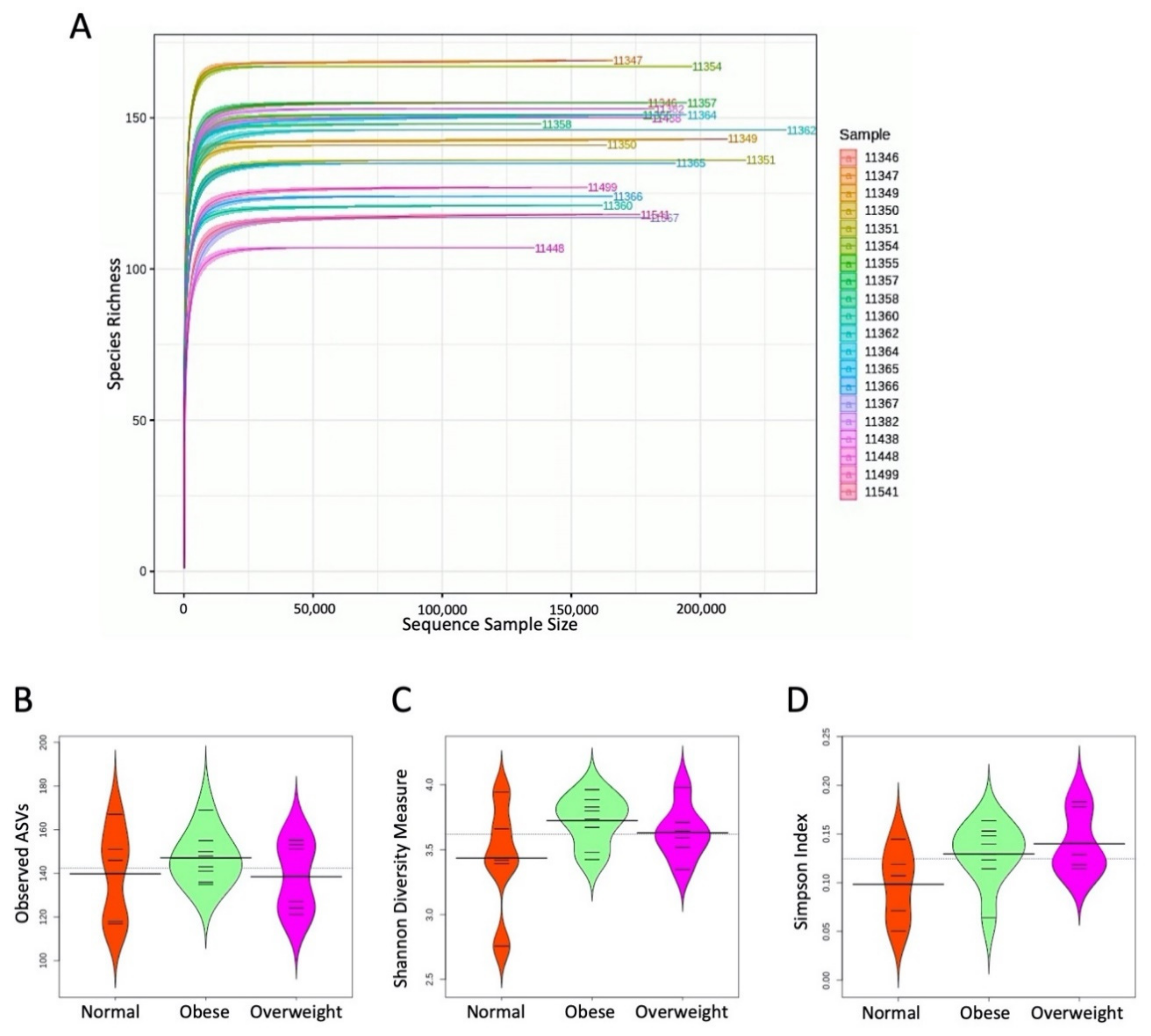
3.3. Alpha Diversity

The dataset was rarefied to estimate the alpha diversity indices. The rarefaction curves for the samples are shown in Figure 1A. The results suggest that the sequencing depth generated per sample is sufficient to infer the microbiome community structure. The distribution of observed ASVs, Shannon diversity and Simpson diversity indices were studied in each group, and the results are shown in Figure 1B–D, respectively. Although the alpha diversity indices were found to vary across different groups, the differences were not significant.

3.4. Beta Diversity

The beta diversity was assessed based on the unweighted UniFrac matrix, which uses phylogenetic information derived from the microbiome composition. The principal coordinate analysis plot and dendrogram based on unweighted UniFrac distance are shown in Figure 2A,B, respectively. The results show that most of the samples were clustered together with other samples of their respective groups. Nonetheless, some samples were found to be clustered with other groups.

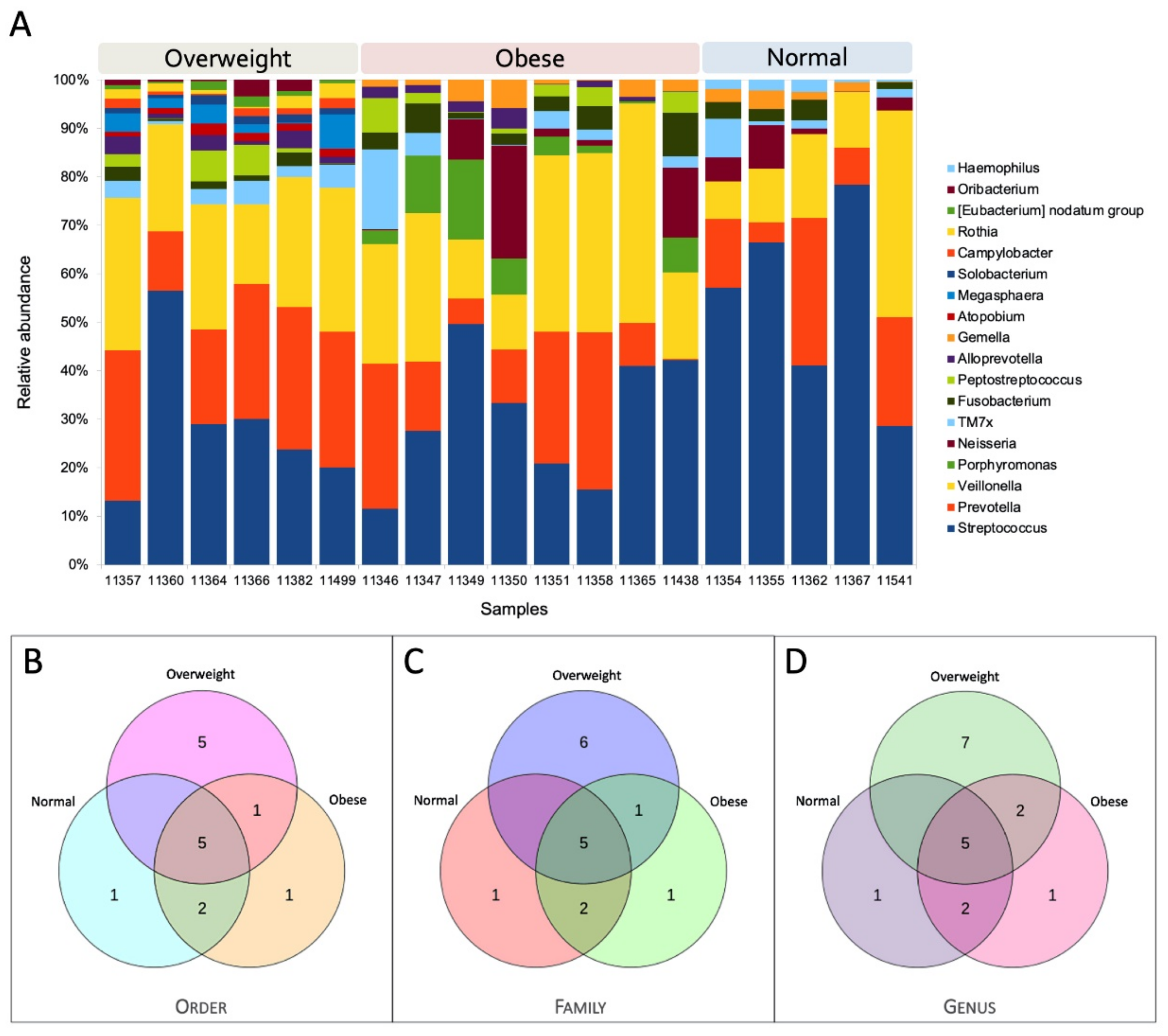
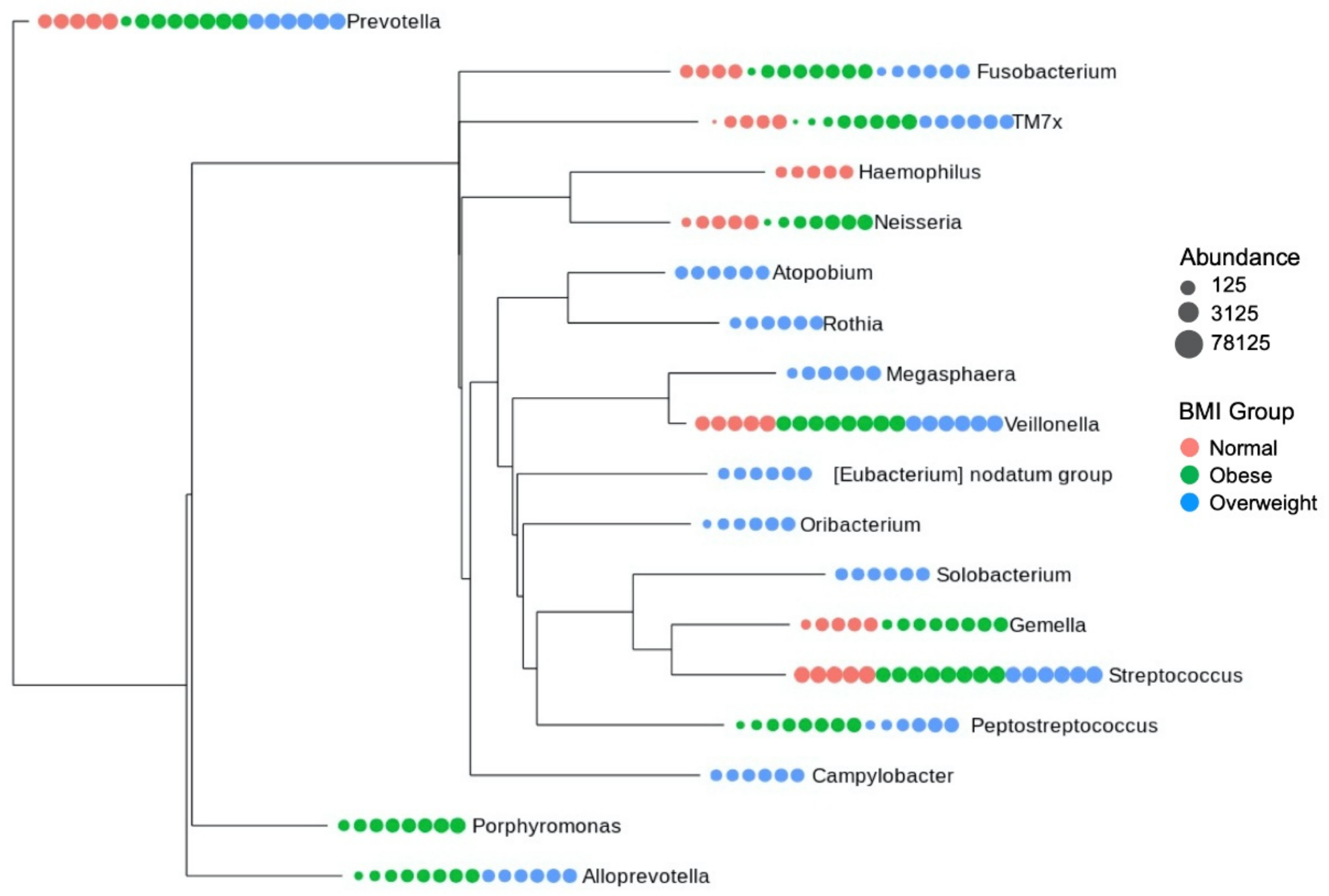
3.5. Core Microbiome Structure

The core microbiome structure of each BMI group was analyzed. The analyses show that the composition of core microbiome taxa varies across different groups. The core microbiome composition for each sample is shown in Figure 3A. The comparison of core microbiome taxa indicates that each group uniquely exhibits some taxa (Figure 3B–D and Figure 4). For instance, Haemophilus exists only in the normal BMI group, whereas the obese group exclusively had Porphyromonas as one of the core microbial taxa. Similarly, Atopobium, Rothia, Megasphaera, Eubacterium nodatum group, Oribacterium, Solobacterium, and Campylobacter were found only in the overweight group.
Further, the differential abundance of core microbial taxa between different groups was evaluated based on the log2 fold changes. The normal vs. overweight analyses presented a higher abundance of Gemella, Haemophillus, and Neisseria in the normal group. In contrast, the higher abundance of Eubacterium notatum group, Alloprevotella, Atopobium, Campylobacter, Megasphaera, Oribacterium, Peptostreptococcus, Prevotella, Rothia, and Solobacterium were noticed in the overweight group (Figure 5A). The normal vs. obese showed significantly increased abundance of Haemophilus and Streptococcus genera in the normal group, while an overabundance of Alloprevotella, Peptostreptococcus, and Porphyromonas was exhibited by the obese group (Figure 5B). Finally, overweight vs. obese revealed a significantly increased abundance of Eubacterium nodatum group, Atopobium, Campylobacter, Megasphaera, Prevotella, Rothia, and Solobacterium in the overweight group and increased abundance of Gemella, Neisseria, and Porphyromonas in the obese group (Figure 5C). The PERMANOVA analyses showed that the composition of core microbiome across different BMI groups is significantly different (p-value < 0.001).

4. Discussion

The interest in exploring salivary microbiota has been escalating due to its potential role in human health and disease [9,25]. The immune system has evolved to manage the microorganisms inside and around the human body for nutrition and against possible damage made by those microorganisms [26,27]. Due to the immune reactions such as phagocytosis and xenophagy [28], the human microbiome becomes the prey of human immunity and directly provides essential nutrients to the human body [29], and microbial dysbiosis (the overgrowth of any microbial species at any site of the human body) is the direct cause of the host’s inflammatory and metabolic disorders [30]. Hence, we conducted this NGS-based pilot study to understand the salivary microbial diversity in Kuwaiti adolescents enrolled in a longitudinal study. This preliminary analysis promisingly illustrated a distinct variation of salivary microbiome composition among adolescents grouped based on BMI, i.e., obese, overweight, and normal.
Though the alpha diversity was different across the three BMI groups (Figure 1B–D), the differences were not significant. It is intriguing to note that the obese and overweight groups had an increased number of observed ASVs compared to the normal group, which contrasts with earlier salivary [10,31] and gut [5,32,33,34] microbiome studies which reported lower alpha diversity in obese individuals. However, the association between alpha diversity and obesity varied in different populations. For example, a gut microbiome study [35] illustrated lower alpha diversity in White obese individuals, whereas the obese individuals with Hispanic and African ancestry had higher alpha diversity. Thus, our observation of higher alpha diversity in obese and overweight groups indicates possible localization in the study population. The beta diversity index clustered most of the normal, overweight, and obese individuals with their respective group but some of the individuals were found to be closely related with samples of other BMI groups, which is not surprising, as earlier microbiome studies also noted unexpected clustering patterns of samples with different BMI groups [35].
The analyses of core microbiome composition showed Firmicutes, Bacteroidota, Proteobacteria, Patescibacteria, Fusobacteriota, Actinobacteriota, and Campylobacterota as the common phyla found in the Kuwaiti adolescent population. A large-scale salivary microbiome profiling of the Qatari population also demonstrated Bacteroidetes, Firmicutes, Actinobacteria, and Proteobacteria as the common taxa [15]. Further analyses of core microbiome taxa showed distinct core microbiome structures in different BMI groups, and the differences were found to be significant. The overweight group was especially found with a higher number of distinct taxa than other groups. Concerning the proportion of microbial populations between groups, obese and normal individuals highly differed in assent with previous studies [10,11,13,31], while the obese and overweight difference was nominal as expected, indicating the plausible progression from overweight to obesity.
In details of the occurrence of a particular bacterial population, Streptococcus was found to be common in all the sampled adolescents with varying percentages (12–78%) (Figure 3A); this may indicate the symptoms of dental caries in the analyzed individuals with a likelihood of association between dental caries and obesity, which is unclear at this stage [36]. However, it is to be noted that Streptococcus is one of the common bacterial communities found in the human oral microbiome [37,38]. Notably, Haemophilus was observed solely in the normal weight Kuwaiti adolescents, which agrees with a previous study that recorded its depletion in obese Chinese individuals [10]. The presence of Campylobacter only in overweight individuals is intriguing, as it has been linked with gastrointestinal health [39] and reported to be highly present in obese adolescents of Sweden [40]. Similarly, Porphyromonas species was noted exclusively in Kuwaiti adolescents with obese BMI, one of the key periodontal pathogens [41]. In this considerable background, it is plausible that a microbial community could potentially indicate or predispose the obesity status in a Kuwaiti individual. For example, Prevotella is one of the core microbiome taxa significantly enriched in individuals with overweight BMI (median relative abundance: normal = 0.142; overweight = 0.278; obese = 0.126) that could be a potential marker to identify individuals at risk of developing obesity as suggested by prior investigations that demonstrated a significant association between BMI and Prevotella [31,35]. However, salivary microbiome profiling at a larger scale is required to confirm the validity of the microbial markers.
Our profiling identified the salivary core bacterial microbiota affiliated to the genus Haemophilus, Oribacterium, Eubacterium nodatum group, Rothia, Campylobacter, Solobacterium, Megasphaera, Atopobium, Gemella, Alloprevotella, Peptostreptococcus, Fusobacterium, TM7x, Neisseria, Porphyromonas, Veillonella, Prevotella, and Streptococcus in Kuwaiti adolescents (Figure 3A). A recent study reported slightly different core salivary bacterial microbiota in Brazilian adolescents, including Streptococcus, Rothia, Neisseria, Haemophilus, Prevotella, Actinomyces, Porphyromonas, Lautropia, Granulicatella, Gemella, Pseudomonas, Oribacterium, and Fusobacterium [13]. Likewise, another study suggested a slightly diverse common core salivary microbiome that consists of eight bacterial genera, including Streptococcus, Veillonella, Gemella, Granulicatella, Neisseria, Prevotella, Rothia, and Fusobacterium, by investigating the oral microbiome of multiethnic adolescent twins and siblings [42]. Collectively, these bacterial populations were found to commonly occur in the human oral microbiome according to the Human Oral Microbiome Database [38,43]. While the previous salivary or gut microbiome studies demonstrated an increased Firmicutes/Bacteroidetes (F/B) ratio in obese individuals [44,45], we noted a decreased F/B ratio in obese and overweight individuals when compared to the normal BMI group. In line with this observation, few prior investigations have also recorded an increased F/B ratio in the normal group compared to obese individuals [46,47]. Magne et al. (2020) reviewed the association between the F/B ratio and obesity and argued the difficulties in accepting the F/B ratio as the hallmark of obesity [48].
In the context of microbial composition specific to different BMI groups, we observed distinct core microbiome structures for each BMI group (Figure 3A). Nonetheless, the core microbiome taxa in obese individuals, observed in earlier studies, are slightly different from this study. Wu et al. (2018) noted the genus Prevotella, Granulicatella, Peptostreptococcus, Solobacterium, Catonella, and Mogibacterium as core salivary microbiome in obese Chinese individuals [10]. Similarly, Liu et al. (2012) recorded the following taxa, Actinomyces, Prevotella, Streptococcus, Fusobacterium, Leptotrichia, Corynebacterium, Veillonella, Rothia, Capnocytophaga, Selenomonas, Treponema, and TM7 as core microbiome [49]. The reason for observing slightly different core microbiome taxa in our study than earlier studies could be due to the population level differences [50]. Earlier studies have shown a strong association between ethnicity and microbiome composition [51,52,53,54,55,56,57]. Those studies were conducted in various Western and Asian populations to understand the association between core microbiome and BMI, while our study subjects were from Kuwait, one of the Arabian Gulf nations. Our recent studies disclosed the distinct genetic diversity of Kuwait people compared with the Western and Asian populations [58,59]. Thus, the slight difference observed in the core microbiome composition in our study compared to earlier studies could be attributed to the population genetics in addition to the lifestyle and environmental variables. However, further detailed studies are warranted to confirm the core microbiome taxa localized in the Kuwait population.

5. Conclusions

This pilot study profiled the salivary microbiome of Kuwaiti adolescents and unraveled its diversity, providing an overview for future studies. Most likely, our observations on microbial diversity may reflect the oral health of the sampled adolescent individuals. This pilot study’s limitation was a small sample size, which could have contributed to the fact that no associations were observed. This study is preliminary and further directed toward the large-scale cataloging of the salivary microbiome in the Kuwait population.

Author Contributions

Conceptualization, M.E. and F.A.-M.; methodology, M.P.R. and M.E.; formal analysis, M.P.R. and M.E.; investigation, H.A., R.N. and S.J.; data curation, H.A. and S.D.; writing—original draft preparation, M.E. and M.P.R.; writing—review and editing, M.E., M.P.R., H.A. and F.A.-M.; project administration, F.A.-M., H.A. and S.D.; funding acquisition, F.A.-M. and H.A.; supervision, F.A.-M. All authors have read and agreed to the published version of the manuscript.

Funding

This research was funded by a research grant to the Dasman Diabetes Institute from the Kuwait Foundation for the Advancement of Sciences, grant number RA-HM-2017-025.

Institutional Review Board Statement

The study was conducted according to the guidelines of the Declaration of Helsinki and approved by the Institutional Ethical Review Committee of Dasman Diabetes Institute in Kuwait (document RA/058/2019 dated 20 February 2019), authorized by the Kuwait Ministry of Health.

Informed Consent Statement

Informed consent was obtained from all the subjects’ parents (or guardians) and assent from the children themselves involved in the study.

Data Availability Statement

The data presented in this study are available upon reasonable request from the corresponding author.

Acknowledgments

We are thankful to all the participants for providing saliva samples to conduct the present study. M.P.R. acknowledges CSIR-NEERI for providing necessary support to carry out the analyses. The manuscript draft is submitted in the Institute repository under the KRC No.: CSIR-NEERI/KRC/2021/APRIL/HZC/2.

Conflicts of Interest

The authors declare no conflict of interest. The funders had no role in the design of the study; in the collection, analyses, or interpretation of data; in the writing of the manuscript, or in the decision to publish the results.

References

  1. World Health Organization Noncommunicable Diseases Country Profiles 2018; World Health Organization: Geneva, Switzerland, 2018.
  2. Cani, P.D.; Amar, J.; Iglesias, M.A.; Poggi, M.; Knauf, C.; Bastelica, D.; Neyrinck, A.M.; Fava, F.; Tuohy, K.M.; Chabo, C.; et al. Metabolic Endotoxemia Initiates Obesity and Insulin Resistance. Diabetes 2007, 56, 1761–1772. [Google Scholar] [CrossRef] [Green Version]
  3. Manco, M.; Putignani, L.; Bottazzo, G.F. Gut Microbiota, Lipopolysaccharides, and Innate Immunity in the Pathogenesis of Obesity and Cardiovascular Risk. Endocr. Rev. 2010, 31, 817–844. [Google Scholar] [CrossRef] [PubMed] [Green Version]
  4. Larsen, J.M. The Immune Response to Prevotella Bacteria in Chronic Inflammatory Disease. Immunology 2017, 151, 363–374. [Google Scholar] [CrossRef] [PubMed] [Green Version]
  5. Le Chatelier, E.; Nielsen, T.; Qin, J.; Prifti, E.; Hildebrand, F.; Falony, G.; Almeida, M.; Arumugam, M.; Batto, J.-M.; Kennedy, S.; et al. Richness of Human Gut Microbiome Correlates with Metabolic Markers. Nature 2013, 500, 541–546. [Google Scholar] [CrossRef] [PubMed]
  6. Olsen, I.; Yamazaki, K. Can Oral Bacteria Affect the Microbiome of the Gut? J. Oral Microbiol. 2019, 11, 1586422. [Google Scholar] [CrossRef] [Green Version]
  7. Schmidt, T.S.; Hayward, M.R.; Coelho, L.P.; Li, S.S.; Costea, P.I.; Voigt, A.Y.; Wirbel, J.; Maistrenko, O.M.; Alves, R.J.; Bergsten, E.; et al. Extensive Transmission of Microbes along the Gastrointestinal Tract. Elife 2019, 8. [Google Scholar] [CrossRef]
  8. Human Microbiome Project Consortium Structure, Function and Diversity of the Healthy Human Microbiome. Nature 2012, 486, 207–214. [CrossRef] [Green Version]
  9. Belstrøm, D. The Salivary Microbiota in Health and Disease. J. Oral Microbiol. 2020, 12, 1723975. [Google Scholar] [CrossRef] [Green Version]
  10. Wu, Y.; Chi, X.; Zhang, Q.; Chen, F.; Deng, X. Characterization of the Salivary Microbiome in People with Obesity. PeerJ 2018, 6, e4458. [Google Scholar] [CrossRef] [Green Version]
  11. Tam, J.; Hoffmann, T.; Fischer, S.; Bornstein, S.; Gräßler, J.; Noack, B. Obesity Alters Composition and Diversity of the Oral Microbiota in Patients with Type 2 Diabetes Mellitus Independently of Glycemic Control. PLoS ONE 2018, 13, e0204724. [Google Scholar] [CrossRef]
  12. Mervish, N.A.; Hu, J.; Hagan, L.A.; Arora, M.; Frau, C.; Choi, J.; Attaie, A.; Ahmed, M.; Teitelbaum, S.L.; Wolff, M.S. Associations of the Oral Microbiota with Obesity and Menarche in Inner City Girls. J Child. Obes 2019, 4. [Google Scholar] [CrossRef]
  13. De Andrade, P.A.M.; Giovani, P.A.; Araujo, D.S.; de Souza, A.J.; Pedroni-Pereira, A.; Kantovitz, K.R.; Andreote, F.D.; Castelo, P.M.; Nociti, F.H., Jr. Shifts in the Bacterial Community of Saliva Give Insights on the Relationship between Obesity and Oral Microbiota in Adolescents. Arch. Microbiol. 2020, 202, 1085–1095. [Google Scholar] [CrossRef]
  14. Araujo, D.S.; Klein, M.I.; Scudine, K.G.d.O.; de Sales Leite, L.; Parisotto, T.M.; Ferreira, C.M.; Fonseca, F.L.A.; Perez, M.M.; Castelo, P.M. Salivary Microbiological and Gingival Health Status Evaluation of Adolescents With Overweight and Obesity: A Cluster Analysis. Front. Pediatr. 2020, 8, 429. [Google Scholar] [CrossRef]
  15. Murugesan, S.; Al Ahmad, S.F.; Singh, P.; Saadaoui, M.; Kumar, M.; Al Khodor, S. Profiling the Salivary Microbiome of the Qatari Population. J. Transl. Med. 2020, 18, 127. [Google Scholar] [CrossRef] [PubMed]
  16. Alqaderi, H.; Al-Ozairi, E.; Bin-Hasan, S.; Tavares, M.; Goodson, J.M.; Alsumait, A.; Abu-Farha, M.; Abubaker, J.; Devarajan, S.; Almuhana, N.; et al. Mediation Effect of C-Reactive Protein in the Relationship between Abdominal Obesity and Intermediate Hyperglycemia in Kuwaiti Adolescents. Biomark. Med. 2020, 14, 1427–1437. [Google Scholar] [CrossRef]
  17. Martin, M. Cutadapt Removes Adapter Sequences from High-Throughput Sequencing Reads. EMBnet.journal 2011, 17, 10–12. [Google Scholar] [CrossRef]
  18. Callahan, B.J.; McMurdie, P.J.; Rosen, M.J.; Han, A.W.; Johnson, A.J.A.; Holmes, S.P. DADA2: High-Resolution Sample Inference from Illumina Amplicon Data. Nat. Methods 2016, 13, 581–583. [Google Scholar] [CrossRef] [PubMed] [Green Version]
  19. McMurdie, P.J.; Holmes, S. Phyloseq: An R Package for Reproducible Interactive Analysis and Graphics of Microbiome Census Data. PLoS ONE 2013, 8, e61217. [Google Scholar] [CrossRef] [Green Version]
  20. Love, M.I.; Huber, W.; Anders, S. Moderated Estimation of Fold Change and Dispersion for RNA-Seq Data with DESeq2. Genome Biol. 2014, 15, 550. [Google Scholar] [CrossRef] [PubMed] [Green Version]
  21. Caruso, V.; Song, X.; Asquith, M.; Karstens, L. Performance of Microbiome Sequence Inference Methods in Environments with Varying Biomass. mSystems 2019, 4. [Google Scholar] [CrossRef] [Green Version]
  22. Murali, A.; Bhargava, A.; Wright, E.S. IDTAXA: A Novel Approach for Accurate Taxonomic Classification of Microbiome Sequences. Microbiome 2018, 6, 140. [Google Scholar] [CrossRef]
  23. Gloor, G.B.; Macklaim, J.M.; Pawlowsky-Glahn, V.; Egozcue, J.J. Microbiome Datasets Are Compositional: And This Is Not Optional. Front. Microbiol. 2017, 8, 2224. [Google Scholar] [CrossRef] [PubMed] [Green Version]
  24. Ramakodi, M.P. Effect of Amplicon Sequencing Depth in Environmental Microbiome Research. Curr. Microbiol. 2021, 78, 1026–1033. [Google Scholar] [CrossRef]
  25. Xiao, J.; Fiscella, K.A.; Gill, S.R. Oral Microbiome: Possible Harbinger for Children’s Health. Int. J. Oral Sci. 2020, 12, 12. [Google Scholar] [CrossRef] [PubMed]
  26. McFall-Ngai, M.; Hadfield, M.G.; Bosch, T.C.G.; Carey, H.V.; Domazet-Lošo, T.; Douglas, A.E.; Dubilier, N.; Eberl, G.; Fukami, T.; Gilbert, S.F.; et al. Animals in a Bacterial World, a New Imperative for the Life Sciences. Proc. Natl. Acad. Sci. USA 2013, 110, 3229–3236. [Google Scholar] [CrossRef] [PubMed] [Green Version]
  27. Broderick, N.A. A Common Origin for Immunity and Digestion. Front. Immunol. 2015, 6, 72. [Google Scholar] [CrossRef]
  28. Klionsky, D.J.; Abdel-Aziz, A.K.; Abdelfatah, S.; Abdellatif, M.; Abdoli, A.; Abel, S.; Abeliovich, H.; Abildgaard, M.H.; Abudu, Y.P.; Acevedo-Arozena, A.; et al. Guidelines for the Use and Interpretation of Assays for Monitoring Autophagy (4th edition) 1. Autophagy 2021, 17, 1–382. [Google Scholar] [PubMed]
  29. Dewi, L.; Rosidi, A.; Noer, E.R.; Ayuningtyas, A. Prospect for Type 2 Diabetes Mellitus in the Combination of Exercise and Synbiotic: A Perspective. Curr. Diabetes Rev. 2021. [Google Scholar] [CrossRef]
  30. Troisi, J.; Venutolo, G.; Pujolassos Tanyà, M.; Delli Carri, M.; Landolfi, A.; Fasano, A. COVID-19 and the Gastrointestinal Tract: Source of Infection or Merely a Target of the Inflammatory Process Following SARS-CoV-2 Infection? World J. Gastroenterol. 2021, 27, 1406–1418. [Google Scholar] [CrossRef]
  31. Raju, S.C.; Lagström, S.; Ellonen, P.; de Vos, W.M.; Eriksson, J.G.; Weiderpass, E.; Rounge, T.B. Gender-Specific Associations Between Saliva Microbiota and Body Size. Front. Microbiol. 2019, 10, 767. [Google Scholar] [CrossRef] [Green Version]
  32. Turnbaugh, P.J.; Hamady, M.; Yatsunenko, T.; Cantarel, B.L.; Duncan, A.; Ley, R.E.; Sogin, M.L.; Jones, W.J.; Roe, B.A.; Affourtit, J.P.; et al. A Core Gut Microbiome in Obese and Lean Twins. Nature 2009, 457, 480–484. [Google Scholar] [CrossRef] [Green Version]
  33. Sze, M.A.; Schloss, P.D. Looking for a Signal in the Noise: Revisiting Obesity and the Microbiome. MBio 2016, 7. [Google Scholar] [CrossRef] [PubMed] [Green Version]
  34. Peters, B.A.; Shapiro, J.A.; Church, T.R.; Miller, G.; Trinh-Shevrin, C.; Yuen, E.; Friedlander, C.; Hayes, R.B.; Ahn, J. A Taxonomic Signature of Obesity in a Large Study of American Adults. Sci. Rep. 2018, 8. [Google Scholar] [CrossRef] [PubMed] [Green Version]
  35. Stanislawski, M.A.; Dabelea, D.; Lange, L.A.; Wagner, B.D.; Lozupone, C.A. Gut Microbiota Phenotypes of Obesity. NPJ Biofilms Microbiomes 2019, 5, 18. [Google Scholar] [CrossRef] [PubMed]
  36. Alshihri, A.A.; Rogers, H.J.; Alqahtani, M.A.; Aldossary, M.S. Association between Dental Caries and Obesity in Children and Young People: A Narrative Review. Int. J. Dent. 2019, 2019, 9105759. [Google Scholar] [CrossRef]
  37. Aas, J.A.; Paster, B.J.; Stokes, L.N.; Olsen, I.; Dewhirst, F.E. Defining the Normal Bacterial Flora of the Oral Cavity. J. Clin. Microbiol. 2005, 43, 5721–5732. [Google Scholar] [CrossRef] [PubMed] [Green Version]
  38. Chen, T.; Dewhirst, F. Human Oral Microbiome Database (HOMD). Encycl. Metagenom. 2015, 271–291. [Google Scholar]
  39. Man, S.M. The Clinical Importance of Emerging Campylobacter Species. Nat. Rev. Gastroenterol. Hepatol. 2011, 8, 669–685. [Google Scholar] [CrossRef]
  40. Zeigler, C.C.; Persson, G.R.; Wondimu, B.; Marcus, C.; Sobko, T.; Modéer, T. Microbiota in the Oral Subgingival Biofilm Is Associated with Obesity in Adolescence. Obesity 2012, 20, 157–164. [Google Scholar] [CrossRef]
  41. Mulhall, H.; Huck, O.; Amar, S. Porphyromonas Gingivalis, a Long-Range Pathogen: Systemic Impact and Therapeutic Implications. Microorganisms 2020, 8. [Google Scholar] [CrossRef]
  42. Stahringer, S.S.; Clemente, J.C.; Corley, R.P.; Hewitt, J.; Knights, D.; Walters, W.A.; Knight, R.; Krauter, K.S. Nurture Trumps Nature in a Longitudinal Survey of Salivary Bacterial Communities in Twins from Early Adolescence to Early Adulthood. Genome Res. 2012, 22, 2146–2152. [Google Scholar] [CrossRef] [PubMed] [Green Version]
  43. Dewhirst, F.E.; Chen, T.; Izard, J.; Paster, B.J.; Tanner, A.C.R.; Yu, W.-H.; Lakshmanan, A.; Wade, W.G. The Human Oral Microbiome. J. Bacteriol. 2010, 192, 5002–5017. [Google Scholar] [CrossRef] [Green Version]
  44. Koliada, A.; Syzenko, G.; Moseiko, V.; Budovska, L.; Puchkov, K.; Perederiy, V.; Gavalko, Y.; Dorofeyev, A.; Romanenko, M.; Tkach, S.; et al. Association between Body Mass Index and Firmicutes/Bacteroidetes Ratio in an Adult Ukrainian Population. BMC Microbiol. 2017, 17, 120. [Google Scholar] [CrossRef] [PubMed] [Green Version]
  45. Sohail, M.U.; Elrayess, M.A.; Al Thani, A.A.; Al-Asmakh, M.; Yassine, H.M. Profiling the Oral Microbiome and Plasma Biochemistry of Obese Hyperglycemic Subjects in Qatar. Microorganisms 2019, 7. [Google Scholar] [CrossRef] [Green Version]
  46. Patil, D.P.; Dhotre, D.P.; Chavan, S.G.; Sultan, A.; Jain, D.S.; Lanjekar, V.B.; Gangawani, J.; Shah, P.S.; Todkar, J.S.; Shah, S.; et al. Molecular Analysis of Gut Microbiota in Obesity among Indian Individuals. J. Biosci. 2012, 37, 647–657. [Google Scholar] [CrossRef]
  47. Tims, S.; Derom, C.; Jonkers, D.M.; Vlietinck, R.; Saris, W.H.; Kleerebezem, M.; de Vos, W.M.; Zoetendal, E.G. Microbiota Conservation and BMI Signatures in Adult Monozygotic Twins. ISME J. 2013, 7, 707–717. [Google Scholar] [CrossRef] [Green Version]
  48. Magne, F.; Gotteland, M.; Gauthier, L.; Zazueta, A.; Pesoa, S.; Navarrete, P.; Balamurugan, R. The Firmicutes/Bacteroidetes Ratio: A Relevant Marker of Gut Dysbiosis in Obese Patients? Nutrients 2020, 12. [Google Scholar] [CrossRef]
  49. Liu, B.; Faller, L.L.; Klitgord, N.; Mazumdar, V.; Ghodsi, M.; Sommer, D.D.; Gibbons, T.R.; Treangen, T.J.; Chang, Y.-C.; Li, S.; et al. Deep Sequencing of the Oral Microbiome Reveals Signatures of Periodontal Disease. PLoS ONE 2012, 7, e37919. [Google Scholar] [CrossRef] [Green Version]
  50. D’Angiolella, G.; Tozzo, P.; Gino, S.; Caenazzo, L. Trick or Treating in Forensics—The Challenge of the Saliva Microbiome: A Narrative Review. Microorganisms 2020, 8, 1501. [Google Scholar] [CrossRef]
  51. Nasidze, I.; Li, J.; Quinque, D.; Tang, K.; Stoneking, M. Global Diversity in the Human Salivary Microbiome. Genome Res. 2009, 19, 636–643. [Google Scholar] [CrossRef] [PubMed] [Green Version]
  52. Li, J.; Quinque, D.; Horz, H.-P.; Li, M.; Rzhetskaya, M.; Raff, J.A.; Hayes, M.G.; Stoneking, M. Comparative Analysis of the Human Saliva Microbiome from Different Climate Zones: Alaska, Germany, and Africa. BMC Microbiol. 2014, 14, 316. [Google Scholar] [CrossRef] [PubMed] [Green Version]
  53. Pylro, V.S.; Roesch, L.F.W.; Ortega, J.M.; do Amaral, A.M.; Tótola, M.R.; Hirsch, P.R.; Rosado, A.S.; Góes-Neto, A.; da Costa da Silva, A.L.; Rosa, C.A.; et al. Brazilian Microbiome Project: Revealing the Unexplored Microbial Diversity—Challenges and Prospects. Microb. Ecol. 2014, 67, 237–241. [Google Scholar] [CrossRef] [PubMed] [Green Version]
  54. Ozga, A.T.; Sankaranarayanan, K.; Tito, R.Y.; Obregon-Tito, A.J.; Foster, M.W.; Tallbull, G.; Spicer, P.; Warinner, C.G.; Lewis, C.M., Jr. Oral Microbiome Diversity among Cheyenne and Arapaho Individuals from Oklahoma. Am. J. Phys. Anthropol. 2016, 161, 321–327. [Google Scholar] [CrossRef] [PubMed]
  55. Gupta, V.K.; Paul, S.; Dutta, C. Geography, Ethnicity or Subsistence-Specific Variations in Human Microbiome Composition and Diversity. Front. Microbiol. 2017, 8, 1162. [Google Scholar] [CrossRef] [PubMed] [Green Version]
  56. Brooks, A.W.; Priya, S.; Blekhman, R.; Bordenstein, S.R. Gut Microbiota Diversity across Ethnicities in the United States. PLoS Biol. 2018, 16, e2006842. [Google Scholar] [CrossRef] [PubMed] [Green Version]
  57. Balakrishnan, B.; Selvaraju, V.; Chen, J.; Ayine, P.; Yang, L.; Ramesh Babu, J.; Geetha, T.; Taneja, V. Ethnic Variability Associating Gut and Oral Microbiome with Obesity in Children. Gut Microbes 2021, 13, 1–15. [Google Scholar] [CrossRef]
  58. Eaaswarkhanth, M.; Pathak, A.K.; Ongaro, L.; Montinaro, F.; Hebbar, P.; Alsmadi, O.; Metspalu, M.; Al-Mulla, F.; Thanaraj, T.A. Unraveling a Fine-Scale High Genetic Heterogeneity and Recent Continental Connections of an Arabian Peninsula Population. Eur. J. Hum. Genet. 2021. [Google Scholar] [CrossRef]
  59. Eaaswarkhanth, M.; Dos Santos, A.L.C.; Gokcumen, O.; Al-Mulla, F.; Thanaraj, T.A. Genome-Wide Selection Scan in an Arabian Peninsula Population Identifies a TNKS Haplotype Linked to Metabolic Traits and Hypertension. Genome Biol. Evol. 2020, 12, 77–87. [Google Scholar] [CrossRef]
Figure 1. Alpha diversity. (A) Rarefaction curve plot showing the sufficient sequencing depth of the samples qualified for diversity analyses; (B) distribution of the observed amplicon sequence variants (ASVs); (C) Shannon diversity measure; (D) Simpson index in each group.
Microorganisms 09 01222 g001
Figure 2. Beta diversity based on unweighted UniFrac measurements. (A) Principal coordinate analysis scatter plot showing the distribution of samples based on the microbiome composition; (B) dendrogram showing the clustering pattern of samples with varying BMI.
Microorganisms 09 01222 g002
Figure 3. Core microbiome structure. (A) Bar plot showing the relative abundance of the core microbiome in each individual; (BD) Venn diagrams showing the number of overlapping core microbial taxa based on the microbial taxonomic hierarchy—(B) order; (C) family; (D) genus.
Microorganisms 09 01222 g003
Figure 4. Dendrogram showing the distribution and phylogenetic relationship of the identified core microbial taxa.
Microorganisms 09 01222 g004
Figure 5. The scatter plots illustrating the differential abundance of core microbial taxa between different BMI groups. (A) Normal vs. overweight; (B) normal vs. obese; (C) overweight vs. obese.
Microorganisms 09 01222 g005
Publisher’s Note: MDPI stays neutral with regard to jurisdictional claims in published maps and institutional affiliations.

