Article

Comparison between the Effects of Adding Vitamins, Trace Elements, and Nanoparticles to SHOTOR Extender on the Cryopreservation of Dromedary Camel Epididymal Spermatozoa

by
Mohamed A. Shahin
1,2,
Wael A. Khalil
1,*,
Islam M. Saadeldin
3,4,*,
Ayman Abdel-Aziz Swelum
3,5 and
Mostafa A. El-Harairy
1
1
Department of Animal Production, Faculty of Agriculture, Mansoura University, Mansoura 35516, Egypt
2
Electron Microscope Unit, Mansoura University, Mansoura 35516, Egypt
3
Department of Animal Production, College of Food and Agricultural Sciences, King Saud University, Riyadh 11451, Saudi Arabia
4
Department of Physiology, Faculty of Veterinary Medicine, Zagazig University, Zagazig 44519, Egypt
5
Department of Theriogenology, Faculty of Veterinary Medicine, Zagazig University, Zagazig 44519, Egypt
*
Authors to whom correspondence should be addressed.
Animals 2020, 10(1), 78; https://doi.org/10.3390/ani10010078
Submission received: 14 November 2019 / Revised: 26 December 2019 / Accepted: 31 December 2019 / Published: 2 January 2020
(This article belongs to the Special Issue Advanced Technology in Animals Reproduction)

Abstract

:

Simple Summary

This is a comprehensive study to compare between the effects of different supplements (vitamins C and E, trace elements Na2SeO3 and ZnSO4, and nanoparticles of zinc oxide and selenium) to the semen extender of camel epididymal spermatozoa during cooling and freezing/thawing cryopreservation. Supplementation of the semen SHOTOR extender with zinc oxide and selenium nanoparticles lead to improved progressive motility, vitality, and anti-oxidative defense, and reduced the ultrastructural abnormalities in camel epididymal spermatozoa.

Abstract

There are several obstacles in camel semen cryopreservation; such as increasing semen viscosity and the reduction in motile spermatozoa after ejaculation. Epididymal spermatozoa offer an efficient alternative to overcome these problems and are well-suited for artificial insemination in camels. In the current study, we compared the effects of supplementation with vitamin C, E, inorganic trace elements of selenium (Na2SeO3) and zinc (ZnSO4), and zinc and selenium nanoparticles (ZnONPs and SeNPs, respectively) on the cryopreservation of dromedary camel epididymal spermatozoa. When the SHOTOR extender was supplemented with ZnONPs and SeNPs; the sperm showed increased progressive motility; vitality; and membrane integrity after cooling at 5 °C for 2 h; when compared to the control and vitamin-supplemented groups. Moreover, the ZnONPs and SeNPs supplementation improved the progressive motility, vitality, sperm membrane integrity, ultrastructural morphology, and decreased apoptosis when frozen and thawed. SeNPs significantly increased reduced glutathione (GSH), superoxide dismutase (SOD), and decreased lipid peroxide malondialdehyde (MDA) levels. The advantageous effects of the trace elements were potentiated by reduction into a nano-sized particle, which could increase bioavailability and reduce the undesired liberation of toxic concentrations. We recommend the inclusion of SeNPs or ZnONPs to SHOTOR extenders to improve the cryotolerance of camel epididymal spermatozoa.

1. Introduction

The camel (Camelus dromedarius) is one of the oldest known mammals to have adapted to desert climates and is used for milk and meat production [1], as well as a sports animal [2]. Compared to other farm animals, studies on camel reproductive physiology are lacking because of the many complex genetic and environmental factors that contribute to the declined fertility in camels. In recent years, the number of studies on the camelid family, in terms of science and research, has greatly increased [3]. There are several ways to improve the productivity and reproductive performance of the Arabian camel [4,5,6,7,8], such as artificial insemination (AI). AI is a useful tool for genetic livestock improvement but has not yet been optimized in camels, owing to the improper protocols for camel semen cryopreservation [9,10]. The obstacles surrounding this protocol include (1) difficulties in collecting semen from aggressive males in rut, (2) the absence of suitable extenders for its storage, (3) low sperm concentration, (4) low sperm motility, and (5) the viscous nature of semen [1,11,12].
Epididymal spermatozoa, on the other hand, could be used as an alternative method for camel AI [11]. Caudal epididymal sperm collection is an important technique in the generation and conservation of animal specimens and is particularly useful when the animal is seriously injured or when collecting from dead specimens [13].
The cryopreservation of camel epididymal spermatozoa involves a suitable cryo-diluent to protect the sperm from cryo-damage [14]. To date, no studies have systematically examined the ability of the epididymis to protect sperm from oxidative stress (e.g., by epidermal fluids containing antioxidants) [11].
The mammalian sperm cell contains a high ratio of polyunsaturated fatty acids, and is therefore highly susceptible to peroxidative damage and oxidative stress (OS), particularly after cryopreservation. The cryopreservation process may result in a loss of membrane integrity, DNA fragmentation, or impaired cell function, all of which may cause decreased sperm movement and reduced fertilization capacity [15,16,17]. OS is a major factor affecting male fertility and results from an imbalance between the production of antioxidant defense mechanisms and reactive oxygen species (ROS) in the cell.
In recent years, studies have also been conducted on camels, cow bulls, and human semen diluents, to supplement anti-oxidants such as glutathione, vitamin C (Vit C), vitamin E (Vit E), zinc (Zn), and selenium (Se) to improve the post-thawed motility, vitality, and membrane integrity of spermatozoa [18,19,20].
Vit C is a highly effective antioxidant and a free radical scavenger in many metabolic processes [21,22]. Vit C increases the percentage of live, acrosome-intact sperm, and decreases the percentage of abnormal sperm during storage at 5 °C. Vit C prevents membrane lipid oxidation during preparation and thus has protective effects. Chinoy et al. [23] reported that the testes and seminal plasma are extremely sensitive to decreased Vit C levels in the body.
Vit E, a lipophilic molecule in the cell membrane, is considered to be a membrane-stabilizer and a strong antioxidant agent that protects the cell membrane from lipid peroxidation and ROS attacks [24]. Vitamin E is the most widely known antioxidant and provides protective effects by reducing or preventing peroxidative damage [25]. Previous research has shown that Vit E can improve the post-thaw sperm quality in bulls [26], rabbits [27], roosters [28], and sheep [29].
Zn has antioxidant properties and reduces ROS released by defective spermatozoa and leucocytes, inhibits lipid peroxidation and reduces circulating anti-sperm antibodies [30,31]. The addition of 100 μM zinc sulfate to semen extender showed a significant increase in the percentage of intact DNA sperm, mitochondrial function, and progressive motility compared to cryopreserved semen samples without zinc supplementation [19]. Dorostkar et al. [32] reported that the addition of 0.288 mg/L zinc sulfate to the extender provides better sperm preservation during freezing processes compared to the control group, which in turn can lead to higher fertility in semen. Ghallab et al. [33] reported that adding 200 μM Zn to semen diluent may improve the quality of frozen/thawed and chilled Arab stallion spermatozoa parameter.
The antioxidant function of Se is mediated through glutathione peroxidase enzyme activity that is known as an important antioxidant and a marker of oxidative stress and protects germ cells, proteins and organic membranes from OS [34]. Extenders containing 1 and 2 μg/mL of sodium selenite significantly improved buffalo frozen/thawed sperm motility, viability, membrane integrity, and total antioxidant capacity and reduced sperm DNA damage [32]. Marai et al. [35] reported that 0.1 ppm sodium selenite improved semen performance in rams by increasing semen volume per ejaculate, sperm motility, and concentration, and by decreasing the percentage of dead sperm, and sperm and acrosome damage abnormalities.
Nanoparticles (NPs), defined by having at least one dimension within the range of 1–100 nm, have become increasingly common in a variety of medical areas [20]. However, there are concerns regarding their biological impact. Recently, nano-elements have drawn great interest due to their low toxicity and high bioavailability. This is because the nanometer particulates exhibit novel characteristics, such as a specific surface area, numerous active surface centers, high surface activity, high catalytic efficiency, and strong adsorption ability [36,37].
Zinc oxide nanoparticles (ZnONPs) increases antioxidant enzymes and improves the quality of sperm [38], and is therefore considered an essential element for spermatogenesis. Zinc (Zn) deficiency in rats triggers a decline in ribonucleic acid (RNA), deoxyribonucleic acid (DNA), and protein levels, and is accompanied by increased ribonuclease activity. Zn functions as a cofactor for DNA and RNA polymerase operations and RNA-dependent DNA polymerase and is therefore important for cell growth [39,40]. Zinc is involved in sperm motility, through adenosine triphosphate processes and phospholipid control, and is highly concentrated in the mature sperm tail [39]. Zinc plays an important role in sperm vitality and motility, affecting protein metabolism, nucleic acid synthesis, and the stabilization of the sperm membrane [41]. Zn can also neutralize the effects of ROS, thereby increasing the efficiency of ATP pathways [42]. Zn has antioxidant properties (it reduces the ROS released by defective spermatozoa and leucocytes), inhibits lipid peroxidation (by phospholipase inhibition), and reduces the amount of circulating anti-sperm antibodies [43,44].
Selenium (Se) is also an important trace element that is fundamental to human health. It plays a role in cell development, apoptosis, and cell signaling mechanisms, and several studies have demonstrated its protective antioxidant characteristics [45]. Se is a powerful antioxidant that alters the expression of selenoproteins-it is incorporated by replacing sulfur in proteins with seleno-amino acids (L-selenomethionine, L-selenocysteine) and selenoenzymes, such as GPxs. Glutathione peroxidases 4 (GPXs4) is a critical component affecting sperm quality and male fertility. Therefore, spermatozoa may be vulnerable to oxidative stress if the Se content of the selenoproteins is low [46]. Several studies have used nano-selenium (SeNPs) as a ROS scavenger to safeguard against oxidative damage in sperm cells. Adding SeNPs to the semen extender enhanced the post-thawing quality and oxidizing rooster semen variables [28,47]. The oral supplementation of SeNPs also protected the spermatozoa quality (motility, DNA integrity) and spermatogenesis against oxidative damage caused by cisplatin, a male reproductive toxicant [48].
Although prior studies have compared various camel semen extenders and measured sperm motility, morphology, and vitality [49,50,51], few have used electron microscopy to examine frozen-thawed camel spermatozoa [52,53,54]. Therefore, the current study aimed to evaluate the freezability and molecular functional integrity of dromedary camel epididymal spermatozoa after dilution by extender supplemented with vit C, vit E, inorganic trace elements (Na2SeO3 and ZnSO4), and nano-sized zinc or selenium.

2. Materials and Methods

2.1. Characteristics of Nano-Sized Elements (Particle Size, Zeta Potential, and Ultramorphology)

SeNPs were purchased from Nanocs (cat. no. Se50-01-5, Nanocs, New York, NY, USA) and Zinc oxide nanoparticles (ZnONPs) were purchased from Sigma-Aldrich (cat. no. 544906, Sigma-Aldrich, Taufkirchen, Germany). Photon correlation spectroscopy Malvern zetasizer Nano-Zs90 was used to measure the particle size and polydispersity index (PDI) of the Zn and Se NPs. The samples were suitably diluted and sonicated to uniformly distribute the particles. All measurements were done in triplicate, and the results were calculated as the mean ± standard deviation (SD). The zeta potential of the Zn and Se NPs was measured by photon correlation spectroscopy at 25 °C. To determine the NPs’ surface charge, the samples were properly diluted with double deionized water, placed in an electrophoretic cell, and measured. The morphological evaluation of Zn and Se NPs was observed by transmission electron microscopy (TEM) (JEOL-JEM-2100) at 160 kV (EM-Unit at Mansoura University, Mansoura, Egypt). For this procedure, one milliliter of the NP sample dispersion was properly diluted with double deionized water and sonicated for 2 min using an ultrasonic bath. After dilution, one drop of the NPs was added to a carbon-coated copper grid, and the excess material was removed, leaving a thin film stretched over the holes. This was allowed to dry at room temperature for 10 min before the image was captured and analyzed in the Gatan software (Version 2.11. 1404.0, Pleasanton, CA, USA).
The average diameter of the ZnONPs and SeNPs was 30.92 ± 1.25 nm and 78.47 ± 17.93 nm, respectively, with low PDI values, suggesting a narrow size distribution (Supplementary Material Table S1). The ZnONPs had a positive zeta potential (32.16 ± 0.252 mV), while the SeNPs had a negative zeta potential (−20.36 ± 1.79 mV). The representative size and zeta potential curves of the ZnONPs and SeNPs are depicted in Supplementary Material Figures S1 and S2, respectively. Moreover, the TEM images exhibited a spherical nanoscopic size for the ZnONPs and SeNPs, in agreement with the Zetasizer measurements (Figure 1).

2.2. Epididymal Camel Spermatozoa Collection

One hundred and two testes from healthy, mature dromedary camels, 6 to 12 years old, were collected from a local abattoir during the breeding season (January–May 2019). There was no member of the research team involved in pre-slaughter live animal handling or in the process of slaughtering, hence ethical approval is not required. The testes were immediately placed in a plastic bag containing normal saline and kept in an air-tight sterile cryo-box at 5 °C. All samples were processed within 6 h after collection. In the laboratory, the testes were washed with sterile warm saline, and the epididymis was isolated. The epididymis was washed three times with warm saline and once with 70% ethyl alcohol. Various incisions in the corpus and tail of epididymis were performed with a scalpel, and the spermatozoa were released by manually pressing the dissected epididymis. The sperm was collected by rinsing 3–4 times with a sterile disposable syringe containing 5 mL warm extender. The recovered spermatozoa were placed in 50 mL tubes.

2.3. Experimental Design

The Tris-based extender, SHOTOR® diluent [51], comprised of 2.6 g Tris, 1.35 g citric acid, 1.2 g glucose, 0.9 g fructose, 1000 IU/mL penicillin, and 1000 µg/mL streptomycin dissolved in 100 mL of deionized water. The osmolality was 330 mOsm/kg, and the pH was 6.9. The extender was composed of 74 mL buffer + 6 mL glycerol + 20 mL egg yolk. The following supplements were added to the SHOTER extender to examine the effects on spermatozoa cryopreservation: Vit E (α tocopherol, T3251) at 200 μM/mL [55], Vit C (ascorbic acid, A4544) at 1 mg/mL [18], SeNPs at 1 μg/mL [47], ZnONPs at 50 μg/mL [20], Na2SeO3 (Loba Chemie Pvt. Ltd., Mumbai, India) at 2 μg/mL [32], and ZnSO4 (Loba Chemie) at 100 μM/mL [19].
Fluid rich spermatozoa collected from the cauda and corpus epididymides were initially evaluated for progressive motility and sperm cell concentration. Fluid rich spermatozoa having ≥ 60% motility was diluted with different extenders (final concentration: 80 × 106 sperm/mL), and the sperm characteristics were evaluated, including progressive motility, vitality, abnormal morphology, and the hypo-osmotic swollen test. The diluted epididymal spermatozoa were cooled to 5 °C for a period of 2 h (for equilibration) before being loaded into 0.25 mL straws. The straw was placed 4 cm above liquid nitrogen vapor for 10 min and then immersed in liquid nitrogen. The straws remained in liquid nitrogen until thawing at 37 °C in a water bath for 30 sec.

2.4. Epididymal Spermatozoa Evaluation

2.4.1. Sperm Progressive Motility

Equilibrated and post-thawed epididymal spermatozoa were evaluated for progressive motility rate under a phase-contrast microscope (Leica DM 500) supplied with a hot stage adjusted to 37 °C [56].

2.4.2. Sperm Vitality and Abnormalities

A smear of the diluted semen samples was placed on a glass slide and stained with a dual staining procedure; eosin (5%) and nigrosin (10%) [57]. A total of 200 spermatozoa from each sample were examined with a light microscope at 400× magnification (Leica DM 500, Leica Mikrosysteme Vertrieb GmbH, Wetzlar, Germany). For each sample, the number of dead spermatozoa (red-stained) was counted, and the morphological abnormalities of the spermatozoa were determined, i.e., spermatozoa bearing head, tail, and cytoplasmic droplets abnormalities [58].

2.4.3. Sperm Plasma Membrane Integrity

The hypo-osmotic swelling test (HOST) was used to evaluate the functional plasma membrane of spermatozoa [59]. Briefly, 10 µL of semen was incubated with 100 µL hypo-osmotic solution (6.75 g/L fructose and 3.67 g/L sodium citrate, for an osmolality of 75 mOsmol/L) at 37 °C for 30 min. Afterward, 10 µL of the mixture was placed on a microscope slide and mounted with a coverslip. A total of 300 spermatozoa (from each sample) were evaluated for swollen and coiled tails under phase-contrast microscopy (Leica DM 500, Leica Mikrosysteme Vertrieb GmbH) at 400× magnification.

2.4.4. Antioxidants Assay

The post-thawed epididymal spermatozoa samples were centrifuged for 15 min at 1500 rpm, and the extender was separated and stored at −20 °C. The concentrations of reduced glutathione (GSH) [60,61], malondialdehyde (MDA), and superoxide dismutase (SOD) [62] were analyzed by commercial kits (Biodiagnostic, Cairo, Egypt) using a spectrophotometer (Spectro UV-VIS Auto, UV-2602, Labomed, Los Angeles, CA, USA).

2.4.5. Assessment of Sperm Apoptosis and Necrosis through Flowcytometric Analysis

The epididymal spermatozoa samples were processed for annexin V staining as described in Chaveiro et al. [63], but with some modifications. Briefly, 1 mL of sperm suspension was added to a 5 mL tube and suspended in a 2 mL binding buffer and thoroughly mixed. 100 µL of the sperm suspension was mixed with 5 µL of annexin V (FITC label) and then 5 µL propidium iodide (PI, PE label), and incubated for at least 15 min, in darkness at room temperature. After incubation, the sperm was suspended in 200 µL binding buffer. The flowcytometric analysis was performed on an Accuri C6 Cytometer (BD Biosciences, San Jose, CA, USA) using the Accuri C6 software (Becton Dickinson) for data acquisition and analysis [64]. The percentages of annexin V negative or positive (A− or A+), PI negative or positive (PI− or PI+), and double-positive cells were evaluated. As described by Peña et al. [65], four different categories of spermatozoa were determined: (1) viable (A−/PI−), with no fluorescence signal, and recorded as live without membrane dysfunction (live sperm); (2) early apoptotic (A+/PI−), but viable spermatozoa, labeled with annexin V but not with PI (live sperm); (3) late apoptotic spermatozoa (A+/PI+) labeled with annexin V and PI and with damaged permeable membranes (dead sperm); (4) necrotic spermatozoa (A−/PI+), labeled by PI but not annexin V, that have completely lost the sperm membrane (dead sperm).

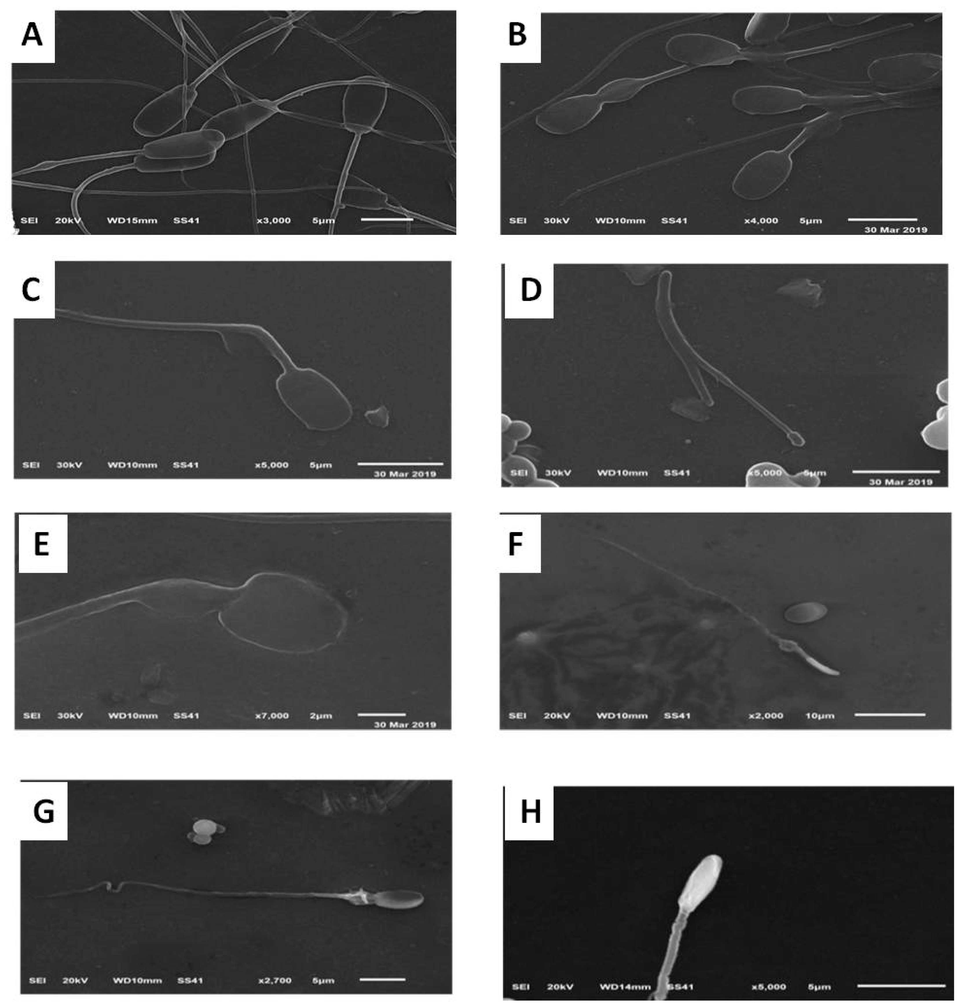
2.4.6. Assessment of Sperm Morphology Using Scanning Electron Microscope (SEM)

The specimens were centrifuged at 500× g for 20 min, and the sperm pellets were collected. The samples were fixed in a solution containing 2.5% (w/v) buffered glutaraldehyde and 2% (w/v) paraformaldehyde in a 0.1 M sodium phosphate buffer (pH 7.4) at 4 °C overnight [66]. The specimens were then washed three times for 15 min each in 0.1 M sodium phosphate and treated with a 2% sodium phosphate-buffered osmium tetroxide (pH 7.4) for 90 min. Finally, the specimens were washed with a 0.1 M sodium phosphate buffer (pH 7.4) and dehydrated in an increasing gradient of ethanol. Four drops of 100% acetone were added to the specimen on small glass plates glued to the specimen stubs of the microscope. After the acetone had evaporated, the specimens were coated with gold-palladium membranes and observed in a Jeol-JSM-6510 L.V SEM. The microscope was operated at 20 kV, and only the central areas of the glass plates were examined.

2.4.7. Assessment of Sperm Ultrastructure Using Transmission Electron Microscope (TEM)

The samples were processed for transmission electron microscopy (TEM) according to Heath et al. [53]. Briefly, the straws from each treatment were washed three times by centrifugation at 1000 rpm for 5 min using Phosphate Buffered Saline, and suspended in a fixative solution of 2.5% (w/v) buffered glutaraldehyde and 2% (w/v) paraformaldehyde in a 0.1 M sodium phosphate buffer (pH 7.4) for 2 h at 4 °C. The samples were then washed and post-fixed in 1% osmium tetroxide for 1 h at room temperature in darkness. The fixed samples were dehydrated in an ethanol gradient, treated with acetone, embedded in an Epon resin (Epon 812; FlukaChemie, Switzerland), and ultrathin-sectioned (60–80 nm) for TEM. Ultrathin sections were observed using a JEOL 2100 TEM at 80 kV. The sperm ultrastructure (acrosome, plasma membrane, and mid-piece) was examined in 100 sperm cells per treatment.

2.5. Statistical Analysis

All data were statistically analyzed by one-way ANOVA design using a software package (SAS, 2007, Cary, NC, USA) [67]. Completely randomized design was used based on the following model: Yij = μ + Gi + eij Where μ = the overall mean, Gi = Treatment (1,2,...7), and eij = residual error. The percentages of values were transformed by arcsine values before analysis. Differences between groups were tested by Duncan’s multiple range test [68] and set at p < 0.05.

3. Results

3.1. Effects on Sperm Quality After Cooling (5 °C for 2 h) and Pre-Freezing

Compared to the control group, supplementation of the extender with nano-sized selenium and zinc resulted in significant increases in sperm progressive motility, vitality, and sperm membrane integrity after cooling at 5 °C for 2 h (Table 1). Supplementation with vitamins C and E did not affect vitality but did show advantageous effects on sperm progressive motility and membrane integrity (Table 1). There were no differences among the experimental groups for sperm abnormality and cytoplasmic droplets.

3.2. Effects on Post-Thawing Sperm Quality

Nano-sized selenium and zinc supplementation significantly improved the sperm properties post-thawing. Sperm progressive motility, vitality, and membrane integrity increased significantly in the nano-sized selenium and zinc treatments when compared to the regular salts, vitamins, and control groups (Table 2). Conversely, the abnormality occurrence was significantly decreased by the supplementation of nano-sized and regular trace elements salts, when compared to the vitamin-supplemented and control groups (Table 2).

3.3. Effects on Sperm Apoptosis and Necrosis (Annexin V/PI Assay) Post-Thawing

Annexin V- and PI-negative sperm were significantly increased in the nano-selenium group, compared to the nano-zinc, vitamin C, vitamin E, sodium selenite, zinc sulfate, and control groups (Table 3). Early apoptotic and apoptotic sperm was significantly increased in the control group when compared to the other groups; the lowest value was observed in the nano-selenium supplemented group (Table 3). Representative pictures of the flow cytometry analysis are shown in Supplementary Material Figure S3.

3.4. Effects on Oxidative Stress of the Extender Post-Thawing

The levels of GSH and the activity of SOD were significantly increased in the nano-selenium and nano-zinc groups when compared to the other experimental and control groups. The lowest values were observed in the control group (Table 4). However, the level of MDA was significantly increased in the control group when compared to the other groups (Table 4).

3.5. Effects on Sperm Ultra-Morphological Characters of Plasma Membrane (PM) and Acrosome Post-Thawing

Figure 2 and Figure 3 show the normal and abnormal ultrastructure of the epididymal camel spermatozoa. There was a significant increase in the percentage of intact plasma membranes in the nano-selenium group, compared to the control and zinc sulfate-supplemented groups (Table 5). The occurrence of abnormal ultra-structures (swollen and slightly swollen plasma membrane) was variable among the experimental groups, but there was a significant increase in the swollen membrane of the control group (Table 5). The lost PM (Table 5) and acrosome ultra-morphology (Table 6) and mid-piece structures (data are not shown) showed no differences among the experimental groups.

4. Discussion

Because of the high level of unsaturated fatty acids in the sperm plasma membrane and inefficient free radical scavenging system because of scanty cytoplasm in the sperm, it is highly vulnerable to the oxidative damage [69]. Sperm cryopreservation and thawing increase ROS formation [70]. ROS increase, together with reduced antioxidants, can lead to oxidative stress (OS) [71], which negatively affects semen quality, induces detrimental effects on spermatozoa [72], which can result in extensive changes, lipid peroxidation, in the plasma membrane concerning their organization as well as the function [73,74,75]. Therefore, OS causes sperm DNA damage and reduces sperm motility, functional integrity, endogenous antioxidant enzyme activity, and fertility [76].
Our results revealed that adding Vit C, Vit E, Na2SeO3, ZnSO4, ZnONPs, or SeNPs can improve the post-thaw quality of the dromedary camel epididymal spermatozoa. This improvement can be explained by their ant-oxidative properties. Therefore, the highest MDA level and the lowest GSH and SOD levels were observed in the control group. This means that these additives can protect sperm from oxidative stress during cryopreservation which reflected on semen quality parameters including progressive motility, vitality, plasma membrane integrity, abnormality, and/or intactness of acrosome and plasma membrane. Oxidative stress induced at the membrane level is correlated to MDA level, which is the commonly used biomarker for membrane lipid peroxidation of omega-3 and omega-6 fatty acids level [72]. The antioxidant system consisting of catalase (CAT), glutathione peroxidase (GSH-PX), and superoxide dismutase (SOD) acts synergistically to increase GSH and reduce MDA, to preserve sperm motility and vitality against OS [15,77].
Nanotechnology, such as nano-zinc and nano-selenium, can be used to achieve bioactive properties in the reproduction, digestion, development, and freezing cells of various elements [78]. Recent studies have shown the beneficial effects of nanoparticles for sperm freezing in humans and animals by lowering the sperm chromatin damage and MDA levels [79,80,81]. However, no studies have examined the impact of zinc oxide or selenium nanoparticles on epididymal camel sperm. The significantly lowest level of MDA and highest level of GSH were observed in the ZnONPs and SeNPs groups in the present study that suggests that the nanoparticles efficiently scavenge the free radicals generated during the freezing/thawing process. Therefore, ZnONPs and SeNPs improved the progressive motility, vitality, and spermatozoa membrane integrity, and decreased apoptosis in frozen-thawed sperm. Nano-sized manufacturing potentiates the effects of these metals when compared to the regular size particles of zinc sulfate, and sodium selenite, respectively. The effect of the NPs is attributable to their small size (surface area and size distribution), chemical composition (purity, crystallinity, and electronic properties), surface structure (surface reactivity, surface groups, inorganic or organic coatings), solubility, shape, and aggregation. These factors give synthetic nanoparticles physicochemical features and a higher surface reactivity than their counterparts of the regular trace elements and vitamins [82,83]. The nanoparticles form ZnONPs thought to be had a more efficient interaction with protamines [79]. According to the TEM results of Isaac et al. [79], the nanoparticles (ZnONPs) were accumulated around the spermatozoa and did not internalize into the sperm. A similar observation was recorded after using the fluorescent ZnONPs; the viable sperm membrane is not permeable to ZnONPs; while, the fluorescence ZnONPs was observed only in the dead spermatozoa. The spermatozoa membrane has a net negative charge and the ZnONPs have a neutral charge. The ZnONPs cannot dissociate to ions which is probably the reason why they cannot enter into the sperm chromatin [79].
Our findings agree with an earlier report by Isaac et al. [79] who reported that ZnONPs have membrane protective function. ZnONPs supplementation leads to an increase in antioxidant enzymes and improves the quality of sperm [20,38,84,85]. The free radical scavenging function of zinc is well established through earlier in vivo and in vitro studies [38,86]. Moreover, it enhanced the post-thawing quality when supplemented to rooster semen extender [28,47] as well as protected the spermatozoa quality (motility, DNA integrity) and spermatogenesis against oxidative damage when administered orally [48]. Therefore, it is probable that the protection of membrane peroxidation by ZnONPs and SeNPs through the reduction of MDA and increasing the SOD and GSH help in mitigating the subsequent damaging effect on the macromolecules like DNA and crucial organelles like mitochondria [38,87,88]. Similar results were recorded by Isaac et al. [79] who concluded that incubation of human ejaculate with ZnONPs improved the quality of cryopreserved human semen, particularly, with respect to chromatin integrity without any adverse effect on their functional competence. The protective effect on sperm chromatin during freezing/thawing process seems to be due to the formation of a protective layer of ZnONPs around the spermatozoa, which can prevent lipid peroxidation at the membrane level [79].
Safa et al. [28] showed that the addition of 5 mg/mL of vitamin E to freezing diluents combined with 1% SeNPs improved the post-thawing sperm motility, vitality, and oxidative variables in rooster semen. Khalil et al. [47] recommended an increase in the supplementation concentration of SeNPs from 0.5 to 1.0 mg/mL to Tris-egg yolk extenders after observing an increase in the percentage of viable sperm, and a decrease in early apoptotic, apoptotic, and necrotic sperm in post-thawed bull semen. However, increasing the concentration of SeNPs to 1.5 mg/mL adversely affected the viable sperm and increased the amount of apoptotic and necrotic sperm.
Our results also revealed that sodium selenite group had a higher level of GSH and SOD and lower level of MDA than zinc sulfate group, which reflect on semen quality. This can be explained by the smaller sizes of sodium selenite particles compared with zinc sulfate in addition to different roles of Zn and Se in the sperm.

5. Conclusions

In spite of supplementation of SHOTOR extender with Vit C, Vit E, Na2SeO3, ZnSO4, ZnONPs or SeNPs can improve the post-thaw quality of dromedary camel epididymal spermatozoa, the selenium and zinc nanoparticles provide the best post-thaw quality. The results presented here highlight favorable sperm properties such as increased motility, vitality, and ultrastructure morphology after cooling or freezing-thawing. We recommend the inclusion of nano-selenium and nano-zinc to extenders to improve the cryotolerance of camel epididymal spermatozoa.

Supplementary Materials

The following are available online at https://www.mdpi.com/2076-2615/10/1/78/s1, Table S1: Characteristics of ZnONPs and SeNPs; Figure S1: Representative particle size distribution (A) and zeta potential (B) curves of ZnONPs; Figure S2: Representative particle size distribution (A) and zeta potential (B) curves of SeNPs; Figure S3: The raw data of two replicates for annexin V flowcytometry.

Author Contributions

Conceptualization, M.A.S., W.A.K. and M.A.E.-H.; methodology, W.A.K., and M.A.S.; software, M.A.S.; investigation, M.A.S. and W.A.K.; data curation, M.A.S., W.A.K., I.M.S. and M.A.E.-H.; writing—original draft preparation, M.A.S., W.A.K., I.M.S. and A.A.-A.S.; writing—review and editing, M.A.S., W.A.K., I.M.S., and A.A.-A.S.; supervision, M.A.E.-H. and W.A.K.; project administration, M.A.E.-H. and W.A.K.; funding acquisition, I.M.S. All authors have read and agreed to the published version of the manuscript.

Funding

The authors extend their appreciation to the Deanship of Scientific Research at King Saud University Research group no. RG-1438-018.

Acknowledgments

We thank the Research Support Unit at KSU for their technical assistance and editing the manuscript. We thank Mahmoud A.E. Hassan (Electron Microscope Unit, Mansoura University) for their valuable help in the part of Sperm ultrastructure.

Conflicts of Interest

The authors declare no conflict of interest.

References

  1. Skidmore, J. Reproduction in dromedary camels: An update. Anim. Reprod. Sci. 2005, 2, 161–171. [Google Scholar]
  2. Spencer, P.; Wilson, K.; Tinson, A. Parentage testing of racing camels (Camelus dromedarius) using microsatellite DNA typing. Anim. Genet. 2010, 41, 662–665. [Google Scholar] [CrossRef]
  3. Swelum, A.A.; Ismael, A.B.; Khalaf, A.F.; Abouheif, M.A. Clinical and laboratory findings associated with naturally occurring babesiosis in dromedary camels. Bull. Vet. Inst. Pulawy 2014, 58, 229–233. [Google Scholar] [CrossRef] [Green Version]
  4. El-Hassanein, E.-S. Prospects of Improving Semen Collection and Preservation from Elite Dromedary Camel Breeds. World Vet. J. 2017, 7, 47–64. [Google Scholar] [CrossRef]
  5. Derar, D.R.; Hussein, H.A.; Ali, A. Reference values for the genitalia of male dromedary before and after puberty using caliper and ultrasonography in subtropics. Theriogenology 2012, 77, 459–465. [Google Scholar] [CrossRef]
  6. Swelum, A.A.; Saadeldin, I.M.; Ba-Awadh, H.; Alowaimer, A.N. Shortened daily photoperiod during the non-breeding season can improve the reproductive performance of camel bulls (Camelus dromedarius). Anim. Reprod. Sci. 2018, 195, 334–344. [Google Scholar] [CrossRef]
  7. Swelum, A.A.; Saadeldin, I.M.; Ba-Awadh, H.; Alowaimer, A.N. Effects of melatonin implants on the reproductive performance and endocrine function of camel (Camelus dromedarius) bulls during the non-breeding and subsequent breeding seasons. Theriogenology 2018, 119, 18–27. [Google Scholar] [CrossRef]
  8. Swelum, A.A.; Saadeldin, I.M.; Ba-Awadh, H.; Al-Mutary, M.G.; Alowaimer, A.N. Effect of short artificial lighting and low temperature in housing rooms during non-rutting season on reproductive parameters of male dromedary camels. Theriogenology 2019, 131, 133–139. [Google Scholar] [CrossRef]
  9. Wani, N.A.; Morton, K.; Billah, M.; Skidmore, J. Biophysical and biochemical characteristics of ejaculated semen of dromedary camel (Camelus dromedarius) and Llama (Llama glama). J. Camel. Pract. Res. 2011, 18, 97–102. [Google Scholar]
  10. Swelum, A.A.; Saadeldin, I.; Ba-Awadh, H.G.; Al-Mutary, M.F.; Moumen, A.N.; Alowaimer, A.; Abdalla, H. Efficiency of Commercial Egg Yolk-Free and Egg Yolk-Supplemented Tris-Based Extenders for Dromedary Camel Semen Cryopreservation. Animals 2019, 9, 999. [Google Scholar] [CrossRef] [Green Version]
  11. Waheed, M.; Al-Eknah, M.; El-Bahr, S. Some biochemical characteristics and preservation of epididymal camel spermatozoa (Camelus dromedarius). Theriogenology 2011, 76, 1126–1133. [Google Scholar] [CrossRef] [PubMed]
  12. Skidmore, J.A.; Malo, C.M.; Crichton, E.G.; Morrell, J.M.; Pukazhenthi, B.S. An update on semen collection, preservation and artificial insemination in the dromedary camel (Camelus dromedarius). Anim. Reprod. Sci. 2018, 194, 11–18. [Google Scholar] [CrossRef] [PubMed]
  13. Dong, Q.; Rodenburg, S.E.; Huang, C.; Vandevoort, C.A. Cryopreservation of rhesus monkey (Macaca mulatta) epididymal spermatozoa before and after refrigerated storage. J. Androl. 2008, 29, 283–292. [Google Scholar] [CrossRef] [PubMed]
  14. Kumaresan, A.; Ansari, M.; Garg, A.; Kataria, M. Effect of oviductal proteins on sperm functions and lipid peroxidation levels during cryopreservation in buffaloes. Animl. Reprod. Sci. 2006, 93, 246–257. [Google Scholar] [CrossRef]
  15. Bucak, M.N.; Ateşşahin, A.; Yüce, A. Effect of anti-oxidants and oxidative stress parameters on ram semen after the freeze-thawing process. Small Rum. Res. 2008, 75, 128–134. [Google Scholar] [CrossRef]
  16. Sardoy, M.; Carretero, M.; Neild, D. Evaluation of stallion sperm DNA alterations during cryopreservation using toluidine blue. Anim. Reprod. Sci. 2008, 3, 349–350. [Google Scholar] [CrossRef]
  17. El-Badry, D.; Azab, A.; Karima Gh, M.; Scholkamy, T. The effect of extender type on freezability of one-humped camel spermatozoa with special reference to their fine structure. In Proceedings of the 25th Annual Conf, Taba, Egypt, 14–17 April 2016. [Google Scholar]
  18. El-Harairy, M.A.; El-Razek, I.M.A.; Shamiah, S.M.; Zaghloul, H.; Khalil, W. Effect of antioxidants on the stored dromedary camel epididymal sperm characteristics. Asian J. Anim. Sci. 2016, 10, 147–153. [Google Scholar] [CrossRef] [Green Version]
  19. Kotdawala, A.P.; Kumar, S.; Salian, S.R.; Thankachan, P.; Govindraj, K.; Kumar, P.; Kalthur, G.; Adiga, S.K. Addition of zinc to human ejaculate prior to cryopreservation prevents freeze-thaw-induced DNA damage and preserves sperm function. J. Assist. Reprod. Genet. 2012, 29, 1447–1453. [Google Scholar] [CrossRef] [Green Version]
  20. Barkhordari, A.; Hekmatimoghaddam, S.; Jebali, A.; Khalili, M.A.; Talebi, A.; Noorani, M. Effect of zinc oxide nanoparticles on viability of human spermatozoa. Iran. J. Reprod. Med. 2013, 11, 767. [Google Scholar]
  21. Dawson, E.B.; Harris, W.A.; Teter, M.C.; Powell, L.C. Effect of ascorbic acid supplementation on the sperm quality of smokers. Fertil. Steril. 1992, 58, 1034–1039. [Google Scholar] [CrossRef]
  22. Abdelazim, A.M.; Saadeldin, I.M.; Swelum, A.A.; Afifi, M.M.; Alkaladi, A. Oxidative Stress in the Muscles of the Fish Nile Tilapia Caused by Zinc Oxide Nanoparticles and Its Modulation by Vitamins C and E. Oxid. Med. Cell. Longev. 2018, 2018, 1–9. [Google Scholar] [CrossRef] [PubMed]
  23. Chinoy, N.; Mehta, R.; Seethalakshmi, L.; Sharma, J.; Chinoy, M. Effects of vitamin C deficiency on physiology of male reproductive organs of guinea pigs. Int. J. Fertil. 1986, 31, 232–239. [Google Scholar] [PubMed]
  24. Benhenia, K.; Lamara, A.; Fatmi, S.; Iguer-Ouada, M. Effect of cyclodextrins, cholesterol and vitamin E and their complexation on cryopreserved epididymal ram semen. Small Rum. Res. 2016, 141, 29–35. [Google Scholar] [CrossRef]
  25. El-Sawy, H.B.I.; Soliman, M.M.; El-Shazly, S.A.; Ali, H.A.-M. Protective effects of camel milk and vitamin E against monosodium glutamate induced biochemical and testicular dysfunctions. Prog. Nutr. 2018, 20, 76–85. [Google Scholar]
  26. Hu, J.-H.; Zhao, X.-L.; Tian, W.-Q.; Zan, L.-S.; Li, Q.-W. Effects of vitamin E supplementation in the extender on frozen-thawed bovine semen preservation. Animal 2011, 5, 107–112. [Google Scholar] [CrossRef] [Green Version]
  27. Yousef, M.; Abdallah, G.; Kamel, K. Effect of ascorbic acid and vitamin E supplementation on semen quality and biochemical parameters of male rabbits. Anim. Reprod. Sci. 2003, 76, 99–111. [Google Scholar] [CrossRef]
  28. Safa, S.; Moghaddam, G.; Jozani, R.J.; Kia, H.D.; Janmohammadi, H. Effect of vitamin E and selenium nanoparticles on post-thaw variables and oxidative status of rooster semen. Anim. Reprod. Sci. 2016, 174, 100–106. [Google Scholar] [CrossRef]
  29. Luo, H.; Jia, Z.; Zhu, S.; Ding, J. Effect of vitamin E on the qualities of fresh and frozen-thawed Ram Semen. China Herbiv. Sci. 2004, 5, 4–16. [Google Scholar]
  30. Zago, M.P.; Oteiza, P.I. The antioxidant properties of zinc: Interactions with iron and antioxidants. Free Radical. Biol. Med. 2001, 31, 266–274. [Google Scholar] [CrossRef]
  31. Fallah, A.; Mohammad-Hasani, A.; Colagar, A.H. Zinc is an essential element for male fertility: A review of zn roles in men’s health, germination, sperm quality, and fertilization. J. Reprod. Infertil. 2018, 19, 69. [Google Scholar]
  32. Dorostkar, K.; Alavi-Shoushtari, S.M.; Mokarizadeh, A. Effects of in vitro selenium addition to the semen extender on the spermatozoa characteristics before and after freezing in water buffaloes (Bubalus bubalis). Vet. Res. Forum 2012, 3, 263–268. [Google Scholar] [PubMed]
  33. Ghallab, A.M.; Shahat, A.M.; Fadl, A.M.; Ayoub, M.M.; Moawad, A.R. Impact of supplementation of semen extender with antioxidants on the quality of chilled or cryopreserved Arabian stallion spermatozoa. Cryobiology 2017, 79, 14–20. [Google Scholar] [CrossRef] [PubMed]
  34. Ursini, F.; Heim, S.; Kiess, M.; Maiorino, M.; Roveri, A.; Wissing, J.; Flohe, L. Dual function of the selenoprotein PHGPx during sperm maturation. Science 1999, 285, 1393–1396. [Google Scholar] [CrossRef] [PubMed] [Green Version]
  35. Marai, I.F.M.; El-Darawany, A.-H.; Ismail, E.; Abdel-Hafez, M.A.M. Reproductive and physiological traits of Egyptian Suffolk rams as affected by selenium dietary supplementation and housing heat radiation effects during winter of the sub-tropical environment of Egypt. Archiv. Anim. Breed. 2009, 52, 402–409. [Google Scholar] [CrossRef]
  36. Shi, L.-G.; Yang, R.-J.; Yue, W.-B.; Xun, W.-J.; Zhang, C.-X.; Ren, Y.-S.; Shi, L.; Lei, F.-L. Effect of elemental nano-selenium on semen quality, glutathione peroxidase activity, and testis ultrastructure in male Boer goats. Anim. Reprod. Sci. 2010, 118, 248–254. [Google Scholar] [CrossRef]
  37. Peters, K.; Unger, R.E.; Kirkpatrick, C.J.; Gatti, A.M.; Monari, E. Effects of nano-scaled particles on endothelial cell function in vitro: Studies on viability, proliferation and inflammation. J. Mat. Sci. Mater. Med. 2004, 15, 321–325. [Google Scholar] [CrossRef]
  38. Afifi, M.; Almaghrabi, O.A.; Kadasa, N.M. Ameliorative effect of zinc oxide nanoparticles on antioxidants and sperm characteristics in streptozotocin-induced diabetic rat testes. BioMed Res. Int. 2015, 2015, 153573. [Google Scholar] [CrossRef]
  39. Hidiroglou, M.; Knipfel, J. Zinc in mammalian sperm: A review. J. Dairy Sci. 1984, 67, 1147–1156. [Google Scholar] [CrossRef]
  40. Cheah, Y.; Yang, W. Functions of essential nutrition for high quality spermatogenesis. Adv. Biosci. Biotechnol. 2011, 2, 182. [Google Scholar] [CrossRef] [Green Version]
  41. Dissanayake, D.; Wijesinghe, P.; Ratnasooriya, W.; Wimalasena, S. Relationship between seminal plasma zinc and semen quality in a subfertile population. J. Hum. Reprod. Sci. 2010, 3, 124. [Google Scholar] [CrossRef]
  42. Baumber, J.; Ball, B.A.; Gravance, C.G.; Medina, V.; Davies-morel, M.C. The effect of reactive oxygen species on equine sperm motility, viability, acrosomal integrity, mitochondrial membrane potential, and membrane lipid peroxidation. J. Androl. 2000, 21, 895–902. [Google Scholar] [PubMed]
  43. Roy, B.; Baghel, R.; Mohanty, T.; Mondal, G. Zinc and male reproduction in domestic animals: A Review. Indian J. Anim. Nutr. 2013, 30, 339–350. [Google Scholar]
  44. Rangari, K.; Shrivastav, T. A correlation study between steroid hormone levels and anti-sperm antibodies in serum and seminal plasma of men with or without reduced sperm motility. J. Endocrinol. Reprod. 2007, 11, 31–35. [Google Scholar]
  45. Balázs, C.; Rácz, K. The role of selenium in endocrine system diseases. Orvosi Hetilap 2013, 154, 1628–1635. [Google Scholar] [CrossRef] [Green Version]
  46. Tareq, K.; Akter, Q.S.; Takagi, Y.; Hamano, K.-I.; Sawada, T.; Tsujii, H. Effect of selenium and vitamin E on acrosome reaction in porcine spermatozoa. Reprod. Med. Biol. 2010, 9, 73–81. [Google Scholar] [CrossRef]
  47. Khalil, W.A.; El-Harairy, M.A.; Zeidan, A.E.; Hassan, M.A. Impact of selenium nano-particles in semen extender on bull sperm quality after cryopreservation. Theriogenology 2019, 126, 121–127. [Google Scholar] [CrossRef]
  48. Rezvanfar, M.A.; Rezvanfar, M.A.; Shahverdi, A.R.; Ahmadi, A.; Baeeri, M.; Mohammadirad, A.; Abdollahi, M. Protection of cisplatin-induced spermatotoxicity, DNA damage and chromatin abnormality by selenium nano-particles. Toxicol. Appl. Pharmacol. 2013, 266, 356–365. [Google Scholar] [CrossRef]
  49. Deen, A.; Vyas, S.; Sahani, M. Semen collection, cryopreservation and artificial insemination in the dromedary camel. Anim. Reprod. Sci. 2003, 77, 223–233. [Google Scholar] [CrossRef]
  50. Hammadi, M.; Zarrouk, O.; Barmat, A.; Trimeche, A.; Khorchani, T.; Khaldi, G. Characterization and conservation of Maghrebi camel semen. In Proceedings of the WBC/ICAR Satellite Meeting on Camelid Reproduction, Budapest, Hungary, 12–13 July 2008. [Google Scholar]
  51. Niasari-Naslaji, A.; Mosaferi, S.; Bahmani, N.; Gharahdaghi, A.; Abarghani, A.; Ghanbari, A.; Gerami, A. Effectiveness of a tris-based extender (SHOTOR diluent) for the preservation of Bactrian camel (Camelus bactrianus) semen. Cryobiology 2006, 53, 12–21. [Google Scholar] [CrossRef]
  52. Marin-Guzman, J.; Mahan, D.; Whitmoyer, R. Effect of dietary selenium and vitamin E on the ultrastructure and ATP concentration of boar spermatozoa, and the efficacy of added sodium selenite in extended semen on sperm motility. J. Anim. Sci. 2000, 78, 1544–1550. [Google Scholar] [CrossRef] [Green Version]
  53. Heath, E.; Mostafa, M.; Karasek, S. Ultrastructure of Camel (Camelus dromedarius) Spermatozoa. Anat. Histol. Embryol. 1986, 15, 147–154. [Google Scholar] [CrossRef] [PubMed]
  54. Tingari, M. Studies on camel semen. III. Ultrastructure of the spermatozoon. Anim. Reprod. Sci. 1991, 26, 333–344. [Google Scholar] [CrossRef]
  55. Taylor, K.; Roberts, P.; Sanders, K.; Burton, P. Effect of antioxidant supplementation of cryopreservation medium on post-thaw integrity of human spermatozoa. Reprod. Biomed. Online 2009, 18, 184–189. [Google Scholar] [CrossRef]
  56. Salisbury, G.W.; VanDemark, N.L.; Lodge, J.R. Physiology of Reproduction and Artificial Insemination of Cattle; WH Freeman and Company: Stuttgart, Germany, 1978. [Google Scholar]
  57. Moskovtsev, S.I.; Librach, C.L. Methods of sperm vitality assessment. Method Mol. Biol. 2013, 927, 13–19. [Google Scholar] [CrossRef]
  58. Menon, A.G.; Thundathil, J.C.; Wilde, R.; Kastelic, J.P.; Barkema, H.W. Validating the assessment of bull sperm morphology by veterinary practitioners. Canad. Vet. J. 2011, 52, 407–408. [Google Scholar]
  59. Caycho, K.; Santolaria, P.; Soler, C.; Yániz, J. Effect of hypoosmotic swelling test and water test on the distribution of sperm subpopulations in bull. Anim. Reprod. Sci. 2016, 169, 101. [Google Scholar] [CrossRef]
  60. Ochseedorf, F.R.; Buhl, R.; Bästlein, A.; Beschmann, H. Glutathione le spermatozoa and seminal plasma of infertile men. Hum. Reprod. 1998, 13, 353–359. [Google Scholar] [CrossRef] [Green Version]
  61. Koziorowska-Gilun, M.; Koziorowski, M.; Fraser, L.; Strzeżek, J. Antioxidant defence system of boar cauda epididymidal spermatozoa and reproductive tract fluids. Reprod. Domest. Anim. 2011, 46, 527–533. [Google Scholar] [CrossRef]
  62. Tuncer, P.B.; Bucak, M.N.; Sarıözkan, S.; Sakin, F.; Yeni, D.; Çiğerci, İ.H.; Ateşşahin, A.; Avdatek, F.; Gündoğan, M.; Büyükleblebici, O. The effect of raffinose and methionine on frozen/thawed Angora buck (Capra hircus ancryrensis) semen quality, lipid peroxidation and antioxidant enzyme activities. Cryobiology 2010, 61, 89–93. [Google Scholar] [CrossRef]
  63. Chaveiro, A.; Santos, P.; Da Silva, F. Assessment of Sperm Apoptosis in Cryopreserved Bull Semen After Swim-up Treatment: A Flow Cytometric Study. Reprod. Domest. Anim. 2007, 42, 17–21. [Google Scholar] [CrossRef]
  64. Masters, A.; Harrison, P. Platelet counting with the BD AccuriTM C6 flow cytometer. Platelets 2014, 25, 175–180. [Google Scholar] [CrossRef] [PubMed]
  65. Peña, F.J.; Johannisson, A.; Wallgren, M. Assessment of fresh and frozen–thawed boar semen using an Annexin-V assay: A new method of evaluating sperm membrane integrity. Theriogenology 2003, 60, 677–689. [Google Scholar] [CrossRef]
  66. Graham, R.C., Jr.; Lundholm, U.; Karnovsky, M.J. Cytochemical demonstration of peroxidase activity with 3-amino-9-ethylcarbazole. J. Histochem. Cytochem. 1965, 13, 150–152. [Google Scholar] [CrossRef] [PubMed]
  67. SAS. Statistical Analysis System. Stat-User’s Guid. Release 9.1.3; SAS Institute: Cary, NC, USA, 2007. [Google Scholar]
  68. Duncan, D.B. Multiple Range and Multiple “F” Test. Biometrics 1955, 11, 1–42. [Google Scholar] [CrossRef]
  69. Aitken, R.J.; Clarkson, J.S.; Fishel, S. Generation of reactive oxygen species, lipid peroxidation, and human sperm function. Biol. Reprod. 1989, 41, 183–197. [Google Scholar] [CrossRef]
  70. Wang, A.W.; Zhang, H.; Ikemoto, I.; Anderson, D.J.; Loughlin, K.R. Reactive oxygen species generation by seminal cells during cryopreservation. Urology 1997, 49, 921–925. [Google Scholar] [CrossRef]
  71. Tatone, C.; Di Emidio, G.; Vento, M.; Ciriminna, R.; Artini, P.G. Cryopreservation and oxidative stress in reproductive cells. Gynecol. Endocrinol. 2010, 26, 563–567. [Google Scholar] [CrossRef]
  72. Esterbauer, H.; Cheeseman, K.H. Determination of aldehydic lipid peroxidation products: Malonaldehyde and 4-hydroxynonenal. Method Enzymol 1990, 186, 407–421. [Google Scholar] [CrossRef]
  73. Simões, R.; Feitosa, W.B.; Siqueira, A.F.P.; Nichi, M.; Paula-Lopes, F.F.; Marques, M.G.; Peres, M.A.; Barnabe, V.H.; Visintin, J.A.; Assumpção, M.E.O. Influence of bovine sperm DNA fragmentation and oxidative stress on early embryo in vitro development outcome. Reproduction 2013, 146, 433–441. [Google Scholar] [CrossRef] [Green Version]
  74. Bucak, M.N.; Sarıözkan, S.; Tuncer, P.B.; Sakin, F.; Ateşşahin, A.; Kulaksız, R.; Çevik, M. The effect of antioxidants on post-thawed Angora goat (Capra hircus ancryrensis) sperm parameters, lipid peroxidation and antioxidant activities. Small Rum. Res. 2010, 89, 24–30. [Google Scholar] [CrossRef]
  75. Aitken, R.J.; Baker, M.A. Oxidative stress, sperm survival and fertility control. Mol. Cell. Endocrinol. 2006, 250, 66–69. [Google Scholar] [CrossRef] [PubMed]
  76. Sarıözkan, S.; Bucak, M.N.; Tuncer, P.B.; Ulutaş, P.A.; Bilgen, A. The influence of cysteine and taurine on microscopic–oxidative stress parameters and fertilizing ability of bull semen following cryopreservation. Cryobiology 2009, 58, 134–138. [Google Scholar] [CrossRef] [PubMed]
  77. Saadeldin, I.M.; Fadel, A.M.; Hamada, M.M.Z.; El-Badry, A.A. Effects of exposure to 50 Hz, 1 Gauss magnetic field on reproductive traits in male albino rats. Acta. Vet. Brno. 2011, 80, 107–111. [Google Scholar] [CrossRef] [Green Version]
  78. Pelyhe, C.; Mézes, M. Myths and facts about the effects of nano selenium in farm animals–mini-review. Eur. Chem. Bull. 2013, 2, 1049–1052. [Google Scholar]
  79. Isaac, A.V.; Kumari, S.; Nair, R.; Urs, D.R.; Salian, S.R.; Kalthur, G.; Adiga, S.K.; Manikkath, J.; Mutalik, S.; Sachdev, D.; et al. Supplementing zinc oxide nanoparticles to cryopreservation medium minimizes the freeze-thaw-induced damage to spermatozoa. Biochem. Biophys. Res. Commun. 2017, 494, 656–662. [Google Scholar] [CrossRef]
  80. Heidari, G.; Seifdavati, J.; Mohebodini, H.; Seyed Sharifi, R.; Abdi Benemar, H. Effect of Nano Zinc Oxide on Post-Thaw Variables and Oxidative Status of Moghani Ram Semen. Kafkas Üniversitesi Veteriner Fakültesi Dergisi 2018, 25, 71–76. [Google Scholar] [CrossRef]
  81. Falchi, L.; Khalil, W.A.; Hassan, M.; Marei, W.F.A. Perspectives of nanotechnology in male fertility and sperm function. Int. J. Vet. Sci. Med. 2019, 6, 265–269. [Google Scholar] [CrossRef] [Green Version]
  82. Nel, A.; Xia, T.; Mädler, L.; Li, N. Toxic potential of materials at the nanolevel. Science 2006, 311, 622–627. [Google Scholar] [CrossRef] [Green Version]
  83. Handy, R.D.; Owen, R.; Valsami-Jones, E. The ecotoxicology of nanoparticles and nanomaterials: Current status, knowledge gaps, challenges, and future needs. Ecotoxicology 2008, 17, 315–325. [Google Scholar] [CrossRef]
  84. Hussein, M.M.A.; Ali, H.A.; Saadeldin, I.M.; Ahmed, M.M. Querectin Alleviates Zinc Oxide Nanoreprotoxicity in Male Albino Rats. J. Bioch. Mol. Toxicol. 2016, 30, 489–496. [Google Scholar] [CrossRef]
  85. Abbasalipourkabir, R.; Moradi, H.; Zarei, S.; Asadi, S.; Salehzadeh, A.; Ghafourikhosroshahi, A.; Mortazavi, M.; Ziamajidi, N. Toxicity of zinc oxide nanoparticles on adult male Wistar rats. Food Chem. Toxicol. 2015, 84, 154–160. [Google Scholar] [CrossRef] [PubMed]
  86. Celino, F.T.; Yamaguchi, S.; Miura, C.; Ohta, T.; Tozawa, Y.; Iwai, T.; Miura, T. Tolerance of Spermatogonia to Oxidative Stress Is Due to High Levels of Zn and Cu/Zn Superoxide Dismutase. PLoS ONE 2011, 6, e16938. [Google Scholar] [CrossRef] [PubMed] [Green Version]
  87. El-Maddawy, Z.; Abd El Naby, W.S.H. Protective effects of zinc oxide nanoparticles against doxorubicin induced testicular toxicity and DNA damage in male rats. Toxicol. Res. 2019, 8, 654–662. [Google Scholar] [CrossRef] [PubMed]
  88. Asri-Rezaei, S.; Nourian, A.; Shalizar-Jalali, A.; Najafi, G.; Nazarizadeh, A.; Koohestani, M.; Karimi, A. Selenium supplementation in the form of selenium nanoparticles and selenite sodium improves mature male mice reproductive performances. Iran. J. Basic Med. Sci. 2018, 21, 577–585. [Google Scholar] [PubMed]
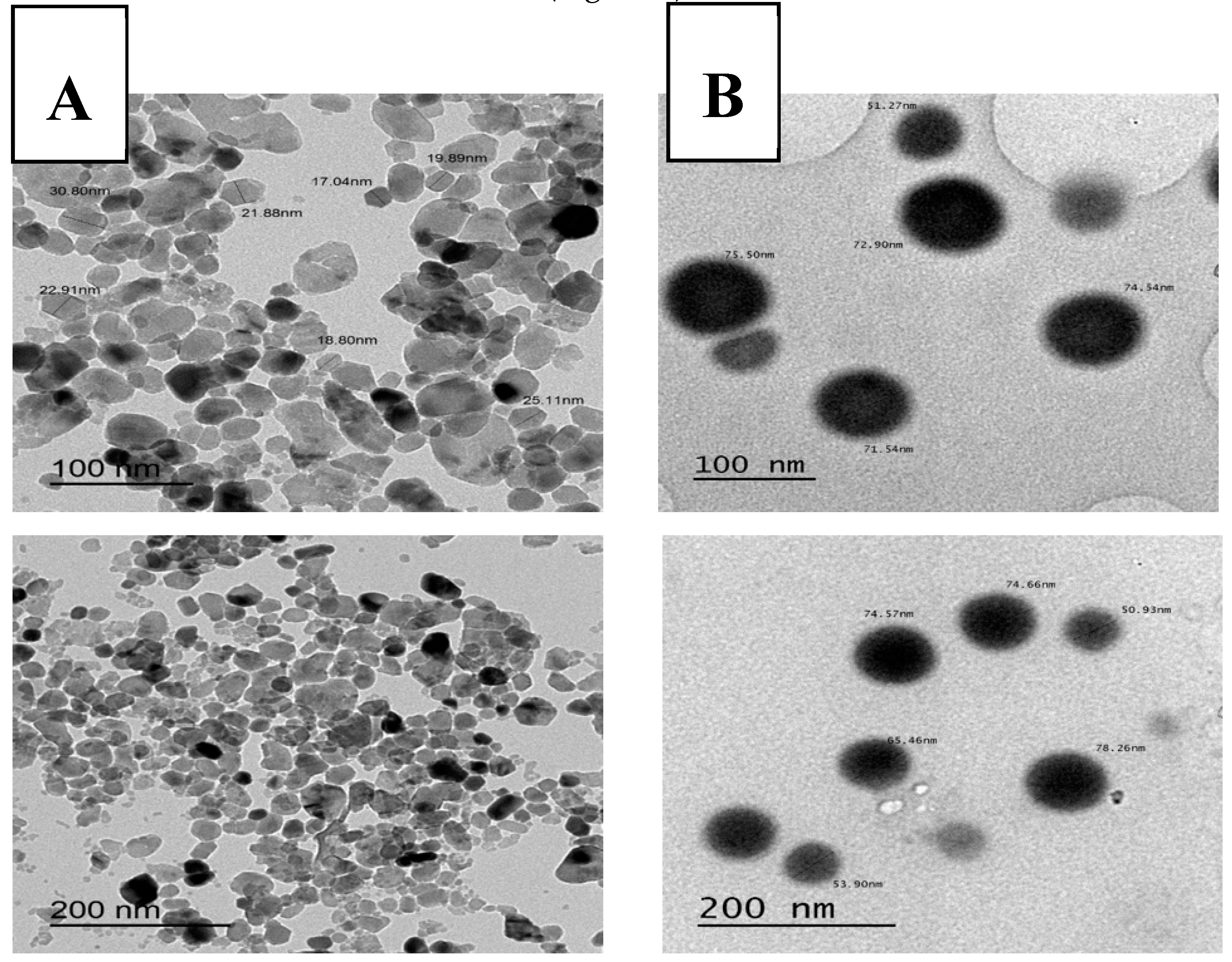
Figure 1. Transmission electron microscopy of (A) ZnONPs and (B) SeNPs with different magnifications.
Animals 10 00078 g001
Figure 2. Representative scanning electron micrographs (SEM) showing the epididymal sperm morphology. Images (A,B) depict normal sperm cells while images (CH) show abnormalities in the head and mid-piece (C). detached acrosomes; (D). detached head; (E), damaged plasma membrane and swelled head; (F). thin mid-piece and pen head; (G). small head; (H). abnormal head and mid-piece size).
Animals 10 00078 g002
Figure 3. Transmission electron micrographs (80 kV) of longitudinal and ultrathin cross-sections of different epididymal camel spermatozoa after thawing. (A) Sperm with intact plasma membrane, (B) Sperm with slightly swollen plasma membrane, (C) Sperm with swollen plasma membrane, (D) Sperm with lost plasma membrane, (E) Sperm with intact acrosome, (F) Sperm with typical acrosome reaction, (G) Sperm with atypical acrosome reaction and (H) Sperm with lost acrosome.
Animals 10 00078 g003
Table 1. Effect of supplementing SHOTOR extender with vitamins, trace elements, and nanoparticles on epididymal camel spermatozoa characteristics (%) after an equilibration period (2 h at 5 °C).
TreatmentProgressive MotilityVitalityPlasma Membrane IntegrityAbnormalityCytoplasmic Droplet
Control60.8 ± 2.01 c63.2 ± 2.91 b60.2 ± 2.04 c12.3 ± 1.8736.2 ± 1.51
Vitamin C67.5 ± 2.14 ab70.2 ± 3.39 ab68.5 ± 1.61 ab11.0 ± 1.2936.7 ± 1.69
Vitamin E63.3 ± 1.67 bc65.5 ± 1.45 ab65.8 ± 0.31 b14.2 ± 1.6834.5 ± 0.62
SeNPs72.5 ± 1.44 a73.3 ± 0.85 a73.0 ± 1.83 a15.5 ± 1.5035.3 ± 1.11
Na2SeO368.0 ± 1.22 ab69.8 ± 1.77 ab69.2 ± 2.48 ab11.6 ± 1.6933.8 ± 1.71
ZnONPs70.8 ± 2.01 a73.3 ± 2.14 a71.3 ± 1.87 ab12.5 ± 0.9234.5 ± 1.48
ZnSO468.8 ± 2.39 ab72.0 ± 2.35 a70.5 ± 2.84 ab15.8 ± 2.5333.8 ± 0.75
a–c Means denoted within the same column with different superscripts are significantly different at p < 0.05.
Table 2. Effect of supplementing SHOTOR extender with vitamins, minerals, and nanoparticles on sperm characteristics (%) in post-thawed epididymal camel spermatozoa.
TreatmentProgressive MotilityVitalityPlasma Membrane IntegrityAbnormalityCytoplasmic Droplet
Control27.0 ± 1.22 c28.8 ± 1.32 c28.4 ± 1.12 c26.2 ± 1.02 a21.6 ± 2.84
Vitamin C30.0 ± 1.58 c32.2 ± 2.11 c30.4 ± 2.36 c18.6 ± 0.75 b21.0 ± 2.61
Vitamin E31.0 ± 1.00 c33.8 ± 0.86 c31.0 ± 0.89 c24.8 ± 0.86 a21.2 ± 2.71
SeNPs48.3 ± 1.67 a50.7 ± 2.33 a50.0 ± 1.15 a18.7 ± 0.88 b20.7 ± 2.40
Na2SeO340.0 ± 2.04 b42.3 ± 2.14 b41.5 ± 2.87 b19.5 ± 0.65 b21.3 ± 2.46
ZnONPs49.0 ± 1.87 a52.2 ± 2.15 a51.0 ± 2.19 a18.8 ± 0.58 b19.4 ± 1.81
ZnSO436.7 ± 1.67 b39.7 ± 1.76 b38.3 ± 0.33 b18.7 ± 1.20 b22.3 ± 2.91
a–c Means denoted within the same column with different superscripts are significantly different at p < 0.05.
Table 3. Effect of supplementing SHOTOR extender with vitamins, trace elements, and nanoparticles on viable, early apoptotic, apoptotic, and necrotic sperm in post-thawed epididymal camel spermatozoa using Annexin V/PI assay.
TreatmentViable (%)
(A−/PI−)
Early Apoptosis (%)
(A+/PI−)
Late Apoptosis (%)
(A+/PI+)
Necrosis (%)
(A−/PI+)
Control26.4 ± 1.10 g38.4 ± 0.26 a31.0 ± 1.30 a4.3 ± 0.07
Vitamin C57.4 ± 0.92 c24.0 ± 0.55 c15.7 ± 1.30 c2.9 ± 0.18
Vitamin E52.4 ± 0.58 d24.2 ± 0.61 c15.9 ± 0.18 c7.5 ± 0.20
SeNPs78.1 ± 0.58 a12.0 ± 0.43 d4.3 ± 0.17 e5.6 ± 0.03
Na2SeO347.0 ± 0.09 e38.2 ± 0.32 a12.8 ± 0.30 d2.0 ± 0.12
ZnONPs69.6 ± 0.26 b4.5 ± 0.35 e20.0 ± 0.35 b5.9 ± 0.26
ZnSO442.5 ± 0.64 f35.0 ± 0.23 b21.4 ± 0.43 b1.2 ± 0.03
a–g Means denoted within the same column with different superscripts are significantly different at p < 0.05.
Table 4. Effect of supplementing SHOTOR extender with vitamins, trace elements, and nanoparticles on the antioxidant (GSH, SOD) and oxidative biomarkers (malondialdehyde, MDA) in the extender of post-thawed epididymal camel spermatozoa.
TreatmentGSH (mg/dL)SOD (U/mL)MDA (nmol/mL)
Control0.45 ± 0.03 e48.3 ± 5.93 f30.1 ± 2.15 a
Vitamin C0.59 ± 0.03 dc113.4 ± 5.66 dc14.8 ± 0.26 d
Vitamin E0.64 ± 0.02 bc124.3 ± 3.94 c14.8 ± 0.17 d
SeNPs0.77 ± 0.04 a168.0 ± 6.67 a12.7 ± 0.70 d
Na2SeO30.55 ± 0.01 d106.8 ± 1.70 d18.4 ± 1.07 c
ZnONPs0.70 ± 0.00 ab148.2 ± 2.33 b13.6 ± 0.48 d
ZnSO40.51 ± 0.03 de67.2 ± 6.28 e22.6 ± 1.32 b
a–f Means denoted within the same column with different superscripts are significantly different at p < 0.05.
Table 5. Effects of supplementing SHOTOR extender with vitamins, trace elements, and nanoparticles on the sperm plasma membrane (PM) post-thawing.
TreatmentIntact PMSlightly Swollen PMSwollen PMLost PM
Control38 ± 4.88 b12 ± 3.27 ab37 ± 4.85 a13 ± 3.38
Vitamin C58 ± 4.96 ab7 ± 2.56 b23 ± 4.23 b12 ± 3.27
Vitamin E55 ± 5.00 ab8 ± 2.73 b26 ± 4.41 ab11 ± 3.14
SeNPs68 ± 4.69 a9 ± 2.88a b15 ± 3.59 b8 ± 2.73
Na2SeO353 ± 5.02 ab18 ± 3.86 a18 ± 3.86 b11 ± 3.14
ZnONPs65 ± 4.79 ab7 ± 2.56 b17 ± 3.78 b11 ± 3.14
ZnSO451 ± 5.02 bc16 ± 3.68 ab21 ± 4.09 b12 ± 3.27
a–c Means denoted within the same column with different superscripts are significantly different at p < 0.05.
Table 6. Effects supplementing SHOTOR extender with vitamins, trace elements, and nanoparticles on the sperm acrosomes post-thawing.
TreatmentIntact AcrosomeTypical ARAtypical ARLost Acrosome
Control61 ± 4.9024 ± 4.2310 ± 2.885 ± 2.19
Vitamin C68 ± 4.6916 ± 3.6812 ± 3.274 ± 1.97
Vitamin E67 ± 4.7318 ± 3.8610 ± 3.025 ± 2.19
SeNPs78 ± 4.1610 ± 3.028 ± 2.734 ± 1.97
Na2SeO370 ± 4.6116 ± 3.689 ± 2.885 ± 2.19
ZnONPs74 ± 4.4113 ± 3.3810 ± 3.023 ± 1.71
ZnSO472 ± 4.5112 ± 3.2710 ± 3.026 ± 2.39
Intact acrosome: where sperm heads exhibited intact acrosomal membrane surrounding the acrosomal ground substance; Acrosome reaction (AR): a swelling of acrosomal ground substance with vesicles of fused plasma and outer acrosomal membranes; Atypical AR: sperm head presenting swelling of acrosomal ground substance dispersed under the swollen outer acrosomal.