Article

Lactobacillus reuteri and Enterococcus faecium from Poultry Gut Reduce Mucin Adhesion and Biofilm Formation of Cephalosporin and Fluoroquinolone-Resistant Salmonella enterica

1
Atta Ur Rahman School of Applied Biosciences (ASAB), National University of Sciences and Technology (NUST), Islamabad 44000, Pakistan
2
Institute of Food and Nutritional Sciences, PMAS-Arid Agriculture University, Rawalpindi 46000, Pakistan
3
Department of Biosciences, Faculty of Sciences, COMSATS University Islamabad (CUI), Park Road, Islamabad 45550, Pakistan
4
Department of Animal Sciences, The Huck Institute of Life Sciences, Penn State University, University Park, PA 16802, USA
*
Author to whom correspondence should be addressed.
Animals 2021, 11(12), 3435; https://doi.org/10.3390/ani11123435
Submission received: 3 August 2021 / Revised: 29 August 2021 / Accepted: 3 September 2021 / Published: 1 December 2021
(This article belongs to the Special Issue Antimicrobial Resistance: Surveillance and New Strategies)

Abstract

:

Simple Summary

Enteric infections, such as Salmonella spp., are common in the poultry sector. Even though the European Union has banned the use of growth-promoting antibiotics; many countries continue to use these synthetic medications, leading to the emergence of antibiotic resistance (especially cephalosporin and fluoroquinolones) in non-typhoidal Salmonella, limiting treatment options. Probiotics are beneficial bacteria that reside in the intestine and help improve the host’s health; they are also one of the most popular antibiotic alternatives. As a result, we set out to collect lactic acid bacteria from the poultry gut that had never been fed a medicated diet and conduct in vitro probiotic studies. L. reuteri PFS4, E. faecium PFS13, and E. faecium PFS14 were screened as potential probiotic candidates. The obtained strains show good aggregation, mucin adherence, antibiofilm, and anti-salmonella activities. More research is now being conducted to determine the strain’s efficacy in commercial poultry.

Abstract

Non-typhoidal Salmonella (NTS) can cause infection in poultry, livestock, and humans. Although the use of antimicrobials as feed additives is prohibited, the previous indiscriminate use and poor regulatory oversight in some parts of the world have resulted in increased bacterial resistance to antimicrobials, including cephalosporins and fluoroquinolones, which are among the limited treatment options available against NTS. This study aimed to isolate potential probiotic lactic acid bacteria (LAB) strains from the poultry gut to inhibit fluoroquinolone and cephalosporin resistant MDR Salmonella Typhimurium and S. Enteritidis. The safety profile of the LAB isolates was evaluated for the hemolytic activity, DNase activity, and antibiotic resistance. Based on the safety results, three possible probiotic LAB candidates for in vitro Salmonella control were chosen. Candidate LAB isolates were identified by 16S rDNA sequencing as Lactobacillus reuteri PFS4, Enterococcus faecium PFS13, and Enterococcus faecium PFS14. These strains demonstrated a good tolerance to gastrointestinal-related stresses, including gastric acid, bile, lysozyme, and phenol. In addition, the isolates that were able to auto aggregate had the ability to co-aggregate with MDR S. Typhimurium and S. Enteritidis. Furthermore, LAB strains competitively reduced the adhesion of pathogens to porcine mucin Type III in co-culture studies. The probiotic combination of the selected LAB isolates inhibited the biofilm formation of S. Typhimurium FML15 and S. Enteritidis FML18 by 90% and 92%, respectively. In addition, the cell-free supernatant (CFS) of the LAB culture significantly reduced the growth of Salmonella in vitro. Thus, L. reuteri PFS4, E. faecium PFS13, and E. faecium PFS 14 are potential probiotics that could be used to control MDR S. Typhimurium and S. Enteritidis in poultry. Future investigations are required to elucidate the in vivo potential of these probiotic candidates as Salmonella control agents in poultry and animal feed.

1. Introduction

Non-typhoidal Salmonella serovars S. Typhimurium and S. Enteritidis cause economic losses in poultry by reducing growth and egg production and pose a food safety risk to humans due to potential carcass contamination in slaughterhouses [1]. In fact, poultry products such as meat and eggs have been implicated as a major cause of non-typhoidal Salmonella (NTS) in humans [2]. The poultry industry is Pakistan’s most important agricultural activity, contributing 1.3% to the country’s gross domestic product [3]. In addition to its economic importance, the Pakistani poultry sector contributes significantly to efforts to close the gap between the demand and supply of animal protein. However, the poultry industry faces several challenges, including the emergence of multidrug-resistant foodborne pathogens, which can significantly endanger animal and human health and negatively impact economic output [4]. Antibiotics and vaccines are the primary control strategies to combat salmonellosis in poultry farms [5], but ever-increasing antimicrobial resistance results in antibiotics becoming less effective, while vaccine efficacy remains suboptimal [6].
Antibiotics have been widely used as a prophylactic measure and as growth promoters in poultry production [7]. The use of clinically significant antibiotics as feed additives in poultry led to the emergence and spread multi-drug-resistant enteropathogenic bacteria, including Salmonella serovars [8,9]. Many studies have reported that multidrug-resistant Salmonella serovars S. Typhimurium and S. Enteritidis had been isolated from poultry and clinical samples [10,11]. Routine monitoring in 2018–2019 revealed that Salmonella spp. isolated from animals and food was resistant to ampicillin, tetracyclines, and sulfonamides, similar to what was observed in Salmonella isolates reported from human cases during the same time period [12]. Resistance to quinolones was also very high among Salmonella spp. recovered from broilers, finishing turkeys, and poultry carcasses/meat during 2018, substantiating the threat these pathogens pose to the poultry industry [13].
Cephalosporins and fluoroquinolones (enrofloxacin and ciprofloxacin) are the drugs of choice for the treatment of S. Typhimurium and S. Enteritidis caused infections in poultry and humans [14,15]. S. Enteritidis, a serovar predominantly associated with poultry, demonstrated increasing trends in resistance in eight countries between 2015 and 2019 [16]. Ciprofloxacin-resistant Salmonella spp. was also isolated from human samples in 2019 [17]. Of relevance to Pakistan, rising cephalosporin resistance in S. Typhimurium and other S. enterica serovars from food samples have also been reported [18]. Due to the widespread problem of antimicrobial resistance to key antibiotics, there is a substantial need for antibiotic alternatives in the control and prevention of Salmonella [19]. According to the Organization for Economic Cooperation and Development (OECD), probiotics are a promising alternative therapy to topical antibiotics [20].
Probiotics have been shown to improve host health and to protect the host from enteric bacteria that can cause GIT infections. [21,22]. Probiotics control pathogens through various mechanisms, including producing antimicrobial substances, enhancing mucosal barrier function, competing with pathogens for adhesion sites, and interacting with the host’s immune system [23,24,25]. Probiotics also have the potential to be used in place of antibiotics as growth promoters, further reducing the selective pressure and spread of antimicrobial resistance. To effectively control pathogens and adapt to the host GIT, the source of probiotic bacteria is an essential feature. Thus, the selection of host-specific probiotic strains is critical for optimal probiotic production.
The in vitro characterization of potential probiotics is considered as a practical approach to evaluate isolates against multidrug-resistant (MDR) pathogens [26,27,28]. Competitive inhibition, growth kinetics, and co-aggregation are some of the most used methods to investigate the potential of probiotics for pathogen control. Several in vitro and in vivo studies have provided evidence that probiotic bacteria can help control Salmonella in food animals [29,30]. As part of the one health concept, controlling Salmonella in food producing animals and food production systems using probiotics could ultimately reduce pathogen spread to humans. Probiotic-mediated control of cephalosporin- and quinolone-resistant NTS could help contain the spread of antimicrobial resistance to other pathogens, further benefiting animals and humans.
Although routinely used as human probiotics, lactic acid bacteria (LAB) isolated from the poultry gut have not been extensively studied as potential probiotics for use in the poultry industry. To the best of our knowledge, very little information on the potential antagonistic activity of LAB against third generation cephalosporin and quinolone resistant NTS is available. The current study aims to screen probiotic LAB isolated from poultry gut for their potential inhibitory effect against MDR S. Typhimurium and S. Enteritidis of a poultry origin.

2. Methodology

2.1. Isolation of Lactic Acid Bacteria

Cloacal swab samples were collected from broilers (n = 10) in three local poultry farms located in the Islamabad capital territory. The birds’ health was certified by the resident veterinarian before sampling. The birds were reared on an antibiotic-free diet. Samples were immediately stored at 4 °C and were processed within 6 h. The fecal samples were processed as previously described [31]. Three to five suspected lactic acid bacteria colonies were characterized by Gram-staining and catalase testing, followed by microscopic examination. Only Gram-positive, catalase-negative isolates with a rod or coccus shape were selected for further studies.

2.2. Test Pathogens

S. Typhimurium FML15 and S. Enteritidis FML18, previously isolated from poultry feces, were selected as the test pathogens. Both serovars were multi-drug resistant (MDR), and resistance was observed against third generation cephalosporin and quinolone, as characterized in our previous study [32].

2.3. Safety Assessments

2.3.1. Hemolytic Activity

The hemolytic activity was determined as described previously [25]. Overnight-grown bacterial cultures were streaked on Blood agar (Oxoid, Hampshire, UK). Agar plates were incubated at 37 °C for 48–72 h and were observed for the formation of any clean (α-hemolysis), greenish (β-hemolysis) hemolytic zones or no zone (γ-hemolysis) around the LAB colonies [33]. Staphylococcus aureus was used as a positive control.

2.3.2. DNase Activity

For the DNA degradation activity, bacterial colonies were streaked on DNase agar (Oxoid, Hampshire, UK). The plates were incubated for 48–72 h at 37 °C and were observed for the clear zone of the DNase activity [34]. DNase activity was considered positive in the clear or pinkish zones surrounding the colonies [35]. S. aureus was used as a positive control.

2.3.3. Antibiotic Resistance Profiling

Antibiotic resistance profiling of LAB isolates was determined using the disk diffusion method as per CLSI guidelines [36]. The pattern of antibiotic resistance was tested against widely used antibiotics in poultry production and clinical practices. The following 15 antibiotics were tested: Tetracycline 30 µg, Kanamycin 30 µg, Ciprofloxacin 5 µg, Chloramphenicol 30 µg, Gentamicin 10 µg, Amoxicillin/clavulanic acid 30 µg, Vancomycin 30 µg, Rifampicin 10 µg, Meropenem 5 µg, Imipenem 5 µg, Cefepime 10 µg, Cefixime 10 µg, Streptomycin 30 µg, Sulfamethoxazole-Trimethoprim 25 µg, Linezolid 10 µg, Amikacin 10 µg, and Nalidixic acid 10 µg. An overnight-grown 100 µL bacterial suspension was inoculated on MRS and M17 agar plates. Antibiotic discs were then placed on the agar plates and incubated at 37 °C for 24 h. After incubation, the zone of inhibition was measured. The resistance pattern of the LAB isolates was interpreted as follows: a diameter of ≥21 mm indicating a susceptible (S) strain, a diameter of 16–20 mm revealed an intermediate (I) strain, and a diameter ≤15 mm indicating a resistant (R) strain [37].

2.4. Identification of the Lactic Acid Bacterial Isolates

The selected lactic acid bacteria were identified by partial sequencing of the 16S rDNA genes. The genomic DNA was extracted using the phenol–chloroform extraction method, as described previously [38]. The DNA was quantified using a nanodrop spectrophotometer (Thermo Scientific) as per the user manual. The extracted DNA was used as a template in PCR targeted at the 16S rRNA gene using primers 9F (GAGTTTGATCCTGGCTCAG) and 1510R (GGCTACCTTGTTACGA), with the following PCR conditions: 1 cycle of 94 °C for 5 min followed by 35 cycles of 94 °C for 30 s, 54 °C for 45 s, and 72 °C for 30 s, and finally 1 cycle of 7 min at 72 °C [39]. The purified PCR products of the selected LAB isolates were sequenced [40]. The strains were identified by comparing the sequences with the GenBank database using the Basic Local Alignment Search Tool [41].

2.5. Screening of Probiotic Properties of LAB Isolates

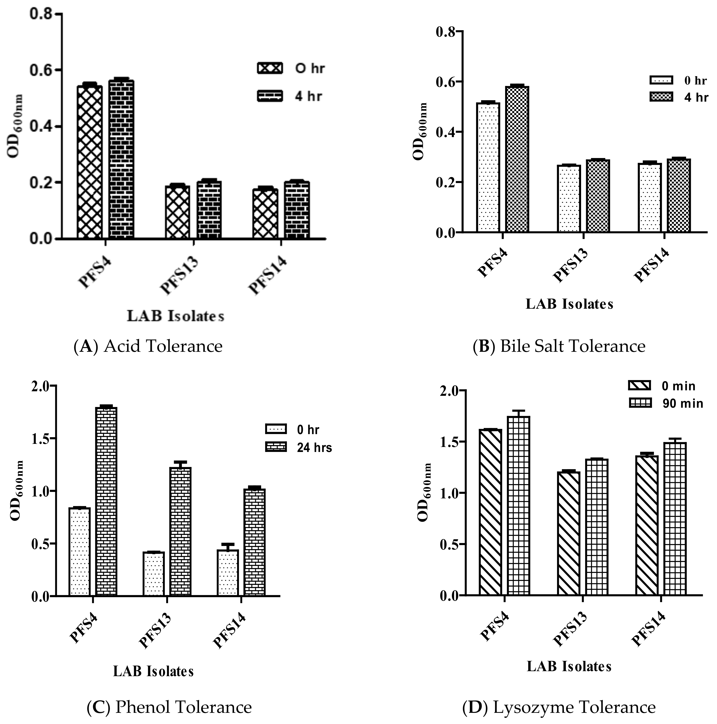
2.5.1. Acid Tolerance

The resistance to a low pH was assessed in vitro to determine the isolates’ ability to withstand passage through the gastric cavity, as described previously [39]. LAB isolates were grown for 24 h at 37 °C in MRS and M17 broth. In addition, 30 µL overnight-grown LAB isolates were suspended in 150 μL of MRS broth and M17 broth adjusted to pH 2.0 by 1M HCl. The cell suspensions were incubated anaerobically at 37 °C for 4 h. After incubation, bacterial growth was monitored by measuring the absorbance at 600 nm. All experiments were performed in triplicate.

2.5.2. Bile Tolerance

Bile tolerance tests for LAB isolates were performed as described [39]. First, 30 μL of overnight grown LAB isolates were suspended in 150 μL of MRS, and M17 broth with 0.3% bile salt (Oxoid, Hampshire, UK) was added to each well, while mixtures without bile salt were used as a control. The cell suspensions were incubated anaerobically at 37 °C for 4 h. Optical density (OD) at 600 nm was measured for monitoring the growth kinetics on a microtiter plate reader (Bio-Rad Laboratories, CA, USA). All of the experiments were performed in triplicate.

2.5.3. Lysozyme Tolerance

The lysozyme tolerance test was performed as described previously [39]. The overnight-grown cultures were harvested by centrifugation at 3500× g for 5 min. The bacterial cells were washed with phosphate buffer saline (PBS) and were re-suspended in a sterile PBS solution. The cell suspension (10 µL) was then inoculated into a PBS solution containing lysozyme (100 mg/L). Isolates inoculated in PBS without lysozyme were used as a control. LAB isolates were incubated at 37 °C. OD was measured at 600 nm after 30 and 90 min.

2.5.4. Phenol Tolerance

Gut microbes can break down aromatic amino acids, which result in the formation of phenol, which can inhibit the growth of LAB. Therefore, resistance to phenol tolerance by LAB is vital for their survival in GIT. Overnight-grown bacterial cultures were inoculated in respective broths supplemented with 0.4% phenol. OD was measured at 600 nm [39].

2.6. Cell Surface Properties

2.6.1. Auto Aggregation and Co-Aggregation Assay

The aggregation ability of LAB strains was studied as described previously [42]. The overnight grown culture was harvested by centrifugation at 5000× g for 20 min at 4 °C, and was washed and then suspended in PBS. Optical densities were adjusted at 0.25 ± 0.05 OD to maintain the number of bacterial cells (107–108 CFU/mL). For auto-aggregation, the OD of the LAB strains was recorded at 0 h and 24 h at 37 °C. The aggregation percentage was expressed as [1 − (ATime/A0) × 100], where ATime represents the absorbance of the mixture at 24 h and A0 represents the absorbance at 0 h (21). The criteria for the auto-aggregation ability determination were (low, 16–35%; medium, 35–50%; and high, 50% and above) [43].
For the co-aggregation assay, bacterial suspensions of LAB strains, as described above, were mixed with an equal volume (4 mL) of the overnight grown cultures of Salmonella in a sterile falcon tube; the mixtures were incubated at 37 °C. After 24 h, the OD620 nm was measured, and the percent co-aggregation was calculated as follows: [(Apathog + ALAB)/2 − (Amix)/(Apathog C ALAB)/2] × 100, where Apathog and ALAB represent the absorbance in the tubes containing only the pathogen and LAB strain, respectively, and Amix represents the absorbance of the mixture at 24 h [42].

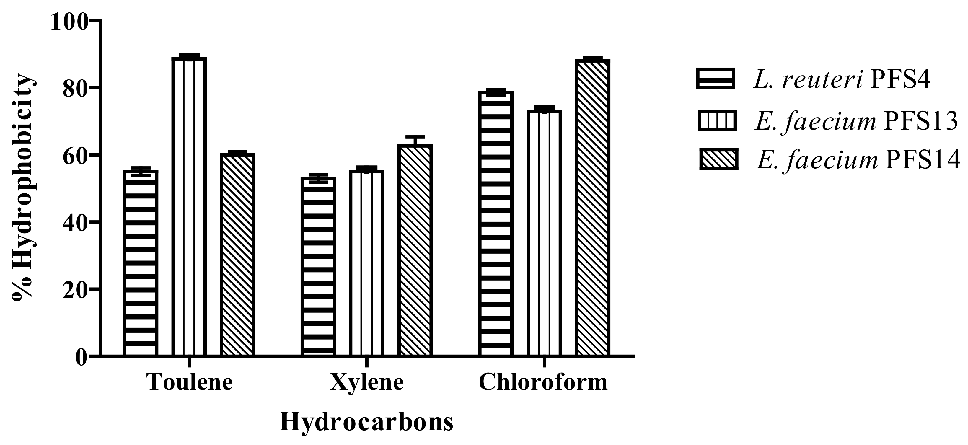
2.6.2. Microbial Adhesion to Hydrocarbon Test (MATH)

A bacterial cell surface hydrophobicity assay was performed with some modifications [44]. LAB isolates cultivated in MRS and M17 broth at 37 °C for 24 h were harvested by centrifugation at 8000× g for 10 min, and the cell pellet was washed thrice with sterile PBS and was re-suspended in the same media. The optical densities were maintained 1 ± 0.05 at 600 nm. Initially, 3 mL of the bacterial cell suspension was transferred to a sterile falcon tube, followed by the addition of 1 mL of hydrocarbons. Three hydrocarbons were tested in this study, namely: toluene (apolar, aromatic), xylene (apolar, aromatic), and chloroform (polar, aliphatic). The tubes were incubated at 37 °C for 10 min for temperature equilibrium, followed by 15 s vortexing, and were then set at 37 °C for 20 min for the phase separation. The lower aqueous phase was carefully collected in a separate glass tube, and OD600 nm was recorded.
% affinity = (ODi − ODt/ODi) × 100
ODi is the initial OD of the cell suspension, and ODt is OD of the aqueous phase recorded at 600 nm after 20 min.

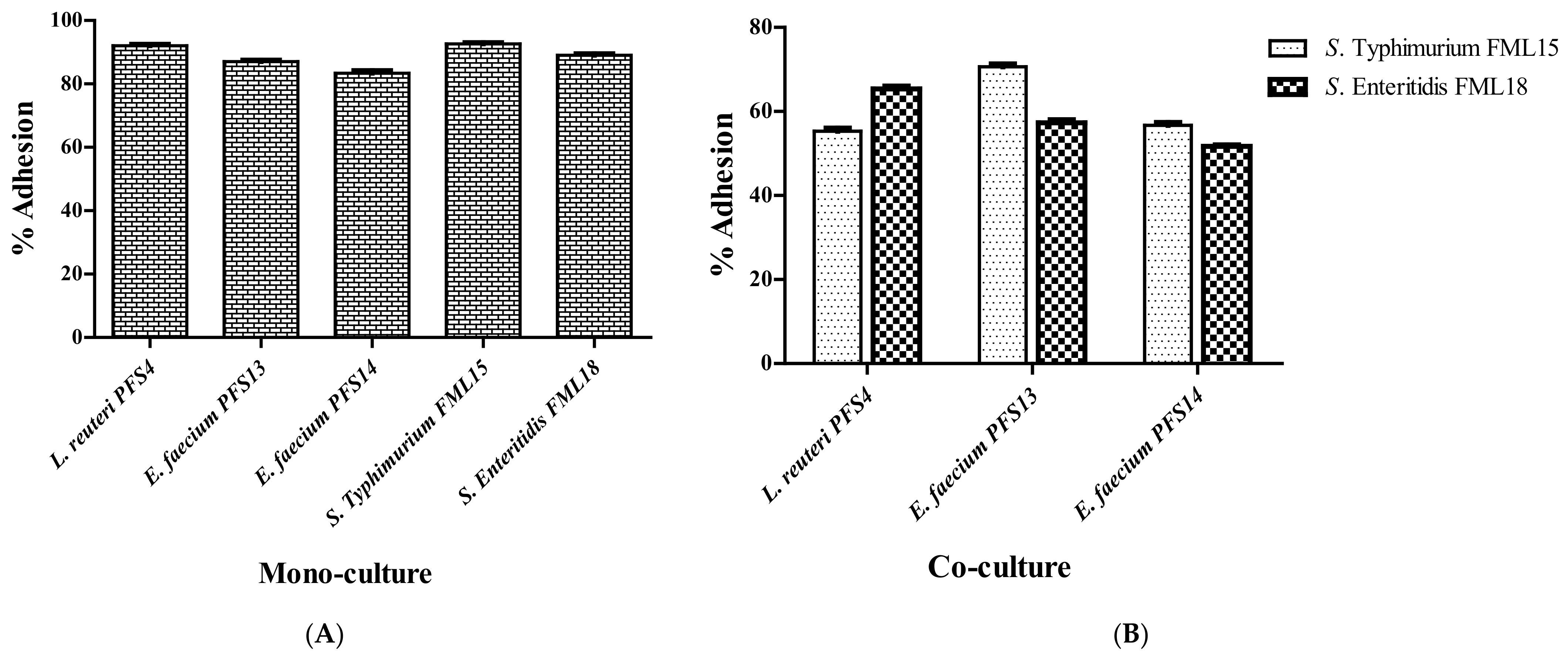
2.6.3. Mucin Adhesion Assay

The previously reported method was used to determine the in vitro mucin adhesion assay [45]. Flat-bottom 96 well microtiter plates were coated with 300 µL porcine mucin type III (10 mg/mL (Sigma-Aldrich, Inc. St. Louis, MO, USA) suspended in sterile PBS kept overnight at 4 °C. Plates were washed thrice with sterile PBS to remove unbound mucin from the wells. For competitive mucin adhesion, 100 µL of LAB culture with absorbance adjusted to 0.25 ± 0.05 and 100 µL of each of the Salmonella serovars were added to the mucin-coated wells simultaneously and were co-incubated for 90 min. After incubation, the wells were washed five times with PBS. Adhered bacterial cells were then treated with 300 µL Triton X in a sterile phosphate buffer solution to the separate bacterial cells. The viable cell count was measured by enumerating the LAB strains and Salmonella serovars on MRS agar, M17 agar, and SS agar. The % relative adhesion was calculated by (CFU/mL after adhesion/CFU/mL before adhesion) × 100.

2.6.4. Biofilm Formation Potential of Lactic Acid Bacteria

The quantification of the biofilm formation by lactic acid bacteria was determined as described previously [21]. Briefly, 150 μL of MRS and M17 broth were added to each well of 96 flat-bottom microtiter plates along with 50 μL of an overnight culture of L. reuteri and E. faecium. The 96 well microtiter plates were incubated at 37 °C for 48 h. To quantify the biofilm formation, the wells were gently washed thrice with PBS. Biofilms were then fixed with 2 mL methanol for 15 min, and the microplates were dried at room temperature. Then 2 mL of the 2% v/v crystal violet solution was added to each well and was incubated at room temperature for 5 min. The excess stain was removed by washing with sterilized water. The adherent cells were removed with 2 mL of 33% v/v glacial acetic acid. Each well’s optical density (OD) was measured at 600 nm using a plate reader (Bio-Rad, Hercules, CA, USA). The experiment was performed in triplicate. The cut-off (ODC) was defined as the mean OD value of the negative control. On the basis of OD, isolates were classified as follows: (4 × ODC) < OD = strongly adherent, (2 × ODC) < OD ≤ (4 × ODC) = moderately adherent, ODC < OD ≤ (2 × ODC) = weakly adherent, and OD ≤ ODC = non-adherent [46].

2.7. Antimicrobial Potential Assessment

2.7.1. Inhibition of Pathogenic Biofilm Formation

The effect of cell-free supernatant (CFS) of the LAB strains on the biofilm formation of S. Typhimurium and S. Enteritidis was evaluated as previously described [47]. The modified crystal violet assay was performed in 96-well microtiter plates for the biofilm quantification. First, 100 µL overnight-grown Salmonella strains were added in a well in the presence of 100 µL of pH neutralized CFS of the LAB isolates. The cultures were incubated at 37 °C for 48 h. The Salmonella test pathogens without CFS were used as a positive control. After incubation, the developed biofilm was washed three times with 200 μL of distilled water and was air dried. Then, 100 μL of 0.2% crystal violet (Merck KGaA, Gernsheim, Germany) was added to each well, and the plate was incubated for 20 min to allow for biofilm staining. Subsequently, the wells were washed three times with distilled water, and were air-dried and treated with 200 μL of 95% ethanol (Sigma-Aldrich, Inc. St. Louis MO USA) to dissolve the crystal violet crystals. The plate was incubated for 30 min, and the intensity of the crystal violet was measured at 600 nm using a microplate reader (Bio-Rad, Hercules, CA, USA). The ability of CFS to affect biofilm formation was calculated by comparing the absorbance values of the CFS treated wells versus the untreated control wells. The experiments were performed in triplicate. To estimate the reduction percent of CFS, the following formula was used:
Percentage inhibition = 100 − [(OD600 of wells in the presence of CFS × 100)/OD600 of wells in the presence of MRS]

2.7.2. Scanning Electron Microscopy

Scanning electron microscopy (SEM) was used to identify any changes or ruptures in the cell morphology of the Salmonella due to the effect of the CFS of the LAB strains, as described previously [48]. The overnight-grown culture of both Salmonella serovars were treated with CFS of L. reuteri PFS4, E. faecium PFS13, and E. faecium PFS14, alone and in combination, in a sterile 24-well microtiter plate (Corning, Corning, NY, USA) with a 12-mm round cover glass and were incubated for 48 h at 37 °C. The overnight-grown culture of both Salmonella enterica serovars in a tryptic soy broth (TSB) media with 12-mm round cover glass was used as the control. After incubation, the microtiter plate was gently washed to remove the non-adherent cells before fixation with 2.5% glutaraldehyde in a potassium phosphate buffer (50 mM, pH 7). The cover glass was dehydrated by chilled ethanol in a series (v/v) ranging from 30, 50, 70, 90, and 100%. For SEM, the specimens were dried to the critical point, coated with gold, and photographed with a scanning electron microscope (JSM-IT500HR, JEOL, Akishima, Japan).

2.7.3. Determination of the Antimicrobial Activity of the LAB Isolates

Aa time kill assay was performed as described previously [22]. Salmonella test pathogens were grown in a Luria broth (LB) media for 24 h at 37 °C. One hundred microliters of overnight-grown S. enterica serovars and pH neutralized CFS of LAB strains were co-cultured in a 96-well sterile microtiter plate and incubated at 37 °C. The number of viable bacteria was counted on an SS agar plate 0, 4, 8, 12, 24, and 48 h after incubation. Salmonella enterica strains without the cell-free supernatant of the LAB strains were taken as the positive control. The experiment was performed in triplicate.

2.8. Statistical Analysis

All of the experiments were carried out in triplicate. The standard deviations were determined with Microsoft Excel (Microsoft Corp., Albuquerque, NM, USA). A t-test was performed at the 95% confidence interval with SPSS20 (IBM Co., New York, NY, USA) to determine the statistical significance of the data.

3. Results

3.1. Bacterial Isolates

A total of 17 potential LAB strains were selected from the gut of broiler chickens. Among the 17 isolates, nine were chosen from MRS agar plates and eight were chosen from M17 agar plates. The isolates were identified as LAB based on the following characteristics: Gram-positive, catalase negative, non-motile, and cocci- or rod-shaped.

3.2. Safety Assessment

Hemolytic, DNase, and Antibiotic Susceptibility Assay

All 17 isolates showed negative results for hemolytic (γ-hemolysis) and DNase activity. However, a high resistance to most of the tested antibiotics, e.g., ciprofloxacin, chloramphenicol, third and fourth generation cephalosporin, amikacin, gentamicin, and vancomycin, was observed in the study (Table 1). Most of the strains were sensitive to rifampin, imipenem, meropenem, linezolid, and nalidixic acid. The criteria for selecting potential probiotics were as follows: isolates must be susceptible to clinically significant antibiotics, i.e., quinolones, ampicillin, amoxicillin-clavulanic acid, and cephalosporin [49]. Only three isolates, PFS4, PFS13, and PFS14, were chosen for further investigation based on the selection criteria.

3.3. Evaluation of Probiotic Properties

Tolerance to GIT Related Stresses

The survival of isolates in the GIT is an essential feature of probiotics. All 17 isolates showed tolerance to GIT related stresses as shown in (Figure S1). After selection of three LAB isolates on the basis of their safety profile, the tolerance to GIT-related stresses of selected LAB strains was shown in (Figure 1A–D). Acid tolerance is a vital characteristic for all probiotics. Bile salt tends to damage the cell membrane structure, so tolerance to bile salt is an essential characteristic of probiotics. All LAB isolates survived to exposure of pH 2 and 0.3% bile for 4 h (Figure 1A,B). The results indicate that all of the tested strains showed a high resistance to acid and bile. These isolates were subsequently evaluated for phenol resistance, where these showed increased tolerance to 0.4% phenol with OD values >1.000 after 24 h of incubation (Figure 1C). All of the tested LAB isolates showed significant resistance to lysozyme (100 mg/L) after 90 min (Figure 1D).

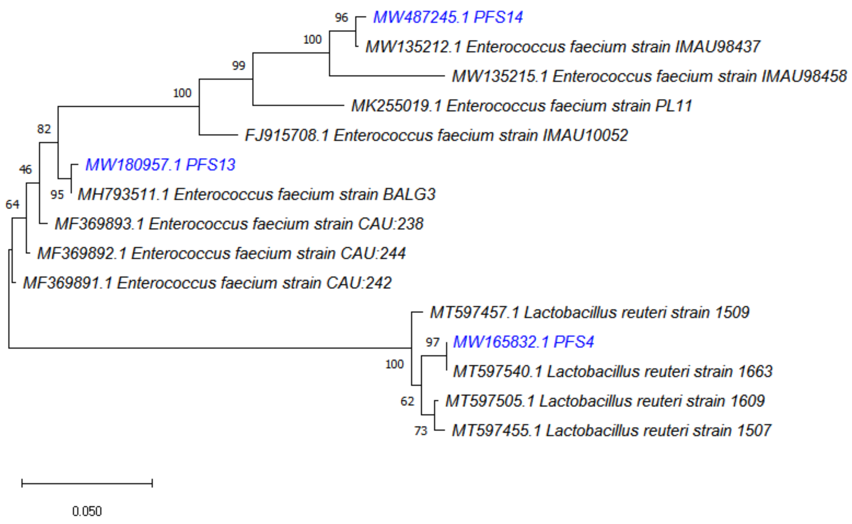
3.4. Molecular Identification of LAB Strains

The 16S rRNA sequences obtained were compared using the public database of NCBI. As a result, the LAB isolates were identified as L. reuteri PFS4, E. faecium PFS13, and E. faecium PFS14. A phylogenetic tree of bacterial isolates was created based on the minimum evolutionary distance to obtain a well-resolved tree. It was found that isolate PFS4 was under the group of Lactobacillus spp. and was closely related to identified organism L. reutei strain 1663 with a strong similarity of a 97% branch value. Isolates PFS13 and PFS14 was under the group of Enterococcus spp. and were closely related to the identified organism Enterococcus faecium strain BALG3 and Enterococcus faecium strain IMAU98437, with a strong similarity of 95% and 96% branch value, respectively (Figure 2).

3.5. Cell Surface Hydrophobicity

The microbial adhesion hydrocarbon test indicates the adherence capability of probiotic strains to intestinal cells. It is an essential characteristic that helps probiotics colonize, control enteric pathogens, and modulate the host immune system. LAB isolates showed variable hydrophobicity towards xylene (53–64%), toluene (59–88%), and chloroform (78–89%) (Figure 2). L. reuteri PFS4, E. faecium PFS13, and E. faecium PFS14 showed a maximum affinity towards chloroform at 89%, 78%, and 83%, respectively. E. faecium PFS13 showed a high affinity (88%) towards toluene, while L. reuteri PFS4 showed less affinity (53%) towards xylene in (Figure 3).

3.6. Auto-Aggregation and Co-Aggregation Assay

Auto-aggregation helps with adherence to epithelial cells in the intestine, and thus provides competitive adhesion against gut pathogens. All of the strains tested in this study exhibited some degree of auto-aggregation, ranging from 71.2% to 80.1%. The results indicate that each strain showed a good auto-aggregation ability according to the criteria described by [43]. Among these probiotic strains, L. reuteri PFS4 isolates showed the highest auto-aggregation capacity (80.1 ± 3.1%) at 24 h.
The co-aggregation ability of selected LAB isolates with Salmonella depends on the serovars (Table 2). E. faecium PFS13 showed the maximum co-aggregation ability (66 ± 1.1% and 71 ± 1.5%) with S. Typhimurium and S. Enteritidis, respectively, while L. reuteri PFS4 and E. faecium PFS14 exhibited moderate co-aggregation with both Salmonella serovars. A combination of LAB isolates showed a high co-aggregation percentage (52.2% and 68.6%) with S. Typhimurium FML15 and S. Enteritidis FML18, respectively.

3.7. In Vitro Mucin Adhesion Assay

All probiotic strains and Salmonella strains demonstrated a good mucin adhesion profile in the monoculture. The adhesion to porcine mucin films was >80% for all of the probiotic and Salmonella strains. The results revealed that L. reuteri PFS4 exhibited the highest percentage adhesion (92.6%), followed by E. faecium PFS13 (86.3%) and E. faecium PFS14 (84%) (Figure 4). However, the adhesion of Salmonella strains was significantly reduced in the presence of LAB isolates. The adhesion percentage of pathogenic strains, S. Typhimurium PF40 and S. Enteritidis PF76, was low (55.3% to 70.6%, and 51.6% to 65.3%, respectively) in the presence of LAB isolates compared with the untreated wells.

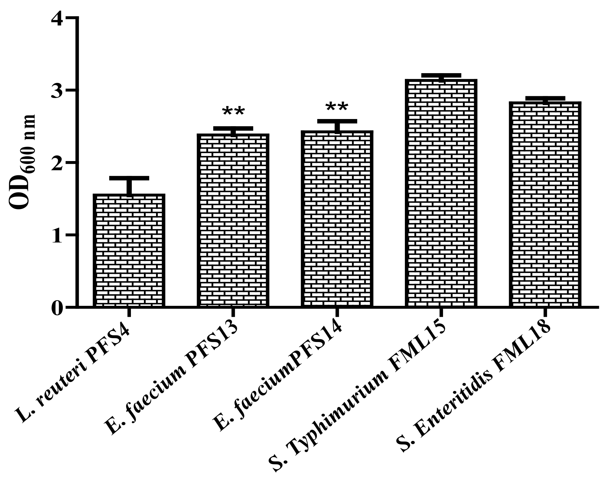
3.8. Biofilm Formation Assay

The biofilm formation by the probiotics is helpful in the competitive exclusion of gut pathogens in the intestine. Hence, the potential of probiotic candidates to form biofilms was evaluated in an in vitro assay. All of the probiotic candidate strains were capable of developing a moderate to strong biofilm (Figure 5). Both E. faecium strains exhibited a strong biofilm formation (OD > 2.0) potential, whereas L. reuteri PFS4 showed a moderate biofilm formation (OD < 2.0).

3.9. Effect of CFS on Biofilm Formation Ability of Salmonella enterica

The ability of probiotics to reduce Salmonella biofilms is an important characteristic for therapeutic probiotics post Salmonella infection. Therefore, the Salmonella biofilms were treated with pH neutralized probiotic CFS to assess the biofilm inhibition potential. Maximum inhibition was observed in the case of probiotic combination (90 and 92%) followed by L. reuteri PFS4 (87 and 89%), E. faecium PFS13 (80 and 87%), and E. faecium PFS14 (79 and 83%) for both S. Typhimurium and S. Enteritidis, respectively. The Salmonella biofilm was significantly inhibited (p ≤ 0.05) by the CFS of the three probiotics strains, alone or combined (Figure 6). However, the combination of CFS from all strains was better than the single strain CFS.

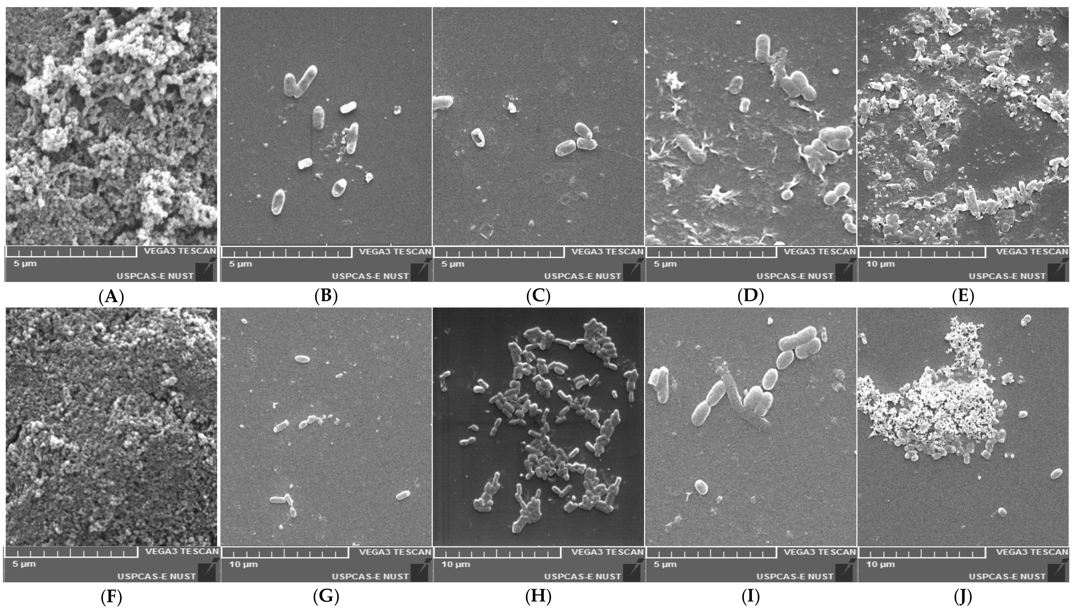
3.10. Scanning Electron Microscopic Study

The biofilm produced by S. enterica serovars was confirmed by SEM. It was observed that the control sample (S. enterica serovars cultured into sterile TSB) showed a high cell density and aggregation (Figure 7A,F). Compared with the control, the neutralizing CFS of the LAB strains reduced the adherence and aggregation of the tested Salmonella enterica strains after 48 h of incubation. These results agree with the findings of the crystal violet technique, which showed a significant reduction in bacterial adherence and biofilm formation (Figure 6).

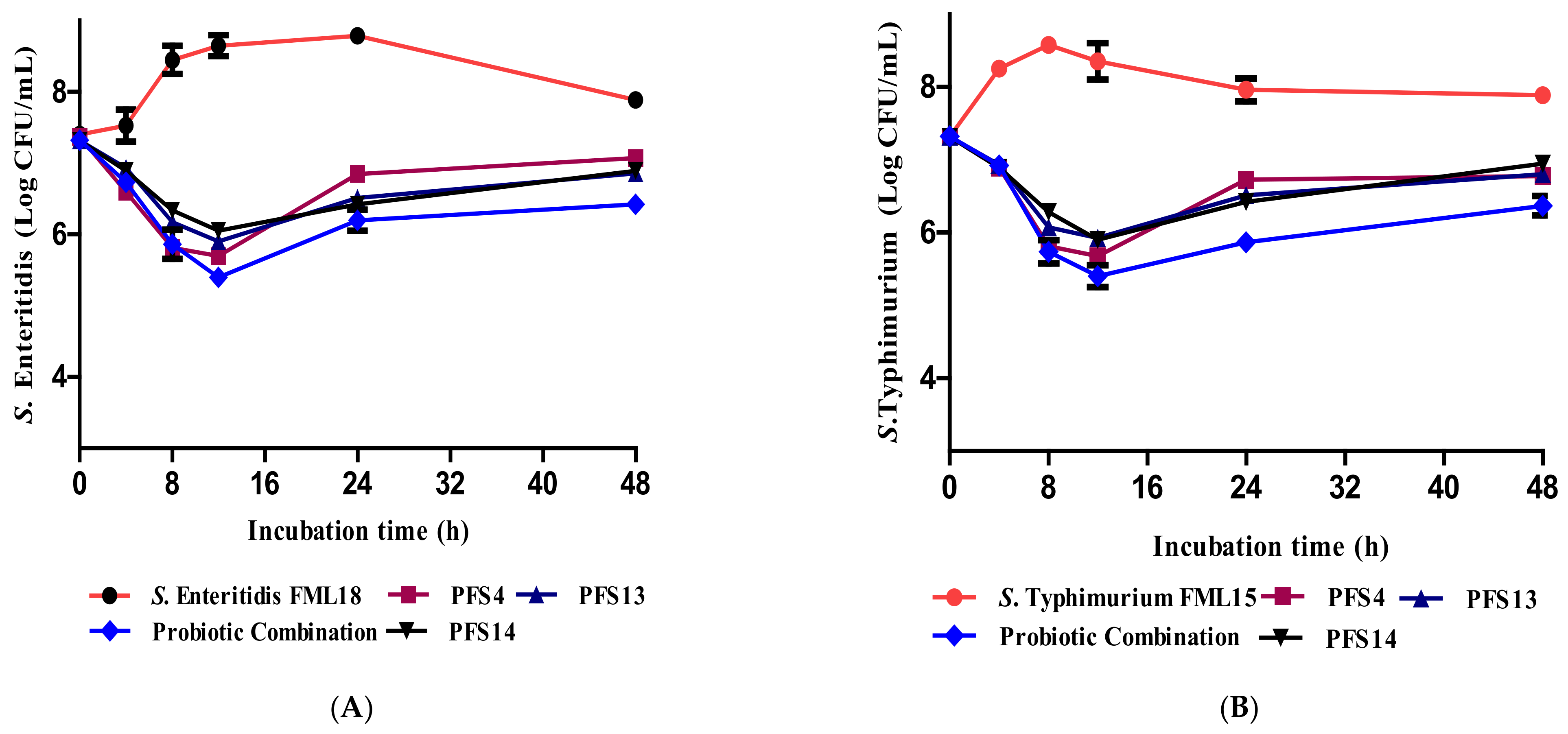
3.11. Effect of CFS on the Growth Kinetics of Salmonella enterica Serovars

The results of the time-kill assay indicated the ability of CFS to reduce the growth of Salmonella enterica serovars (Figure 8). The growth of S. Typhimurium FML15 and S. Enteritidis FML18 was significantly inhibited after culturing with CFS in all of the tested time points compared with the control cultures incubated without CFS (p ≤ 0.05). The inhibitory effect was more prominent on both serovars, as indicated by the lower CFU, particularly after 12 h of incubation. The CFS of the LAB combination significantly reduced the count of both Salmonella serovars by 2 log compared with the control Salmonella without any CFS after 12 h of incubation.

4. Discussion

The emergence of multi drug resistant (MDR) Salmonella enterica serovars Typhimurium and Enteritidis constitutes a significant health and economic threat for poultry and humans [8]. Therefore, the application of probiotics in the poultry industry as a suitable alternative to antibiotics in order to control enteric pathogens has received tremendous attention in recent years [50]. Thus, we investigated potential probiotics from the chicken gut to control clinically significant antibiotic-resistant Salmonella enterica.
Initially, 17 isolates from 10 poultry fecal samples were isolated based on their colony morphology, Gram staining, and biochemical characteristics. However, only three strains, L. reuteri PFS4, E. faecium PFS13, and E. faecium PFS14, were retained based on the selection criteria. The findings were consistent with previous studies, which found L. reuteri [51] and E. faecium [52] in the chicken gut. According to the previous study, L. reuteri is the most common Lactobacillus species in the chicken GIT, primarily found in the crop and cecum [53]. In addition, many previous studies have reported on the protective effect of L. reuteri [54,55] and E. faecium [56] against Salmonella serovars and other gut pathogens.
Potential probiotic LAB strains should withstand digestive system stress to benefit the health of their hosts. The ability to tolerate acid, bile salts, lysozyme, phenol, and the production of antimicrobial agents are all good indicators of a strain’s survival in the GIT [57]. In this study, all 17 isolates tested for GIT-related stress tolerance exhibited a high potential to tolerate a low pH (2), 0.3% bile salt concentration, lysozyme (100 mg/mL), and 0.4 % phenol. The ability of LAB isolates to tolerate a low pH and bile is not a surprise, as one of LAB’s natural habitat is the gastrointestinal tract of humans or animals [58]. Our results are in agreement with previous findings that reported on the survival of different LAB strains, including Enterococcus spp., L. salivarius, L. johnsonii, and L. reuteri, isolated from healthy chicken gut, being able to survive at pH 2 and 0.3% bile concentration [59,60,61]. Another study reported the resistance of LAB strains to lysozyme and 0.4% phenol isolated from poultry gut at varying degrees [51].
Lactobacilli is a probiotic genus widely employed in animal production [62]. Most Lactobacillus species (L. acidophilus, L. rhamnosus, and L. reuteri) strains demonstrate no pathogenicity or acute oral toxicity in animals [63]. Lactobacillus are considered GRAS “Generally Recognized as Safe” because of their long history of safe use in animal feed [64]. However, some researchers reported some enterococcal species as opportunistic pathogens that can cause infections in humans and animals. Thus, determining the safety of these strains is critical for selecting these strains for industrial purposes and, more importantly, as probiotics. However, in several investigations, E. faecium has also been used as a probiotic candidate in poultry birds [65,66].
All of the initially screened 17 isolates showed negative results for a hemolytic DNase activity and demonstrated variable antimicrobial resistance patterns towards the tested antibiotics (Table 2). These results are following the previous study where E. faecium and L. reuteri showed a negative hemolytic and DNase activity [67]. The isolated LAB should be sensitive to antibiotics in order to avoid any potential transfer of undesirable antibiotic resistance to the intestinal microbiota [68,69,70]. Recently, many researchers have revealed that commensal microbes, including LAB isolates, contain antibiotic resistance genes similar to those found in pathogens [71]. Genes conferring resistance to tetracycline and cephalosporin detected and characterized in the Lactococcus spp. and Lactobacilli constitute a reservoir of resistance for potential food and gut pathogens, thus presenting safety issues [72,73]. These results are in agreement with previous findings [74,75,76], which reported the safety and sensitivity of L. reuteri and Enterococcus strains isolated from poultry gut towards common antibiotics. The high intrinsic resistance and susceptibility of probiotic microorganisms to a variety of antibiotics are quite significant. Some LAB species, including L. reuteri, L. fermentum, L. rhamnosus, E. faecium, E. faecalis, and L. acidophilus, have intrinsic resistance to vancomycin, sulfamethoxazole/trimethoprim, and nalidixic acid [77]. LAB with acquired/transferable drug resistance genes are not GRAS (generally recognized as safe) and should not be added to probiotic foods or animal feed. Conjugative plasmids typically carry antibiotic resistance genes that can be passed on to other bacteria, leading to antibiotic-resistant enteropathogenic bacteria [78]. Therefore, due to safety issues, the antimicrobial resistance pattern of the bacterial species is an essential criterion for their selection as probiotic candidates.
Bacterial adhesion to intestinal surfaces could be initiated by non-specific physical interactions such as hydrophobic interactions, followed by adhesion by specific cell wall components at a second level [79]. Microbial cell surface hydrophobicity (MATH) is the most commonly used in vitro method to assess cell surface hydrophobicity in lactic acid bacteria [80]. Some researchers have reported a correlation between hydrophobicity and adhesion [81]. The hydrophobicity test can be considered as a pre-test for probiotic bacteria’s ability to adhere to epithelial cells, preventing pathogens’ colonization (Falah et al., 2019). Hydrophobicity is an important property for enhancing the first contact between the bacteria and host cells. The hydrophobicity of the cell surface of L. reuteri and E. faecium is determined in order to know the cell surface properties responsible for aggregation and adhesion. In our study, LAB isolates showed variable hydrophobicity with different hydrocarbons (53–89%). Low to high hydrophobicity abilities were observed in L. reuteri LR11, LR19, and LR26 (30–71.10%) isolated from the poultry gut [60]. In addition, [82] obtained a high hydrophobicity (>80%) among Enterococcus spp. isolated from poultry. Some authors have considered that L. reuteri and E. faecium, with a high hydrophobicity, have a better ability to bind to epithelial cells [83]. Aggregation helps probiotic strains colonize in the intestine and significantly attaches intestinal epithelial cells, preventing Salmonella adhesion. This test is essential for the selection of probable probiotic bacteria from the gut [84].
Auto-aggregation allows microorganisms of the same species to form self-forming communities, and this process is most commonly associated with microorganisms adhering to the intestinal mucosa [85]. Our data revealed that LAB isolates showed a high auto-aggregation ability ranging from 71–80%. Similar findings were observed in previous studies, where L. reuteri, E. faecium, and other Lactobacillus spp. showed high auto-aggregation (70–90%) abilities [86,87]. In addition, some in vivo studies reported that the ability of high auto-aggregation of L. reuteri and E. faecium was strongly associated with adhesion [88,89].
Co-aggregation is the intercellular adhesion of different strains that are linked to the ability to associate with pathogens. The co-aggregation potential of gut probiotics enables the host to limit enteric pathogens like S. Typhimurium and S. Enteritidis [90]. In our study, E. faecium PFS13 had the most remarkable ability to co-aggregate with Salmonella Typhimurium FML15 and Salmonella Enteritidis FML18 (66.1% and 71.5%, respectively). Therefore, all three LAB strains in our study, with moderate to high co aggregation abilities, are potential probiotic candidates. The results obtained in our study correspond with previous findings, where L. reuteri, Enterococcus spp., and L. acidophilus showed a moderate to high co-aggregation (40–60%) ability with enteric pathogens, including S. enterica [42,91]. Many of the guidelines for probiotics’ health benefits regard their ability to stay viable and conform to the host’s intestine as a critical factor. Adherence allows probiotics to live longer in the GI tract and improves bacterial–host interactions, but it also aids in reducing gastric motility. As a result, probiotics’ ability to adhere to mucosal surfaces and epithelial cells is essential [92].
Bacterial adhesion to the host mucin is thought to be critical in helping probiotic species colonize in any setting, the ability to exclude food-borne pathogens competitively, and to improve their ability to stimulate the immune system. The mucus layer not only serves as the first physical barrier to bacterial invasion of the gut epithelium, but it also serves as the first point of contact for gut bacteria with host cells. For many pathogens like Salmonella, mucin adhesion is an important element in virulence in the host. [93]. The mucus of the small and large intestines may contain many receptors that mediate the adherence of beneficial or harmful microorganisms [94]. All LAB isolates in this study showed good adhesion (84–92.6%) to porcine mucin Type III. In a previous study, L. reuteri showed a better adhesion (74%) ability than other LAB strains to the intestinal mucin [95].
On the other hand, the adhesion of Salmonella serovars was significantly reduced when they co-cultured with LAB strains. The previous findings of an in vitro mucin adhesion assay revealed that L. reuteri, L. rhamnosus, and L. casei competitively excluded (13.26% to 25%) S. paratyphi A, S. Typhimurium SA2093, S. flexneri, and S. aureus TISTR from adhesion to porcine mucin. The inhibition of pathogen adhesion to the mucin is reported to prevent translocation and subsequently infection [96,97].
Pathogenic and non-pathogenic microorganisms form biofilms responsible for maintaining such microorganisms in healthy ecosystems in vivo [98]. The biofilm formation ability can be considered beneficial for probiotic strains, promoting colonization and long-term permanence on the host’s mucosa [99]. Our data showed that all three LAB isolates are strong biofilm producers. Moderate to strong biofilm production by L. plantarum, L. reuteri, L. acidophilus, L. casei subsp. Pseudoplantarum. and E. faecium have been observed in previous studies [23,100]. Scanning electron microscope (SEM) images demonstrated a strong adherence and aggregation of S. Typhimurium and S. Enteritidis bacterial cells. Our SEM images also showed that the adherence of cells was minimized after CFS treatment. Similar findings were observed in a previous study where the CFS of different probiotic strains reduced biofilm formation [22]. The results of the in vitro mucin adhesion assay, aggregation abilities, cell surface hydrophobicity, and biofilm formation capability of our LAB isolates showed strong adhesion abilities. They can prevent NTS in poultry gut through a competitive adhesion mechanism. Some of the Lactobacillus spp. with a low adhesion and co-aggregation ability were excluded in a previous study, as they could not be selected as potential probiotic candidates [101]. Our findings provide the foundation for understanding L. reuteri and E. faecium adhesion mechanisms and for predicting adherence in various host models. The authors of [102] revealed that S. Enteritidis growth in poultry birds was considerably reduced by L. reuteri, with a good coaggregation ability and mucin adhesion profile.
Lactic acid bacteria produce various antimicrobial agents, including organic acids, ethanol, diacetyl, hydrogen peroxide, and antimicrobial peptides [103,104]. The inhibition of pathogenic bacterial biofilms through probiotics is an attractive target for therapeutic intervention [105]. Probiotics have received significant attention in recent years, leading to the discovery of biofilm inhibitors to counter foodborne pathogens, including S. Typhimurium and S. Enteritidis [42,106]. Our data suggest that pH neutralized CFS of selected LAB strains alone and in combination significantly reduced the biofilm formation (up to 84%) of Salmonella strains compared with the control. The anti-biofilm property of CFS of L. reuteri and E. faecium against Salmonella enterica has been reported in previous studies [23,42,106]. The antagonistic activity of certain microbes and their extracellular antibacterial compounds found in cell-free supernatants (CFSs) provides an excellent potential for controlling foodborne pathogens, including E. coli and Salmonella enterica [107]. The findings of the growth kinetic study revealed that all three LAB isolates, individually and in combination, significantly reduced the growth of Salmonella test pathogens for up to 12 h. These results of the antagonistic activity against Salmonella could be due to various antimicrobial substances produced by probiotic strains. L. reuteri and E. faecium produced various antimicrobial substances such as lactic acid, acetic acid, hydrogen peroxide, bacteriocins, and bacteriocins, like inhibitory substances. The authors of [108,109,110,111] studied the efficacy of CFS on MDR Salmonella and other Enterobacteracae species and revealed that CFS significantly inhibits the growth of pathogens.

5. Conclusions

Based on our findings, we may infer that the LAB strains isolated from poultry gut exhibit endurance to GIT related stress. L. reuteri and E. faecium strains demonstrated a co-aggregation potential and competitively reduced the mucin adhesion of S. Typhimurium and S. Enteritidis. This study provides evidence that isolated probiotic strains from poultry gut have an in vitro antagonistic activity towards extended spectrum cephalosporin and fluoroquinolone (key antibiotics for salmonellosis) resistant Salmonella serovars. Therefore, L. reuteri PFS4, E. faecium PFS13, and E. faecium PFS14 can be potential candidates to control extended spectrum cephalosporin and fluoroquinolones resistant Salmonella serovars after being subjected to in vivo studies. In vivo studies using these strains can be performed to elucidate their safety profile, growth, and disease prevention ability. As part of the One Health approach, resistant-specific pathogen control can help mitigate the resistant pathogen load in animals and it’s spread to the associated food ecosystem.

Supplementary Materials

The following are available online at https://www.mdpi.com/article/10.3390/ani11123435/s1, Figure S1: Survivability of gastrointestinal tract (GIT) related stresses of 17 LAB strains (A) Acidic pH (pH 2) tolerance. (B) 0.3% Bile salts tolerance. (C) Phenol tolerance. (D) Lysozyme tolerance.

Author Contributions

A.S. performed the experiments and wrote the manuscript. S.A. performed the experiments. F.A., M.A. and M.I. completed the data analysis and technical assistance. A.A. and A.R.—planned and executed the experiments, analyzed the data, and wrote the manuscript. E.G., provided comments and wrote the manuscript. All authors have read and agreed to the published version of the manuscript.

Funding

This study was financially supported through the ICT Endowment scholarship by National University of Science and Technology, Islamabad, Pakistan.

Institutional Review Board Statement

The ethical approval of the study was approved by the Institutional Review Board of the Atta Ur Rahman School of Applied Biosciences, National University of Science and Technology, (ref no IRB-132, Dated: 06-12-2018).

Data Availability Statement

The 16S rRNA sequences have been deposited in NCBI under the accession numbers; MW165832, MW180957 and MW487245.

Conflicts of Interest

All of the authors declare no competing interests.

References

  1. Adetoye, A.; Pinloche, E.; Adeniyi, B.A.; Ayeni, F.A. Characterization and anti-salmonella activities of lactic acid bacteria isolated from cattle faeces. BMC Microbiol. 2018, 18, 96. [Google Scholar] [CrossRef] [Green Version]
  2. Nhung, N.; Van, N.; Van Cuong, N.; Duong, T.; Nhat, T.; Hang, T.; Nhi, N.; Kiet, B.; Hien, V.; Ngoc, P.; et al. Antimicrobial residues and resistance against critically important antimicrobials in non-typhoidal Salmonella from meat sold at wet markets and supermarkets in Vietnam. Int. J. Food Microbiol. 2018, 266, 301–309. [Google Scholar] [CrossRef] [PubMed]
  3. Azam, A.; Shafique, M. Agriculture in Pakistan and its Impact on Economy. Int. J. Adv. Sci. Technol. 2017, 103, 47–60. [Google Scholar] [CrossRef]
  4. Rafique, M.; Potter, R.F.; Ferreiro, A.; Wallace, M.A.; Rahim, A.; Malik, A.A.; Siddique, N.; Abbas, M.A.; D’Souza, A.W.; Burnham, C.; et al. Genomic characterization of antibiotic resistant Escherichia coli isolated from domestic chickens in Pakistan. Front. Microbiol. 2020, 10, 3052. [Google Scholar] [CrossRef]
  5. Cox, J.; Pavic, A. Advances in enteropathogen control in poultry production. J. Appl. Microbiol. 2010, 108, 745–755. [Google Scholar] [CrossRef] [PubMed]
  6. Cortés-Sánchez, A.d.J.; Garcia-Barrientos, R.; Minor-Pérez, H.; Dublán-García, O.; San Martin-Azocar, A. Food Safety and Antimicrobial Resistance an Approach to the Genus Salmonella spp. J. Biosci. Med. 2017, 5, 55. [Google Scholar]
  7. Heredia, N.; García, S. Animals as sources of food-borne pathogens: A review. Anim. Nutr. 2018, 4, 250–255. [Google Scholar] [CrossRef]
  8. Gut, A.M.; Vasiljevic, T.; Yeager, T.; Donkor, O.N. Salmonella infection–prevention and treatment by antibiotics and probiotic yeasts: A review. Microbiology 2018, 164, 1327–1344. [Google Scholar] [CrossRef] [PubMed]
  9. Hur, J.; Jawale, C.; Lee, J.H. Antimicrobial resistance of Salmonella isolated from food animals: A review. Food Res. Int. 2012, 45, 819–830. [Google Scholar] [CrossRef]
  10. Andoh, L.A.; Ahmed, S.; Olsen, J.E.; Obiri-Danso, K.; Newman, M.J.; Opintan, J.A.; Barco, L.; Dalsgaard, A. Prevalence and characterization of Salmonella among humans in Ghana. Trop. Med. Health 2017, 45, 3. [Google Scholar] [CrossRef] [Green Version]
  11. Breurec, S.; Reynaud, Y.; Frank, T.; Farra, A.; Costilhes, G.; Weill, F.-X.; Le Hello, S. Serotype distribution and antimicrobial resistance of human Salmonella enterica in Bangui, Central African Republic, from 2004 to 2013. PLoS Negl. Trop. Dis. 2019, 13, e0007917. [Google Scholar] [CrossRef] [Green Version]
  12. EFSA. The European Union Summary Report on Antimicrobial Resistance in zoonotic and indicator bacteria from humans, animals and food in 2017/2018. EFSA J. 2020, 18, 6007. [Google Scholar]
  13. EFSA. The European Union Summary Report on Antimicrobial Resistance in zoonotic and indicator bacteria from humans, animals and food in 2018/2019. EFSA J. 2021, 19, 6490. [Google Scholar]
  14. Prestinaci, F.; Pezzotti, P.; Pantosti, A. Antimicrobial resistance: A global multifaceted phenomenon. Pathog. Glob. Health 2015, 109, 309–318. [Google Scholar] [CrossRef] [Green Version]
  15. Wang, H.; Ye, K.; Wei, X.; Cao, J.; Xu, X.; Zhou, G. Occurrence, antimicrobial resistance and biofilm formation of Salmonella isolates from a chicken slaughter plant in China. Food Control 2013, 33, 378–384. [Google Scholar] [CrossRef]
  16. Peruzy, M.F.; Capuano, F.; Proroga, Y.T.R.; Cristiano, D.; Carullo, M.R.; Murru, N. Antimicrobial Susceptibility Testing for Salmonella Serovars Isolated from Food Samples: Five-Year Monitoring (2015–2019). Antibiotics 2020, 9, 365. [Google Scholar] [CrossRef]
  17. Lunn, A.D.; Fàbrega, A.; Sánchez-Céspedes, J.; Vila, J. Prevalence of mechanisms decreasing quinolone-susceptibility among Salmonella spp. clinical isolates. Int. Microbiol. 2010, 13, 15–20. [Google Scholar] [PubMed]
  18. Siddiqui, T.R.; Bibi, S.; Mustufa, M.A.; Ayaz, S.M.; Khan, A. High prevalence of typhoidal Salmonella enterica serovars excreting food handlers in Karachi-Pakistan: A probable factor for regional typhoid endemicity. J. Health Popul. Nutr. 2015, 33, 27. [Google Scholar] [CrossRef] [PubMed] [Green Version]
  19. Venter, H.; Henningsen, M.L.; Begg, S.L. Antimicrobial resistance in healthcare, agriculture and the environment: The biochemistry behind the headlines. Essays Biochem. 2017, 61, 1–10. [Google Scholar] [CrossRef]
  20. Fijan, S.; Frauwallner, A.; Langerholc, T.; Krebs, B.; ter Haar née Younes, J.A.; Heschl, A.; Mičetić Turk, D.; Rogelj, I. Efficacy of using probiotics with antagonistic activity against pathogens of wound infections: An integrative review of literature. BioMed Res. Int. 2019, 2019, 7585486. [Google Scholar] [CrossRef] [PubMed] [Green Version]
  21. Hossain, M.I.; Mizan, M.F.R.; Ashrafudoulla, M.; Nahar, S.; Joo, H.; Jahid, I.; Park, S.; Kim, K.; Ha, S. Inhibitory effects of probiotic potential lactic acid bacteria isolated from kimchi against Listeria monocytogenes biofilm on lettuce, stainless-steel surfaces, and MBEC™ biofilm device. LWT 2020, 118, 108864. [Google Scholar] [CrossRef]
  22. Prabhurajeshwar, C.; Chandrakanth, K. Evaluation of antimicrobial properties and their substances against pathogenic bacteria in-vitro by probiotic Lactobacilli strains isolated from commercial yoghurt. Clin. Nutr. Exp. 2019, 23, 97–115. [Google Scholar] [CrossRef] [Green Version]
  23. Merino, L.; Trejo, F.M.; De Antoni, G.; Golowczyc, M.A. Lactobacillus strains inhibit biofilm formation of Salmonella sp. isolates from poultry. Food Res. Int. 2019, 123, 258–265. [Google Scholar] [CrossRef]
  24. Oyewole, O.F.; Maria, C.O.; Tope, P.S.; Funmi, O.O. In vitro Study of Potential Probiotic Lactic Acid Bacteria Isolated from The Gut of Chickens in Abeokuta, Nigeria. Alex. J. Vet. Sci. 2018, 58, 73–84. [Google Scholar] [CrossRef]
  25. Sanders, M.E.; Akkermans, L.M.; Haller, D.; Hammerman, C.; Heimbach, J.T.; Hörmannsperger, G.; Huys, G. Safety assessment of probiotics for human use. Gut Microbes 2010, 1, 164–185. [Google Scholar] [CrossRef]
  26. Drago, L.; Gismondo, M.R.; Lombardi, A.; De Haën, C.; Gozzini, L. Inhibition of in vitro growth of enteropathogens by new Lactobacillus isolates of human intestinal origin. FEMS Microbiol. Lett. 1997, 153, 455–463. [Google Scholar] [CrossRef] [PubMed]
  27. Gharbi, Y.; Fhoula, I.; Ruas-Madiedo, P.; Afef, N.; Boudabous, A.; Gueimonde, M.; Ouzari, H. In-vitro characterization of potentially probiotic Lactobacillus strains isolated from human microbiota: Interaction with pathogenic bacteria and the enteric cell line HT29. Ann. Microbiol. 2019, 69, 61–72. [Google Scholar] [CrossRef]
  28. Pellegrino, M.S.; Frola, I.D.; Natanael, B.; Gobelli, D.; Nader-Macias, M.E.; Bogni, C. In vitro characterization of lactic acid bacteria isolated from bovine milk as potential probiotic strains to prevent bovine mastitis. Probiotics Antimicrob. Proteins 2019, 11, 74–84. [Google Scholar] [CrossRef] [PubMed]
  29. Filho, R.A.; Higgins, J.; Higgins, S.; Gaona, G.; Wolfenden, A.; Tellez, G.; Hargis, B. Ability of bacteriophages isolated from different sources to reduce Salmonella enterica serovar Enteritidis in vitro and in vivo. Poult. Sci. 2007, 86, 1904–1909. [Google Scholar] [CrossRef]
  30. Vinderola, G.; Gueimonde, M.; Gomez-Gallego, C.; Delfederico, L.; Salminen, S. Correlation between in vitro and in vivo assays in selection of probiotics from traditional species of bacteria. Trends Food Sci. Technol. 2017, 68, 83–90. [Google Scholar] [CrossRef]
  31. Ashraf, R.; Shah, N.P. Selective and differential enumerations of Lactobacillus delbrueckii subsp. bulgaricus, Streptococcus thermophilus, Lactobacillus acidophilus, Lactobacillus casei and Bifidobacterium spp. in yoghurt—A review. Int. J. Food Microbiol. 2011, 149, 194–208. [Google Scholar] [CrossRef] [PubMed]
  32. Siddique, A.; Azim, S.; Ali, A.; Andleeb, S.; Ahsan, A.; Imran, M.; Rahman, A. Antimicrobial Resistance Profiling of Biofilm Forming Non Typhoidal Salmonella enterica Isolates from Poultry and Its Associated Food Products from Pakistan. Antibiotics 2021, 10, 785. [Google Scholar] [CrossRef]
  33. Torshizi, M.; Rahimi, S.; Mojgani, N.; Esmaeilkhanian, S.; Grimes, J. Screening of indigenous strains of lactic acid bacteria for development of a probiotic for poultry. Asian-Australas. J. Anim. Sci. 2008, 21, 1495–1500. [Google Scholar] [CrossRef]
  34. Taheur, F.B.; Kouidhi, B.; Fdhila, K.; Elabed, H.; Slama, R.B.; Mahdouani, K.; Bakhrouf, A.; Chaieb, K. Anti-bacterial and anti-biofilm activity of probiotic bacteria against oral pathogens. Microb. Pathog. 2016, 97, 213–220. [Google Scholar] [CrossRef]
  35. Singhal, N.; Singh, N.S.; Mohanty, S.; Singh, P.; Virdi, J.S. Evaluation of probiotic characteristics of lactic acid bacteria isolated from two commercial preparations available in Indian market. Indian J. Microbiol. 2019, 59, 112–115. [Google Scholar] [CrossRef]
  36. Jorgensen, J.H.; Turnidge, J.D. Susceptibility test methods: Dilution and disk diffusion methods. In Manual of Clinical Microbiology, 11th ed.; American Society of Microbiology: Washington, DC, USA, 2015; pp. 1253–1273. [Google Scholar]
  37. Vlková, E.; Rada, V.; Popelářová, P.; Trojanová, I.; Killer, J. Antimicrobial susceptibility of bifidobacteria isolated from gastrointestinal tract of calves. Livest. Sci. 2006, 105, 253–259. [Google Scholar] [CrossRef]
  38. Cheng, H.-R.; Jiang, N. Extremely rapid extraction of DNA from bacteria and yeasts. Biotechnol. Lett. 2006, 28, 55–59. [Google Scholar] [CrossRef] [PubMed]
  39. Turchi, B.; Mancini, S.; Fratini, F.; Pedonese, F.; Nuvoloni, R.; Bertelloni, F.; Ebani, V.V.; Cerri, D. Preliminary evaluation of probiotic potential of Lactobacillus plantarum strains isolated from Italian food products. World J. Microbiol. Biotechnol. 2013, 29, 1913–1922. [Google Scholar] [CrossRef] [PubMed]
  40. Rajoka, M.S.R.; Hayat, H.; Sarwar, S.; Mehwish, H.; Ahmad, F.; Hussain, N.; Shah, S.; Khurshid, M.; Siddiqu, M.; Shi, J. Isolation and evaluation of probiotic potential of lactic acid bacteria isolated from poultry intestine. Microbiology 2018, 87, 116–126. [Google Scholar] [CrossRef]
  41. Altschul, S.F.; Gish, W.; Miller, W.; Myers, E.W.; Lipman, D.J. Basic local alignment search tool. J. Mol. Biol. 1990, 215, 403–410. [Google Scholar] [CrossRef]
  42. Gómez, N.C.; Ramiro, J.M.; Quecan, B.X.; Franco, B.D.d.M. Use of potential probiotic lactic acid bacteria (LAB) biofilms for the control of Listeria monocytogenes, Salmonella typhimurium, and Escherichia coli O157: H7 biofilms formation. Front. Microbiol. 2016, 7, 863. [Google Scholar] [CrossRef] [Green Version]
  43. Sharma, S.; Kandasamy, S.; Kavitake, D.; Shetty, P.H. Probiotic characterization and antioxidant properties of Weissella confusa KR780676, isolated from an Indian fermented food. LWT 2018, 97, 53–60. [Google Scholar] [CrossRef]
  44. Nachtigall, C.; Weber, C.; Rothenburger, S.; Jaros, D.; Rohm, H. Test parameters and cell chain length of Streptococcus thermophilus affect the microbial adhesion to hydrocarbons assay: A methodical approach. FEMS Microbiol. Lett. 2019, 366, fnz150. [Google Scholar] [CrossRef]
  45. Valeriano, V.; Parungao-Balolong, M.; Kang, D.K. In vitro evaluation of the mucin-adhesion ability and probiotic potential of Lactobacillus mucosae LM 1. J. Appl. Microbiol. 2014, 117, 485–497. [Google Scholar] [CrossRef] [PubMed]
  46. Saxena, S.; Banerjee, G.; Garg, R.; Singh, M. Comparative study of biofilm formation in Pseudomonas aeruginosa isolates from patients of lower respiratory tract infection. J. Clin. Diagn. Res. 2014, 8, DC09. [Google Scholar] [PubMed]
  47. Kaur, S.; Sharma, P.; Kalia, N.; Singh, J.; Kaur, S. Anti-biofilm Properties of the Fecal Probiotic Lactobacilli Against Vibrio spp. Front. Cell. Infect. Microbiol. 2018, 8, 120. [Google Scholar] [CrossRef] [Green Version]
  48. Tareb, R.; Bernardeau, M.; Gueguen, M.; Vernoux, J. In vitro characterization of aggregation and adhesion properties of viable and heat-killed forms of two probiotic Lactobacillus strains and interaction with foodborne zoonotic bacteria, especially Campylobacter jejuni. J. Med. Microbiol. 2013, 62, 637–649. [Google Scholar] [CrossRef]
  49. Das, D.J.; Shankar, A.; Johnson, J.B.; Thomas, S. Critical insights into antibiotic resistance transferability in probiotic Lactobacillus. Nutrition 2020, 69, 110567. [Google Scholar] [CrossRef]
  50. Ficoseco, C.A.; Mansilla, F.I.; Maldonado, N.C.; Miranda, H.; Nader-Macias, M.E.F.; Vignolo, G.M. Safety and growth optimization of lactic acid bacteria isolated from feedlot cattle for probiotic formula design. Front. Microbiol. 2018, 9, 2220. [Google Scholar] [CrossRef]
  51. Musikasang, H.; Tani, A.; H-kittikun, A.; Maneerat, S. Probiotic potential of lactic acid bacteria isolated from chicken gastrointestinal digestive tract. World J. Microbiol. Biotechnol. 2009, 25, 1337–1345. [Google Scholar] [CrossRef]
  52. Hammerum, A.M. Enterococci of animal origin and their significance for public health. Clin. Microbiol. Infect. 2012, 18, 619–625. [Google Scholar] [CrossRef] [PubMed]
  53. Wang, L.; Fang, M.; Hu, Y.; Yang, Y.; Yang, M.; Chen, Y. Characterization of the most abundant Lactobacillus species in chicken gastrointestinal tract and potential use as probiotics for genetic engineering. Acta Biochim. Biophys. Sin. 2014, 46, 612–619. [Google Scholar] [CrossRef] [PubMed] [Green Version]
  54. Kuleaşan, H.; Çakmakçı, M. Effect of reuterin, produced by Lactobacillus reuteri on the surface of sausages to inhibit the growth of Listeria monocytogenes and Salmonella spp. Food/Nahrung 2002, 46, 408–410. [Google Scholar] [CrossRef]
  55. Shi, S.; Qi, Z.; Sheng, T.; Tu, J.; Shao, Y.; Qi, K. Antagonistic trait of Lactobacillus reuteri S5 against Salmonella enteritidis and assessment of its potential probiotic characteristics. Microb. Pathog. 2019, 137, 103773. [Google Scholar] [CrossRef] [PubMed]
  56. Audisio, M.C.; Oliver, G.; Apella, M. Protective effect of Enterococcus faecium J96, a potential probiotic strain, on chicks infected with Salmonella pullorum. J. Food Prot. 2000, 63, 1333–1337. [Google Scholar] [CrossRef] [PubMed]
  57. Reuben, R.C.; Roy, P.C.; Sarkar, S.L.; Alam, R.-U.; Jahid, I. Isolation, characterization, and assessment of lactic acid bacteria toward their selection as poultry probiotics. BMC Microbiol. 2019, 19, 253. [Google Scholar] [CrossRef] [PubMed] [Green Version]
  58. Amin, M.; Adams, M.B.; Burke, C.M.; Bolch, C. Isolation and screening of lactic acid bacteria associated with the gastrointestinal tracts of abalone at various life stages for probiotic candidates. Aquac. Rep. 2020, 17, 100378. [Google Scholar] [CrossRef]
  59. Blajman, J.; Gaziano, C.; Zbrun, M.V.; Soto, L.; Astesana, D.; Berisvil, A.; Scharpen, A.R.; Signorini, M.; Frizzo, L. In vitro and in vivo screening of native lactic acid bacteria toward their selection as a probiotic in broiler chickens. Res. Vet. Sci. 2015, 101, 50–56. [Google Scholar] [CrossRef]
  60. García-Hernández, Y.; Pérez-Sánchez, T.; Boucourt, R.; Balcázar, J.L.; Nicoli, J.R.; Moreira-Silva, J.; Rodríguez, Z.; Fuertes, H.; Nuñez, O.; Albelo, N.; et al. Isolation, characterization and evaluation of probiotic lactic acid bacteria for potential use in animal production. Res. Vet. Sci. 2016, 108, 125–132. [Google Scholar] [CrossRef] [PubMed]
  61. Musikasang, H.; Sohsomboon, N.; Tani, A.; Maneerat, S. Bacteriocin-producing lactic acid bacteria as a probiotic potential from Thai indigenous chickens. Czech J. Anim. Sci. 2012, 57, 137–149. [Google Scholar] [CrossRef] [Green Version]
  62. Dowarah, R.; Verma, A.; Agarwal, N. The use of Lactobacillus as an alternative of antibiotic growth promoters in pigs: A review. Anim. Nutr. 2017, 3, 1–6. [Google Scholar] [CrossRef]
  63. Bernardeau, M.; Vernoux, J.P.; Gueguen, M. Safety and efficacy of probiotic lactobacilli in promoting growth in post-weaning Swiss mice. Int. J. Food Microbiol. 2002, 77, 19–27. [Google Scholar] [CrossRef]
  64. Anadón, A.; Martínez-Larrañaga, M.R.; Martínez, M. Probiotics for animal nutrition in the European Union. Regulation and safety assessment. Regul. Toxicol. Pharmacol. 2006, 45, 91–95. [Google Scholar] [CrossRef]
  65. Cao, G.; Zeng, X.; Chen, A.; Zhou, L.; Zhang, L.; Xiao, Y.; Yang, C. Effects of a probiotic, Enterococcus faecium, on growth performance, intestinal morphology, immune response, and cecal microflora in broiler chickens challenged with Escherichia coli K88. Poult. Sci. 2013, 92, 2949–2955. [Google Scholar] [CrossRef] [PubMed]
  66. Park, J.; Jeong, J.; Lee, S.; Kim, I. Effect of dietary supplementation with a probiotic (Enterococcus faecium) on production performance, excreta microflora, ammonia emission, and nutrient utilization in ISA brown laying hens. Poult. Sci. 2016, 95, 2829–2835. [Google Scholar] [CrossRef] [PubMed]
  67. Babot, J.D.; Argañaraz-Martínez, E.; Saavedra, L.; Apella, M.C.; Chaia, A.P. Compatibility and safety of five lectin-binding putative probiotic strains for the development of a multi-strain protective culture for poultry. Benef. Microbes 2018, 9, 927–935. [Google Scholar] [CrossRef]
  68. Huys, G.; D’HAENE, K.; Danielsen, M.; Maettoe, J.; Egervärn, M.; Vandamme, P. Phenotypic and molecular assessment of antimicrobial resistance in Lactobacillus paracasei strains of food origin. J. Food Prot. 2008, 71, 339–344. [Google Scholar] [CrossRef] [PubMed]
  69. Sornplang, P.; Soikum, C. Effect of Lactobacillus spp. Supplementation on Spoilage Bacteria Decontamination in Chicken Meat. In Proceedings of the International Conference on Beneficial Microbes 2016, Phuket, Thailand, 31 May–2 June 2016. [Google Scholar]
  70. Toomey, N.; Bolton, D.; Fanning, S. Characterisation and transferability of antibiotic resistance genes from lactic acid bacteria isolated from Irish pork and beef abattoirs. Res. Microbiol. 2010, 161, 127–135. [Google Scholar] [CrossRef] [PubMed]
  71. Wang, H.H.; Manuzon, M.; Lehman, M.; Wan, K.; Luo, H.; Wittum, T.E.; Yousef, A.; Bakaletz, L. Food commensal microbes as a potentially important avenue in transmitting antibiotic resistance genes. FEMS Microbiol. Lett. 2006, 254, 226–231. [Google Scholar] [CrossRef] [PubMed]
  72. Ouoba, L.I.I.; Lei, V.; Jensen, L.B. Resistance of potential probiotic lactic acid bacteria and bifidobacteria of African and European origin to antimicrobials: Determination and transferability of the resistance genes to other bacteria. Int. J. Food Microbiol. 2008, 121, 217–224. [Google Scholar] [CrossRef]
  73. Pan, L.; Hu, X.; Wang, X. Assessment of antibiotic resistance of lactic acid bacteria in Chinese fermented foods. Food Control 2011, 22, 1316–1321. [Google Scholar] [CrossRef]
  74. Ashraf, R.; Shah, N.P. Antibiotic resistance of probiotic organisms and safety of probiotic dairy products. Int. Food Res. J. 2011, 18, 837–853. [Google Scholar]
  75. Giraffa, G. Selection and design of lactic acid bacteria probiotic cultures. Eng. Life Sci. 2012, 12, 391–398. [Google Scholar] [CrossRef]
  76. Harzallah, D.; Belhadj, H. Lactic acid bacteria as probiotics: Characteristics, selection criteria and role in immunomodulation of human GI muccosal barrier. In Lactic Acid Bacteria—R & D for Food, Health and Livestock Purposes; Kongo, M., Ed.; InTech: London, UK, 2013; pp. 197–216. [Google Scholar]
  77. Dicks, L.M.; Todorov, S.D.; Franco, B. Current Status of Antibiotic Resistance in Lactic Acid Bacteria; Nova Publisher: New York, NY, USA, 2011; pp. 379–425. [Google Scholar]
  78. Gueimonde, M.; Sánchez, B.; de Los Reyes-Gavilán, C.G.; Margolles, A. Antibiotic resistance in probiotic bacteria. Front. Microbiol. 2013, 4, 202. [Google Scholar] [CrossRef] [PubMed] [Green Version]
  79. Pringsulaka, O.; Rueangyotchanthana, K.; Suwannasai, N.; Watanapokasin, R.; Amnueysit, P.; Sunthornthummas, S.; Sukkhum, S.; Sarawaneeyaruk, S.; Rangsiruji, A. In vitro screening of lactic acid bacteria for multi-strain probiotics. Livest. Sci. 2015, 174, 66–73. [Google Scholar] [CrossRef]
  80. Pedersen, K.; Tannock, G. Colonization of the porcine gastrointestinal tract by lactobacilli. Appl. Environ. Microbiol. 1989, 55, 279–283. [Google Scholar] [CrossRef] [Green Version]
  81. Han, Q.; Kong, B.; Chen, Q.; Sun, F.; Zhang, H. In vitro comparison of probiotic properties of lactic acid bacteria isolated from Harbin dry sausages and selected probiotics. J. Funct. Foods 2017, 32, 391–400. [Google Scholar] [CrossRef]
  82. Ehrmann, M.A.; Kurzak, P.; Bauer, J.; Vogel, R.F. Characterization of lactobacilli towards their use as probiotic adjuncts in poultry. J. Appl. Microbiol. 2002, 92, 966–975. [Google Scholar] [CrossRef]
  83. Grajek, K.; Sip, A.; Foksowicz-Flaczyk, J.; Dobrowolska, A.; Wita, A. Adhesive and hydrophobic properties of the selected LAB isolated from gastrointestinal tract of farming animals. Acta Biochim. Pol. 2016, 63, 311–314. [Google Scholar] [CrossRef] [Green Version]
  84. Sharma, K.; Attri, S.; Goel, G. Selection and evaluation of probiotic and functional characteristics of autochthonous lactic acid bacteria isolated from fermented wheat flour dough babroo. Probiotics Antimicrob. Proteins 2019, 11, 774–784. [Google Scholar] [CrossRef]
  85. Lukic, J.; Strahinic, I.; Milenkovic, M.; Nikolic, M.; Tolinacki, M.; Kojic, M.; Begovic, J. Aggregation factor as an inhibitor of bacterial binding to gut mucosa. Microb. Ecol. 2014, 68, 633–644. [Google Scholar] [CrossRef]
  86. Klopper, K.B.; Deane, S.M.; Dicks, L.M. Aciduric strains of Lactobacillus reuteri and Lactobacillus rhamnosus, isolated from human feces, have strong adhesion and aggregation properties. Probiotics Antimicrob. Proteins 2018, 10, 89–97. [Google Scholar] [CrossRef] [PubMed]
  87. Veljović, K.; Popović, N.; Miljković, M.; Tolinački, M.; Terzić-Vidojević, A.; Kojić, M. Novel aggregation promoting factor AggE contributes to the probiotic properties of Enterococcus faecium BGGO9-28. Front. Microbiol. 2017, 8, 1843. [Google Scholar] [CrossRef] [PubMed] [Green Version]
  88. Chaucheyras-Durand, F.; Durand, H. Probiotics in animal nutrition and health. Benef. Microbes 2010, 1, 3–9. [Google Scholar] [CrossRef] [PubMed]
  89. Dhama, K.; Verma, V.; Sawant, P.; Tiwari, R.; Vaid, R.; Chauhan, R. Applications of probiotics in poultry: Enhancing immunity and beneficial effects on production performances and health: A review. J. Immunol. Immunopathol. 2011, 13, 1–19. [Google Scholar]
  90. Zhang, W.; Liu, M.; Dai, X. Biological characteristics and probiotic effect of Leuconostoc lactis strain isolated from the intestine of black porgy fish. Braz. J. Microbiol. 2013, 44, 685–691. [Google Scholar] [CrossRef] [PubMed] [Green Version]
  91. TAS, G.I.A.-C.; Korany, A.; Bustos, I.; de Cadi, N. Anos LPGO, Requena T, Pel A Ez C, Mart I Nez-Cuesta MC. Adhesion abilities of dairy Lactobacillus plantarum strains showing an aggregation phenotype. Food Res. Int. 2014, 57, 44–50. [Google Scholar]
  92. Saulnier, D.M.; Ringel, Y.; Heyman, M.B.; Foster, J.A.; Bercik, P.; Shulman, R.J.; Versalovic, J.; Verdu, E.F.; Dinan, T.G.; Hecht, G.; et al. The intestinal microbiome, probiotics and prebiotics in neurogastroenterology. Gut Microbes 2013, 4, 17–27. [Google Scholar] [CrossRef]
  93. Li, X.; Yue, L.; Guan, X.; Qiao, S. The adhesion of putative probiotic lactobacilli to cultured epithelial cells and porcine intestinal mucus. J. Appl. Microbiol. 2008, 104, 1082–1091. [Google Scholar] [CrossRef]
  94. Martens, E.C.; Neumann, M.; Desai, M.S. Interactions of commensal and pathogenic microorganisms with the intestinal mucosal barrier. Nat. Rev. Microbiol. 2018, 16, 457–470. [Google Scholar] [CrossRef]
  95. Uraipan, S.; Hongpattarakere, T. Antagonistic characteristics against food-borne pathogenic bacteria of lactic acid bacteria and bifidobacteria isolated from feces of healthy Thai infants. Jundishapur J. Microbiol. 2015, 8, e18264. [Google Scholar] [CrossRef] [Green Version]
  96. Jin, L.; Marquardt, R.; Zhao, X. A strain of Enterococcus faecium (18C23) inhibits adhesion of enterotoxigenic Escherichia coli K88 to porcine small intestine mucus. Appl. Environ. Microbiol. 2000, 66, 4200–4204. [Google Scholar] [CrossRef] [Green Version]
  97. Jonsson, H.; Ström, E.; Roos, S. Addition of mucin to the growth medium triggers mucus-binding activity in different strains of Lactobacillus reuteri in vitro. FEMS Microbiol. Lett. 2001, 204, 19–22. [Google Scholar] [CrossRef] [Green Version]
  98. Ventolini, G.; Mitchell, E.; Salazar, M. Biofilm formation by vaginal Lactobacillus in vivo. Med. Hypotheses 2015, 84, 417–420. [Google Scholar] [CrossRef] [PubMed]
  99. Terraf, M.L.; Tomás, M.J.; Nader-Macías, M.; Silva, C. Screening of biofilm formation by beneficial vaginal lactobacilli and influence of culture media components. J. Appl. Microbiol. 2012, 113, 1517–1529. [Google Scholar] [CrossRef] [PubMed]
  100. Dec, M.; Nowaczek, A.; Urban-Chmiel, R.; Stępień-Pyśniak, D.; Wernicki, A. Probiotic potential of Lactobacillus isolates of chicken origin with anti-Campylobacter activity. J. Vet. Med. Sci. 2018, 80, 1195–1203. [Google Scholar] [CrossRef] [Green Version]
  101. Balakrishna, A. In vitro evaluation of adhesion and aggregation abilities of four potential probiotic strains isolated from guppy (Poecilia reticulata). Braz. Arch. Biol. Technol. 2013, 56, 793–800. [Google Scholar] [CrossRef] [Green Version]
  102. Nakphaichit, M.; Sobanbua, S.; Siemuang, S.; Vongsangnak, W.; Nakayama, J.; Nitisinprasert, S. Protective effect of Lactobacillus reuteri KUB-AC5 against Salmonella Enteritidis challenge in chickens. Benef. Microbes 2019, 10, 43–54. [Google Scholar] [CrossRef] [PubMed]
  103. Arena, M.P.; Capozzi, V.; Spano, G.; Fiocco, D. The potential of lactic acid bacteria to colonize biotic and abiotic surfaces and the investigation of their interactions and mechanisms. Appl. Microbiol. Biotechnol. 2017, 101, 2641–2657. [Google Scholar] [CrossRef]
  104. Camargo, A.C.; Todorov, S.D.; Chihib, N.E.; Drider, D.; Nero, L.A. Lactic acid bacteria (LAB) and their bacteriocins as alternative biotechnological tools to control Listeria monocytogenes biofilms in food processing facilities. Mol. Biotechnol. 2018, 60, 712–726. [Google Scholar] [CrossRef]
  105. Bjarnsholt, T. The role of bacterial biofilms in chronic infections. Apmis 2013, 121, 1–58. [Google Scholar] [CrossRef] [PubMed]
  106. Janssens, J.C.; Steenackers, H.; Robijns, S.; Gellens, E.; Levin, J.; Zhao, H.; Hermans, K.; De Coster, D.; Verhoeven, T.L.; Marchal, K.; et al. Brominated furanones inhibit biofilm formation by Salmonella enterica serovar Typhimurium. Appl. Environ. Microbiol. 2008, 74, 6639–6648. [Google Scholar] [CrossRef] [Green Version]
  107. Kecerová, K.; Pristaš, P.; Javorský, P. Bacteriocin production and sensitivity. Folia Microbiol. 2004, 49, 172. [Google Scholar] [CrossRef] [PubMed]
  108. Lima, E.T.; Filho, R.L.A.; Okamoto, A.S.; Noujaim, J.C.; Barros, M.R.; Crocci, A. Evaluation in vitro of the antagonistic substances produced by Lactobacillus spp. isolated from chickens. Can. J. Vet. Res. 2007, 71, 103–107. [Google Scholar]
  109. Martin, R.; Olivares, M.; Marin, M.; Xaus, J.; Fernández, L.; Rodríguez, J. Characterization of a reuterin-producing Lactobacillus coryniformis strain isolated from a goat’s milk cheese. Int. J. Food Microbiol. 2005, 104, 267–277. [Google Scholar] [CrossRef] [PubMed]
  110. Chen, C.-C.; Lai, C.-C.; Huang, H.-L.; Huang, W.-Y.; Toh, H.-S.; Weng, T.-C.; Chuang, Y.-C.; Lu, Y.-C.; Tang, H.-J. Antimicrobial activity of Lactobacillus species against carbapenem-resistant Enterobacteriaceae. Front. Microbiol. 2019, 10, 789. [Google Scholar] [CrossRef] [PubMed]
  111. El-Mokhtar, M.A.; Hassanein, K.M.; Ahmed, A.S.; Gad, G.F.; Amin, M.M.; Hassanein, O. Antagonistic activities of cell-free supernatants of lactobacilli against extended-spectrum β-lactamase producing Klebsiella pneumoniae and Pseudomonas aeruginosa. Infect. Drug Resist. 2020, 13, 543–552. [Google Scholar] [CrossRef] [Green Version]
Figure 1. Survivability of probiotics in gastrointestinal tract (GIT) related stresses for LAB isolates: (A) acidic pH (pH 2) tolerance; (B) 0.3% bile salts tolerance; (C) phenol tolerance; (D) lysozyme tolerance. The standard error (n = 3 independent experiments) is indicated in the error bar.
Animals 11 03435 g001
Figure 2. Phylogenetic tree showing the position of L. reuteri PFS4, Enterococcus faecium PFS13, and Enterococcus faecium PFS14 (in blue color) relative to the other phylogenetically close neighbors. Sequence alignment and phylogenetic inferences were obtained using the maximum likelihood method with MEGA X University Park PA, USA software. The numbers at the nodes are the percentages of bootstrap values obtained by repeating the analysis 1000 times to generate a majority consensus tree.
Animals 11 03435 g002
Figure 3. Percentage hydrophobicity of probiotic isolates against various solvents. Each value is the mean ± SD of three separate experiments.
Animals 11 03435 g003
Figure 4. Percent adhesion values of the (A) monoculture of LAB strains and tested pathogens, and the (B) co-culture of LAB strains with tested Salmonella pathogens to porcine mucin Type III performed in microtiter plates. Each isolate’s relative adhesion (%) is calculated from the CFU values before and after the adhesion assay. Error bars represent the standard deviation of the trials.
Animals 11 03435 g004
Figure 5. Biofilm forming abilities of LAB strains and the tested Salmonella pathogens (S. Typhimurium FML15 and S. Enteritidis FML18) evaluated using a crystal violet assay performed in a microtiter plate at OD 600 nm wavelength. Bars represent the mean, and error bars represent the standard deviation of three independent experiments. (**) Values show non-significance (p ≤ 0.05).
Animals 11 03435 g005
Figure 6. Percentage inhibition of biofilm formation of the tested Salmonella pathogens (S. Typhimurium FML15 and S. Enteritidis FML18) by pH neutralized CFS of LAB isolates evaluated by a modified crystal violet assay. Bars are representative of the mean, and error bars are representative of the standard deviation of three independent experiments.
Animals 11 03435 g006
Figure 7. Scanning electron microscope images of the biofilm formed by S. enterica serovars Typhimurium and Enteritidis after 48 h at 37 °C. (A,F) Positive biofilm of S. Typhimurium and S. Enteritidis, respectively. (B,G) S. Typhimurium and S. Enteritidis treated with CFS of a probiotic combination, respectively. (C,H) S. Typhimurium and S. Enteritidis treated with CFS of L. reuteri PFS 4, respectively. (D,I) S. Typhimurium and S. Enteritidis treated with CFS of E. faecium PFS 13, respectively. (E,J) S. Typhimurium and S. Enteritidis treated with CFS of E. faecium PFS 14, respectively.
Animals 11 03435 g007
Figure 8. Growth kinetics of (A) S. Enteritidis FML18 and (B) S. Typhimurium FML15 monoculture and co-culture with neutralized CFS for three LAB strains alone and for the probiotic combination. Values are the means of triplicate experiments.
Animals 11 03435 g008
Table 1. Antibiotic susceptibility profile of the LAB strains isolated from the poultry gut.
IsolatesCNCIPKVACSTMIPMMEMNACFMFEPLZDSXTRIFAKTEAMC
PFS1RRRRSRSSSRRSRSRRS
PFS2RRRRSRSSSRRSSSRSS
PFS3RSRRRRSISRRSRSRSI
PFS4SSRRSRSSSSSSSSSSS
PFS5RSRRRRSSSRRSRSRRS
PFS6RRRRSSSSSSRSRSSSI
PFS7RSRRRRSSSRRSRSRRI
PFS8RRRRSRSSSRRSISRSS
PFS9RSRRIRSSSRISISRRR
PFS10RSRSSRSSSRISISRSI
PFS11SSRSSRSSSRRSRSRSR
PFS12RSRSRSSSSRRSISRSS
PFS13SSRSSSSSSSSSSSSRS
PFS14SSRSSRSSSSSSSSSSS
PFS15SRRRSRSSSRRSRSRRS
PFS16SSRRSRSRSRISISRIS
PFS17SSRSSRSISRRSISRIS
Abbreviations: C—chloramphenicol (30 μg); AMC—amoxicillin-clavulanic acid (10 μg); CIP—ciprofloxacin(10 μg); CN—gentamicin (10 μg); SXT—trimethoprim/sulfamethoxazole (25 μg); K—kanamycin (30 μg); MEM—meropenem (10 μg); IPM—imipenem (10 μg); FEP—cefepime (30 μg); CFM—cefixime (5 μg); LZD—linezolid (30 μg); NA—nalidixic acid (30 μg); VA—vancomycin (30 μg); RIF—rifampicin (30 μg); Ak—amikacin (10 μg); TE—tetracycline (30 μg); STM—streptomycin (5 μg); R—resistance; S—sensitivity; I—intermediate.
Table 2. Auto and co-aggregation of potential LAB strains isolated from poultry.
Auto-Aggregation (%)Co-Aggregation (%)
StrainsS. Typhimurium
FML15
S. Enteritidis
FML18
L. reuteri PFS480.1 ± 3.1 *39.3 ± 4.142.1 ± 1.1 *
E. faecium PF1371.2 ± 2.166.5 ± 1.1 *71.4 ± 1.5 *
E. faecium PF1476.2 ± 1.8 *40.1 ± 1.8 *43.5 ± 1.1
Probiotic combination61.1 ± 0.852.2 ± 2.3 *68.6 ± 1.1 *
Values are means ± SD of the triplicate independent experiment. (*) values are significantly different (p < 0.05).
Publisher’s Note: MDPI stays neutral with regard to jurisdictional claims in published maps and institutional affiliations.

