Cranial Structure of Varanus komodoensis as Revealed by Computed-Tomographic Imaging
Abstract
:Simple Summary
Abstract
1. Introduction
2. Materials and Methods
2.1. Animals
2.2. CT Technique
3. Results
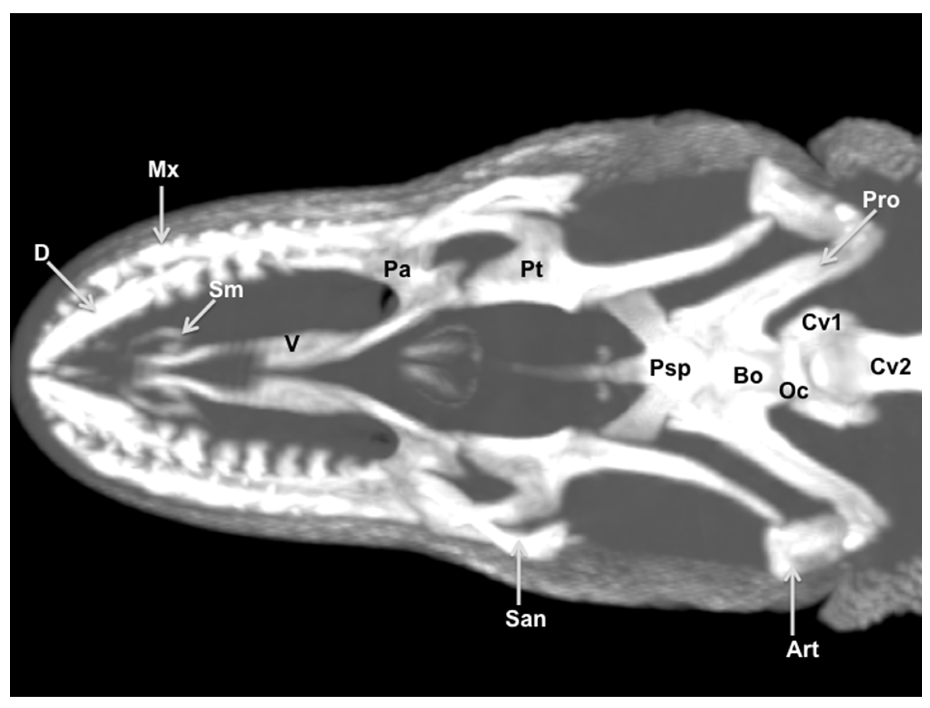
3.1. Transverse Computed Tomography Images
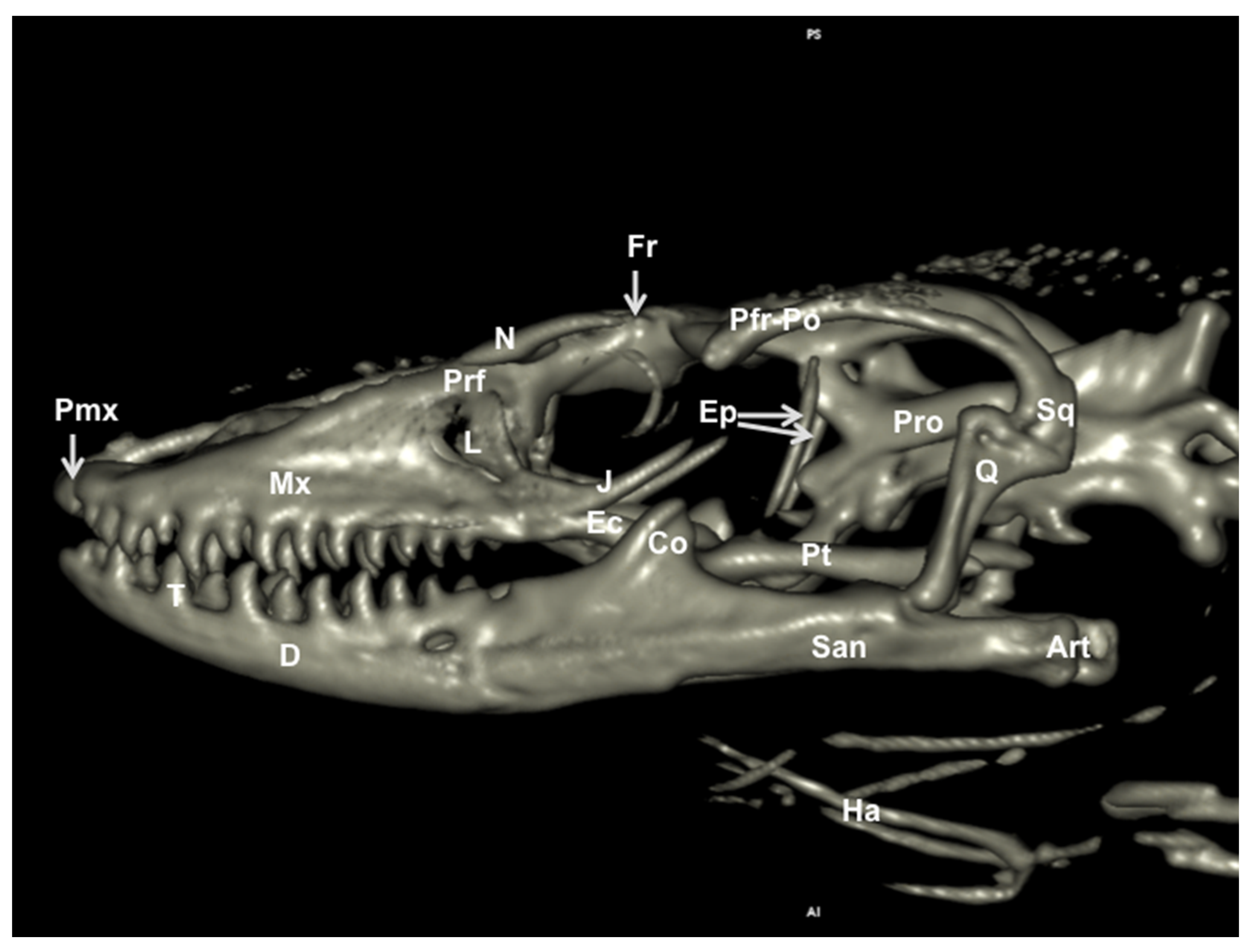
3.2. Head Volume-Rendered Reconstructed Images
3.3. Maximum Intensity Projection (MIP) Images
4. Discussion
Author Contributions
Funding
Institutional Review Board Statement
Data Availability Statement
Acknowledgments
Conflicts of Interest
References
- Banzato, T.; Russo, E.; Di Toma, A.; Palmisano, G.; Zotti, A. Anatomic imaging of the Boa constrictor head: A comparison between radiography, computed tomography and cadaver anatomy. Am. J. Vet. Res. 2011, 72, 1592–1599. [Google Scholar] [CrossRef] [PubMed]
- Lauridsen, H.; Hansen, K.; Wang, T.; Agger, P.; Andersen, J.L.; Knudsen, P.S.; Rasmussen, A.S.; Uhrenholt, L.; Pedersen, M. Inside Out: Modern Imaging Techniques to Reveal Animal Anatomy. PLoS ONE 2011, 6, e17879. [Google Scholar] [CrossRef] [PubMed] [Green Version]
- Deem, S.L. Role of the zoo veterinarian in the conservation of captive and free ranging wildlife. Int. Zoo Yearb. 2007, 41, 3–11. [Google Scholar] [CrossRef]
- IUCN/SSC. IUCN Red List of Threatened Species. 2002. Available online: www.redlist.org (accessed on 1 December 2020).
- Banzato, T.; Selleri, P.; Veladiano, I.A.; Martin, A.; Zanetti, E.; Zotti, A. Comparative evaluation of the cadaveric, radiographic and computed tomographic anatomy of the heads of green iguana (Iguana iguana), common tegu (Tupinambis merianae) and bearded dragon (Pogona vitticeps). BMC Vet. Res. 2012, 11, 53. [Google Scholar] [CrossRef] [PubMed]
- Arencibia, A.; Rivero, M.; De Miguel, I.; Contreras, S.; Cabrero, A.; Oros, J. Computed tomographic anatomy of the head of the loggerhead sea turtle (Caretta caretta). Res. Vet. Sci. 2006, 81, 165–169. [Google Scholar] [CrossRef] [PubMed]
- Banzato, T.; Selleri, P.; Veladiano, I.A.; Zotti, A. Comparative Evaluation of the Cadaveric and Computed Tomographic Features of the Coelomic Cavity in the Green Iguana (Iguana iguana), Black and White Tegu (Tupinambis merianae) and Bearded Dragon (Pogona vitticeps). Anat. Histol. Embryol. 2013, 42, 453–460. [Google Scholar] [CrossRef] [PubMed]
- Pees, M.; Kiefer, I.; Thielebein, J.; Oechtering, G.; Krautwald-Junghanns, M.E. Computed tomography of the lung of healthy snakes of the species Python regius, Boa constrictor, Python reticulatus, Morelia viridis, Epicrates cenchria and Morelia spilota. Vet. Radiol. Ultrasound 2009, 50, 487–491. [Google Scholar] [CrossRef] [PubMed]
- Hernández, C.; Peloso, P.L.; Bolívar, W.; Daza, J.D. Skull Morphology of the Lizard Ptychoglossus vallensis (Squamata: Alopoglossidae) with Comments on the Variation within Gymnophthalmoidea. Anat. Rec. 2019, 302, 1074–1092. [Google Scholar] [CrossRef] [PubMed]
- Maisano, J.A.; Rieppel, O. The skull of the Round Island boa, Casarea dussumieri schlegel, based on high-resolution X-ray computed tomography. J. Morphol. 2007, 268, 371–384. [Google Scholar] [CrossRef] [PubMed]
- Maisano, J.A.; Laduc, T.J.; Bell, C.J.; Barber, D. The Cephalic Osteoderms of Varanus komodoensis Revealed by High-Resolution X-ray Computed Tomography. Anat. Rec. 2019, 302, 1675–1680. [Google Scholar] [CrossRef] [PubMed]
- Moreno, K.; Wroe, S.; Clausen, P.; McHenry, C.; D’ Amore, D.C.; Rayfield, E.J.; Cunningham, E. Cranial performance in the Komodo dragon ( Varanus komodoensis) as revealed by high-resolution 3-D finite element analysis. J. Anat. 2008, 212, 736–746. [Google Scholar] [CrossRef] [PubMed]
- Fishman, E.K.; Ney, D.R.; Heath, D.G.; Corl, F.M.; Horton, K.M.; Johnson, P.T. Volume rendering versus maxi mum intensity projection in CT angiography: What works best, when, and why. Radiographics 2006, 26, 905–922. [Google Scholar] [CrossRef] [PubMed] [Green Version]
- Knipe, F.M. Principles of neurological imaging of exotic animal species. Vet. Clin. N. Am. Exot. Anim. Pract. 2007, 10, 893–907. [Google Scholar] [CrossRef] [PubMed]
- Banzato, T.; Hellebuyck, T.; Van Caelenberg, A.; Saunders, J.H.; Zotti, A. A review of diagnostic imaging of snakes and lizards. Vet. Rec. 2013, 173, 43–49. [Google Scholar] [CrossRef] [PubMed]
- George, T.F., II; Smallwood, J.E. Anatomic atlas for computed tomography in the mesaticephalic dog: Head and neck. Vet. Radiol. Ultrasound 1992, 33, 217–240. [Google Scholar] [CrossRef]
- Wilken, A.T.; Middleton, K.M.; Sellers, K.C.; Cost, I.N.; Holliday, C.M. The roles of joint tissues and jaw muscles in palatal biomechanics of the savannah monitor (Varanus exanthematicus) and their significance for cranial kinesis. J. Exp. Biol. 2019, 222, jeb201459. [Google Scholar] [CrossRef] [PubMed] [Green Version]
- Mertens, R. Die Familie der Warane (Varanidae). 2, Der Schädel. Senck. Naturf. Ges. 1942, 465, 1–118. [Google Scholar]
- Evans, S. The skull of lizards and Tuatara. Biol. Reptil. 2008, 20, 1–347. [Google Scholar]


Publisher’s Note: MDPI stays neutral with regard to jurisdictional claims in published maps and institutional affiliations. |
© 2021 by the authors. Licensee MDPI, Basel, Switzerland. This article is an open access article distributed under the terms and conditions of the Creative Commons Attribution (CC BY) license (https://creativecommons.org/licenses/by/4.0/).
Share and Cite
Pérez, S.; Encinoso, M.; Corbera, J.A.; Morales, M.; Arencibia, A.; González-Rodríguez, E.; Déniz, S.; Melián, C.; Suárez-Bonnet, A.; Jaber, J.R. Cranial Structure of Varanus komodoensis as Revealed by Computed-Tomographic Imaging. Animals 2021, 11, 1078. https://doi.org/10.3390/ani11041078
Pérez S, Encinoso M, Corbera JA, Morales M, Arencibia A, González-Rodríguez E, Déniz S, Melián C, Suárez-Bonnet A, Jaber JR. Cranial Structure of Varanus komodoensis as Revealed by Computed-Tomographic Imaging. Animals. 2021; 11(4):1078. https://doi.org/10.3390/ani11041078
Chicago/Turabian StylePérez, Sara, Mario Encinoso, Juan Alberto Corbera, Manuel Morales, Alberto Arencibia, Eligia González-Rodríguez, Soraya Déniz, Carlos Melián, Alejandro Suárez-Bonnet, and José Raduan Jaber. 2021. "Cranial Structure of Varanus komodoensis as Revealed by Computed-Tomographic Imaging" Animals 11, no. 4: 1078. https://doi.org/10.3390/ani11041078
APA StylePérez, S., Encinoso, M., Corbera, J. A., Morales, M., Arencibia, A., González-Rodríguez, E., Déniz, S., Melián, C., Suárez-Bonnet, A., & Jaber, J. R. (2021). Cranial Structure of Varanus komodoensis as Revealed by Computed-Tomographic Imaging. Animals, 11(4), 1078. https://doi.org/10.3390/ani11041078


