Communication

Dietary Copper Improves Intestinal Morphology via Modulating Intestinal Stem Cell Activity in Pigs

1
Laboratory of Animal Nutrition and Human Health, Hunan Provincial Key Laboratory of Animal Intestinal Function and Regulation, Hunan International Joint Laboratory of Animal Intestinal Ecology and Health, College of Life Sciences, Hunan Normal University, Changsha 410081, China
2
Hunan Provincial Key Laboratory of Animal Nutritional Physiology and Metabolic Process, Institute of Subtropical Agriculture, Chinese Academy of Sciences, Changsha 410125, China
3
State Key Laboratory of Developmental Biology of Freshwater Fish, College of Life Sciences, Hunan Normal University, Changsha 410081, China
*
Author to whom correspondence should be addressed.
Lanmei Yin and Qing Yang should be considered joint first authors.
Animals 2021, 11(9), 2513; https://doi.org/10.3390/ani11092513
Submission received: 20 July 2021 / Revised: 19 August 2021 / Accepted: 23 August 2021 / Published: 26 August 2021
(This article belongs to the Section Pigs)

Abstract

:

Simple Summary

Copper (Cu) is one of the essential trace elements for animal growth. Piglet intestinal epithelium undergoes a complete renewal driven by intestinal stem cells located at the crypt base. However, whether Cu improves intestinal morphology and development via modulating intestinal stem cell activity in pigs remains unclear. This study aimed to investigate the effects of dietary Cu during the weaning period on the intestinal morphology and development of weaned and finished pigs and explored the potential mechanism by culturing the intestinal organoids and treated them with Cu in vitro. The results indicated that Cu supplementation during the weaning period improves the intestinal morphology in finishing pigs by modulating the activity of intestinal stem cells.

Abstract

Copper (Cu) is an essential micronutrient for animals. Many studies have been conducted on the effects of dietary Cu on growth performance, intestinal morphology, and function of piglets. However, the underlying mechanism remains to be explored. Intestinal stem cells (ISC) drive the development and constant renewal of intestinal epithelium. Therefore, we hypothesized that dietary Cu affects piglets’ intestinal development via modulating ISC activity. A total of eighty-five 21-day-old piglets were randomly assigned to five groups, where 25, 50, 75, 100, and 125 mg CuSO4/kg on a dry matter basis were supplemented to the basal diet at phase 1 (day 0 to 21). Increasing the dietary Cu concentration decreased (p < 0.05) villus width but increased (p < 0.001) the number of Ki67-positive cells. At phase 2 (day 22 to 163), the other 45 pigs were offered the same diets. Villus height in the 125 mg/kg Cu group was greater (p < 0.001) than in the other groups. Moreover, the effects of Cu on ISC activity in vitro were tested to explore the underlying mechanism. Compared to the control group, 10 μmol/L CuSO4·5H2O increased (p < 0.001) the organoid budding efficiency, crypt depth, and crypts per organoid. Dietary Cu improved the intestinal morphology of finishing pigs via promoting cell proliferation and modulating ISC activity.

1. Introduction

Copper (Cu) is an essential trace element for animal growth [1]. The dietary Cu requirement of weanling pigs is 5–6 mg/kg [2]. In commercial diets, 10–20 mg of inorganic Cu/kg is usually added [3]. Cu supplementation at pharmacological doses (150–250 mg/kg, resulted in piglets’ average body weight of 5.45 kg) improved growth performance and reduced the occurrence of diarrhea in weaned piglets [4,5]. Various studies have shown that high dietary Cu improves intestinal digestive and absorptive functions, which enhances growth performance, as the digestibility of energy, fat, and amino acids increases in pigs fed high Cu diets [6,7,8,9,10].
However, whether dietary Cu improves intestinal digestibility by affecting intestinal morphology is not clear because of the inconsistency of results. Dietary supplementation with 150 mg/kg of CuSO4 in the unprotected form increased duodenal villus height (VH) in weaned piglets [11]. However, 225 mg of CuSO4/kg in the diet had adverse effects on duodenal and jejunal VH in weaned piglets [12]. Radecki et al. found that 250 mg Cu/kg of dry matter did not affect intestinal the VH and crypt depth (CD) in weaned piglets [13]. Therefore, it still needs to be clarified whether Cu supplementation improves intestinal morphology in pigs. Moreover, intestinal morphology is dominated by the continual renewal of the epithelial cells along the crypt–villus axis [14]. It has been pointed out that dietary fiber increased goblet cells and altered the expression of the nutrient receptors and transporters to modulate intestinal epithelium differentiation in growing pigs, but it did not affect intestinal morphology [15]. Thus, dietary Cu may regulate cell renewal and morphology in pigs.
Cu is also required for the growth of various organs and the whole body of animals [16]. Intestinal stem cells (ISC) play a central role in modulating intestinal epithelium renewal and intestinal development [17]. Piglet intestinal epithelium undergoes a complete renewal every two to three days, which is driven by the ISC located at the crypt base [18]. Therefore, we hypothesize that dietary Cu affects intestinal development and functions via the ISC. The intestinal organoid is a newly developed technology that can simulate the characteristics of intestinal tissues, as it recapitulates in part the intestinal crypt–villus anatomy and can be used to detect the effects of substances on intestinal epithelial renewal and development [19]. Accordingly, the intestinal organoid is a good model for studying the effects of Cu on ISC activity in vitro. The present study was conducted to investigate how dietary Cu affects piglets’ intestinal morphology and development via modulating ISC activity.

2. Materials and Methods

The experimental protocol was reviewed and approved (Approval number 2016-093) by the Animal Care and Use Committee of Hunan Normal University.

2.1. Animals and Experimental Treatments

A total of eighty-five 21-day-old weaned piglets (Duroc × (Landrace × Yorkshire)) with an initial body weight of 4.98 ± 0.09 kg were randomly assigned to five dietary treatments. Previous studies have shown that dietary Cu affects the small intestine epithelium of weaned piglets offered the diets for two weeks, and the pigs were slaughtered for sampling at 100 kg during the finishing period [11,12,13,20]. Accordingly, the present trial lasted 163 days and included two phases: (day 1 to 21 and day 22 to 163). During phase 1, diets were supplemented with 25, 50, 75, 100, and 125 mg Cu/kg dry matter, respectively; the basal diets are displayed in Table 1. During phase 2, pigs from different treatments were given the same diet with Cu concentration according to NRC (2012) recommendations. The requirement of Cu for weaned piglets is 6 mg/kg according to the NRC (2012), thus, in the present experiment, percentage of Cu supplementation was 400%, 800%, 1200%, 1600%, and 2000%, relative to the Cu requirement indicated by the NRC (2012). All of the pigs were housed individually in a 1.2 × 0.5 m pen and were fed four times per day at 7:00, 11:00, 15:00, and 19:00. Feed and water were available to pigs throughout the experimental period. The room temperature was maintained at around 28–30 °C, and relative humidity was 60%.

2.2. Sample Collection

A total of eight randomly selected piglets from each treatment group with an average final body weight of 6.75 ± 0.18 kg were euthanized with 4% sodium pentobarbital solution for tissue sampling on day 21. Additionally, other pig group with an average body weight of 95.4 ± 1.9 kg were slaughtered by electrical stunning on d 163 according to standard commercial procedures [21]. After slaughtering, the small intestine was separated from the large intestine and was divided into three segments following the methodology of Wu et al. [22]. The duodenum was 10 cm from the pylorus, the distal ileum was 5 cm proximal to the ileocecal junctions, and the jejunum was separated from the middle segment. These segments were then rinsed with physiological saline. After removing the contents, the length and weight of the small intestine were measured. A 2 cm jejunal segment was fixed in 4% neutral-buffered formalin and was stored at room temperature before analysis.

2.3. Morphological Analysis

The jejunum sections were serially dehydrated with graded ethanol, cleared in xylene, and embedded in paraffin wax. Sections of 4-μm thickness were stained with hematoxylin and eosin and were examined under a light microscope (Leica DM3000; Wetzlar, Germany). Measurements were performed blindly using Image-Pro Plus 6.0 software (Media Cybernetics, San Diego, CA, USA), and the VH, CD, and villus width (VW) were measured [15,23]. Mean values of 15 fields, 30 well-oriented, complete villus-crypt structures were calculated for each pig.

2.4. Immunohistochemistry for Ki67

The slides were dried, dewaxed, rehydrated, and treated with 3% hydrogen peroxide (H2O2) in methanol for 10 min. Antigen retrieval was performed by boiling the slides twice in a sodium citrate buffer (0.01 mol/L, pH 6.0). A 5% bovine serum albumin (BSA; Boster Biological Technology Co. Ltd, Wuhan, China) was used in a 1:10 dilution during a 30 min incubation period at 37 °C. There was a two-hour incubation period with the Ki67 antibody (Abcam, ab15580; 1:800 dilution) at 37 °C. Sections were treated with a goat anti-rabbit IgG secondary antibody (ZSGB-BIO, Beijing, China) for one hour at 37 °C. With the exception of the blocking step, at every step, washing was completed thrice in PBS for 5 min. Positive cells were observed with a diaminobenzidine (DAB) Kit (ZSGB-BIO, Beijing, China), stained with hematoxylin, and made into permanent pieces. A total of 15 microscopic fields per sample were captured using a light microscope under 20× magnification (Leica DM3000, Leica Microsystems, Wetzlar, Germany). The number of Ki67-positive cells, at least 30 well-oriented complete crypts per sample was counted manually for analysis [24].

2.5. Cell Shedding Analysis

A total of thirty pictures per sample were captured using a light microscope under 10× magnification (Leica DM3000, Leica Microsystems, Wetzlar, Germany). The rate of cell shedding is expressed as the proportion of shedding villus in 100 intact villi [25,26].

2.6. Porcine Crypt Isolation, Organoid Culture, and Measurement

After euthanizing the piglets, the 3 cm segments of anterior jejunum best suited for organoid growth were removed and immediately rinsed with cold PBS to remove the intestinal contents. The fat and mesentery attached to the jejunum were removed with forceps and scissors. Then, the jejunum was dissected, cut longitudinally, and washed with ice-cold PBS. The mucosal surface was scraped with a glass coverslip to remove the villi [27]. The jejunum was then cut into small pieces (2–5 mm) and transferred to a 50 mL tube conical tube, and washed three times with ice-cold PBS. Epithelial isolation was performed by incubating the tissues in PBS supplemented with 2.5 mmol/L ethylenediaminetetraacetic acid disodium salt (Sigma-Aldrich, St. Louis, MO, USA) for 30 min at 4 °C on a rotator. Thereafter, the tissues were thoroughly suspended by being pipetted up and down 20 times with a 5 mL tip to loosen the crypts and were then filtered through a 70 μm cell strainer. An amount of 10% FBS was added to the crypt suspension and was spun down at 1200 rpm for 5 min. The supernatant was discarded, and the crypts were re-suspended in 2 mL complete medium, which consisted of advanced DMEM/F12 (Gibco, Grand Island, NE, USA), 1% of GlutaMAXTM Supplement (Gibco), 10 mmol/L of HEPES (Gibco), 100 U/mL of penicillin, 100 μg/mL streptomycin, and 1000 × antibiotic (Gibco). The crypts were then collected by means of centrifugation at 600 rpm for 5 min. Approximately 500 crypts were suspended in 40 μL cold Matrigel (Corning, Bedford, OH, USA). After that, one droplet of Matrigel/crypts mix was placed in the center of each well of a pre-warmed 24-well plate and was subsequently incubated at 37 °C with 5% CO2 for 15 min. A 500 μL amount of culture medium was added per well after the Matrigel was solidified. The culture medium was supplemented with Wnt3a, R-spondin 1, and Noggin (WRN) conditioned medium, FBS, N2 supplement (Gibco), B27 supplement (Gibco), n-acetyl-L-cysteine (Invitrogen, Carlsbad, CA, USA), nicotinamide (Sigma-Aldrich), epidermal growth factor (EGF, Sigma-Aldrich), A83-01 (TGF-beta inhibitor, Tocris, Bristol, UK), SB202190 (p38 inhibitor, R & D Systems, Minneapolis, MN, USA), 10 μmol/L Y27632 (Rho-kinase inhibitor, R & D Systems), and 2.5 μmol/L the glycogen synthase kinase 3 inhibitor (GSK3i, CHIR99021; Sigma-Aldrich). The culture medium was refreshed every two days, and the organoids were passaged every 6–7 days [28,29]. Li et al. treated porcine intestinal epithelial cells (IPEC-J2) with two doses (30 and 120 μmol/L) of CuSO4 [30]; therefore, the porcine organoid was incubated with 10 and 100 μmol/L CuSO4·5H2O (Sangon Biotech, A100330, Shanghai, China) for 72 h after passage. The organoid activity was determined by organoid budding efficiency, budding crypt depth, and crypts per organoid [31,32]. Organoid budding efficiency was calculated as a ratio of the budding organoid number to the total organoid number. Budding crypt depth was measured using Image-Pro Plus 6.0 software (Media Cybernetics, San Diego, CA, USA), while the number of crypts per organoid was expressed by counting the average number of buds.

2.7. Statistical Analysis

Data were analyzed using SPSS statistics 20 (SPSS Inc., Chicago, IL, USA). Before analysis, all of the data were tested for normality using histograms and the Shapiro–Wilk test. Any value that departed more than two standard deviations from the standardized mean was eliminated. One-way analysis of variance (ANOVA) was used if the data followed a normal distribution; otherwise, the Kruskal–Wallis test (VH, CD, and relative small intestine length at phase 1; CD at phase 2) was performed as the non-parametric testing. Values were expressed as means ± SEM. Individual piglets were the experimental units. Linear and quadratic contrasts of treatment were investigated. Differences among the treatments were examined using Duncan’s multiple comparisons, with p < 0.05 considered statistically significant and 0.05 < p < 0.10 considered as tending towards significance. All of figures in this study were drawn using Graphpad Prism 6.0 (GraphPad Inc., San Diego, CA, USA).

3. Results

3.1. Relative Small Intestinal Index

The small intestinal index of weaned and finishing pigs are shown in Table 2. Dietary Cu tended (ANOVA, p = 0.064; Liner, p = 0.026) to decrease the length of the small intestine. Dietary Cu did not affect the relative small intestine length and relative small intestine weight of either the weaned or finished pigs.

3.2. Intestinal Morphology

The gut morphology is presented in Table 3. On day 21 of age, no difference (p > 0.05) was observed in the VH. Moreover, the VW decreased (p = 0.037) with increases in dietary Cu supplementation. Offering 125 mg Cu/kg DM during the weaning period increased (p < 0.001) the jejunal VH in finished pigs.

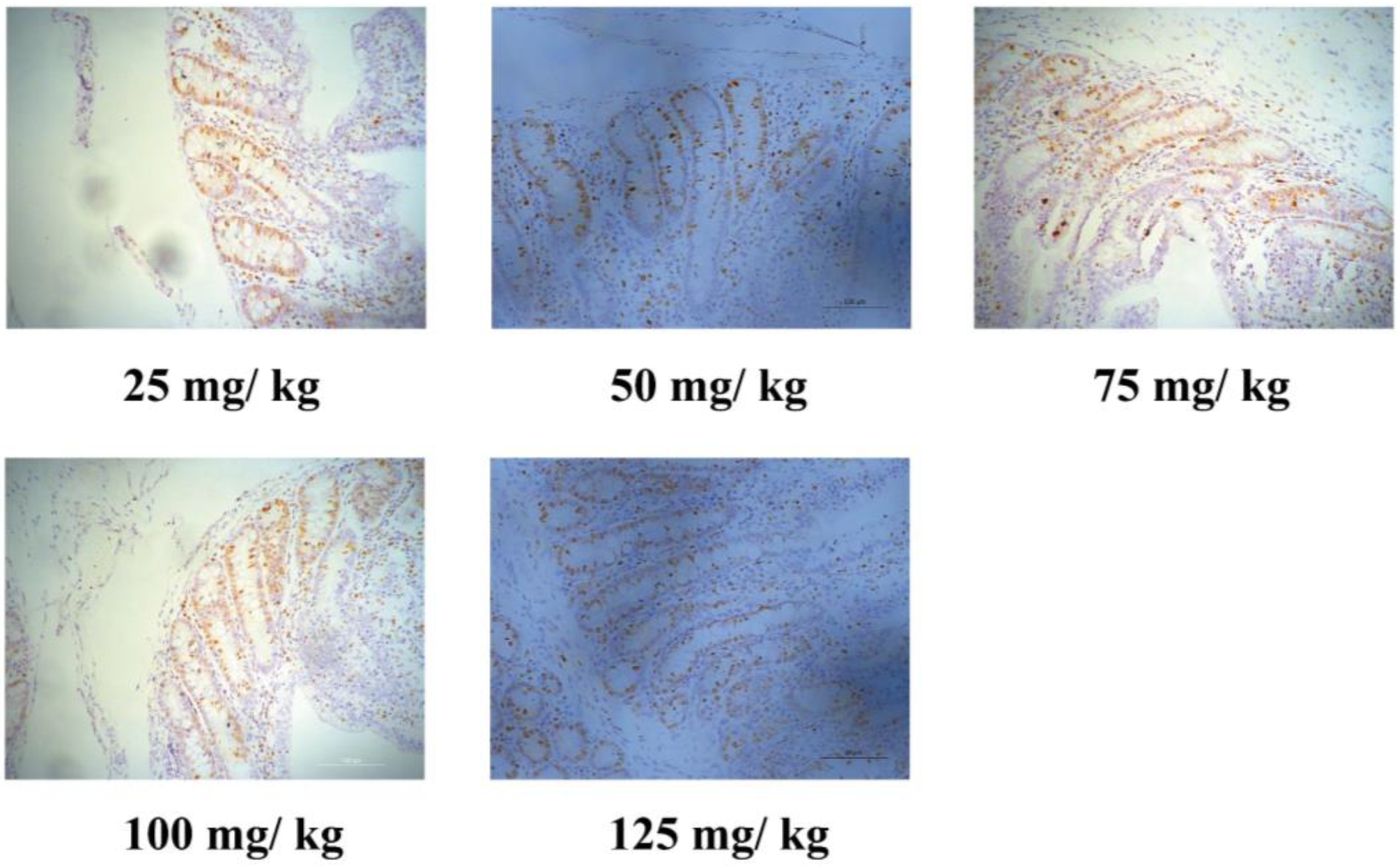
3.3. Intestinal Epithelium Cell Proliferation and Cell Shedding

Enterocyte proliferation and cell shedding are displayed in Table 4. The number of Ki67+ cells in the crypt increased (p < 0.001) with higher dietary Cu. Moreover, increasing dietary Cu tended to increase (quadratic, p = 0.060) the cell shedding rate. Representative images of the Ki67-positive cells are captured in Figure 1.

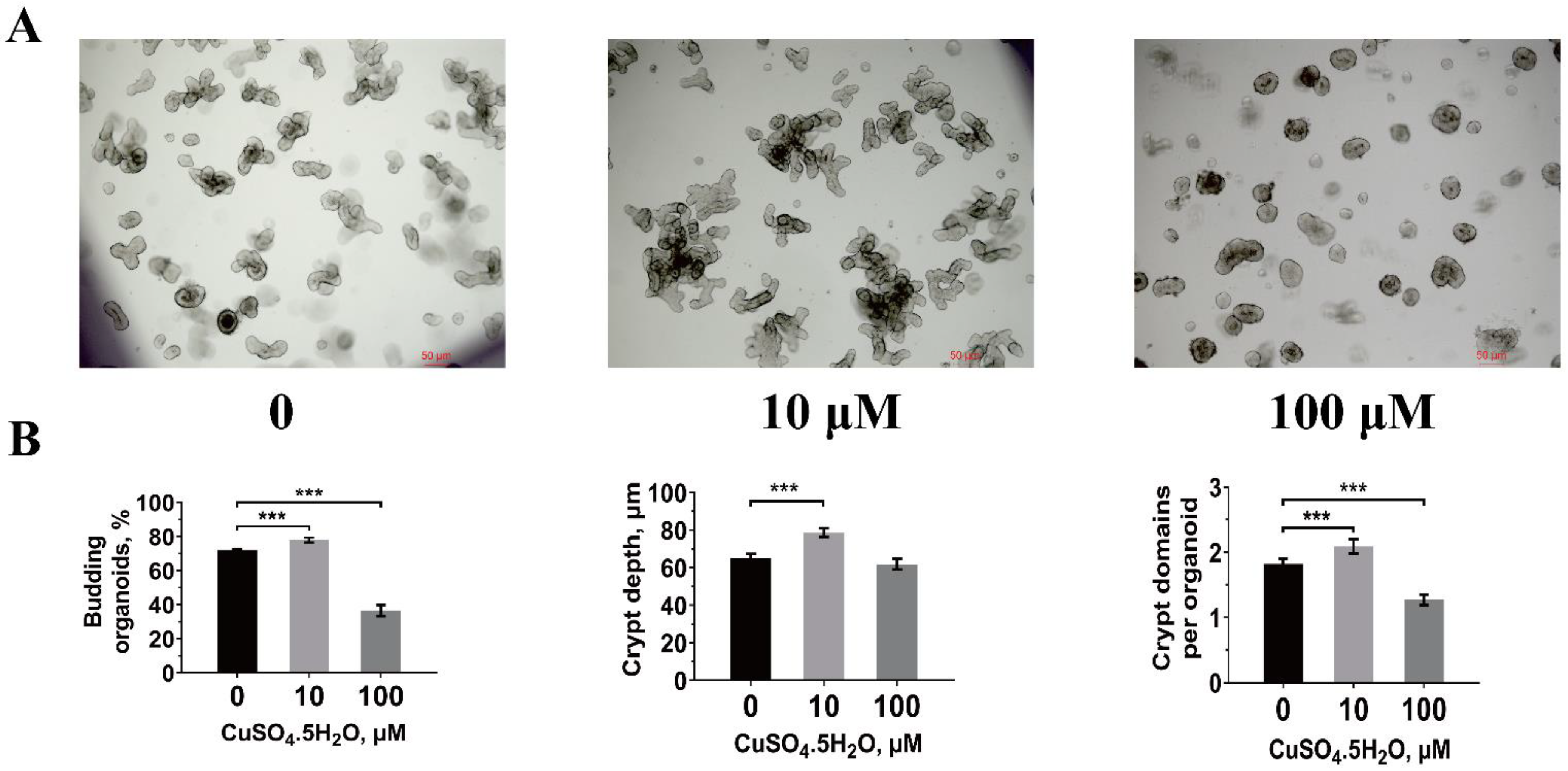
3.4. Jejunal Intestinal Organoid Activity

In vitro intestinal stem cell activity obtained from piglets treated with 10 and 100 μmol/L CuSO4·5H2O is presented in Figure 2. We found that 10 μmol/L CuSO4·5H2O significantly increased (p < 0.001) organoid budding efficiency, crypt depth, and crypts per organoid compared to the control pigs (Figure 2B). However, 100 μmol/L CuSO4·5H2O inhibited (p < 0.001) the organoid budding efficiency and the crypts per organoid.

4. Discussion

Small intestine morphology, including VH, CD, and their ratio, have commonly been used as indicators of piglets’ intestinal health [33]. Giancamillo et al. showed that dietary Cu could increase intestinal VH in weaned piglets, which is consistent with our results [11]. The intestinal epithelium is composed of a monolayer of epithelial cells that undergo continual renewal along the crypt–villus axis, and VH and epithelial cell populations are highly correlated [14]. Changes in intestinal morphology are usually accompanied by the process of epithelial cell renewal [34]. Our previous study showed a positive relationship between cell proliferation and jejunal VH and CD in weaned piglets [24]. To determine whether the changes in intestinal morphology resulted from the alteration in epithelium renewal by dietary Cu, we counted Ki67-positive proliferating cells to the crypt base and cell shedding rates at the villus tips in weaned piglets. The number of Ki67+ cells in the crypt increased with increasing the amount of dietary Cu, which indicated that high Cu ingestion increased intestinal epithelial cell proliferation. Ki67, a marker for proliferating cells, labeled the undifferentiated proliferating transit-amplifying (TA) cells (progenitors) at the crypt, which stem from the ISC and differentiated into functional epithelial cells [35,36,37]. Moreover, increasing the dietary Cu tended to increase the cell shedding rates. These results indicated that dietary Cu might modulate the ISC to increase cell proliferation and affect intestinal cell renewal in weaned piglets, which may regulate intestinal morphology. Length and weight are primary indices of intestinal development, which are highly related to intestinal digestion and absorption capacity [38,39]. Dietary Cu supplementation only tended to decrease the length of the small intestine and had no effects on the relative small intestinal index of the weaned piglets, which indicates that dietary Cu has minor effects on the intestinal development of weaned piglets.
The weaning period is an important developmental window for the small intestine of animals, and any effects on their developmental programming during early life, especially during the developmental windows, will have lifelong implications [40,41]. Previous studies tested the effects of dietary Cu during the nursery or finishing period, but no experiments had tested the effects of dietary Cu during early life on the intestinal development of the finishing period. The results of the present study showed that dietary supplementation with 125 mg Cu/kg DM during the weaning period increased jejunal VH in finishing pigs at 163 d of age. Dietary Cu did not affect the relative small intestine length or the relative small intestine weight. These results indicate that dietary Cu during the weaning period improves the intestinal morphology in finishing pigs.
Piglet intestinal epithelium undergoes a complete renewal every two to three days, which is driven by the ISC located at the crypt base, which first generate transit-amplifying (TA) cells that further differentiate into absorptive (enterocytes) or secretory cell lineages (Paneth cells, goblet cells, and enteroendocrine cells) [18]. Enteroids cultured from ISC partially recreate the villus-crypt anatomy of the native intestine and mimic hallmarks of in vivo epithelium [42]. The ISC play a crucial role in maintaining gut morphology and intestinal development [17]. To test whether the effects of dietary Cu on intestinal morphology and development acted through the ISC, the jejunal organoids from 21-day-old piglets were treated with different concentrations of CuSO4·5H2O. Organoid budding may be similar to the expansion of the intestinal stem cell compartment and the formation of new crypts through crypt fission [43]. A greater crypt depth in the organoids suggests increased stem cell proliferation and differentiation, which resembles the increase in cell renewal along the crypt–villus axis in the intestinal mucosa [44]. We found that dietary Cu stimulated the epithelial proliferation with increased Ki67+ cells at the crypt in vivo, and enteroid growth with increased budding efficiency, crypt depth, and crypts per organoid under Cu exposure in vitro. Wang et al. indicated that enterocyte proliferation was positively related to mucosal enzyme activities and nutrient digestibility [24]; whether dietary Cu improves intestinal morphology by altering the expression of nutrient receptors and transporters to modulate intestinal differentiation in growing pigs needs to be further explored, although this was demonstrated by Saqui-Salces et al. in study on fiber [15]. These results indicate that Cu modulates the activity of the ISC and that dietary Cu may, by increasing intestinal epithelial cell proliferation and impacting the ISC, improve intestinal morphology in finishing pigs. Unfortunately, the commercially available antibodies used to detect the intestinal stem cells, Lgr5, did not demonstrate cross-reactivity with the active CBC stem cells in the porcine jejunal tissue. Further experiments are needed to test the potential mechanism of Cu to modulate ISC activity in vitro.

5. Conclusions

Dietary Cu supplementation during the weaning period improved intestinal morphology of finishing pigs, which may be achieved by promoting cell proliferation and increasing the activity of ISC.

Author Contributions

L.Y. analyzed the experimental data and wrote the manuscript; Q.Y. conducted the experiments; Y.Z. performed the animal feeding and sample collections; D.W. and Y.Y. (Yuebang Yin) contributed to experimental concepts and design and provided scientific direction; Q.W., J.H. and J.L. performed the project administration; H.Y. and Y.Y. (Yulong Yin) reviewed and edited the manuscript and provided financial support. All authors have read and agreed to the published version of the manuscript.

Funding

This work was supported by the Key Programs of Frontier Scientific Research of the Chinese Academy of Sciences (Grant No. QYZDY-SSW-SMC008) and the Natural Science Foundation of Hunan Province (Grant No. 2017JJ1020).

Institutional Review Board Statement

The experimental protocol was reviewed and approved (Approval number 2016-093) by the Animal Care and Use Committee of Hunan Normal University.

Data Availability Statement

All data are available from the corresponding author on reasonable request.

Acknowledgments

We would like to thank Yuebang Yin who provided detailed support in culturing the intestinal organoids in this study.

Conflicts of Interest

The authors declare no conflict of interest.

References

  1. Zheng, P.; Pu, B.; Yu, B.; He, J.; Yu, J.; Mao, X.B.; Luo, Y.H.; Luo, J.Q.; Huang, Z.Q.; Luo, C.G.; et al. The differences between copper sulfate and tribasic copper chloride on growth performance, redox status, deposition in tissues of pigs, and excretion in feces. Asian-Australas J. Anim. Sci. 2018, 31, 873–880. [Google Scholar] [CrossRef] [PubMed]
  2. Okonkwo, A.C.; Ku, P.K.; Miller, E.R.; Keahey, K.K.; Ullrey, D.E. Copper requirement of baby pigs fed purified diets. J. Nutr. 1979, 109, 939–948. [Google Scholar] [CrossRef]
  3. Cromwell, G.L.; Monegue, H.J.; Stahly, T.S. Long-term effects of feeding a high copper diet to sows during gestation and lactation. J. Anim. Sci. 1993, 71, 2996–3002. [Google Scholar] [CrossRef] [Green Version]
  4. Smith, J.W.; Tokach, M.D.; Goodband, R.D.; Nelssen, J.L.; Richert, B.T. Effects of the interrelationship between zinc oxide and copper sulfate on growth performance of early-weaned pigs. J. Anim. Sci. 1997, 75, 1861–1866. [Google Scholar] [CrossRef]
  5. Pérez, V.G.; Waguespack, A.M.; Bidner, T.D.; Southern, L.L.; Fakler, T.M.; Ward, T.L.; Steidinger, M.; Pettigrew, J.E. Additivity of effects from dietary copper and zinc on growth performance and fecal microbiota of pigs after weaning. J. Anim. Sci. 2011, 89, 414–425. [Google Scholar] [CrossRef]
  6. Luo, X.G.; Dove, C.R. Effect of Dietary Copper and Fat on Nutrient Utilization, Digestive Enzyme Activities, and Tissue Mineral Levels in Weanling Pigs. J. Anim. Sci. 1996, 74, 1888–1896. [Google Scholar] [CrossRef]
  7. Gonzales-Eguia, A.; Fu, C.M.; Lu, F.Y.; Lien, T.F. Effects of nanocopper on copper availability and nutrients digestibility, growth performance and serum traits of piglets. Livest. Sci. 2009, 126, 122–129. [Google Scholar] [CrossRef]
  8. Coble, K.F.; Burnett, D.D.; DeRouchey, J.M.; Tokach, M.D.; Gonzalez, J.M.; Wu, F.Z.; Dritz, S.S.; Goodband, R.D.; Woodworth, J.C.; Pluske, J.R. Effect of diet type and added copper on growth performance, carcass characteristics, energy digestibility, gut morphology, and mucosal mRNA expression of finishing pigs. J. Anim. Sci. 2018, 96, 3288–3301. [Google Scholar] [CrossRef] [Green Version]
  9. Rochell, S.J.; Usry, J.L.; Parr, T.M.; Parsons, C.M.; Dilger, R.N. Effects of dietary copper and amino acid density on growth performance, apparent metabolizable energy, and nutrient digestibility in Eimeria acervulina-challenged broilers. Poult. Sci. 2016, 96, 602–610. [Google Scholar] [CrossRef]
  10. Cappai, M.G.; Alesso, G.A.; Nieddu, G.; Sanna, M.; Pinna, W. Electron microscopy and composition of raw acorn starch in relation to in vivo starch digestibility. Food Funct. 2013, 4, 917–922. [Google Scholar] [CrossRef]
  11. Di Giancamillo, A.; Rossi, R.; Martino, P.A.; Aidos, L.; Maghin, F.; Domeneghini, C.; Corino, C. Copper sulphate forms in piglet diets: Microbiota, intestinal morphology and enteric nervous system glial cells. Anim. Sci. J. 2018, 89, 616–624. [Google Scholar] [CrossRef] [PubMed]
  12. Fry, R.S.; Ashwell, M.S.; Lloyd, K.E.; O’Nan, A.T.; Flowers, W.L.; Stewart, K.R.; Spears, J.W. Amount and source of dietary copper affects small intestine morphology, duodenal lipid peroxidation, hepatic oxidative stress, and mRNA expression of hepatic copper regulatory proteins in weanling pigs. J. Anim. Sci. 2012, 90, 3112–3119. [Google Scholar] [CrossRef] [PubMed]
  13. Radecki, S.V.; Ku, P.K.; Bennink, M.R.; Yokoyama, M.T.; Miller, E.R. Effect of dietary copper on intestinal mucosa enzyme activity, morphology, and turnover rates in weanling pigs. J. Anim. Sci. 1992, 70, 1424–1431. [Google Scholar] [CrossRef] [PubMed]
  14. Wright, N.A. The experimental analysis of changes in proliferative and morphological status on the intestine. Scand. J. Gastroenterol. Suppl. 1982, 74, 3–10. [Google Scholar]
  15. Saqui-Salces, M.; Huang, Z.; Ferrandis Vila, M.; Li, J.; Mielke, J.A.; Urriola, P.E.; Shurson, G.C. Modulation of intestinal cell differentiation in growing pigs is dependent on the fiber source in the diet. J. Anim. Sci. 2017, 95, 1179–1190. [Google Scholar] [CrossRef]
  16. Kuo, Y.M.; Zhou, B.; Cosco, D.; Gitschier, J. The copper transporter CTR1 provides an essential function in mammalian embryonic development. Proc. Natl. Acad. Sci. USA 2001, 98, 6836–6841. [Google Scholar] [CrossRef] [Green Version]
  17. Barker, N.; van de Wetering, M.; Clevers, H. The intestinal stem cell. Genes Dev. 2008, 22, 1856–1864. [Google Scholar] [CrossRef] [Green Version]
  18. Verdile, N.; Mirmahmoudi, R.; Brevini, T.A.L.; Gandolfi, F. Evolution of pig intestinal stem cells from birth to weaning. Animal 2019, 13, 2830–2839. [Google Scholar] [CrossRef]
  19. Pierson, H.; Yang, H.J.; Lutsenko, S. Copper Transport and Disease: What Can We Learn from Organoids? Annu. Rev. Nutr. 2019, 39, 75–94. [Google Scholar] [CrossRef]
  20. Remus, A.; Del Castillo, J.R.E.; Pomar, C. Improving the estimation of amino acid requirements to maximize nitrogen retention in precision feeding for growing-finishing pigs. Animal 2020, 14, 2032–2041. [Google Scholar] [CrossRef]
  21. Zhou, X.H.; Liu, Y.H.; Zhang, L.Y.; Kong, X.F.; Li, F.N. Serine-to-glycine ratios in low-protein diets regulate intramuscular fat by affecting lipid metabolism and myofiber type transition in the skeletal muscle of growing-finishing pigs. Anim. Nutr. 2021, 7, 384–392. [Google Scholar] [CrossRef]
  22. Wu, Y.P.; Jiang, Z.Y.; Zheng, C.T.; Wang, L.; Zhu, C.; Yang, X.F.; Wen, X.L.; Ma, X.Y. Effects of protein sources and levels in antibiotic-free diets on diarrhea, intestinal morphology, and expression of tight junctions in weaned piglets. Anim. Nutr. 2015, 1, 170–176. [Google Scholar] [CrossRef]
  23. Zong, E.Y.; Yan, S.L.; Wang, M.W.; Yin, L.M.; Wang, Q.Y.; Yin, J.; Li, J.Z.; Li, Y.L.; Ding, X.Q.; Huang, P.F.; et al. The effects of dietary supplementation with hyodeoxycholic acid on the differentiation and function of enteroendocrine cells and the serum biochemical indices in weaned piglets1. J. Anim. Sci. 2019, 97, 1796–1805. [Google Scholar] [CrossRef]
  24. Wang, L.X.; Yan, S.L.; Li, J.Z.; Li, Y.L.; Ding, X.Q.; Yin, J.; Xiong, X.; Yin, Y.L.; Yang, H.S. Rapid Communication: The relationship of enterocyte proliferation with intestinal morphology and nutrient digestibility in weaning piglets. J. Anim. Sci. 2019, 97, 353–358. [Google Scholar] [CrossRef]
  25. Bullen, T.F.; Forrest, S.; Campbell, F.; Dodson, A.R.; Hershman, M.J.; Pritchard, D.M.; Turner, J.R.; Montrose, M.H.; Watson, A.J. Characterization of epithelial cell shedding from human small intestine. Lab. Investig. 2006, 86, 1052–1063. [Google Scholar] [CrossRef] [PubMed]
  26. Williams, J.M.; Duckworth, C.A.; Burkitt, M.D.; Watson, A.J.; Campbell, B.J.; Pritchard, D.M. Epithelial cell shedding and barrier function: A matter of life and death at the small intestinal villus tip. Vet. Pathol. 2015, 52, 445–455. [Google Scholar] [CrossRef] [PubMed] [Green Version]
  27. Khalil, H.A.; Lei, N.Y.; Brinkley, G.; Scott, A.; Wang, J.F.; Kar, U.K.; Jabaji, Z.B.; Lewis, M.; Martin, M.G.; Dunn, J.C.; et al. A novel culture system for adult porcine intestinal crypts. Cell Tissue Res. 2016, 365, 123–134. [Google Scholar] [CrossRef] [PubMed] [Green Version]
  28. Yin, Y.B.; Bijvelds, M.; Dang, W.; Xu, L.; van der Eijk, A.A.; Knipping, K.; Tuysuz, N.; Dekkers, J.F.; Wang, Y.; de Jonge, J.; et al. Modeling rotavirus infection and antiviral therapy using primary intestinal organoids. Antivir. Res. 2015, 123, 120–131. [Google Scholar] [CrossRef] [PubMed]
  29. Zhou, J.Y.; Huang, D.G.; Qin, Y.C.; Li, X.G.; Gao, C.Q.; Yan, H.C.; Wang, X.Q. mTORC1 signaling activation increases intestinal stem cell activity and promotes epithelial cell proliferation. J. Cell Physiol. 2019, 234, 19028–19038. [Google Scholar] [CrossRef]
  30. Li, R.X.; Wen, Y.; Lin, G.; Meng, C.Z.; He, P.L.; Wang, F.L. Different Sources of Copper Effect on Intestinal Epithelial Cell: Toxicity, Oxidative Stress, and Metabolism. Metabolites 2019, 10, 11. [Google Scholar] [CrossRef] [Green Version]
  31. Li, X.G.; Zhu, M.; Chen, M.X.; Fan, H.B.; Fu, H.L.; Zhou, J.Y.; Zhai, Z.Y.; Gao, C.Q.; Yan, H.C.; Wang, X.Q. Acute exposure to deoxynivalenol inhibits porcine enteroid activity via suppression of the Wnt/β-catenin pathway. Toxicol. Lett. 2019, 305, 19–31. [Google Scholar] [CrossRef]
  32. Zhou, J.Y.; Wang, Z.; Zhang, S.W.; Lin, H.L.; Gao, C.Q.; Zhao, J.C.; Yang, C.B.; Wang, X.Q. Methionine and Its Hydroxyl Analogues Improve Stem Cell Activity To Eliminate Deoxynivalenol-Induced Intestinal Injury by Reactivating Wnt/beta-Catenin signaling. J. Agric. Food Chem. 2019, 67, 11464–11473. [Google Scholar] [CrossRef]
  33. Han, F.; Hu, L.; Xuan, Y.; Ding, X.M.; Luo, Y.H.; Bai, S.P.; He, S.Y.; Zhang, K.Y.; Che, L.Q. Effects of high nutrient intake on the growth performance, intestinal morphology and immune function of neonatal intra-uterine growth-retarded pigs. Br. J. Nutr. 2013, 110, 1819–1827. [Google Scholar] [CrossRef] [Green Version]
  34. Li, J.; Yin, L.M.; Wang, L.; Huang, P.F.; Yang, H.S.; Yin, Y.L. Effects of vitamin B6 on growth, diarrhea rate, intestinal morphology, function, and inflammatory factors expression in a high-protein diet fed to weaned piglets1. J. Anim. Sci. 2019, 97, 4865–4874. [Google Scholar] [CrossRef] [PubMed]
  35. Chivukula, R.R.; Shi, G.; Acharya, A.; Mills, E.W.; Zeitels, L.R.; Anandam, J.L.; Abdelnaby, A.A.; Balch, G.C.; Mansour, J.C.; Yopp, A.C.; et al. An essential mesenchymal function for miR-143/145 in intestinal epithelial regeneration. Cell 2014, 157, 1104–1116. [Google Scholar] [CrossRef] [Green Version]
  36. Ito, S.; Nada, S.; Yamazaki, D.; Kimura, T.; Kajiwara, K.; Miki, H.; Okada, M. p18/Lamtor1-mTORC1 Signaling Controls Development of Mucin-producing Goblet Cells in the Intestine. Cell Struct. Funct. 2020, 45, 93–105. [Google Scholar] [CrossRef] [PubMed]
  37. Barker, N.; Bartfeld, S.; Clevers, H. Tissue-resident adult stem cell populations of rapidly self-renewing organs. Cell Stem Cell 2010, 7, 656–670. [Google Scholar] [CrossRef] [Green Version]
  38. Efird, R.C.; Armstrong, W.D.; Herman, D.L. The development of digestive capacity in young pigs: Effects of age and weaning system. J. Anim. Sci. 1983, 55, 1380–1387. [Google Scholar] [CrossRef] [PubMed] [Green Version]
  39. Wang, M.; Yang, C.; Wang, Q.Y.; Li, J.Z.; Li, Y.L.; Ding, X.Q.; Yin, J.; Yang, H.S.; Yin, Y.L. The growth performance, intestinal digestive and absorptive capabilities in piglets with different lengths of small intestines. Animal 2019, 14, 1196–1203. [Google Scholar] [CrossRef]
  40. Langley-Evans, S.C. Developmental programming of health and disease. Proc. Nutr. Soc. 2006, 65, 97–105. [Google Scholar] [CrossRef] [Green Version]
  41. Meyer, A.M.; Caton, J.S. Role of the small intestine in developmental programming: Impact of maternal nutrition on the dam and offspring. Adv. Nutr. 2016, 7, 169–178. [Google Scholar] [CrossRef]
  42. Yin, Y.B.; de Jonge, H.R.; Wu, X.; Yin, Y.L. Enteroids for Nutritional Studies. Mol. Nutr. Food Res. 2019, 63, e1801143. [Google Scholar] [CrossRef]
  43. Fuller, M.K.; Faulk, D.M.; Sundaram, N.; Shroyer, N.F.; Henning, S.J.; Helmrath, M.A. Intestinal crypts reproducibly expand in culture. J. Surg. Res. 2012, 178, 48–54. [Google Scholar] [CrossRef] [Green Version]
  44. Yin, Y.B.; Guo, S.G.; Wan, D.; Wu, X.; Yin, Y.L. Enteroids: Promising in Vitro Models for Studies of Intestinal Physiology and Nutrition in Farm Animals. J. Agric. Food Chem. 2019, 67, 2421–2428. [Google Scholar] [CrossRef]
Figure 1. Representative immunohistochemical images of the Ki67-positive cells in the jejunum of weaned piglets. Pigs were supplemented with 25, 50, 75, 100, and 125 mg CuSO4/kg dry matter at the weaning phage (phase 1). Note Scale bars, 100 μm (magnification 200×).
Animals 11 02513 g001
Figure 2. Effect of copper concentration on intestinal stem cells activity of piglets. (A) Representative images of organoid morphology after 0, 10, and 100 μmol/L CuSO4·5H2O treatment in vitro. Scale bars, 50 μm (magnification 50×). (B) Organoid budding efficiency, budding crypt depth, and crypts per organoid at three days after CuSO4·5H2O treatments were quantified. Statistical significance was determined by one-way ANOVA. The results are expressed as the means and SEM (n = 6 wells per treatment; *** p < 0.001).
Animals 11 02513 g002
Table 1. Basal diet ingredient and nutritional component (as-fed basis).
IngredientsContent (%)
Corn grain37.16
Extruded corn20.00
Soybean meal (43% CP)8.00
Soy protein concentrate powder7.00
Whey powder10.00
Fish meal (63% CP)5.00
Spray-dried plasma protein4.50
Glucose2.00
Soybean oil2.00
Vitamin and mineral premix a4.34
Total100.00
Calculated nutrient levels
Crude protein, %19.00
ME, MJ/kg13.25
NDF, %6.40
ADF, %2.30
Calcium, %0.75
Available phosphorous, %0.38
Lysine, %1.38
Metionine, %0.40
Met + Cys, %0.80
Threonine, %0.86
Triptophane, %0.25
Cu, mg/kg5.04
a Vitamin-mineral premix supplied per kilogram of feed: Vitamin A: 2200 IU; Vitamin D3: 220 IU; Vitamin E: 16 IU; Vitamin K3: 0.5 mg; Vitamin B12: 0.0175 mg; Riboflavin: 3.5 mg; Niacin: 30 mg; D-pantothenic acid: 10 mg; Biotin: 0.05 mg; Folic acid: 0.3 mg; Thiamine: 1.0 mg; FeSO4: 150 mg; ZnSO4: 100 mg; MnSO4: 30 mg; KIO3: 0.5 mg; CoSO4: 0.3 mg; Na2SeO3: 0.3 mg; Ethoxyquin: 4.0 mg.
Table 2. Effects of dietary copper supplementation during the weaning period on the small intestinal index of weaned and finished pigs 1.
VariablesDietary Copper, mg/kg Dry MatterSEM 4p-Value
255075100125ANOVALinearQuadratic
Phase 1 (day 1 to 21) 2
Small intestine length, m12.412.511.511.711.80.830.0640.0260.244
Small intestine weight, g516.3537.7494.6529.8478.455.840.1900.1790.355
Relative small intestine length, m/kg1.61.91.71.81.80.060.4640.4020.624
Relative small intestine weight, g/kg71.083.176.476.874.52.410.7050.9650.309
Phase 2 (day 22 to 163) 3
Small intestine length, m19.018.619.018.318.70.240.8970.6240.671
Small intestine weight, kg1.71.71.81.61.70.040.7680.5210.573
Relative small intestine length, m/kg18.920.320.219.120.90.340.2960.2590.987
Relative small intestine weight, kg/kg16.818.618.916.918.30.350.1980.5510.306
1 Pigs were supplemented with 25, 50, 75, 100, 125 mg CuSO4/kg dry matter at the weaning phage (phase 1), and all pigs from different treatments were given the same diet with Cu concertation according to the NRC (2012) recommendations for finishing pigs (phase 2). 2 n = 8. 3 n = 9. 4 SEM, pooled standard error of the mean. Relative small intestine weight = the ratio of total small intestine weight to body weight at slaughter; Relative small intestine length = the ratio of total small intestine length to body weight at slaughter.
Table 3. Effects of dietary copper supplementation during the weaning period on intestinal morphology of weaned and finished pigs 1.
VariablesDietary Copper, mg/kg DMSEM 4p-Value
255075100125ANOVALinearQuadratic
Phase 1 (day 1 to 21) 2
Villus height, μm305.9359.5303.5333.4346.09.300.1600.4130.959
Crypt depth, μm319.6324.5327.4321.6296.010.390.7530.5210.449
Villus height: crypt depth, μm:μm1.01.21.01.11.10.050.6460.5870.864
Villus width, μm175.9 a146.0 bc153.7 bc161.3 ac147.7 bc3.420.0370.0750.226
Intestinal villus surface area, mm20.20.20.10.20.20.010.5980.7060.471
Phase 2 (day 22 to 163) 3
Villus height, μm409.7 b379.1 b393.8 b368.6 b512.1 a13.83<0.0010.0160.002
Crypt depth, μm443.1402.5414.9407.6467.39.930.1580.4240.030
Villus height:crypt depth, μm:μm0.90.91.00.91.10.030.2450.1750.368
1 Pigs were supplemented with 25, 50, 75, 100, 125 mg CuSO4/kg dry matter at the weaning phase (phase 1), and all pigs from different treatments were given the same diet with Cu concentration according to the NRC (2012) recommendations at the finishing phase (phase 2). Intestinal morphology was measured in the jejunum. 2 n = 8. 3 n = 9. 4 SEM, pooled standard error of the mean. a, b, c Within rows, means labeled with different superscripts differ (p < 0.05). Intestinal villus surface area = π * villus height * villus width.
Table 4. Effects of dietary copper supplementation on intestinal epithelium renewal of weaned pigs 1.
VariablesDietary Copper, mg/kg DMSEM 2p-Value
255075100125ANOVALinearQuadratic
Ki67-positive cells per crypt, n22.3 b27.4 a25.5 ac24.6 bc27.7 ad0.50<0.0010.0070.381
Cell shedding rate, %25.429.128.828.428.30.480.1340.1350.060
1 Pigs were supplemented with 25, 50, 75, 100, 125 mg CuSO4/kg dry matter. 2 SEM, pooled standard error of the mean (n = 8). a, b, c, d Within rows, means labeled with different superscripts differ (p < 0.05).
Publisher’s Note: MDPI stays neutral with regard to jurisdictional claims in published maps and institutional affiliations.

