
Intestinal S100/Calgranulin Expression in Cats with Chronic Inflammatory Enteropathy and Intestinal Lymphoma

 , , ,
, , , 
Abstract
:Simple Summary
Abstract
1. Introduction
2. Materials and Methods
2.1. Ethics Approval
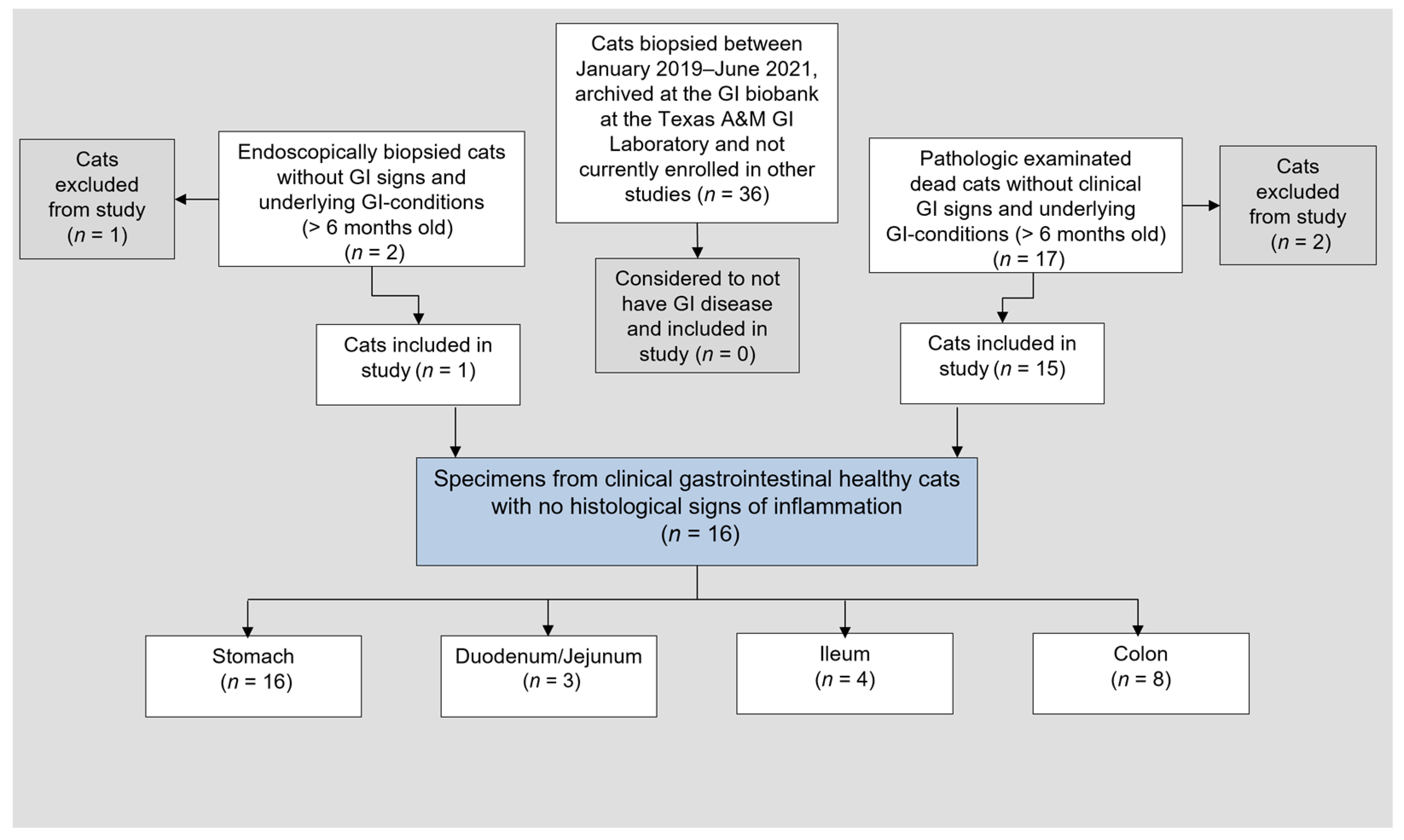
2.2. Study Population and Routine Diagnostics
2.3. Tissue Sample Collection and Routine Evaluation
2.4. IHC for Tissue S100A8/A9 and S100A12 Protein–Protocol Adjustment for Feline Tissues
2.5. S100A8/A9 and S100A12 IHC Analysis of Feline GI Tissue Biopsies
2.6. Statistical Analyses
3. Results
3.1. Patient Clinical Data
3.2. Clinicopathologic Parameters
3.3. Endoscopy, Histopathology, and Clonality Testing
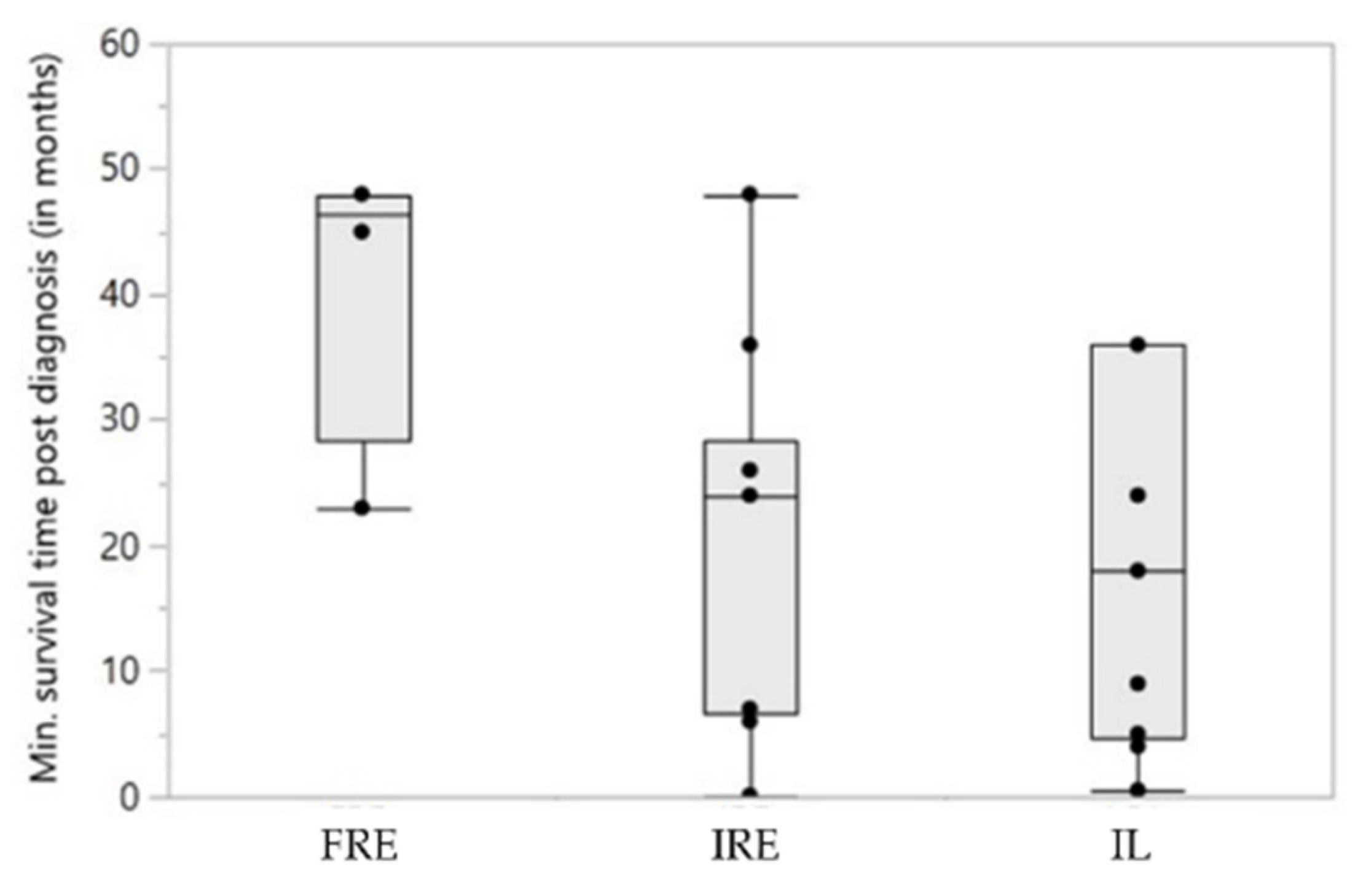
3.4. Follow-Up and Response to Treatment
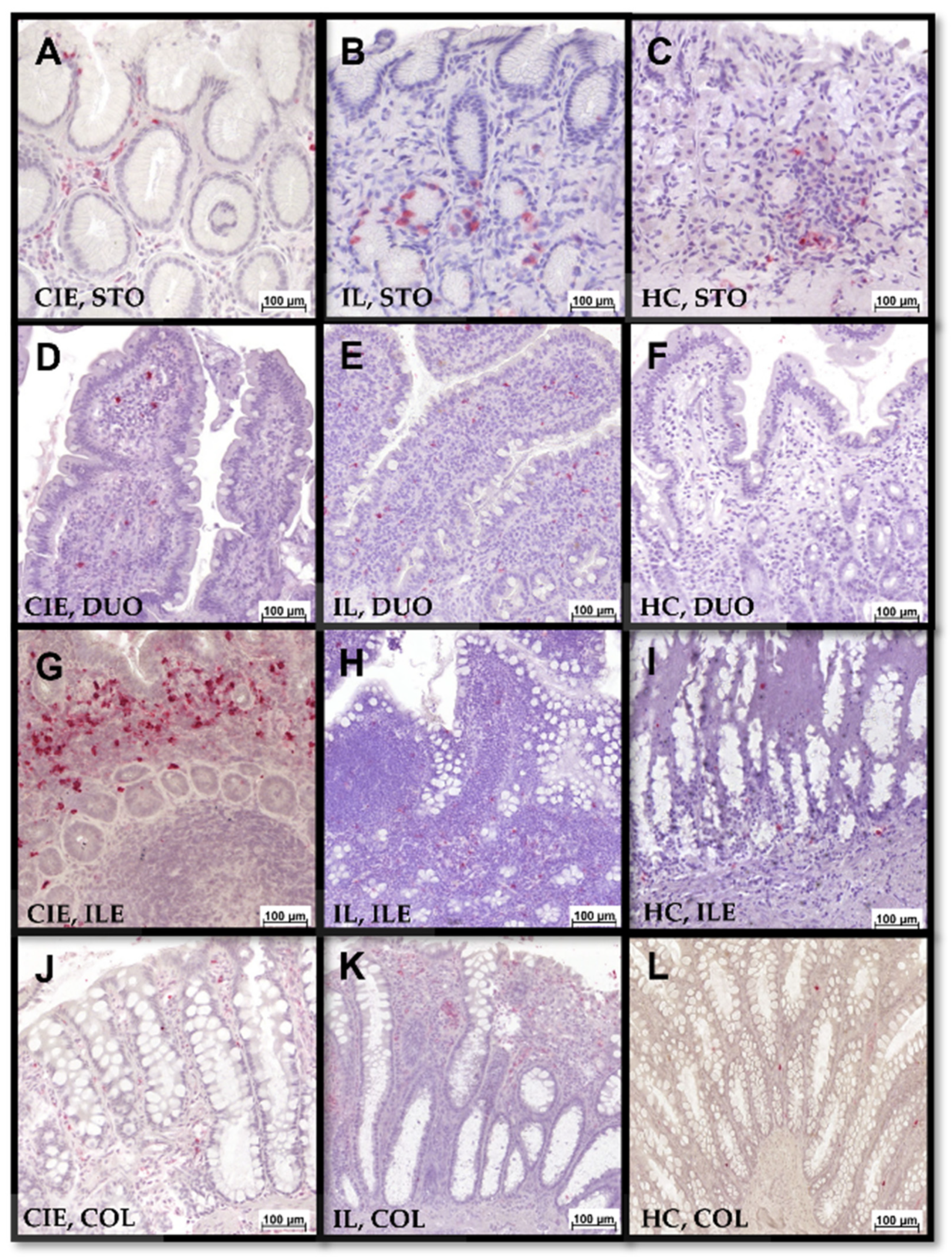
3.5. Gastrointestinal S100/Calgranulin Expression
3.6. Association of Patient Characteristics with Gastrointestinal S100/Calgranulin Expression
4. Discussion
5. Conclusions
Supplementary Materials
Author Contributions
Funding
Institutional Review Board Statement
Informed Consent Statement
Data Availability Statement
Acknowledgments
Conflicts of Interest
References
- Jergens, A.E. Feline idiopathic inflammatory bowel disease: What we know and what remains to be unraveled. J. Fel. Med. Surg. 2012, 14, 445–458. [Google Scholar] [CrossRef] [PubMed]
- Richter, K.P. Feline gastrointestinal lymphoma. Vet. Clin. North. Am. Small. Anim. Pract. 2003, 33, 1083–1098. [Google Scholar] [CrossRef]
- Washabau, R.J.; Day, M.J.; Willard, M.D.; Hall, E.J.; Jergens, A.E.; Mansell, J.; Minami, T.; Bilzer, T.W. Endoscopic, biopsy, and histopathologic guidelines for the evaluation of gastrointestinal inflammation in companion animals. J. Vet. Intern. Med. 2010, 24, 10–26. [Google Scholar] [CrossRef]
- Jergens, A.E.; Simpson, K.W. Inflammatory bowel disease in veterinary medicine. Front. Biosci. 2012, 4, 1404–1419. [Google Scholar] [CrossRef]
- Jergens, A.E.; Crandell, J.M.; Evans, R.; Ackermann, M.; Miles, K.G.; Wang, C. A clinical index for disease activity in cats with chronic enteropathy. J. Vet. Intern. Med. 2010, 24, 1027–1033. [Google Scholar] [CrossRef]
- Marsilio, S. Differentiating inflammatory bowel disease from alimentary lymphoma in cats: Does it matter? Vet. Clin. North. Am. Small. Anim. Pract. 2021, 51, 93–109. [Google Scholar] [CrossRef] [PubMed]
- Albert, E.J. Inflammatory bowel disease: Current perspectives. Vet. Clin. North. Am. Small Anim. Pract. 1999, 29, 501–521. [Google Scholar] [CrossRef]
- Ewald, N.; Rödler, F.; Heilmann, R.M. Chronische Enteropathien bei der Katze—Diagnostische und therapeutische Aspekte. Tierarztl. Prax. Ausg. K. Kleintiere. Heimtiere. 2021, 49, 363–376. [Google Scholar] [CrossRef] [PubMed]
- Barrs, V.R.; Beatty, J.A. Feline alimentary lymphoma: 2. Further diagnostics, therapy and prognosis. J. Fel. Med. Surg. 2012, 14, 191–201. [Google Scholar] [CrossRef]
- Vail, D.M.; Moore, A.S.; Ogilvie, G.K.; Volk, L.M. Feline lymphoma (145 cases): Proliferation indices, cluster of differentiation 3 immunoreactivity, and their association with prognosis in 90 cats. J. Vet. Intern. Med. 1998, 12, 349–354. [Google Scholar] [CrossRef] [PubMed]
- Zwahlen, C.H.; Lucroy, M.D.; Kraegel, S.A.; Madewell, B.R. Results of chemotherapy for cats with alimentary malignant lymphoma: 21 cases (1993-1997). J. Am. Vet. Med. Assoc. 1998, 213, 1144–1149. [Google Scholar] [PubMed]
- Heilmann, R.M.; Suchodolski, J.S. Is inflammatory bowel disease in dogs and cats associated with a Th1 or Th2 polarization? Vet. Immunol. Immunopathol. 2015, 168, 131–134. [Google Scholar] [CrossRef]
- Lingard, A.E.; Briscoe, K.; Beatty, J.A.; Moore, A.S.; Crowley, A.M.; Krockenberger, M.; Churcher, R.K.; Canfield, P.J.; Barrs, V.R. Low-grade alimentary lymphoma: Clinicopathological findings and response to treatment in 17 cases. J. Fel. Med. Surg. 2009, 11, 692–700. [Google Scholar] [CrossRef] [PubMed]
- Kiselow, M.A.; Rassnick, K.M.; McDonough, S.P.; Goldstein, R.E.; Simpson, K.W.; Weinkle, T.K.; Erb, H.N. Outcome of cats with low-grade lymphocytic lymphoma: 41 cases (1995–2005). J. Am. Vet. Med. Assoc. 2008, 232, 405–410. [Google Scholar] [CrossRef] [PubMed]
- Marsilio, S.; Pilla, R.; Sarawichitr, B.; Chow, B.; Hill, S.L.; Ackermann, M.R.; Estep, J.S.; Lidbury, J.A.; Steiner, J.M.; Suchodolski, J.S. Characterization of the fecal microbiome in cats with inflammatory bowel disease or alimentary small cell lymphoma. Sci. Rep. 2019, 9, 19208. [Google Scholar] [CrossRef] [PubMed]
- Norsworthy, G.D.; Estep, J.S.; Hollinger, C.; Steiner, J.M.; Lavallee, J.O.; Gassler, L.N.; Restine, L.M.; Kiupel, M. Prevalence and underlying causes of histologic abnormalities in cats suspected to have chronic small bowel disease: 300 cases (2008–2013). J. Am. Vet. Med. Assoc. 2015, 247, 629–635. [Google Scholar] [CrossRef] [PubMed]
- Burke, K.F.; Broussard, J.D.; Ruaux, C.G.; Suchodolski, J.S.; Williams, D.A.; Steiner, J.M. Evaluation of fecal α1-proteinase inhibitor concentrations in cats with idiopathic inflammatory bowel disease and cats with gastrointestinal neoplasia. Vet. J. 2013, 196, 189–196. [Google Scholar] [CrossRef] [PubMed]
- Waly, N.E.; Gruffydd-Jones, T.J.; Stokes, C.R.; Day, M.J. Immunohistochemical diagnosis of alimentary lymphomas and severe intestinal inflammation in cats. J. Comp. Pathol. 2005, 133, 253–260. [Google Scholar] [CrossRef]
- Reed, N.; Gunn-Moore, D.; Simpson, K. Cobalamin, folate and inorganic phosphate abnormalities in ill cats. J. Fel. Med. Surg. 2007, 9, 278–288. [Google Scholar] [CrossRef] [PubMed]
- Dennis, J.S.; Kruger, J.M.; Mullaney, T.P. Lymphocytic/plasmacytic gastroenteritis in cats: 14 cases (1985–1990). J. Am. Vet. Med. Assoc. 1992, 200, 1712–1718. [Google Scholar] [PubMed]
- Jergens, A.E.; Moore, F.M.; Haynes, J.S.; Miles, K.G. Idiopathic inflammatory bowel disease in dogs and cats: 84 cases (1987–1990). J. Am. Vet. Med. Assoc. 1992, 201, 1603–1608. [Google Scholar]
- Norsworthy, G.D.; Scot Estep, J.; Kiupel, M.; Olson, J.C.; Gassler, L.N. Diagnosis of chronic small bowel disease in cats: 100 cases (2008–2012). J. Am. Vet. Med. Assoc. 2013, 243, 1455–1461. [Google Scholar] [CrossRef] [PubMed]
- Moore, P.F.; Woo, J.C.; Vernau, W.; Kosten, S.; Graham, P.S. Characterization of feline T cell receptor gamma (TCRG) variable region genes for the molecular diagnosis of feline intestinal T cell lymphoma. Vet. Immunol. Iimmunopathol. 2005, 106, 167–178. [Google Scholar] [CrossRef]
- Moore, P.F.; Rodriguez-Bertos, A.; Kass, P.H. Feline gastrointestinal lymphoma: Mucosal architecture, immunophenotype, and molecular clonality. Vet. Pathol. 2012, 49, 658–668. [Google Scholar] [CrossRef] [PubMed]
- Kiupel, M.; Smedley, R.C.; Pfent, C.; Xie, Y.; Xue, Y.; Wise, A.G.; DeVaul, J.M.; Maes, R.K. Diagnostic algorithm to differentiate lymphoma from inflammation in feline small intestinal biopsy samples. Vet. Pathol. 2011, 48, 212–222. [Google Scholar] [CrossRef] [PubMed]
- Marsilio, S.; Newman, S.J.; Estep, J.S.; Giaretta, P.R.; Lidbury, J.A.; Warry, E.; Flory, A.; Morley, P.S.; Smoot, K.; Seeley, E.H.; et al. Differentiation of lymphocytic-plasmacytic enteropathy and small cell lymphoma in cats using histology-guided mass spectrometry. J. Vet. Intern. Med. 2020, 34, 669–677. [Google Scholar] [CrossRef]
- Heilmann, R.M.; Allenspach, K. Pattern-recognition receptors: Signaling pathways and dysregulation in canine chronic enteropathies-brief review. J. Vet. Diagn. Investig. 2017, 29, 781–787. [Google Scholar] [CrossRef] [PubMed]
- Heilmann, R.M.; Volkmann, M.; Otoni, C.C.; Grützner, N.; Kohn, B.; Jergens, A.E.; Steiner, J.M. Fecal S100A12 concentration predicts a lack of response to treatment in dogs affected with chronic enteropathy. Vet. J. 2016, 215, 96–100. [Google Scholar] [CrossRef]
- Heilmann, R.M.; Berghoff, N.; Mansell, J.; Grützner, N.; Parnell, N.K.; Gurtner, C.; Suchodolski, J.S.; Steiner, J.M. Association of fecal calprotectin concentrations with disease severity, response to treatment, and other biomarkers in dogs with chronic inflammatory enteropathies. J. Vet. Intern. Med. 2018, 32, 679–692. [Google Scholar] [CrossRef]
- Heilmann, R.M.; Grellet, A.; Allenspach, K.; Lecoindre, P.; Day, M.J.; Priestnall, S.L.; Toresson, L.; Procoli, F.; Grützner, N.; Suchodolski, J.S.; et al. Association between fecal S100A12 concentration and histologic, endoscopic, and clinical disease severity in dogs with idiopathic inflammatory bowel disease. Vet. Immunol. Immunopathol. 2014, 158, 156–166. [Google Scholar] [CrossRef]
- Otoni, C.C.; Heilmann, R.M.; García-Sancho, M.; Sainz, A.; Ackermann, M.R.; Suchodolski, J.S.; Steiner, J.M.; Jergens, A.E. Serologic and fecal markers to predict response to induction therapy in dogs with idiopathic inflammatory bowel disease. J. Vet. Intern. Med. 2018, 32, 999–1008. [Google Scholar] [CrossRef]
- Heilmann, R.M.; Steiner, J.M. Clinical utility of currently available biomarkers in inflammatory enteropathies of dogs. J. Vet. Intern. Med. 2018, 32, 1495–1508. [Google Scholar] [CrossRef] [PubMed]
- Goyette, J.; Geczy, C.L. Inflammation-associated S100 proteins: New mechanisms that regulate function. Amino Acids 2011, 41, 821–842. [Google Scholar] [CrossRef]
- Foell, D.; Frosch, M.; Sorg, C.; Roth, J. Phagocyte-specific calcium-binding S100 proteins as clinical laboratory markers of inflammation. Clin. Chim. Acta. 2004, 344, 37–51. [Google Scholar] [CrossRef]
- Heilmann, R.M.; Nestler, J.; Schwarz, J.; Grützner, N.; Ambrus, A.; Seeger, J.; Suchodolski, J.S.; Steiner, J.M.; Gurtner, C. Mucosal expression of S100A12 (calgranulin C) and S100A8/A9 (calprotectin) and correlation with serum and fecal concentrations in dogs with chronic inflammatory enteropathy. Vet. Immunol. Immunopathol. 2019, 211, 64–74. [Google Scholar] [CrossRef]
- Vogl, T.; Tenbrock, K.; Ludwig, S.; Leukert, N.; Ehrhardt, C.; van Zoelen, M.A.D.; Nacken, W.; Foell, D.; van der Poll, T.; Sorg, C.; et al. Mrp8 and Mrp14 are endogenous activators of Toll-like receptor 4, promoting lethal, endotoxin-induced shock. Nature. Med. 2007, 13, 1042–1049. [Google Scholar] [CrossRef]
- Hofmann, M.A.; Drury, S.; Fu, C.; Qu, W.; Taguchi, A.; Lu, Y.; Avila, C.; Kambham, N.; Bierhaus, A.; Nawroth, P.; et al. RAGE mediates a novel proinflammatory axis. Cell 1999, 97, 889–901. [Google Scholar] [CrossRef]
- Cabrera-García, A.I.; Suchodolski, J.S.; Steiner, J.M.; Heilmann, R.M. Association between serum soluble receptor for advanced glycation end-products (RAGE) deficiency and severity of clinicopathologic evidence of canine chronic inflammatory enteropathy. J. Vet. Diagn. Investig. 2020, 32, 664–674. [Google Scholar] [CrossRef]
- Day, M.J.; Bilzer, T.; Mansell, J.; Wilcock, B.; Hall, E.J.; Jergens, A.; Minami, T.; Willard, M.; Washabau, R. Histopathological standards for the diagnosis of gastrointestinal inflammation in endoscopic biopsy samples from the dog and cat: A report from the World Small Animal Veterinary Association Gastrointestinal Standardization Group. J. Comp. Pathol. 2008, 138, S1–S43. [Google Scholar] [CrossRef] [PubMed]
- Tucker, S.; Penninck, D.G.; Keating, J.H.; Webster, C.R.L. Clinicopathological and ultrasonographic features of cats with eosinophilic enteritis. J. Fel. Med. Surg. 2014, 16, 950–956. [Google Scholar] [CrossRef] [PubMed]
- German, A.J.; Hall, E.J.; Kelly, D.F.; Watson, A.D.; Day, M.J. An immunohistochemical study of histiocytic ulcerative colitis in boxer dogs. J. Comp. Pathol. 2000, 122, 163–175. [Google Scholar] [CrossRef]
- Wagner, A.; Junginger, J.; Lemensieck, F.; Hewicker-Trautwein, M. Immunohistochemical characterization of gastrointestinal macrophages/phagocytes in dogs with inflammatory bowel disease (IBD) and non-IBD dogs. Vet. Immunol. Immunopathol. 2018, 197, 49–57. [Google Scholar] [CrossRef] [PubMed]
- Truar, K.; Nestler, J.; Schwarz, J.; Grützner, N.; Gabris, C.; Kock, K.; Niederberger, C.; Heilmann, R.M. Feasibility of measuring fecal calprotectin concentrations in dogs and cats by the fCAL® turbo immunoassay. abstract. J. Vet. Intern. Med. 2018, 32, 580. [Google Scholar]
- Zornow, K.A.; Slovak, J.E.; Lidbury, J.A.; Suchodolski, J.S.; Steiner, J.M. Fecal S100A12 (calgranulin C) concentrations in cats with chronic enteropathies. J. Vet. Int. Med. 2021, 3151. [Google Scholar]
- Hsu, K.; Passey, R.J.; Endoh, Y.; Rahimi, F.; Youssef, P.; Yen, T.; Geczy, C.L. Regulation of S100A8 by glucocorticoids. J. Immunol. 2005, 174, 2318–2326. [Google Scholar] [CrossRef] [PubMed]
- Guttin, T.; Walsh, A.; Durham, A.C.; Reetz, J.A.; Brown, D.C.; Rondeau, M.P. Ability of ultrasonography to predict the presence and location of histologic lesions in the small intestine of cats. J. Vet. Intern. Med. 2019, 33, 1278–1285. [Google Scholar] [CrossRef]
- Freiche, V.; Fages, J.; Paulin, M.V.; Bruneau, J.; Couronné, L.; German, A.J.; Penninck, D.; Hermine, O. Clinical, laboratory and ultrasonographic findings differentiating low-grade intestinal T-cell lymphoma from lymphoplasmacytic enteritis in cats. J. Vet. Intern. Med. 2021, 35, 2685–2696. [Google Scholar] [CrossRef] [PubMed]
- Freiche, V.; Paulin, M.V.; Cordonnier, N.; Huet, H.; Turba, M.-E.; Macintyre, E.; Molina, T.-J.; Hermine, O.; Couronné, L.; Bruneau, J. Histopathologic, phenotypic, and molecular criteria to discriminate low-grade intestinal T-cell lymphoma in cats from lymphoplasmacytic enteritis. J. Vet. Intern. Med. 2021, 35, 2673–2684. [Google Scholar] [CrossRef]
- Heilmann, R.M.; Suchodolski, J.S.; Steiner, J.M. Development and analytic validation of a radioimmunoassay for the quantification of canine calprotectin in serum and feces from dogs. Am. J. Vet. Res. 2008, 69, 845–853. [Google Scholar] [CrossRef] [PubMed]
- Heilmann, R.M.; Suchodolski, J.S.; Steiner, J.M. Purification and partial characterization of canine calprotectin. Biochimie 2008, 90, 1306–1315. [Google Scholar] [CrossRef]
- Heilmann, R.M.; Suchodolski, J.S.; Steiner, J.M. Purification and partial characterization of canine S100A12. Biochimie 2010, 92, 1914–1922. [Google Scholar] [CrossRef] [PubMed]
- Heilmann, R.M.; Cranford, S.M.; Ambrus, A.; Grützner, N.; Schellenberg, S.; Ruaux, C.G.; Suchodolski, J.S.; Steiner, J.M. Validation of an enzyme-linked immunosorbent assay (ELISA) for the measurement of canine S100A12. Vet. Clin. Pathol. 2016, 45, 135–147. [Google Scholar] [CrossRef] [PubMed]
- Heilmann, R.M.; Lanerie, D.J.; Ruaux, C.G.; Grützner, N.; Suchodolski, J.S.; Steiner, J.M. Development and analytic validation of an immunoassay for the quantification of canine S100A12 in serum and fecal samples and its biological variability in serum from healthy dogs. Vet. Immunol. Immunopathol. 2011, 144, 200–209. [Google Scholar] [CrossRef]
- Heilmann, R.M.; Grützner, N.; Handl, S.; Suchodolski, J.S.; Steiner, J.M. Preanalytical validation of an in-house radioimmunoassay for measuring calprotectin in feline specimens. Vet. Clin. Pathol. 2018, 47, 100–107. [Google Scholar] [CrossRef]
- Bridges, C.S.; Grützner, N.; Suchodolski, J.S.; Steiner, J.M.; Heilmann, R.M. Analytical validation of an enzyme-linked immunosorbent assay for the quantification of S100A12 in the serum and feces of cats. Vet. Clin. Pathol. 2019, 48, 754–761. [Google Scholar] [CrossRef]
- Fukunaga, S.; Kuwaki, K.; Mitsuyama, K.; Takedatsu, H.; Yoshioka, S.; Yamasaki, H.; Yamauchi, R.; Mori, A.; Kakuma, T.; Tsuruta, O.; et al. Detection of calprotectin in inflammatory bowel disease: Fecal and serum levels and immunohistochemical localization. Int. J. Mol. Med. 2018, 41, 107–118. [Google Scholar] [CrossRef]
- Tamoutounour, S.; Henri, S.; Lelouard, H.; de Bovis, B.; de Haar, C.; van der Woude, C.J.; Woltman, A.M.; Reyal, Y.; Bonnet, D.; Sichien, D.; et al. CD64 distinguishes macrophages from dendritic cells in the gut and reveals the Th1-inducing role of mesenteric lymph node macrophages during colitis. Eur. J. Immunol. 2012, 42, 3150–3166. [Google Scholar] [CrossRef]
- Nolte, A.; Junginger, J.; Baum, B.; Hewicker-Trautwein, M. Heterogeneity of macrophages in canine histiocytic ulcerative colitis. Innate. Immun. 2017, 23, 228–239. [Google Scholar] [CrossRef]
- Zwadlo, G.; Brüggen, J.; Gerhards, G.; Schlegel, R.; Sorg, C. Two calcium-binding proteins associated with specific stages of myeloid cell differentiation are expressed by subsets of macrophages in inflammatory tissues. Clin. Exp. Immunol. 1988, 72, 510–515. [Google Scholar]
- Dandrieux, J.R.; Martinez Lopez, L.M.; Stent, A.; Jergens, A.; Allenspach, K.; Nowell, C.J.; Firestone, S.M.; Kimpton, W.; Mansfield, C.S. Changes in duodenal CD163-positive cells in dogs with chronic enteropathy after successful treatment. Innate Immun. 2018, 24, 400–410. [Google Scholar] [CrossRef] [PubMed]
- Bain, C.C.; Mowat, A.M. Intestinal macrophages—Specialised adaptation to a unique environment. Eur. J. Immunol. 2011, 41, 2494–2498. [Google Scholar] [CrossRef] [PubMed]
- Bujko, A.; Atlasy, N.; Landsverk, O.J.B.; Richter, L.; Yaqub, S.; Horneland, R.; Øyen, O.; Aandahl, E.M.; Aabakken, L.; Stunnenberg, H.G.; et al. Transcriptional and functional profiling defines human small intestinal macrophage subsets. J. Exp. Med. 2018, 215, 441–458. [Google Scholar] [CrossRef] [PubMed]
- Dandrieux, J.R.; Bornand, V.F.; Doherr, M.G.; Kano, R.; Zurbriggen, A.; Burgener, I.A. Evaluation of lymphocyte apoptosis in dogs with inflammatory bowel disease. Am. J. Vet. Res. 2008, 69, 1279–1285. [Google Scholar] [CrossRef] [PubMed]
- Leach, S.T.; Yang, Z.; Messina, I.; Song, C.; Geczy, C.L.; Cunningham, A.M.; Day, A.S. Serum and mucosal S100 proteins, calprotectin (S100A8/S100A9) and S100A12, are elevated at diagnosis in children with inflammatory bowel disease. Scand. J. Gastroenterol. 2007, 42, 1321–1331. [Google Scholar] [CrossRef] [PubMed]
- Marsilio, S.; Ackermann, M.R.; Lidbury, J.A.; Suchodolski, J.S.; Steiner, J.M. Results of histopathology, immunohistochemistry, and molecular clonality testing of small intestinal biopsy specimens from clinically healthy client-owned cats. J. Vet. Intern. Med. 2019, 33, 551–558. [Google Scholar] [CrossRef]
- Jergens, A.E.; Evans, R.B.; Ackermann, M.; Hostetter, J.; Willard, M.; Mansell, J.; Bilzer, T.; Wilcock, B.; Washabau, R.; Hall, E.J.; et al. Design of a simplified histopathologic model for gastrointestinal inflammation in dogs. Vet. Pathol. 2014, 51, 946–950. [Google Scholar] [CrossRef]
- Harjola, V.-P.; Mullens, W.; Banaszewski, M.; Bauersachs, J.; Brunner-La Rocca, H.-P.; Chioncel, O.; Collins, S.P.; Doehner, W.; Filippatos, G.S.; Flammer, A.J.; et al. Organ dysfunction, injury and failure in acute heart failure: From pathophysiology to diagnosis and management. A review on behalf of the Acute Heart Failure Committee of the Heart Failure Association (HFA) of the European Society of Cardiology (ESC). Eur. J. Heart. Fail. 2017, 19, 821–836. [Google Scholar] [CrossRef] [PubMed]
- Tsukamoto, T.; Chanthaphavong, R.S.; Pape, H.-C. Current theories on the pathophysiology of multiple organ failure after trauma. Injury 2010, 41, 21–26. [Google Scholar] [CrossRef] [PubMed]
- Chott, A.; Dragosics, B.; Radaszkiewicz, T. Peripheral T-cell lymphomas of the intestine. Am. J. Pathol. 1992, 141, 1361–1371. [Google Scholar]
- Dieter, R.S.; Duque, K. Enterotherapy associated T-cell lymphoma: A case report and literature review. WMJ 2000, 99, 28–31. [Google Scholar] [PubMed]
- Hanifeh, M.; Sankari, S.; Rajamäki, M.M.; Syrjä, P.; Kilpinen, S.; Suchodolski, J.S.; Heilmann, R.M.; Guadiano, P.; Lidbury, J.; Steiner, J.M.; et al. S100A12 concentrations and myeloperoxidase activities are increased in the intestinal mucosa of dogs with chronic enteropathies. BMC. Vet. Res. 2018, 14, 125. [Google Scholar] [CrossRef] [PubMed]
- Schoepfer, A.M.; Beglinger, C.; Straumann, A.; Trummler, M.; Vavricka, S.R.; Bruegger, L.E.; Seibold, F. Fecal calprotectin correlates more closely with the Simple Endoscopic Score for Crohn’s disease (SES-CD) than CRP, blood leukocytes, and the CDAI. Am. J. Gastroenterol. 2010, 105, 162–169. [Google Scholar] [CrossRef] [PubMed]
- Schoepfer, A.M.; Beglinger, C.; Straumann, A.; Trummler, M.; Renzulli, P.; Seibold, F. Ulcerative colitis: Correlation of the Rachmilewitz endoscopic activity index with fecal calprotectin, clinical activity, C-reactive protein, and blood leukocytes. Inflamm. Bowel. Dis. 2009, 15, 1851–1858. [Google Scholar] [CrossRef] [PubMed]
- Önal, İ.K.; Beyazit, Y.; Şener, B.; Savuk, B.; Özer Etık, D.; Sayilir, A.; Öztaş, E.; Torun, S.; Özderın Özın, Y.; Tunç Demırel, B.; et al. The value of fecal calprotectin as a marker of intestinal inflammation in patients with ulcerative colitis. Turk. J. Gastroenterol. 2012, 23, 509–514. [Google Scholar] [CrossRef] [PubMed]
- Welter, J.; Duckova, T.; Groiss, S.; Wolfesberger, B.; Fuchs-Baumgartinger, A.; Rütgen, B.C.; Hammer, S.E. Revisiting lymphocyte clonality testing in feline B-cell lymphoma. Vet. Immunol. Immunopathol. 2021, 242, 110350. [Google Scholar] [CrossRef] [PubMed]
- Eaden, J.; Abrams, K.; McKay, H.; Denley, H.; Mayberry, J. Inter-observer variation between general and specialist gastrointestinal pathologists when grading dysplasia in ulcerative colitis. J. Pathol. 2001, 194, 152–157. [Google Scholar] [CrossRef]
- Bonsembiante, F.; Martini, V.; Bonfanti, U.; Casarin, G.; Trez, D.; Gelain, M.E. Cytomorphological description and intra-observer agreement in whole slide imaging for canine lymphoma. Vet. J. 2018, 236, 96–101. [Google Scholar] [CrossRef]
- Wilson, H.M. Feline alimentary lymphoma: Demystifying the enigma. Top. Companion. Anim. Med. 2008, 23, 177–184. [Google Scholar] [CrossRef]
- Willard, M.D.; Mansell, J.; Fosgate, G.T.; Gualtieri, M.; Olivero, D.; Lecoindre, P.; Twedt, D.C.; Collett, M.G.; Day, M.J.; Hall, E.J.; et al. Effect of sample quality on the sensitivity of endoscopic biopsy for detecting gastric and duodenal lesions in dogs and cats. J. Vet. Intern. Med. 2008, 22, 1084–1089. [Google Scholar] [CrossRef]
- Jergens, A.E.; Willard, M.D.; Allenspach, K. Maximizing the diagnostic utility of endoscopic biopsy in dogs and cats with gastrointestinal disease. Vet. J. 2016, 214, 50–60. [Google Scholar] [CrossRef]
- Lux, C.N.; Roberts, S.; Grimes, J.A.; Benitez, M.E.; Culp, W.T.N.; Ben-Aderet, D.; Brown, D.C. Evaluation of short-term risk factors associated with dehiscence and death following full-thickness incisions of the large intestine in cats: 84 cases (1993-2015). J. Am. Vet. Med. Assoc. 2021, 259, 162–171. [Google Scholar] [CrossRef] [PubMed]
- Jergens, A.E.; Schreiner, C.A.; Frank, D.E.; Niyo, Y.; Ahrens, F.E.; Eckersall, P.D.; Benson, T.J.; Evans, R. A scoring index for disease activity in canine inflammatory bowel disease. J. Vet. Intern. Med. 2003, 17, 291–297. [Google Scholar] [CrossRef] [PubMed]
- Allenspach, K.; Wieland, B.; Gröne, A.; Gaschen, F. Chronic enteropathies in dogs: Evaluation of risk factors for negative outcome. J. Vet. Intern. Med. 2007, 21, 700. [Google Scholar] [CrossRef] [PubMed]


| Epithelial S100A8/A9 Positivity (Cell Counts) | Lamina Propria S100A8/A9 Positivity (Cell Counts) | Submucosal S100A8/A9 Positivity (Cell Counts) | ||||||||
|---|---|---|---|---|---|---|---|---|---|---|
| Histologic Lesion | n | Range in All Tissue Biopsies | Median (Range) | p | Range in All Tissue Biopsies | Median (Range) | p | Range in All Tissue Biopsies | Median (Range) | p |
| Stomach | 34 | |||||||||
| normal | 19 | 0–1 | 0 (0) A | 0.011 | 0–10 | 1.5 (0–5) | 0.326 | − | − | – |
| inflammation | 13 | 0–10 | 0 (0–4) B | 0–13 | 1.5 (0.5–3) | − | − | |||
| lymphoma | 2 | 0–3 | 1 (0–1) B | 0–3 | 0.5 (0.5–1) | – | – | |||
| Duodenum/Jejunum | 27 | |||||||||
| normal | 2 | 0–2 | 0 (0–0.5) | 0.837 | 0–5 | 1.5 (0.5–2.5) A | 0.045 | 0 * | 0 (0) * | 0.749 |
| inflammation | 16 | 0–12 | 0 (0–2) | 0–38 | 6 (0–21.5) A,B | 0–6 $ | 0 (0–1) $ | |||
| lymphoma | 9 | 0–6 | 0.5 (0–0.5) | 0–27 | 6.5 (3–12) B | 0–8 † | 0 (0–2.5) † | |||
| Ileum | 14 | |||||||||
| normal | 2 | 0 | 0 (0) | 0.271 | 2–13 | 6 (3–9) | 0.901 | 0–1 | 0.5 (0–0.5) | 0.719 |
| inflammation | 8 | 0–1 | 0 (0–0.5) | 0–24 | 3.5 (0.5–12) | 0–4 | 0.5 (0–1) | |||
| lymphoma | 4 | 0 | 0 (0) | 0–16 | 5 (3.5–7.5) | 0–1 ‡ | 0.5 (0–0.5) ‡ | |||
| Colon | 17 | |||||||||
| normal | 7 | 0 | 0 (0) | 0.222 | 0–19 | 6.5 (2.5–8.5) | 0.239 | 0–11 | 1.5 (0.5–2.5) A | 0.004 |
| inflammation | 9 | 0–2 | 0 (0–0.5) | 0–23 | 4 (1–10.5) | 0–1 | 0 (0–0.5) B,§ | |||
| lymphoma | 1 | 0 | 0 (0) | 0–21 | 10 (10) | 0–2 | 0.5 (0.5) A,B | |||
| Epithelial S100A12 Positivity (Cell Counts) | Lamina Propria S100A12 Positivity (Cell Counts) | Submucosal S100A12 Positivity (Cell Counts) | ||||||||
|---|---|---|---|---|---|---|---|---|---|---|
| Histologic Lesion | n | Range in All Tissue Biopsies | Median (Range) | p | Range in All Tissue Biopsies | Median (Range) | p | Range in All Tissue Biopsies | Median (Range) | p |
| Stomach | 34 | |||||||||
| normal | 19 | 0–1 | 0 (0) | 0.102 | 0–17 | 1 (0–6) A | 0.032 | − | − | – |
| inflammation | 13 | 0–10 | 0 (0–1) | 0–6 | 0.5 (0–2) B | − | − | |||
| lymphoma | 2 | 0 | 0 (0) | 0–1 | 0.5 (0–1) A,B | – | – | |||
| Duodenum/Jejunum | 27 | |||||||||
| normal | 2 | 0 | 0 (0) | 0.153 | 0–7 | 1.5 (0.5–3) | 0.342 | 0 | 0 (0) * | 0.574 |
| inflammation | 16 | 0–13 | 0.5 (0–3.5) | 0–23 | 4 (0.5–11) | 0–2 | 0 (0–0.5) $ | |||
| lymphoma | 9 | 0–1 | 0 (0–0.5) | 0–23 | 3.5 (1.5–8) | 0–2 | 0 (0–0.5) † | |||
| Ileum | 14 | |||||||||
| normal | 2 | 0 | 0 (0) | 0.544 | 0–10 | 4 (2–6) | 0.852 | 0–1 | 0.5 (0–0.5) | 0.635 |
| inflammation | 8 | 0–1 | 0 (0–0.5) | 0–17 | 2.5 (0.5–10) | 0–1 | 0.5 (0–0.5) | |||
| lymphoma | 4 | 0–1 | 0 (0–0.5) | 0–30 | 3 (0.5–7) | 0–1 ‡ | 0.5 (0–0.5) ‡ | |||
| Colon | 17 | |||||||||
| normal | 7 | 0 | 0 (0) | 0.641 | 0–30 | 6 (0.5–11) | 0.599 | 0–22 | 0.5 (0–4) A | 0.041 |
| inflammation | 9 | 0–3 | 0 (0–1) | 0–24 | 3 (0.5–9) | 0–1 § | 0 (0–0.5) B,§ | |||
| lymphoma | 1 | 0 | 0 (0) | 0–12 | 5.5 (5.5) | 0–2 | 0.5 (0.5) A,B | |||
| Parameter | Correlated with | Spearman ρ Correlation Coefficient (p-Value, Pcorr) | |||
|---|---|---|---|---|---|
| Epithelial S100A8/A9+ (Cell Counts) | Lamina Propria S100A8/A9+ (Cell Counts) | Epithelial S100A12+ (Cell Counts) | Lamina Propria S100A12+ (Cell Counts) | ||
| Stomach (n = 13) | Stomach (n = 13) | ||||
| Stomach (composite score) # | 0.14 (0.655) | 0.08 (0.801) | 0.06 (0.844) | 0.33 (0.279) | |
| Morphologic criteria (sum) | 0.49 (0.092) | −0.16 (0.609) | 0.38 (0.199) | 0.18 (0.559) | |
| N/A | N/A | N/A | N/A | |
| N/A | N/A | N/A | N/A | |
| 0.33 (0.916) | −0.26 (0.394) | 0.21 (0.494) | −0.09 (0.780) | |
| Inflammatory criteria (sum) | −0.02 (0.948) | 0.25 (0.403) | −0.08 (0.791) | 0.38 (0.206) | |
| −0.36 (0.221) | 0.12 (0.689) | –0.54 (0.059) | −0.25 (0.420) | |
| −0.01 (0.965) | 0.19 (0.528) | −0.18 (0.555) | 0.42 (0.155) | |
| N/A | N/A | N/A | N/A | |
| 0.27 (0.380) | 0.12 (0.705) | 0.47 (0.104) | 0.31 (0.303) | |
| 0.27 (0.380) | 0.12 (0.705) | 0.47 (0.104) | 0.31 (0.303) | |
| 0.59 (0.034; 0.204) | 0.06 (0.852) | 0.28 (0.357) | 0.32 (0.295) | |
| Duodenum/proximal jejunum (n = 16) | Duodenum/proximal jejunum (n = 16) | ||||
| Duodenum/proximal jejunum (composite score) | 0.09 (0.747) | 0.18 (0.515) | −0.08 (0.766) | 0.13 (0.643) | |
| Morphologic criteria (sum) | −0.02 (0.942) | 0.11 (0.679) | −0.14 (0.614) | 0.12 (0.671) | |
| −0.03 (0.927) | 0.16 (0.546) | −0.06 (0.837) | 0.14 (0.594) | |
| −0.04 (0.880) | 0.13 (0.632) | 0.12 (0.667) | 0.27 (0.320) | |
| 0.07 (0.795) | −0.23 (0.401) | −0.06 (0.816) | −0.22 (0.423) | |
| −0.29 (0.289) | 0.41 (0.115) | −0.42 (0.103) | 0.21 (0.446) | |
| 0.02 (0.931) | 0.08 (0.763) | −0.19 (0.481) | 0.25 (0.359) | |
| Inflammatory criteria (sum) | 0.34 (0.196) | 0.44 (0.087) | 0.19 (0.474) | 0.29 (0.282) | |
| 0.12 (0.672) | 0.06 (0.822) | −0.13 (0.620) | −0.13 (0.623) | |
| 0.14 (0.619) | 0.23 (0.395) | 0.01 (0.991) | 0.14 (0.619) | |
| 0.10 (0.719) | 0.26 (0.333) | 0.22 (0.418) | 0.17 (0.529) | |
| 0.42 (0.102) | 0.41 (0.115) | 0.25 (0.344) | 0.45 (0.080) | |
| 0.23 (0.402) | 0.14 (0.605) | 0.03 (0.916) | 0.20 (0.467) | |
| Ileum (n = 8) | Ileum (n = 8) | ||||
| Ileum (composite score) | –0.70 (0.053) | −0.34 (0.399) | −0.54 (0.168) | −0.35 (0.399) | |
| Morphologic criteria (sum) | −0.58 (0.132) | −0.39 (0.346) | −0.61 (0.112) | −0.28 (0.506) | |
| −0.43 (0.284) | 0.06 (0.882) | −0.44 (0.280) | −0.06 (0.882) | |
| −0.43 (0.289) | −0.02 (0.971) | 0.05 (0.899) | 0.09 (0.826) | |
| −0.23 (0.585) | −0.55 (0.157) | −0.61 (0.111) | −0.35 (0.395) | |
| −0.28 (0.496) | 0.25 (0.555) | −0.29 (0.493) | −0.25 (0.555) | |
| −0.02 (0.971) | −0.44 (0.275) | −0.07 (0.867) | −0.15 (0.721) | |
| Inflammatory criteria (sum) | 0.12 (0.783) | 0.37 (0.367) | 0.53 (0.176) | 0.12 (0.786) | |
| 0.13 (0.768) | 0.55 (0.162) | −0.19 (0.654) | 0.22 (0.604) | |
| −0.26 (0.528) | 0.38 (0.359) | 0.11 (0.805) | 0.40 (0.326) | |
| −0.23 (0.577) | 0.03 (0.952) | 0.31 (0.455) | −0.17 (0.694) | |
| 0.66 (0.074) | −0.25 (0.555) | 0.57 (0.139) | 0.08 (0.846) | |
| 0.66 (0.074) | −0.25 (0.555) | 0.57 (0.139) | 0.08 (0.846) | |
| Colon (n = 9) | Colon (n = 9) | ||||
| Colon (composite score) | 0.24 (0.527) | 0.44 (0.241) | 0.49 (0.179) | 0.80 (0.009; 0.009) | |
| Morphologic criteria (sum) | −0.32 (0.399) | 0.12 (0.760) | −0.31 (0.430) | 0.49 (0.184) | |
| −0.37 (0.329) | 0.00 (1.000) | −0.19 (0.626) | 0.41 (0.268) | |
| −0.24 (0.527) | −0.27 (0.476) | −0.13 (0.749) | 0.14 (0.725) | |
| −0.37 (0.333) | 0.05 (0.907) | −0.19 (0.626) | 0.30 (0.438) | |
| 0.06 (0.875) | 0.21 (0.593) | −0.19 (0.626) | 0.21 (0.593) | |
| Inflammatory criteria (sum) | 0.31 (0.427) | 0.48 (0.197) | 0.60 (0.088) | 0.68 (0.046; 0.092) | |
| 0.62 (0.077) | −0.26 (0.500) | 0.32 (0.407) | −0.35 (0.361) | |
| 0.07 (0.856) | 0.51 (0.157) | 0.53 (0.144) | 0.84 (0.005; 0.025) | |
| 0.65 (0.058) | 0.55 (0.127) | 1.00 (<0.001; <0.005) | 0.55 (0.127) | |
| −0.24 (0.527) | 0.41 (0.272) | −0.13 (0.749) | 0.27 (0.476) | |
| –0.24 (0.527) | 0.41 (0.272) | –0.13 (0.749) | 0.27 (0.476) | |
Publisher’s Note: MDPI stays neutral with regard to jurisdictional claims in published maps and institutional affiliations. |
© 2022 by the authors. Licensee MDPI, Basel, Switzerland. This article is an open access article distributed under the terms and conditions of the Creative Commons Attribution (CC BY) license (https://creativecommons.org/licenses/by/4.0/).
Share and Cite
Riggers, D.S.; Gurtner, C.; Protschka, M.; Böttcher, D.; von Bomhard, W.; Alber, G.; Winter, K.; Steiner, J.M.; Heilmann, R.M. Intestinal S100/Calgranulin Expression in Cats with Chronic Inflammatory Enteropathy and Intestinal Lymphoma. Animals 2022, 12, 2044. https://doi.org/10.3390/ani12162044
Riggers DS, Gurtner C, Protschka M, Böttcher D, von Bomhard W, Alber G, Winter K, Steiner JM, Heilmann RM. Intestinal S100/Calgranulin Expression in Cats with Chronic Inflammatory Enteropathy and Intestinal Lymphoma. Animals. 2022; 12(16):2044. https://doi.org/10.3390/ani12162044
Chicago/Turabian StyleRiggers, Denise S., Corinne Gurtner, Martina Protschka, Denny Böttcher, Wolf von Bomhard, Gottfried Alber, Karsten Winter, Joerg M. Steiner, and Romy M. Heilmann. 2022. "Intestinal S100/Calgranulin Expression in Cats with Chronic Inflammatory Enteropathy and Intestinal Lymphoma" Animals 12, no. 16: 2044. https://doi.org/10.3390/ani12162044
APA StyleRiggers, D. S., Gurtner, C., Protschka, M., Böttcher, D., von Bomhard, W., Alber, G., Winter, K., Steiner, J. M., & Heilmann, R. M. (2022). Intestinal S100/Calgranulin Expression in Cats with Chronic Inflammatory Enteropathy and Intestinal Lymphoma. Animals, 12(16), 2044. https://doi.org/10.3390/ani12162044