Article

Investigation of Sperm and Seminal Plasma Candidate MicroRNAs of Bulls with Differing Fertility and In Silico Prediction of miRNA-mRNA Interaction Network of Reproductive Function

by
Vanmathy Kasimanickam
1,
Nishant Kumar
2 and
Ramanathan Kasimanickam
3,*
1
Center for Reproductive Biology, College of Veterinary Medicine, Washington State University, Pullman, WA 99164, USA
2
National Dairy Research Institute, Indian Council of Agricultural Research, Karnal 132001, India
3
Department of Veterinary Clinical Sciences, College of Veterinary Medicine, Washington State University, Pullman, WA 99164, USA
*
Author to whom correspondence should be addressed.
Animals 2022, 12(18), 2360; https://doi.org/10.3390/ani12182360
Submission received: 16 August 2022 / Revised: 2 September 2022 / Accepted: 2 September 2022 / Published: 9 September 2022
(This article belongs to the Section Animal Reproduction)

Abstract

:

Simple Summary

The objective of this study was to identify differentially expressed (DE) sperm and seminal plasma microRNAs (miRNAs) in high- and low-fertile Holstein bulls (four bulls per group), integrate miRNAs to their target genes, and categorize target genes based on predicted biological processes. Out of 84 bovine-specific, prioritized miRNAs analyzed by RT-PCR, 30 were differentially expressed in high-fertile sperm and seminal plasma compared to low-fertile sperm and seminal plasma, respectively (p ≤ 0.05, fold regulation ≥5 magnitudes). Interestingly, expression levels of DE-miRNAs in sperm and seminal plasma followed a similar pattern. Highly scored integrated genes of DE-miRNAs predicted various biological and molecular functions, cellular process, and pathways. Further in silico analysis revealed categorized genes may have a plausible association with pathways regulating sperm structure and function, fertilization, and embryo and placental development. In conclusion, highly DE-miRNAs in bovine sperm and seminal plasma could be used as a tool for predicting reproductive functions. Since the identified miRNA-mRNA interactions were mostly based on predictions from public databases, the causal regulations of miRNA-mRNA and the underlying mechanisms require further functional characterization in future studies.

Abstract

Recent advances in high-throughput in silico techniques portray experimental data as exemplified biological networks and help us understand the role of individual proteins, interactions, and their biological functions. The objective of this study was to identify differentially expressed (DE) sperm and seminal plasma microRNAs (miRNAs) in high- and low-fertile Holstein bulls (four bulls per group), integrate miRNAs to their target genes, and categorize the target genes based on biological process predictions. Out of 84 bovine-specific, prioritized miRNAs analyzed by RT-PCR, 30 were differentially expressed in high-fertile sperm and seminal plasma compared to low-fertile sperm and seminal plasma, respectively (p ≤ 0.05, fold regulation ≥ 5 magnitudes). The expression levels of DE-miRNAs in sperm and seminal plasma followed a similar pattern. Highly scored integrated genes of DE-miRNAs predicted various biological and molecular functions, cellular process, and pathways. Further, analysis of the categorized genes showed association with pathways regulating sperm structure and function, fertilization, and embryo and placental development. In conclusion, highly DE-miRNAs in bovine sperm and seminal plasma could be used as a tool for predicting reproductive functions. Since the identified miRNA-mRNA interactions were mostly based on predictions from public databases, the causal regulations of miRNA-mRNA and the underlying mechanisms require further functional characterization in future studies.

1. Introduction

MicroRNAs (miRNAs) are small non-coding RNAs, expressed in various tissues [1,2,3] and in biofluids [3,4,5,6] that regulate the target genes’ expression post-transcriptionally and translationally. Extracellular miRNAs are detected in various body fluids, including blood serum and plasma, urine, saliva, semen, and milk [7,8]. Although miRNAs are potential biomarkers for developmental stages and diseases, neither the origin of extracellular miRNAs nor their communication between cells of origin and the target cells, are well known. Such studies are hindered by the challenges of isolating and characterizing RNA. Recently, we characterized mature miRNA expression patterns in boar sperm and seminal plasma [9,10].
Seminal plasma is a multifaceted biofluid that provides a nutritious and protein-rich environment that is necessary for the proper functioning of sperm such as sperm maturation, metabolism, motility, modification of sperm membranes, capacitation, acrosome reaction, interaction with oviductal and uterine epithelium, and fertilization [11,12]. Components of seminal plasma are energy substrates (fructose, sorbitol, pyruvate, and glyceryl phosphocholine), organic compounds (citric acid, peptides, proteins, lipid, hormones, and cytokines) including essential (threonine, tryptophan, and lysine) and non-essential amino acids (arginine, glycine, serine, tyrosine, and phenylalanine), nitrogenous compounds (ammonia, urea, uric acid, and creatinine), ions (Na+, K+, Zn2+, Ca2+, Mg2+, Cl, and PO43−), reducing substances (ascorbic acids and hypo-taurine), and reactive oxygen species scavenging enzymes (glutathione peroxidase 5, thioredoxin, glutathione-s-transferase M1, superoxide dismutase 1, and peroxiredoxin) [13,14,15,16].
MicroRNAs are highly stable in biofluids. Expression of miRNAs is detectable in sperm and seminal plasma of all animal species [17,18,19,20,21,22]. The miRNA expression is greater in sperm compared to seminal plasma and follows a similar expression trend [9,10]. The miRNAs may transmigrate between sperm and seminal plasma [23,24]. Bovine sperm and seminal plasma consist of or are associated with several miRNAs; however, functions of most miRNAs remain unknown, and several miRNAs are involved in fertilization [25,26]. Absence, presence, under-, or over-expression of specific genes regulated by miRNAs could alter sperm functions, reduce fertilizing ability, and thus lower the fertility of an ejaculate. The objective of this study was to elucidate differentially expressed (DE) miRNAs in sperm and seminal plasma of high- and-low fertile bulls, integrate miRNAs to their target genes, and categorize target genes to predict biological processes. The hypothesis was that sperm and seminal plasma miRNAs expression differ between high- and low-fertile bulls. Differentially expressed miRNAs of high-fertile bulls and their top-ranked integrated target genes will be involved in the biological process critical for regulating sperm function, fertilization, and embryo development.

2. Materials and Methods

2.1. Ethics Statement

This study was performed in strict accordance with the ethics, standard operating procedure, and use of the animal cells and biofluids for research. The protocol was approved by the institutional animal care and use committee of the Washington State University.

2.2. Semen Sample Processing

Fresh bull semen was collected during the spring season using an artificial vagina. Holstein bulls (n = 8; age 3.4 ± 0.11 year) with high (n = 4) and low (n = 4) fertility based on sire conception rates (https://www.aipl.arsusda.gov/reference/arr-scr1.htm and https://uscdcb.com) (accessed on 10 August and 7 December 2016) were selected [Table S1; high-fertility bulls with SCR ≥ 4 (mean reliability: 91%); low fertility bulls with SCR ≤ −2 (mean reliability: 82%), 6 percentage points mean difference in sire conception rates between high vs. low fertile bulls in this study]. Sire conception rate (SCR) evaluation is released for active artificial insemination Holstein bulls that have a minimum of 300 breeding in the last 48 months (at least 100 breeding in the last 12 months) in at least 10 herds. Immediately after the collection, % progressive motility and % abnormal spermatozoa were determined using Society for Theriogenology standards. None of the semen samples contained either immature germ or somatic cells. Undiluted semen was centrifuged at 1000× g at 4 °C for 20 min. The sperm pellet was washed in PBS and centrifuged three times to ensure the removal of seminal plasma. Thereafter, the seminal plasma supernatant was centrifuged at 16,000× g at 4 °C to ensure the removal of residual sperm. Aliquots of the sperm pellets and seminal plasma supernatant were flash-frozen to −196 °C and stored at −80 °C. Prior to use, it was thawed at room temperature for 15 min. All of the procedures were performed for sperm and seminal plasma samples from each bull separately.

2.2.1. Individual Progressive Motility (%)

Individual progressive motility was assessed in an ejaculate diluted with warmed semen extender. A drop of diluted sperm was placed on a slide, covered with a coverslip, and examined at 400× using phase contrast microscopy. The proportion of sperm that were moving progressively across the field of view was estimated.

2.2.2. Abnormal Sperm (%)

The morphology of individual sperm was determined by examining an eosin-nigrosin stained semen smear under oil immersion (1000×). A 4 or 5 mm drop of warm stain near the end of a warm microscopic slide and a drop of semen near the stain were placed, mixed using a Pasteur pipette, and a smear was prepared by pulling a drop of stained semen slowly across the slide. Percentage abnormal sperm was determined by estimating morphology of at least 100 sperm.

2.3. RNA Isolation

2.3.1. Sperm

RNA was isolated from sperm [9,10] using RNeasy plus Universal Mini Kit (Qiagen Inc., Valencia, CA, USA), following the manufacturer’s instructions. In brief, 750 μL QIAzol Lysis Reagent (Qiagen Sciences Inc., Germantown, MD, USA) was added to the sperm pellet (approximately 100 × 106 sperm), completely homogenized, and held at room temperature for 5 min to help dissociation of nucleoprotein complexes. Then, 100 μL genomic DNA (gDNA) eliminator solution was added, the mixture was shaken vigorously to eliminate gDNA, and then 150 μL of chloroform was added. Following vigorous shaking and 2 to 3 min incubation at room temperature, the mixture was centrifuged (12,000× g, 15 min, 4 °C), the upper aqueous phase was harvested, 1.5 volume of 100% ethanol was added, and the mixture was mixed thoroughly by vigorous pipetting. The sample was then layered on a RNeasy mini spin column (included in the RNeasy Plus Universal Mini Kit), centrifuged (≥8000× g, 15 s, room temperature) and bound RNA was washed by centrifugation (≥8000× g, 15 s, room temperature) using buffers RWT and RPE consecutively. The RNA then was eluted using 60 × L RNase-free water. The concentration was measured in a Thermo Scientific NanoDrop 1000 Spectrophotometer (Thermo Fisher Scientific Inc., Waltham, MA, USA). The purity of RNA was determined by determining the ratio of absorbance at 260 and 280 nm and samples were stored at −80 °C.

2.3.2. Seminal Plasma

Small RNAs were purified from seminal plasma [9,10] using a miRNeasy serum/plasma kit (Qiagen Inc., Valencia, CA, USA). The kit included phenol/guanidine-based lysis of samples and silica-membrane column-based isolation of small RNAs. The kit was designed to isolate cell-free small RNAs. QIAzol reagent (750 μL) was added to 150 μL of thawed samples, mixed thoroughly, incubated for 5 min at room temperature, and then 3.5 μL miRNeasy serum/plasma spike-in-controls (lyophilized C. elegans miR-39 mimic; 1.6 × 10⁸ copies/μL) and 150 μL chloroform were added. The mixture was shaken vigorously for 15 s, incubated for 2 to 3 min at room temperature, and then centrifuged (12,000× g for 15 min at 4 °C). The upper aqueous phase was harvested, avoiding the interphase, mixed with ~1.5 volumes of 100% ethanol and mixed by pipetting. Approximately half of the mix was transferred into a RNeasy MinElute spin column in a 2 mL collection tube and centrifuged (12,000× g, 15 s, room temperature). The flow-through was discarded and this step was repeated with the rest of the sample using the same column. The RNA was bound to the membrane; this membrane with bound RNAs was washed sequentially through 700 μL of buffer RWT and 500 μL of buffer RPE (≥8000× g, 15 s, room temperature). Then, the bound RNA was washed through 500 μL of 80% ethanol by centrifugation (≥8000× g, 2 min, room temperature). The RNeasy MinElute column was dried by centrifugation (12,000× g, 5 min), and the flow-through and collection tube were discarded. Small RNAs were then eluted by placing the membrane in 24 μL RNase-free water and centrifuging (12,000× g, 1 min).

2.4. Complementary DNA Synthesis of Sperm and Seminal Plasma miRNAs

Total RNA containing miRNA was used as starting material. Mature miRNA was reverse transcribed into cDNA using miScript II RT kit (Qiagen Inc., Valencia, CA, USA). In brief, template RNA was thawed on ice, and 10× miScript Nucleics mix, 5× miScript HiSpec buffer, and RNase-free water were thawed at room temperature. Reaction components for a 20 μL reaction were 4 μL of HiSpec buffer, 2 μL Nucleics mix, 2 μL of reverse transcriptase enzyme mix, and 12 μL of RNA template containing 250 ng RNA. Reverse-transcription reaction components were gently mixed, briefly centrifuged (2000× g, 10 s), and kept on ice. The mixture was incubated at 37 °C for 60 min and then at 95 °C for 5 min in a Thermocycler (Thermo Fisher Scientific, San Francisco, CA, USA). After incubation, the product was placed on ice. The product containing cDNA equivalent to 250 ng RNA was diluted with 90 μL nuclease-free water and stored at −20 °C prior to real-time PCR.

2.5. Sperm and Seminal Plasma Mature Mirna Profiling Using Real-Time PCR

Real-time PCR profiling of sperm and seminal plasma mature miRNAs using miScript miRNA PCR arrays in combination with the miScript SYBR Green PCR Kit (Qiagen Sciences Inc., Germantown, MD, USA), which contained the miScript Universal reverse primer and QuantiTect SYBR Green PCR Master Mix, was performed. A bovine miRNome miScript miRNA PCR array 96-well plate 1 (Table 1) was used in this study. The array plate consisted of specific primers to identify 84 highly prioritized bovine mature miRNAs from the most current miRNA genome database, as annotated in miRBase version 20 (www.miRBase.org) (accessed on 19 August 2015) [5]. A set of controls facilitated threshold cycle normalization, assessing reverse transcription performance, and assessing PCR performance. It should be noted that the 84 miRNAs specific to bovine species were selected based on the information available via experiments and computations in the literature and miRbase. In addition, genes involved in male reproductive functions and associated miRNAs were also taken into consideration.
The reaction mix was prepared with 1375 μL of 2× QuantiTect SYBR Green PCR master mix, 275 μL of 10× miScript universal primer, 1000 μL of RNase free water, and 100 μL of template cDNA for each 96-well plate. A 25 μL volume of the reaction mixture was added to each well and the template was amplified in StepOnePlus cycler (Applied Biosystems, Foster City, CA, USA). Cycling conditions consisted of an initial heating step at 95 °C for 15 min. Forty cycles included 15 s denaturation step at 94 °C, 30 s annealing step at 55 °C, and 30 s extension step at 70 °C. Dissociation curve analysis was performed to verify specificity and identity.

2.6. Bovine Mature miRNA PCR Array Analysis

Eighty-four high-priority bovine mature miRNAs were selected from the miRNA genome database to elucidate DE-miRNAs in sperm and seminal plasma samples. Reverse transcription and positive controls were chosen to ensure the efficiency of the array, reagents, and instrument. Raw CT data (.xlsx file format) were uploaded to the data analysis center (http://pcrdataanalysis.sabiosciences.com/pcr/arrayanalysis.php) (accessed on 10 May 2017 and 16 July 2018). Data quality control was examined to assess amplification reproducibility and reverse transcription efficiency, and to detect any other contamination in amplified samples [5,10]. The controls were cel-miR-39-3p, SNORD61, SNORD68, SNORD72, SNORD95, SNORD96A, RNU6-2, miRTC, and PPC. The CT values of samples were calibrated to the CT values of cel-miR-39-3p. Global CT mean of expressed miRNAs was chosen to normalize the target sperm and seminal plasma miRNAs. SNORDs and RNU6-2 served as internal normalizers. Two reverse transcription controls and two positive controls ensured the efficiency of the array, the reagents, and the instrument. The distribution of CT values and raw data average in both groups were reviewed. Average ΔCT, 2−ΔCT, fold change, P-value, and fold regulation were calculated in the web-based program, and p-values were included in subsequent graphical analyses. Average CT values were converted into linear 2−ΔCT values and p-values were calculated with a student’s t-test.

2.7. Bioinformatics Analysis

2.7.1. Conserved Nucleotide Sequences

Nucleotide sequences of DE-miRNAs of bovine, human, and mice were retrieved from miRBase (www.mirbase.org) (accessed on 10 December 2021). Sequences of miRNAs from humans and mice were compared with the sequences of bovine species to ensure the similarity [27,28].

2.7.2. Identification of Target and Predicted Genes of Differentially Expressed miRNAs

The target genes of DE-miRNAs were predicted using miRNet (http://www.mirnet.ca/) (accessed on 16 December 2021) [29]. This tool integrates data from different miRNA databases (TarBase, miRTarBase, and miRecords). The prediction analysis was performed for upregulated and downregulated DE-miRNAs separately.

2.7.3. Construction of Protein-Protein Interaction Network and Screening of Hub Genes

The protein-protein interaction (PPI) network of DE-miRNAs’ predicted target genes was performed by the Search Tool for the Retrieval of Interacting Genes/Proteins (STRING) online database (http://stringdb.org/) (accessed on 16 December 2021) [30]. Gene Ontology (GO) functional annotation for biological process and Kyoto Encyclopedia of Genes and Genomes (KEGG) pathway enrichment analysis for predicted target genes of DE-miRNAs were performed. A p-value of <0.05 was regarded as statistically significant. The PPI interaction was exported to the Cytoscape software (version 3.9) and visualized [31]. The hub genes were selected out as the top 20 nodes of the PPI network using the Maximal Clique Centrality (MCC) method [32], which had a better performance on the precision of predicting top essential proteins. Further analysis was performed using ClueGO (Robertson and Sharkey, 2016) to integrate GO terms as well as KEGG pathways and create a functionally nested or organized GO/pathway term (k score = 3). This task analyzes one set of genes or compares two lists of genes and comprehensively visualizes functionally grouped genes [33].

2.7.4. Gene Ontology and Functional Annotation Analysis

To understand the biological meaning behind DE-miRNAs and integrated genes, biological processes were investigated in the PANTHER (Protein ANalysis THrough Evolutionary Relationships) Classification System. (www.geneontology.org/, www.pantherdb.org/) (accessed on 22 December 2021). Further, the important GO terms and their pertinent cooccurring terms were analyzed using QuickGO (https://www.ebi.ac.uk/QuickGO) (accessed on 22 December 2021).

2.7.5. Real-Time Polymerase Chain Reaction for Determining mRNA Expression of Hub Genes

Genes such as dna methyltransferase 1 (DNMT1), forkhead box P3 (scurfin) (FOXP3), phosphatase and tensin homolog (PTEN) and Zinc Finger E-Box Binding Homeobox 1 (ZEB1) were selected from the group of hub genes to substantiate the mRNA expressions in sperm and seminal plasma samples.
Total RNA extraction and complementary DNA synthesis were performed as previously described [34,35]. In brief, sperm and seminal plasma samples were used to extract RNA by TRizol (Invitrogen, Carlsbad, CA, USA). The RNA concentration and quality were determined using NanoDrop 1000 spectrophotometer and all RNA samples were treated with DNAse I (Invitrogen) to remove the DNA contaminant. Complementary DNA was synthesized using the iScript cDNA synthesis kit (Bio-Rad Laboratories Inc., Hercules, CA, USA) and stored at −20 °C.
Specific primer pairs (Table 2) for the hub genes were designed using primer-BLAST (www.ncbi.nlm.nih.gov/tools/primer-blast/, accessed on 1 July 2022). Prior to real-time PCR, ethidium bromide-stained electrophoresis gel for the amplicon of the expected size was performed (Figure S1). Real-time PCR was conducted using Fast SYBR Green Master Mix (Applied Biosystems, Foster City, CA, USA) as previously described [25] following the manufacturer’s instruction. Endogenous control glyceraldehyde-3-phosphate dehydrogenase (GADPH) was used to normalize the threshold cycle (CT) values. Fold comparisons were made between the high- and low-fertile groups.

2.7.6. Protein Immunoblots

Proteins such as DNMT1, FOXP3, PTEN, and ZEB1, were selected to substantiate the protein expressions in sperm and seminal plasma samples.
Western blots for sperm and seminal plasma samples were performed by methods described previously [36]. In brief, protein extraction methods included: addition of protease and phosphatase inhibitor to sperm and seminal plasma samples accordingly; homogenization; lysate incubation at 4 °C for 45 min; centrifugation (at 12,000× g for 20 min); and determination of protein concentrations. Protein samples were then electrophoresed through 12% SDS-PAGE gel (Bio-Rad Laboratories, Philadelphia, PA, USA) and then transferred onto PVDF membrane (Bio-Rad Laboratories, Hercules, CA, USA). Sixty micrograms of protein lysate were used per lane. The membrane blots were incubated in 10% goat serum in PBS to block non-specific binding. After overnight incubation at 4 °C with primary antibodies (mouse monoclonal to DNMT1 (Catalog # MA5-16169), rabbit polyclonal to PTEN (Catalog # 600-401-859), and mouse polyclonal to ACBT (sc-47778) from Santa Cruz Biotechnology, Santa Cruz, CA, USA), membranes were washed in buffer containing 2% animal serum and 0.1% detergent. The membranes were then incubated in secondary antibodies (goat anti-mouse IgG-FITC for DNMTA1 and ACBT (sc-2010; Santa Cruz Biotechnology, Dallas, TX, USA) and goat anti-rabbit IgG-FITC for PTEN (sc-2012; Santa Cruz Biotechnology, Dallas, TX, USA) for 1 h at room temperature. The blots were then washed and scanned using the Pharos FX Plus system (Bio-Rad Laboratories, Hercules, CA, USA). FITC fluorophore was excited at 488 nm and read at the emission wavelength of 530 nm. All possible negative controls were included.

2.7.7. Statistical Analyses to Determine Differences in mRNA Expression

The average mRNA CT values were converted into linear 2−ΔCT values and differences in relative expressions between high- and low-fertile bulls were calculated with a student’s t-test. Data were analyzed with a statistical software program (SAS version 9.4 for Windows, SAS Institute, Cary, NC, USA). Correlation coefficients were estimated using PROC CORR to determine the association between miRNA and mRNA fold changes. The RT-PCR data were analyzed by t-test, using 2-DDCt values to ascertain statistical significance of any differences in mRNA expressions between high- and low-fertile bulls. For all statistical analyses, p ≤ 0.05 was considered significant.

3. Results

The sire conception rate (SCR), progressive motility (%), and abnormal sperm (%) for high- and low-fertile Holstein bulls used in the study are given in Supplementary File S1.
On miRNA semiquantitative profiling, 32 miRNAs were differentially expressed (p ≤ 0.05; fold change magnitude cut-off at 5, high) in high compared with low-fertile sperm and seminal plasma (Figure 1A). Of these, 20 miRNAs were upregulated (≥5), and 12 miRNAs were down regulated (≤−5) in the high-fertile sperm and seminal plasma relative to low-fertile sperm and seminal plasma (p < 0.05). When fold change cut-off at 2 was considered, there were 56 miRNAs (33 upregulated (≥2) and 23 down regulated (≤−2)) were differentially expressed in high compared with low-fertile sperm and seminal plasma (p ≤ 0.05). Further, when fold change cut-off at 10 (very high) was considered, there were 11 miRNAs (9 upregulated (≥10) and 2 down regulated (≤−10)) were differentially expressed in high compared with low-fertile sperm (p < 0.001); whereas 10 miRNAs (6 upregulated (≥10) and 4 down regulated (≤−10)) were differentially expressed in high compared with low-fertile seminal plasma (Figure 1B; p < 0.001).
Interestingly, the differential expression of miRNAs in high- vs. low-fertile sperm and seminal plasma followed a similar trend. Fold changes were slightly higher for all upregulated and slightly lower for all downregulated miRNAs in sperm compared with seminal plasma. Fold changes for all 84 miRNAs in sperm and seminal plasma for high-fertile bulls (relative to low-fertile bulls) are given in Supplementary Files S1 and S2, respectively. The ratio of sperm: seminal plasma miRNA fold expression differences varied from 0.75 to 1.40 in high-fertile bulls. Since DE-miRNAs (upregulated and downregulated) had a similar pattern for sperm and seminal plasma, the miRNA-mRNA interaction analysis, construction of PPI network, and Gene Ontology and functional annotation analysis were performed once using differentially expressed miRNAs with a fold change cut-off at 5.
Nucleotide sequence similarities for the DE-miRNAs for humans, mice, and cattle are presented in Table S2. Bovine sequences were very similar to human and mouse nucleotide sequences. Therefore, human miRNA IDs were used to construct miRNA-mRNA interaction network and functional enrichment analysis.
The miRNA-mRNA interaction analysis for the upregulated miRNAs resulted in 27881 target genes and 75 predicted genes (Supplementary File S3). The PPI for the 75 predicted genes (73 nodes and 188 edges, PPI enrichment p < 1.0 × 10⁻¹⁶ revealed 299 significantly enriched GO terms biological processes (False Recovery Rate, p < 0.05) and 35 significant (False Recovery Rate, p < 0.05) KEGG enrichment pathways (Supplementary File S4). The miRNA-mRNA interaction analysis for the downregulated miRNAs resulted in 24380 target genes and 57 predicted genes (Supplementary File S5). The PPI for the 57 predicted genes (56 nodes and 186 edges, PPI enrichment p < 1.0 × 10⁻¹⁶) revealed 391 significantly enriched GO terms biological processes (False Recovery Rate, p < 0.05) and 50 significant (False Recovery Rate p < 0.05) KEGG enrichment pathways (Supplementary File S6).
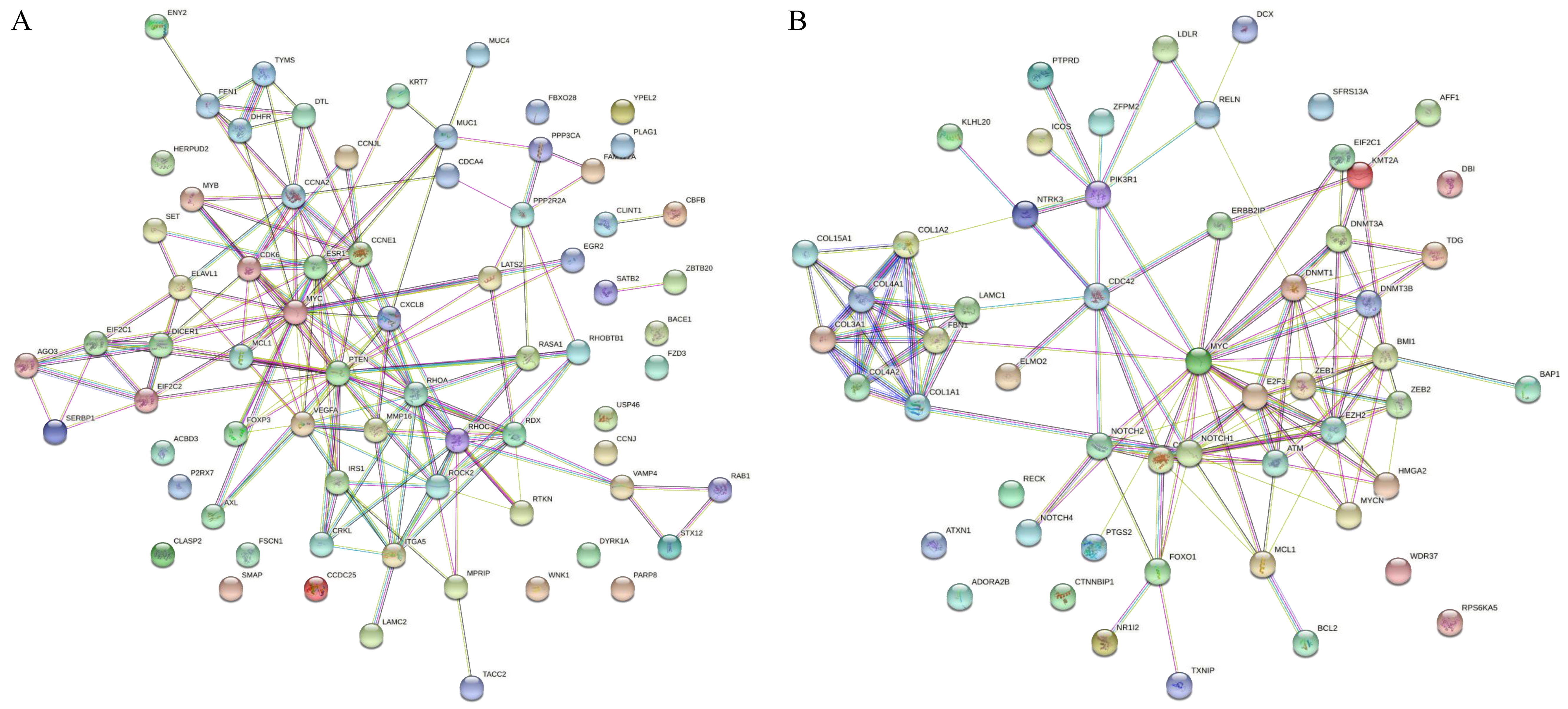
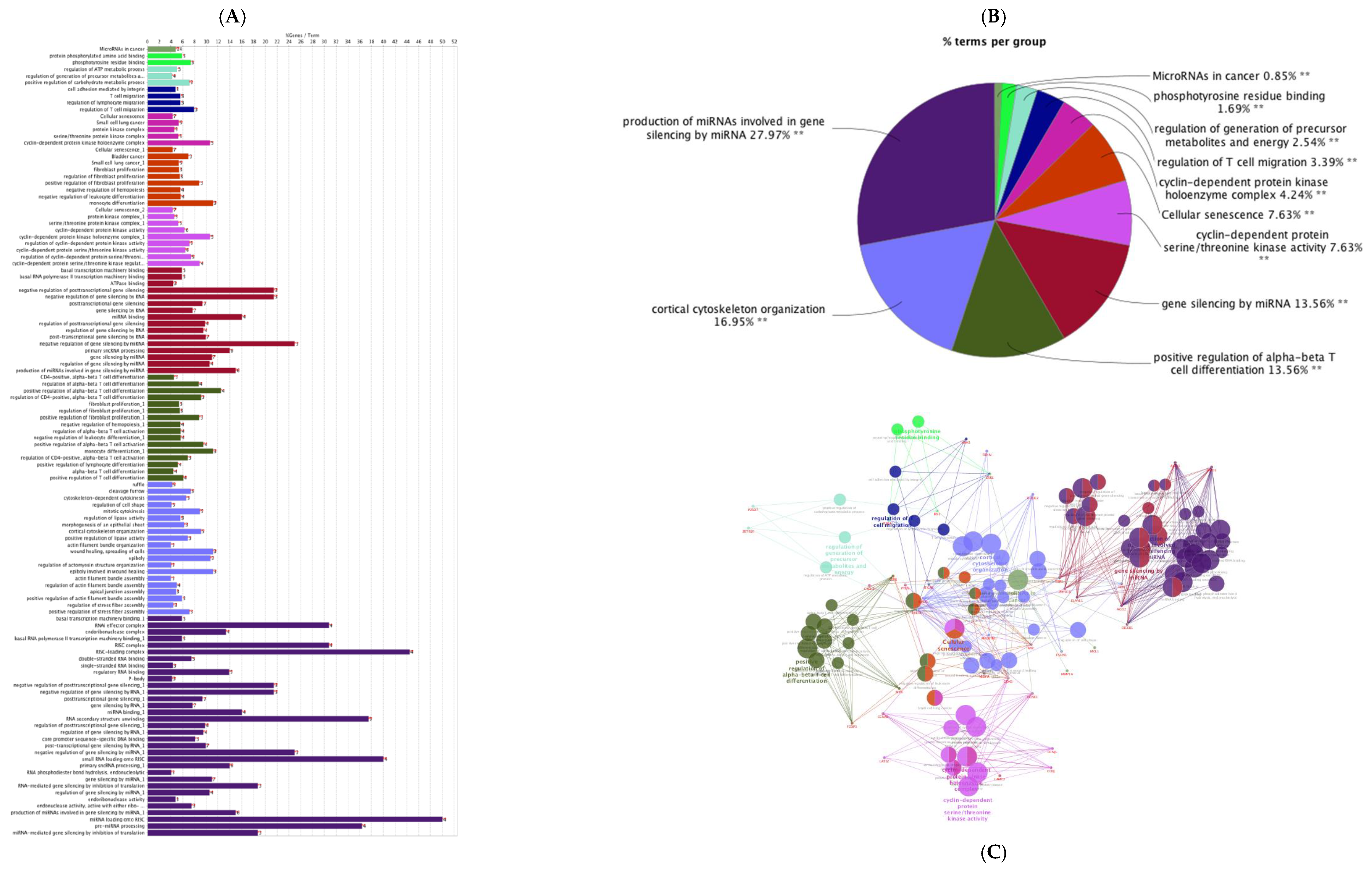
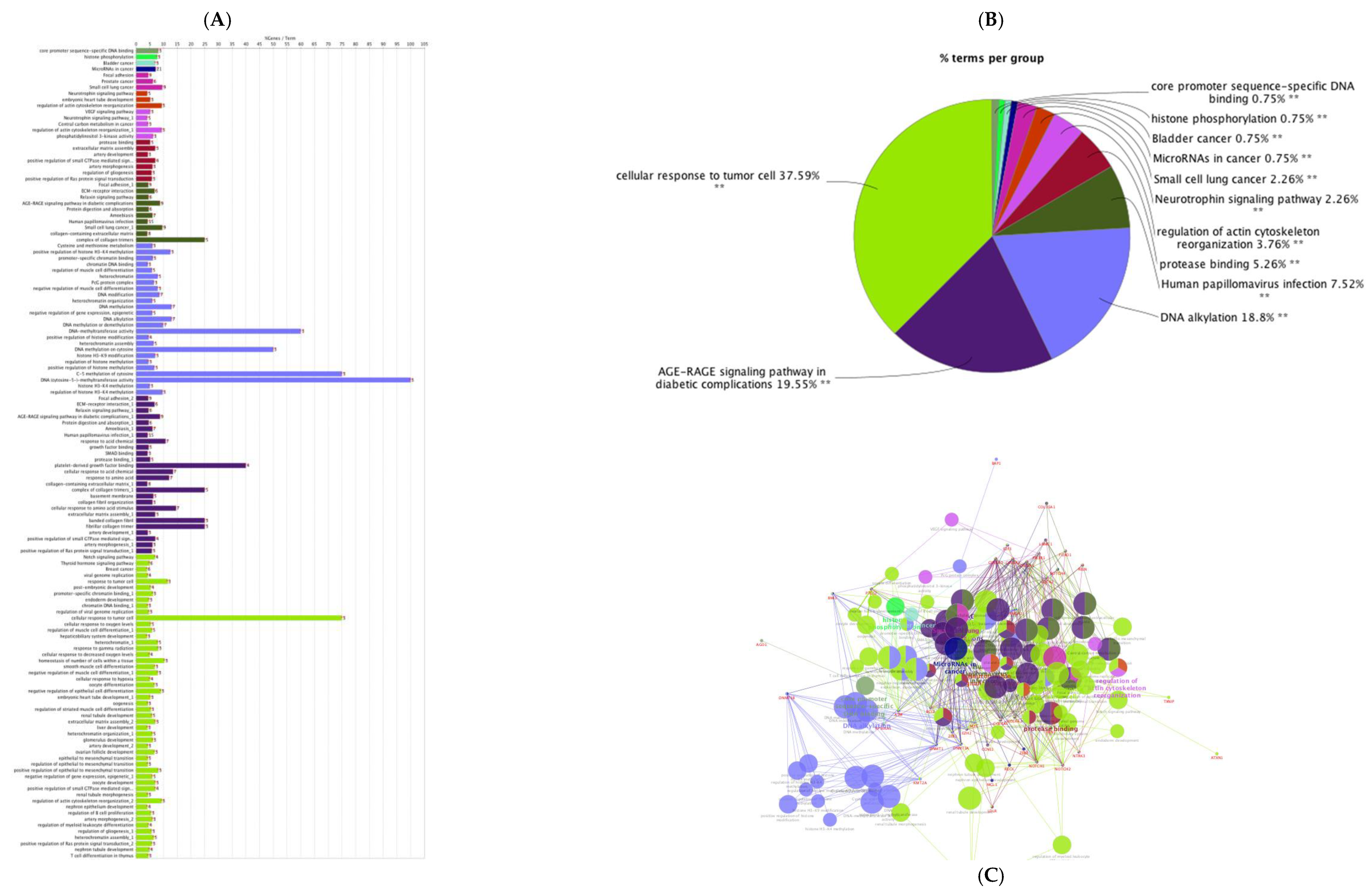
The PPI networks for predicted genes for upregulated miRNAs and downregulated miRNAs were separately constructed (Figure 2A,B) using the STRING database and the Cytoscape software. According to the degree (MCC method), the top 20 hub genes in the networks for upregulated miRNAs and downregulated miRNAs were screened and presented in Figure 3A,B. To decipher functionally nested gene ontology and pathway annotation networks for the predicted genes of upregulated and downregulated miRNAs in higher fertile sperm and seminal plasma, ClueGo nested network analysis was performed, and the results are presented in Figure 4 and Figure 5.
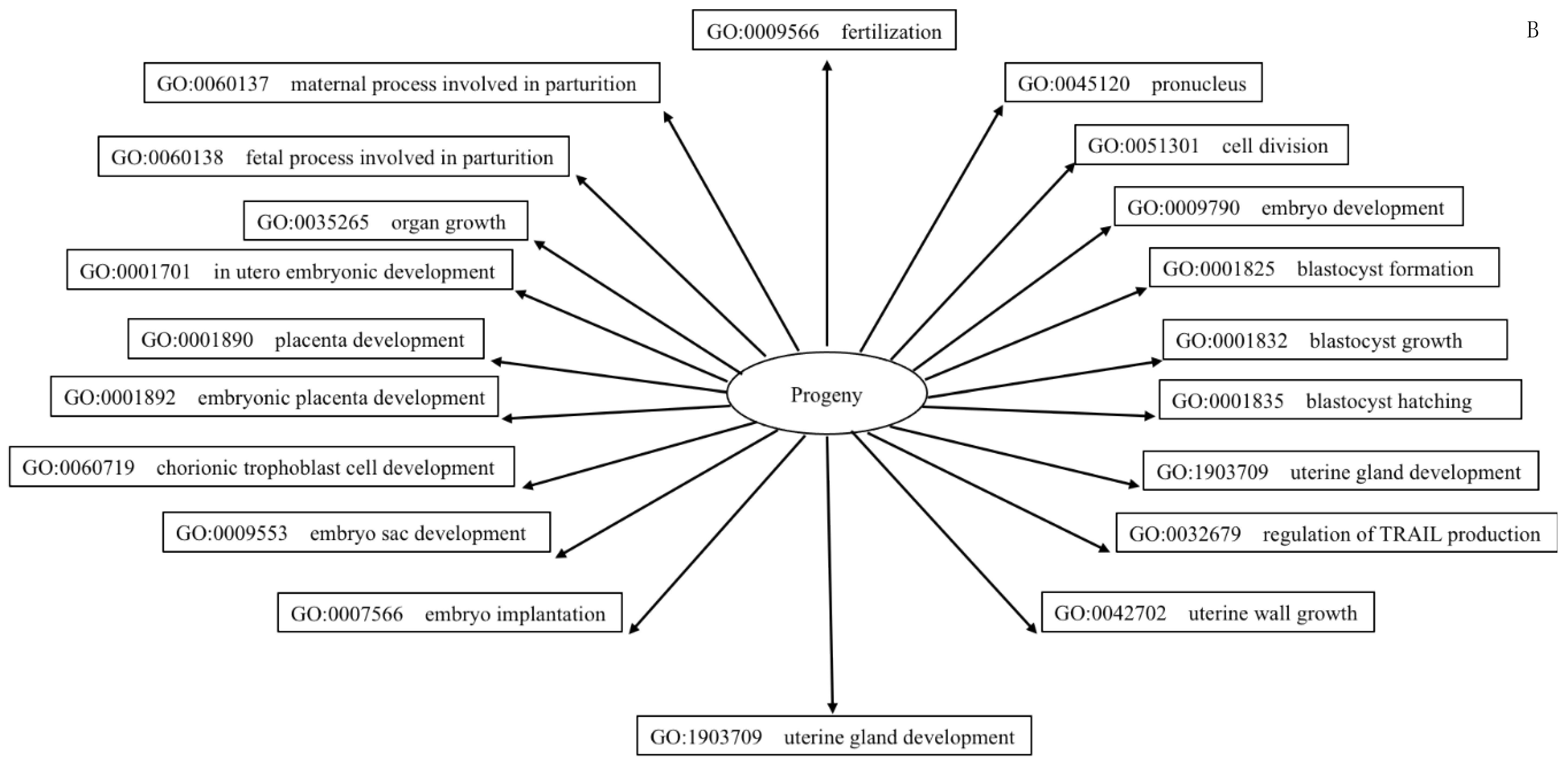
Differentially expressed miRNAs, associated hub genes, and their linked reproductive functions are given in Table 3. Finally, GO terms such as fertilization, implantation, trophoblast formation, placental development, and in utero embryonic development (from the 20 hub gene predicted 640 terms) were used to retrieve (QuickGO) relevant co-occurring GO terms (Supplementary File S7) and from the GO functional annotation terms, fertilization (Figure 6A) and progeny development (Figure 6B) were constructed.
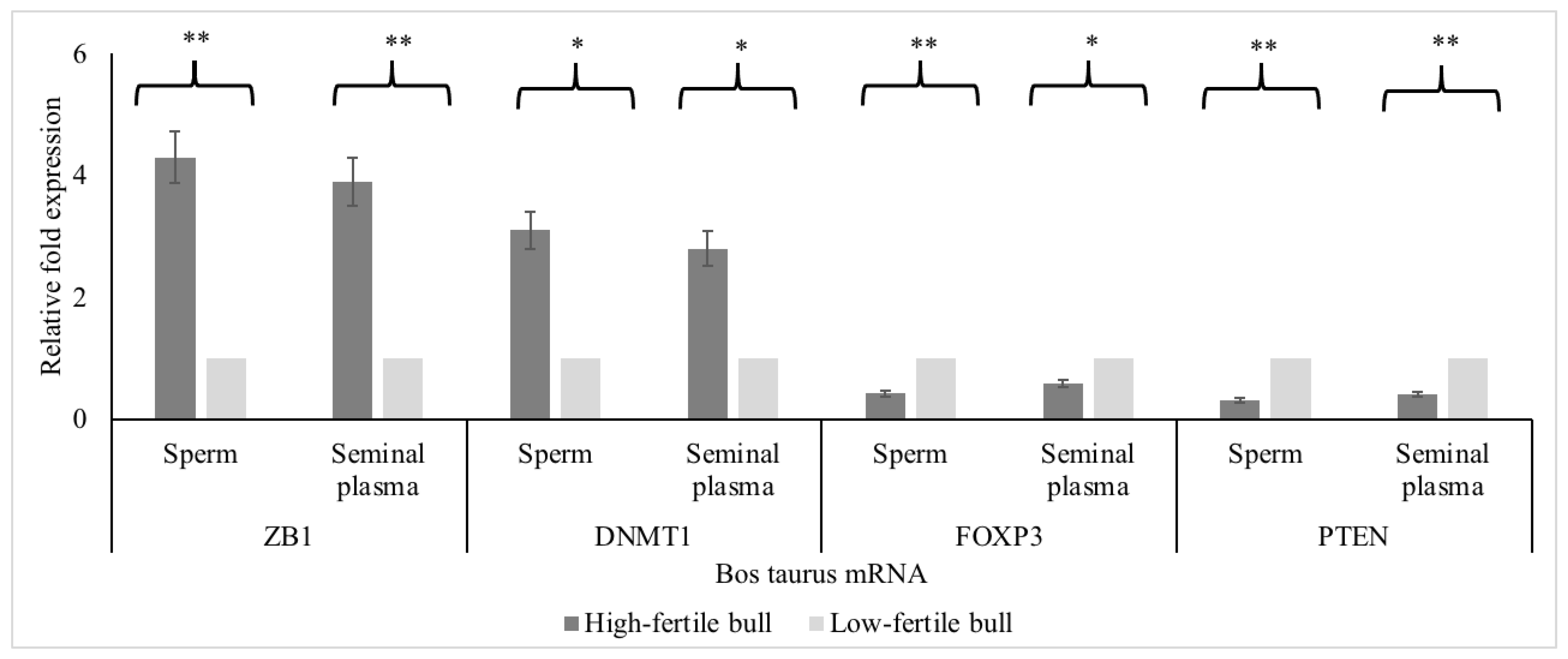
The mRNA expressions for ZB1 and DNMT1 were greater (p < 0.05) in high compared to low-fertile bulls (Figure 7); whereas the mRNA expressions for PTEN and FOXP3 were lower in high compared to low-fertile bulls (p < 0.05; Figure 6). Protein immunoblots were performed to recognize the presence of PTEN, DNMT1, and ACBT (reference gene) proteins in sperm and seminal plasma. The PTEN, DNMT1, and ACBT proteins were 47, ~180, and 42 kDa, respectively (Figure S2). The miRNA relative fold change and associated mRNA relative fold change (miRNA- mRNA pair) showed a negative correlation (Figure 8, r = −0.86; p < 0.05).

4. Discussion

The goal of this current investigation was to elucidate differentially expressed miRNAs in high-fertile sperm and seminal plasma compared to low-fertile sperm and seminal plasma, and to use bioinformatics to investigate how top-ranked integrated genes of DE-miRNAs exerts wide-ranging connotations in sperm function, fertilization, and progeny development. The current study investigated prioritized miRNAs in bull sperm and seminal plasma using real-time PCR. Real-time PCR profiling employed in this study eliminated the need for validation experiments associated with microarray technology. The result showed that 20 miRNAs were highly (≥5 fold) upregulated whereas 12 miRNAs were highly (≤−5 fold) downregulated in high-fertile bull sperm and seminal plasma. It should be noted that highly differentially expressed (≤−5 and ≥5-fold regulation) miRNA were considered for further analysis and for the discussion. Table 3 shows top integrated genes of the DE-miRNAs predicted biological processes demonstrating their critical roles in the regulation of sperm function, fertilization, uterine receptivity, and in the development of embryo, placenta, and fetus.
Seminal plasma has an enriched repertoire of seminal exosomes, which carry a unique profile of miRNAs [110]. A consensus is that seminal exosomes represent a mixed population of extracellular vesicles originating not only from the epididymis but also from accessory sex glands. Nevertheless, seminal exosomes mediators improve sperm motility and induce sperm to have the ability to capacitate and complete acrosomal exocytosis [111,112]. In addition, seminal plasma exosomes modulate the immune response and molecular signaling cascades in the uterus to facilitate embryo implantation [113,114] and these functions are mediated via seminal exosome-carried miRNAs [110].
Free and exosomal miRNAs in seminal plasma can reflect the pathophysiological conditions of the organ of origin. Aberrant cell-free miRNA levels, both in whole seminal plasma [20,109,115] and in seminal plasma exosomes [116] were linked to the poor quality of the sperm. Therefore, the analysis of cell-free miRNAs repertoire could be a specific indicator of alterations of the organ/cells of origin. In the current study, DE-miRNAs in sperm and seminal plasma are found to be common. Previously, we compared expression levels of individual miRNA both in sperm and seminal plasma from the same ejaculate [9,10]. The results revealed that the sperm miRNA level was significantly greater compared with the seminal plasma miRNA level and interestingly, the expressions of DE-miRNAs in sperm and seminal plasma followed similar trends for all miRNAs studied [10]. It also has been reported by others that approximately 75% of miRNAs in boar sperm and seminal plasma are shared [117]. Although a similar pattern of miRNAs’ expression between sperm and seminal plasma exists, the genetic and epigenetic pathways controlled by sperm and seminal plasma could be different. For example, exosomal miRNAs in the seminal plasma seem to not only interact with the spermatozoa but also with cells from the female reproductive tract, modulating their gene expression and influencing the female immune response triggered by the semen [23]. Further, sperm miRNAs are sensitive to environmental stimuli that can modify the sperm miRNA profile and might induce epigenetic modifications in the embryo [118].
The success of artificial insemination (AI) and in vivo and in vitro embryo transfer in cattle indicates that seminal plasma is not required for the establishment of pregnancy. Removal of seminal vesicles had no effect on the fertility of sperm [119]. Intrauterine infusion of 0.5 mL of seminal plasma at artificial insemination in dairy cows did not improve pregnancy/AI (saline, 52.4 (164/313) vs. seminal plasma, 45.5 (143/314) [120] and seminal plasma (37.8) vs. no treatment (33.2%)) [121]. It is plausible that the treatment regimen used in those studies did not cause the amount of inflammation typically induced by natural mating. It should be noted that, following natural mating, an unknown number of seminal molecules are being transferred to the uterus directly by the seminal plasma or carried by sperm [122]. Seminal plasma does not simply protect sperm but can also influence the reproductive events independent of sperm in some species other than cattle. Seminal fluid regulation of female reproductive physiology is shown in hamsters, pigs, and human studies [68,73,123,124,125,126,127,128,129,130,131,132,133,134]. Mating with males lacking seminal vesicles resulted in reduced blastocyst rate, reduced conception rates, and offspring that experienced sex-dependent phenotypic changes postnatally [120,125,135]. However, transformed offspring phenotype was ascribed to sperm damage and absence of seminal fluid [136]. It is evident that seminal plasma and sperm-borne mediators are critical for signaling events that lead to successful pregnancy and healthy offspring; however, there are differences across species [119,127,137] that raise the question of whether seminal plasma is a prerequisite in cattle breeding. It should be noted that the interaction of hub genes of DE-miRNAs in the PPI network was found in this current study.
Sellem et al. (2021) investigated the dynamics of bovine sperm small RNAs from spermatogenesis to final maturation and observed that sperm progressively acquires miRNAs during the epididymal transit with regional specificities [138]. Several miRNAs are involved in cell cycle regulation, spermatogenesis, or embryo development, suggesting the role of epididymal small RNAs in sperm maturation and for ejaculated sperm in embryogenesis. The GO annotation terms from in silico network analysis in this study predicted several functions pertinent to sperm and seminal plasma, and events relevant to fertilization and embryo and placental development. The nested network from in silico interactions revealed that upregulated miRNAs and their integrated genes were involved in cortical cytoskeleton organization, cyclin-dependent protein kinase holoenzyme complex structure, miRNA-mRNA interaction, cyclin-dependent protein serine/threonine kinase activity, phosphotyrosine residue binding, cancer biology, regulation and migration of T-cell, and cellular senescence signaling pathways.
Cytoskeleton organization has been implicated in placental development by regulation of ion transport in humans34, sperm-oocyte interaction [139], and regulation of cell architecture in mouse embryonic stem cells [140]. Actin filaments of the cytoskeleton participate in several vital phases of spermatogenesis including acrosome biogenesis, flagellum formation, nuclear processes modulating capacitation and acrosome reaction, DNA integrity, and fertility [141]. It is interesting to note that protein kinase c (PKC) has a role in the reorganization of the cortical cytoskeleton during the transition from oocyte to the fertilization-competent egg in response to progesterone [142]. Further, PKC-delta regulates bovine embryo development and gene expression in bovine trophoblast cells [143]. Serine/threonine regulation is important for phosphorylation associated with sperm capacitation [75]. Further, it is involved in placental development since the reduction of phosphorylation and endothelial nitric-oxide synthase may cause impaired placenta development, fetal growth retardation, and neonatal mortality [76].
Nested network in silico investigation, especially PANTHER GO prediction for DE miR-20b revealed that T cell migration and regulation are key functions component of uterine receptivity and embryo implantation. The role of seminal plasma in immune regulation of uterine receptivity has been studied in several species [70,124]. Regulatory T cells induced by seminal plasma assist embryo implantation by suppressing inflammation, inhibiting effector immunity towards the embryo, and promoting uterine vascular adaptations that support placental development [70,72,73]. Breeding by seminal plasma-deficient hamsters resulted in reduced fertility, impaired embryonic and fetal development, altered growth trajectories, and increased anxiety in offspring [68,123,125]. Different anti- and pro-tolerogenic factors such as cytokines, pregnancy hormones, seminal fluid, and decidua-specific cells, can modulate the number and the functions of regulatory T cells during pregnancy. Neutrophils and T cells facilitate maternal immune tolerance to paternal alloantigens, and neutrophils interact with T cells to promote angiogenesis at the maternal-fetal interface [72,73,124]. It should be noted that regulatory T cells promote the expansion of uterine mast cells and normalize early pregnancy angiogenesis in abortion-prone mice [26]. In pigs, miRNAs involved in immune cell development and possible regulation in abortion [144]. Significant differences were observed for miRNAs involved in immune cell development and angiogenesis (miR-296-5P, miR-150, miR-17P-5P, miR-18a, and miR-19a) between endometrial lymphocytes associated with healthy and arresting conceptus attachment sites. These pieces of evidence support the mediation of semen miRNAs in T-cell regulation and embryo implantation [72,73].
Protein tyrosine phosphorylation is involved in sperm-oocyte interaction, penetration, and fertilization [127]. Protein tyrosine phosphatase-interacting protein 51 (PTPIP51/RMD3) mRNA expression was seen in syncytiotrophoblast and cytotrophoblast layers of placentae and serves as cellular signaling partner in angiogenesis and vascular remodeling in women [128].
The nested network from in silico interaction analysis revealed that downregulated miRNAs and their predicted genes involved in advanced glycation end products (AGE)- receptor for AGE (RAGE) signaling, protease binding, regulation of actin cytoskeleton reorganization pathway, neurotrophin signaling, and histone phosphorylation pathways. Interfering with these pathways could impact reproductive success.
AGE/RAGE signaling has been shown to increase oxidative stress and inflammation. AGE impaired testicular function and thus affected spermatogenesis and sperm quality in rodents [134]. AGE hindered trophoblast invasion. Placental RAGE was activated during preeclampsia and caused inflammation in the trophoblast [130].
Neurotrophins and hormones influence each other and regulate neuropeptide expression. The PANTHER GO annotation for the DE-miRNAs and integrated genes envisaged neuropeptide and G protein-coupled receptor signaling. Ligands for G protein-coupled receptors (GPCR) are capable of activating mitogenic receptor tyrosine kinases (Trk) [131]. Neurotrophins utilize receptor Trk, namely TrkA, TrkB, and TrkC. Decreased Trk receptor activity by adenosine resulted in increased cell survival [132]. The cellular and molecular basis for the integration of metabolism and reproduction involves a complex interaction of hypothalamic neuropeptides with metabolic hormones (leptin, ghrelin, and IGF) and sex steroids. Neuropeptides including galanin-like peptide (GALP), neuropeptide Y (NPY), products of the proopiomelanocortin (POMC), and kisspeptin, serve as molecular motifs integrating metabolism and reproduction [137]. In heifers, increased rates of BW gain during a prepubertal period can advance puberty [145,146]. Functional changes in the NPY and POMC neuronal pathways appear to contribute to attaining puberty [134]. In humans, miR-21-3p has been implicated in adipose tissue browning and diabetes [147]. Sperm and seminal plasma adiponectins were associated with sperm parameters [148] and circulating adiponectin was linked to body condition, uterine inflammation, and sire conception rate [34,149]. G protein-coupled receptor is involved in spermatogenesis and sperm maturation [150], uterine receptivity [151], embryo implantation and development [152,153], and placental development [154].
Histone modification facilitates histone-to-protamine transition during spermiogenesis. It modulates chromatin compaction and higher-order chromatin structure by ubiquitination and methylation. Flaws in either the replacement or the alteration of histones might cause male infertility such as azoospermia, oligospermia, or teratozoospermia [155]. Disruption of histone variant H3f3a produces abnormal spermatozoa [156,157], and the loss of H3f3b leads to growth defects and death at birth, with surviving H3f3b-null males showing complete infertility [158]. Acrosome proteases trypsin and chymotrypsin play a role in acrosome reaction in bovine sperm. Sperm incubated with the inhibitors of the acrosome proteases reduced the percentage of acrosome reaction [159]. Serine protease PRSS55 affected sperm migration and sperm-egg binding in mice by interfering with ADAM3 [160].
The involvement of DE-miRNAs and their integrated genes in several reproductive functions in high-fertile sperm and seminal plasma in the current study, and various other studies, are presented in Table 3. It is not surprising that DE-miRNAs in the current study, regulate two events, focal adhesion, and cellular senescence. Focal adhesions are contact points that facilitate several cellular processes including proliferation, migration, and differentiation [35]. Several proteins including cadherins are involved in adhesion and signaling [161]. Cadherins are expressed in mature sperm and facilitate capacitation and sperm-oocyte interaction [162]. Cell aggregation, cell to cell contact formation, and cellular tension are important in embryonic development [163]. Programmed cellular senescence is critical to promote tissue remodeling during embryonic development. This process is regulated by the TGF-β/SMAD and PI3K/FOXO pathways [164], which are linked to DE-miRNAs from this study.
Further, DICER1 and AGO2 among the predicted target genes for DE-miRNAs may notably have substantial influence on spermatogenesis and embryo development. Deletion of DICER gene in male germ cells led to impaired differentiation of haploid spermatids, followed by apoptosis and failure of spermatogenesis in the haploid and meiotic stages [165]. Though this is not a sperm/seminal plasma-induced reproductive event, increased expression of DICER1 and AGO2 were observed when porcine blastocysts underwent rapid and radical morphological changes from spherical, through tubular, to filamentous forms [166]. Several sequencing studies also revealed that DE-miRNAs, miR-34a, and miR-200a, inhibited endometrial receptivity and embryo implantation. MicroRNA-34a [167], and miR-200a [98], were downregulated in sperm and seminal plasma of high-fertile bulls in the current study.
Sperm morpho-functional characteristics are not always sufficient to predict male fertility potential. Sperm carry different molecules (proteins, RNAs, and ncRNAs), which are involved in important functions, such as spermatogenesis, sperm maturation, fertilization, and embryo development. The sperm acquire motility and oocyte-fertilizing ability during the epididymal passage. During their transit through the different epididymal segments, sperm come into contact with different repertoires of miRNAs, transcripts, and proteins, that are released from the epididymal epithelium mostly via epididymosomes [24,168,169]. Biogenesis of miRNAs seems to be important in the regulation of epididymal epithelium, sperm maturation, and fertility. While miR-10a/b, -21a, -29c, -196b-5p, -199a, -200b/c, and -208b-3p, accrue in sperm during passage through the epididymis; miR-204b-5p, and miR-375-3p, are more abundant in sperm from the caput and corpus of the epididymis [24,169]. In addition, epididymosomes also display different miRNA profiles and traffic small RNAs to sperm [24], including miR-143, -145, -199, and -214, that are more abundant in epididymosomes of the caput of the bovine epididymis, and miR-395, -654, and -1224, that are more abundant in epididymosomes from the cauda [169]. The miRNAs are acquired during the late steps of spermatogenesis or post-testicular sperm maturation with a potential function at the time of fertilization [24,170] (Sharma et al., 2016; Yuan et al., 2016).
Our previous studies found associations between fertility and sperm parameters. Sire conception rate showed a positive correlation with relative sperm volume shift [35] and acrosome reacted sperm at 5 h post-thaw [34], no correlation with hypoosmotic swelling [35], negative correlation with lipid peroxidation [171], sperm DNA fragmentation, and plasma membrane integrity [172]. Turrri et al. (2021), observed a positive association of field fertility (estimated relative conception rate), semen quality parameters (sperm kinetics and DNA), and specific miRNAs (15 differentially expressed miRNAs (including nine known miR-2285n, miR-378, miR-423-3p, miR-191, miR-2904, miR-378c, miR-431, miR-486, miR-2478)) between high- and low-fertility bulls [21].
In the current study, a bull field fertility parameter (sire conception rate) was utilized to distinguish high- and low-fertile bulls. We observed that 56 out of 84 (66.7%) miRNAs were expressed >2 fold and 32 out of 84 (38%) miRNAs were expressed >5 fold in sperm and seminal plasma by RT-PCR. Keles et al. (2021), used non return rate as fertility parameter and identified 85 differentially expressed sperm miRNAs by RNA sequencing [173]. The miR-2340, miR-26a, miR-425-5p, and miR-151–5p, were moderately correlated to nonreturn rate when conventional semen was used, whereas nine miRNAs were significantly correlated (miR-9-5p, miR-34c, miR-449a, miR-2483-5p, and miR-21–5p were negatively related, and miR-423-5p, miR-1246, miR-92a, and miR-5193-5p, were positively related) when gender selected semen was used.
In the current study, 2 miRNAs (mir-16b and mir-29c) in sperm and 4 miRNAs (mir-16b, mir-29c, 200a, and mir-101) in seminal plasma were very highly down regulated (10 fold; p < 0.001); whereas 9 miRNAs (mir-215, mir-17-5p, mir-199a-3p, mir-193a-3p, mir-142-5p, mir-21-3p, mir-20b, mir-199a-5p, and mir-214) in sperm and 6 miRNAs (mir-193a-3p, mir-142-5p, mir-21-3p, mir-20b, mir-199a-5p, and mir-214) in seminal plasma were highly upregulated (10 fold; p < 0.001). Of these, miR-29c, miR-101, miR-200a, miR-20b, and miR-214, were associated with hub genes and these miRNA-mRNA pairs were involved in spermatogenesis, embryogenesis, placenta development, and embryo implantation. The cluster analysis revealed their involvement in the regulation of spermatogenesis, histone modifications, ovarian follicle development, focal adhesion, cellular senescence, vascular remodeling and placental development, and VEGF and AGE-RAGE signaling.
Previous studies observed positive (upregulated-upregulated) or negative (upregulated-downregulated) association between miRNA and mRNA pairs [174]. The miRNAs are capable of activating gene expression directly or indirectly in response to different cell types and environments and in the presence of distinct cofactors. The biological outcome of miRNA-mRNA interaction can be altered by several factors contributing to the binding strength and repressive effect of a potential target site. Interestingly, miRNA-mRNA associations were complementing in normal tissues and miRNA-mRNA associations were reverse complementing in the same tissues in diseased condition [175]. The correlation between miRNA and hub gene expressions were negative in the current study.
Studies have shown that interaction between seminal plasma and sperm could improve acrosome integrity, sperm quality by modulating sperm potential to evoke in vitro capacitation, and cause progesterone-induced acrosomal exocytosis [176]. During sperm preservation, the impact of seminal plasma on sperm has been unclear. Seminal plasma is diluted/removed prior to cryopreservation to reduce the association between seminal plasma and sperm during storage. An early study revealed that top 10 highly expressed miRNA predicted target genes (across the three libraries such as ejaculated sperm, epididymal sperm, and seminal plasma) could be involved in spermatogenesis, zygote formation, and animal-environment interactions [117]. This shows the importance of contact between sperm and seminal plasma before preservation [177].
There are limitations to this study. First, the study is limited due to small sample size. Though four bulls per group was calculated as adequate to determine fold regulation differences of five, it should be noted that the small number of bulls were represented from general population. We anticipate that more miRNA and genes can be identified in future studies with a large sample size. Second, the identified miRNA-mRNA interactions were mostly based on predictions from public databases. The causal regulations of each pair and the underlying mechanisms require further functional characterization in future studies.

5. Conclusions

In conclusion, we observed miRNAs were differentially expressed in sperm and seminal plasma of high-fertile bulls compared with low-fertile bulls. The DE-miRNAs fold changes were similar in both sperm and seminal plasma. The DE-miRNAs in sperm and seminal plasma elucidated in this study plausibly regulated critical pathways that are required for reproductive success at the transcriptional or post-transcriptional level. In silico network analysis showed association for the tasks of sperm and seminal plasma DE-miRNAs and its predicted genes specific to spermatogenesis, sperm maturation, sperm and seminal plasma interaction, early embryo development, implantation, and organogenesis.

Supplementary Materials

The following supporting information can be downloaded at: https://www.mdpi.com/article/10.3390/ani12182360/s1. Table S1. Sire conception rate, progressive motility (%) and abnormal spermatozoa (%) for Holstein bulls used in the study. Table S2. Nucleotide sequences of human, mouse, and cattle miRs (www.miRBase.org) (accessed on 10 December 2021). Figure S1. The ethidium bromide-stained electrophoresis gel, with amplicons of expected sizes. Figure S2. Representative Western blots of protein isozymes. Supplementary File S1: Fold regulation of differentially expressed 84 bovine-specific miRNAs in sperm of high fertile compared to low-fertile Holstein bulls. Supplementary File S2: Fold regulation of differentially expressed 84 bovine-specific miRNAs in seminal plasma of high-fertile compared to low-fertile Holstein bulls. Supplementary File S3: Identification of associated target and predicted genes of bovine specific upregulated miRNA of high-fertile compared to low-fertile Holstein bulls (http://www.mirnet.ca/) (accessed on 16 December 2021). Supplementary File S4: Biological process gene ontology (GO) functional annotation terms and Kyoto Encyclopedia of Genes and Genomes (KEGG) enrichment pathways for the predicted genes of upregulated miRNA of high-fertile Holstein bulls. Supplementary File S5: Identification of associated target and predicted genes of bovine specific downregulated miRNAs of high-fertile compared to low-fertile Holstein bulls (http://www.mirnet.ca/) (accessed on 16 December 2021). Supplementary File S6: Biological process gene ontology (GO) functional annotation terms and Kyoto Encyclopedia of Genes and Genomes (KEGG) enrichment pathways for the predicted genes of down regulated miRNAs of high-fertile Holstein bulls. Supplementary File S7: Co-occurring gene ontology (GO) terms for hub gene predicted reproductive functional terms (https://www.ebi.ac.uk/QuickGO) (accessed on 22 December 2021).

Author Contributions

Conceptualization, V.K. and R.K.; methodology, V.K., N.K. and R.K.; software, V.K., N.K. and R.K.; validation, V.K. and R.K.; formal analysis, V.K. and R.K.; investigation, V.K., N.K. and R.K.; resources, V.K. and R.K.; data curation, V.K. and R.K.; writing—original draft preparation, V.K., N.K. and R.K.; writing—review and editing, V.K. and R.K.; visualization, V.K., N.K. and R.K.; supervision, V.K. and R.K.; project administration, V.K. and R.K.; funding acquisition, V.K. and R.K. All authors have read and agreed to the published version of the manuscript.

Funding

This research received no external funding.

Institutional Review Board Statement

This study was performed in strict accordance with the ethics, standard operating procedure, and use of the animal cells and biofluids for research. The protocol was approved by the institutional animal care and use committee of the Washington State University.

Informed Consent Statement

Not applicable.

Data Availability Statement

The original contributions presented in the study are included in the article/supplementary material; further inquiries can be directed to the corresponding author.

Acknowledgments

The authors thank College of Veterinary Medicine, Washington State University, Pullman, WA, USA, for the support and Nishant Kumar and Rachel Shutter, DVM class of 2017, for their participation in this study. Nishant Kumar was supported by the Indian Council of Agricultural Research under the National Agricultural Higher Education Project—Institutional Development Plan. Rachel Shutter was supported by College of Veterinary Medicine, Washington State University, Pullman, WA, USA.

Conflicts of Interest

The authors declare no conflict of interest.

References

  1. Fang, X.; Qin, L.; Yu, H.; Jiang, P.; Xia, L.; Gao, Z.; Yang, R.; Zhao, Y.; Yu, X.; Zhao, Z. Comprehensive Analysis of miRNAs and Target mRNAs between Immature and Mature Testis Tissue in Chinese Red Steppes Cattle. Animals 2021, 11, 3024. [Google Scholar] [CrossRef] [PubMed]
  2. Kasimanickam, V.R.; Kasimanickam, R.K. Differential expression of microRNAs in sexually immature and mature canine testes. Theriogenology 2015, 83, 394–398. [Google Scholar] [CrossRef] [PubMed]
  3. Vendrell-Flotats, M.; García-Martínez, T.; Martínez-Rodero, I.; López-Béjar, M.; LaMarre, J.; Yeste, M.; Mogas, T. In vitro maturation in the presence of Leukemia Inhibitory Factor modulates gene and miRNA expression in bovine oocytes and embryos. Sci. Rep. 2020, 10, 17777. [Google Scholar] [CrossRef] [PubMed]
  4. Costa, M.C.; Leitão, A.L.; Enguita, F.J. MicroRNA Profiling in Plasma or Serum Using Quantitative RT-PCR. RNA Mapp. 2014, 1182, 121–129. [Google Scholar] [CrossRef]
  5. Kasimanickam, V.; Kastelic, J. Circulating cell-free mature microRNAs and their target gene prediction in bovine metritis. Sci. Rep. 2016, 6, 29509. [Google Scholar] [CrossRef]
  6. Roberts, T.C.; Coenen-Stass, A.M.L.; Betts, C.A.; Wood, M.J.A. Detection and quantification of extracellular microRNAs in murine biofluids. Biol. Proced. Online 2014, 16, 5. [Google Scholar] [CrossRef]
  7. Weber, J.A.; Baxter, D.H.; Zhang, S.; Huang, D.Y.; Huang, K.H.; Lee, M.J.; Galas, D.J.; Wang, K. The MicroRNA Spectrum in 12 Body Fluids. Clin. Chem. 2010, 56, 1733–1741. [Google Scholar] [CrossRef] [PubMed]
  8. Turchinovich, A.; Weiz, L.; Burwinkel, B. Extracellular miRNAs: The mystery of their origin and function. Trends Biochem. Sci. 2012, 37, 460–465. [Google Scholar] [CrossRef]
  9. Kasimanickam, V.; Kasimanickam, R. An Efficient Approach for RNA Extraction from Boar Sperm and Seminal Plasma. Bio-Protocol 2019, 9, e3284. [Google Scholar] [CrossRef]
  10. Kasimanickam, V.; Buhr, M.; Kasimanickam, R. Patterns of expression of sperm and seminal plasma microRNAs in boar semen. Theriogenology 2019, 125, 87–92. [Google Scholar] [CrossRef]
  11. Rodríguez-Martínez, H.; Kvist, U.; Ernerudh, J.; Sanz, L.; Calvete, J.J. Seminal Plasma Proteins: What role do they play? Am. J. Reprod. Immunol. 2011, 66 (Suppl. 1), 11–22. [Google Scholar] [CrossRef] [PubMed]
  12. Caballero, I.; Parrilla, I.; Almiñana, C.; del Olmo, D.; Roca, J.; Martínez, E.; Vázquez, J. Seminal plasma proteins as modulators of the sperm function and their application in sperm biotechnologies. Reprod. Domest. Anim. 2012, 47 (Suppl. S3), 12–21. [Google Scholar] [CrossRef]
  13. Chabory, E.; Damon, C.; Lenoir, A.; Henry-Berger, J.; Vernet, P.; Cadet, R.; Saez, F.; Drevet, J.R. Mammalian glutathione peroxidases control acquisition and maintenance of spermatozoa integrity 1. J. Anim. Sci. 2010, 88, 1321–1331. [Google Scholar] [CrossRef] [PubMed]
  14. Juyena, N.S.; Stelletta, C. Seminal Plasma: An Essential Attribute to Spermatozoa. J. Androl. 2012, 33, 536–551. [Google Scholar] [CrossRef] [PubMed]
  15. Mann, T. The Biochemistry of Semen and of the Male Reproductive Tract, 2nd ed.; Methuen: London, UK, 1964. [Google Scholar]
  16. Taylor, A.; Robson, A.; Houghton, B.C.; Jepson, C.A.; Ford, W.C.L.; Frayne, J. Epididymal specific, selenium-independent GPX5 protects cells from oxidative stress-induced lipid peroxidation and DNA mutation. Hum. Reprod. 2013, 28, 2332–2342. [Google Scholar] [CrossRef] [PubMed]
  17. Fagerlind, M.; Stålhammar, H.; Olsson, B.; Klinga-Levan, K. Expression of miRNAs in Bull Spermatozoa Correlates with Fertility Rates. Reprod. Domest. Anim. 2015, 50, 587–594. [Google Scholar] [CrossRef]
  18. Selth, L.A.; Roberts, M.; Chow, C.W.K.; Marshall, V.R.; Doi, S.; Vincent, A.D.; Butler, L.; Lavin, M.; Tilley, W.D.; Gardiner, R.A. Human seminal fluid as a source of prostate cancer-specific microRNA biomarkers. Endocr.-Relat. Cancer 2014, 21, L17–L21. [Google Scholar] [CrossRef]
  19. Du, Y.; Wang, X.; Wang, B.; Chen, W.; He, R.; Zhang, L.; Xing, X.; Su, J.; Wang, Y.; Zhang, Y. Deep sequencing analysis of microRNAs in bovine sperm. Mol. Reprod. Dev. 2014, 81, 1042–1052. [Google Scholar] [CrossRef]
  20. Wu, W.; Hu, Z.; Qin, Y.; Dong, J.; Dai, J.; Lu, C.; Zhang, W.; Shen, H.; Xia, Y.; Wang, X. Seminal plasma microRNAs: Potential biomarkers for spermatogenesis status. Mol. Hum. Reprod. 2012, 18, 489–497. [Google Scholar] [CrossRef]
  21. Turri, F.; Capra, E.; Lazzari, B.; Cremonesi, P.; Stella, A.; Pizzi, F. A Combined Flow Cytometric Semen Analysis and miRNA Profiling as a Tool to Discriminate Between High- and Low-Fertility Bulls. Front. Vet. Sci. 2021, 8, 703101. [Google Scholar] [CrossRef]
  22. Nixon, B.; Stanger, S.J.; Mihalas, B.P.; Reilly, J.N.; Anderson, A.L.; Tyagi, S.; Holt, J.E.; McLaughlin, E.A. The microRNA signature of mouse spermatozoa is substantially modified during epididymal maturation. Biol. Reprod. 2015, 93, 91. [Google Scholar] [CrossRef] [PubMed]
  23. Jodar, M. Sperm and seminal plasma RNAs: What roles do they play beyond fertilization? Reproduction 2019, 158, R113–R123. [Google Scholar] [CrossRef] [PubMed]
  24. Sharma, U.; Sun, F.; Conine, C.C.; Reichholf, B.; Kukreja, S.; Herzog, V.A.; Ameres, S.L.; Rando, O.J. Small RNAs are trafficked from the epididymis to developing mammalian sperm. Dev. Cell 2018, 46, 481–494.e6. [Google Scholar] [CrossRef] [PubMed]
  25. Liu, W.-M.; Pang, R.T.K.; Chiu, P.C.N.; Wong, B.P.C.; Lao, K.; Lee, K.-F.; Yeung, W.S.B. Sperm-borne microRNA-34c is required for the first cleavage division in mouse. Proc. Natl. Acad. Sci. USA 2012, 109, 490–494. [Google Scholar] [CrossRef] [PubMed]
  26. Yuan, S.; Schuster, A.; Tang, C.; Yu, T.; Ortogero, N.; Bao, J.; Zheng, H.; Yan, W. Sperm-borne miRNAs and endo-siRNAs are important for fertilization and preimplantation embryonic development. Development 2016, 143, 635–647. [Google Scholar] [CrossRef]
  27. Kozomara, A.; Birgaoanu, M.; Griffiths-Jones, S. miRBase: From microRNA sequences to function. Nucleic Acids Res. 2019, 47, D155–D162. [Google Scholar] [CrossRef] [PubMed]
  28. Kozomara, A.; Griffiths-Jones, S. miRBase: Annotating high confidence microRNAs using deep sequencing data. Nucleic Acids Res. 2014, 42, D68–D73. [Google Scholar] [CrossRef]
  29. Chang, L.; Zhou, G.; Soufan, O.; Xia, J. miRNet 2.0: Network-based visual analytics for miRNA functional analysis and systems biology. Nucleic Acids Res. 2020, 48, W244–W251. [Google Scholar] [CrossRef]
  30. Szklarczyk, D.; Gable, A.L.; Nastou, K.C.; Lyon, D.; Kirsch, R.; Pyysalo, S.; Doncheva, N.T.; Legeay, M.; Fang, T.; Bork, P.; et al. The STRING database in 2021: Customizable protein–protein networks, and functional characterization of user-uploaded gene/measurement sets. Nucleic Acids Res. 2021, 49, D605–D612. [Google Scholar] [CrossRef]
  31. Gustavsen, J.A.; Pai, S.; Isserlin, R.; Demchak, B.; Pico, A.R. RCy3: Network biology using Cytoscape from within R. F1000Research 2019, 8, 1774. [Google Scholar] [CrossRef]
  32. Chin, C.-H.; Chen, S.-H.; Wu, H.-H.; Ho, C.-W.; Ko, M.-T.; Lin, C.-Y. cytoHubba: Identifying hub objects and sub-networks from complex interactome. BMC Syst. Biol. 2014, 8 (Suppl. 4), S11. [Google Scholar] [CrossRef] [PubMed]
  33. Bindea, G.; Mlecnik, B.; Hackl, H.; Charoentong, P.; Tosolini, M.; Kirilovsky, A.; Fridman, W.-H.; Pagès, F.; Trajanoski, Z.; Galon, J. ClueGO: A Cytoscape plug-in to decipher functionally grouped gene ontology and pathway annotation networks. Bioinformatics 2009, 25, 1091–1093. [Google Scholar] [CrossRef] [PubMed]
  34. Kasimanickam, V.R.; Kasimanickam, R.K.; Kastelic, J.P.; Stevenson, J.S. Associations of adiponectin and fertility estimates in Holstein bulls. Theriogenology 2013, 79, 766–777.e3. [Google Scholar] [CrossRef] [PubMed]
  35. Kasimanickam, R.; Kasimanickam, V.; Arangasamy, A.; Kastelic, J. Associations of hypoosmotic swelling test, relative sperm volume shift, aquaporin7 mRNA abundance and bull fertility estimates. Theriogenology 2017, 89, 162–168. [Google Scholar] [CrossRef] [PubMed]
  36. Kasimanickam, V.R.; Kasimanickam, R.K. Sertoli, Leydig, and Spermatogonial Cells’ Specific Gene and Protein Expressions as Dog Testes Evolve from Immature into Mature States. Animals 2022, 12, 271. [Google Scholar] [CrossRef]
  37. Wang, S.; Ma, G.; Zhu, H.; Lv, C.; Chu, H.; Tong, N.; Wu, D.; Qiang, F.; Gong, W.; Zhao, Q.; et al. miR-107 regulates tumor progression by targeting NF1 in gastric cancer. Sci. Rep. 2016, 6, 36531. [Google Scholar] [CrossRef]
  38. Sellem, E.; Marthey, S.; Rau, A.; Jouneau, L.; Bonnet, A.; Perrier, J.-P.; Fritz, S.; Le Danvic, C.; Boussaha, M.; Kiefer, H.; et al. A comprehensive overview of bull sperm-borne small non-coding RNAs and their diversity across breeds. Epigenet. Chromatin 2020, 13, 19. [Google Scholar] [CrossRef]
  39. Liu, T.; Cheng, W.; Gao, Y.; Wang, H.; Liu, Z. Microarray analysis of microRNA expression patterns in the semen of infertile men with semen abnormalities. Mol. Med. Rep. 2012, 6, 535–542. [Google Scholar] [CrossRef]
  40. Khor, E.-S.; Noor, S.M.; Wong, P.-F. MiR-107 inhibits the sprouting of intersegmental vessels of zebrafish embryos. Protoplasma 2022, 259, 691–702. [Google Scholar] [CrossRef]
  41. Abu-Halima, M.; Hammadeh, M.; Schmitt, J.; Leidinger, P.; Keller, A.; Meese, E.; Backes, C. Altered microRNA expression profiles of human spermatozoa in patients with different spermatogenic impairments. Fertil. Steril. 2013, 99, 1249–1255.e16. [Google Scholar] [CrossRef]
  42. Chen, Z.; Lai, T.-C.; Jan, Y.-H.; Lin, F.-M.; Wang, W.-C.; Xiao, H.; Wang, Y.-T.; Sun, W.; Cui, X.; Li, Y.-S.; et al. Hypoxia-responsive miRNAs target argonaute 1 to promote angiogenesis. J. Clin. Investig. 2013, 123, 1057–1067. [Google Scholar] [CrossRef] [PubMed]
  43. Meinhardt, G.; Haider, S.; Kunihs, V.; Saleh, L.; Pollheimer, J.; Fiala, C.; Hetey, S.; Feher, Z.; Szilagyi, A.; Than, N.G.; et al. Pivotal role of the transcriptional co-activator YAP in trophoblast stemness of the developing human placenta. Proc. Natl. Acad. Sci. USA 2020, 117, 13562–13570. [Google Scholar] [CrossRef] [PubMed]
  44. Guo, J.; Shin, K.-T.; Cui, X.-S. Analysis of Cyclin E1 Functions in Porcine Preimplantation Embryonic Development by Fluorescence Microscopy. Microsc. Microanal. 2017, 23, 69–76. [Google Scholar] [CrossRef]
  45. Boissière, A.; Gala, A.; Ferrières-Hoa, A.; Mullet, T.; Baillet, S.; Petiton, A.; Torre, A.; Hamamah, S. Cell-free and intracellular nucleic acids: New non-invasive biomarkers to explore male infertility. Basic Clin. Androl. 2017, 27, 7. [Google Scholar] [CrossRef] [PubMed]
  46. Leung, C.; Zhu, M.; Zernicka-Goetz, M. Polarity in Cell-Fate Acquisition in the Early Mouse Embryo. Curr. Top. Dev. Biol. 2016, 120, 203–234. [Google Scholar] [CrossRef] [PubMed]
  47. Ngondo, R.P.; Cirera-Salinas, D.; Yu, J.; Wischnewski, H.; Bodak, M.; Vandormael-Pournin, S.; Geiselmann, A.; Wettstein, R.; Luitz, J.; Cohen-Tannoudji, M.; et al. Argonaute 2 Is Required for Extra-embryonic Endoderm Differentiation of Mouse Embryonic Stem Cells. Stem Cell Rep. 2018, 10, 461–476. [Google Scholar] [CrossRef] [PubMed]
  48. Zhou, X.; Xiang, C.; Zheng, X. miR-132 serves as a diagnostic biomarker in gestational diabetes mellitus and its regulatory effect on trophoblast cell viability. Diagn. Pathol. 2019, 14, 119. [Google Scholar] [CrossRef]
  49. Kagedan, D.; Lecker, I.; Batruch, I.; Smith, C.; Kaploun, I.; Lo, K.; Grober, E.; Diamandis, E.P.; Jarvi, K.A. Characterization of the seminal plasma proteome in men with prostatitis by mass spectrometry. Clin. Proteom. 2012, 9, 2. [Google Scholar] [CrossRef]
  50. Ding, R.; Guo, F.; Zhang, Y.; Liu, X.-M.; Xiang, Y.-Q.; Zhang, C.; Liu, Z.-W.; Sheng, J.-Z.; Huang, H.-F.; Zhang, J.; et al. Integrated Transcriptome Sequencing Analysis Reveals Role of miR-138-5p/ TBL1X in Placenta from Gestational Diabetes Mellitus. Cell. Physiol. Biochem. 2018, 51, 630–646. [Google Scholar] [CrossRef]
  51. Yin, A.; Chen, Q.; Zhong, M.; Jia, B. MicroRNA-138 improves LPS-induced trophoblast dysfunction through targeting RELA and NF-κB signaling. Cell Cycle 2021, 20, 508–521. [Google Scholar] [CrossRef]
  52. Zhang, J.Y.; Dong, H.S.; Oqani, R.K.; Lin, T.; Kang, J.W.; Jin, D.I. Distinct roles of ROCK1 and ROCK2 during development of porcine preimplantation embryos. Reproduction 2014, 148, 99–107. [Google Scholar] [CrossRef] [PubMed]
  53. Morton, S.U.; Scherz, P.J.; Cordes, K.R.; Ivey, K.N.; Stainier, D.Y.R.; Srivastava, D. microRNA-138 modulates cardiac patterning during embryonic development. Proc. Natl. Acad. Sci. USA 2008, 105, 17830–17835. [Google Scholar] [CrossRef] [PubMed]
  54. Barceló, M.; Castells, M.; Bassas, L.; Vigués, F.; Larriba, S. Semen miRNAs Contained in Exosomes as Non-Invasive Biomarkers for Prostate Cancer Diagnosis. Sci. Rep. 2019, 9, 13772. [Google Scholar] [CrossRef] [PubMed]
  55. Tesfaye, D.; Worku, D.; Rings, F.; Phatsara, C.; Tholen, E.; Schellander, K.; Hoelker, M. Identification and expression profiling of microRNAs during bovine oocyte maturation using heterologous approach. Mol. Reprod. Dev. 2009, 76, 665–677. [Google Scholar] [CrossRef] [PubMed]
  56. Schrader, M.; Müller-Tidow, C.; Ravnik, S.; Müller, M.; Schulze, W.; Diederichs, S.; Serve, H.; Miller, K. Cyclin A1 and gametogenesis in fertile and infertile patients: A potential new molecular diagnostic marker. Hum. Reprod. 2002, 17, 2338–2343. [Google Scholar] [CrossRef] [PubMed]
  57. Murphy, M.; Stinnakre, M.-G.; Senamaud-Beaufort, C.; Winston, N.J.; Sweeney, C.; Kubelka, M.; Carrington, M.; Bréchot, C.; Sobczak-Thépot, J. Delayed early embryonic lethality following disruption of the murine cyclin A2 gene. Nat. Genet. 1997, 15, 83–86. [Google Scholar] [CrossRef]
  58. Kasimanickam, R.; Kasimanickam, V. mRNA Expressions of Candidate Genes in Gestational Day 16 Conceptus and Corresponding Endometrium in Repeat Breeder Dairy Cows with Suboptimal Uterine Environment Following Transfer of Different Quality Day 7 Embryos. Animals 2020, 11, 1092. [Google Scholar] [CrossRef]
  59. Ibrahim, L.A.; Rizo, J.A.; Fontes, P.L.P.; Lamb, G.C.; Bromfield, J.J. Seminal plasma modulates expression of endometrial inflammatory meditators in the bovine†. Biol. Reprod. 2019, 100, 660–671. [Google Scholar] [CrossRef]
  60. Li, X.; Ma, X.; Tian, F.; Wu, F.; Zhang, J.; Zeng, W.; Lin, Y.; Zhang, Y. Downregulation of CCNA2 disturbs trophoblast migration, proliferation, and apoptosis during the pathogenesis of recurrent miscarriage. Am. J. Reprod. Immunol. 2019, 82, e13144. [Google Scholar] [CrossRef]
  61. Stanton, B.R.; Perkins, A.S.; Tessarollo, L.; Sassoon, D.A.; Parada, L.F. Loss of N-myc function results in embryonic lethality and failure of the epithelial component of the embryo to develop. Genes Dev. 1992, 6, 2235–2247. [Google Scholar] [CrossRef]
  62. Abu-Halima, M.; Galata, V.; Backes, C.; Keller, A.; Hammadeh, M.; Meese, E. MicroRNA signature in spermatozoa and seminal plasma of proven fertile men and in testicular tissue of men with obstructive azoospermia. Andrologia 2020, 52, e13503. [Google Scholar] [CrossRef] [PubMed]
  63. Lin, Y.-C.; Kuo, M.-W.; Yu, J.; Kuo, H.-H.; Lin, R.-J.; Lo, W.-L.; Yu, A. c-Myb Is an Evolutionary Conserved miR-150 Target and miR-150/c-Myb Interaction Is Important for Embryonic Development. Mol. Biol. Evol. 2008, 25, 2189–2198. [Google Scholar] [CrossRef] [PubMed]
  64. Sharma, A.; Lagah, S.V.; Nagoorvali, D.; Kumar, B.B.; Singh, M.K.; Singla, S.K.; Manik, R.S.; Palta, P.; Chauhan, M.S. Supplementation of Glial Cell Line-Derived Neurotrophic Factor, Fibroblast Growth Factor 2, and Epidermal Growth Factor Promotes Self-Renewal of Putative Buffalo (Bubalus bubalis) Spermatogonial Stem Cells by Upregulating the Expression of miR-20b, miR-21, and miR-106a. Cell. Reprogram. 2019, 21, 11–17. [Google Scholar] [CrossRef] [PubMed]
  65. Gad, A.; Sánchez, J.M.; Browne, J.A.; Nemcova, L.; Laurincik, J.; Prochazka, R.; Lonergan, P. Plasma extracellular vesicle miRNAs as potential biomarkers of superstimulatory response in cattle. Sci. Rep. 2020, 10, 19130. [Google Scholar] [CrossRef]
  66. Lee, J.; Lee, S.; Son, J.; Lim, H.; Kim, E.; Kim, D.; Ha, S.; Hur, T.; Lee, S.; Choi, I. Analysis of circulating-microRNA expression in lactating Holstein cows under summer heat stress. PLoS ONE 2020, 15, e0231125. [Google Scholar] [CrossRef]
  67. Carreras-Badosa, G.; Bonmatí, A.; Ortega, F.J.; Mercader, J.M.; Guindo-Martínez, M.; Torrents, D.; Prats-Puig, A.; Martinez-Calcerrada, J.-M.; De Zegher, F.; Ibanez, L.; et al. Dysregulation of Placental miRNA in Maternal Obesity Is Associated with Pre- and Postnatal Growth. J. Clin. Endocrinol. Metab. 2017, 102, 2584–2594. [Google Scholar] [CrossRef]
  68. Chan, O.C.; Chow, P.H. Total ablation of paternal accessory sex glands curtails developmental potential in preimplantation embryos in the golden hamster. Anat. Embryol. 2001, 204, 117–122. [Google Scholar] [CrossRef]
  69. Martinez, R.M.; Liang, L.; Racowsky, C.; Dioni, L.; Mansur, A.; Adir, M.; Bollati, V.; Baccarelli, A.A.; Hauser, R.; Machtinger, R. Extracellular microRNAs profile in human follicular fluid and IVF outcomes. Sci. Rep. 2018, 8, 17036. [Google Scholar] [CrossRef]
  70. Nadkarni, S.; Smith, J.; Sferruzzi-Perri, A.N.; Ledwozyw, A.; Kishore, M.; Haas, R.; Mauro, C.; Williams, D.J.; Farsky, S.H.P.; Marelli-Berg, F.M.; et al. Neutrophils induce proangiogenic T cells with a regulatory phenotype in pregnancy. Proc. Natl. Acad. Sci. USA 2016, 113, E8415–E8424. [Google Scholar] [CrossRef]
  71. Martinez, C.A.; Cambra, J.M.; Parrilla, I.; Roca, J.; Ferreira-Dias, G.; Pallares, F.J.; Lucas, X.; Vazquez, J.M.; Martinez, E.A.; Gil, M.A.; et al. Seminal plasma modifies the transcriptional pattern of the endometrium and advances embryo development in pigs. Front. Vet. Sci. 2019, 6, 465. [Google Scholar] [CrossRef] [Green Version]
  72. Robertson, S.A.; Care, A.S.; Moldenhauer, L.M. Regulatory T cells in embryo implantation and the immune response to pregnancy. J. Clin. Investig. 2018, 128, 4224–4235. [Google Scholar] [CrossRef]
  73. Robertson, S.A.; Guerin, L.R.; Moldenhauer, L.M.; Hayball, J.D. Activating T regulatory cells for tolerance in early pregnancy—The contribution of seminal fluid. J. Reprod. Immunol. 2009, 83, 109–116. [Google Scholar] [CrossRef] [PubMed]
  74. O’Flaherty, C.; de Lamirande, E.; Gagnon, C. Phosphorylation of the Arginine-X-X-(Serine/Threonine) motif in human sperm proteins during capacitation: Modulation and protein kinase A dependency. Mol. Hum. Reprod. 2004, 10, 355–363. [Google Scholar] [CrossRef] [PubMed]
  75. Yang, Z.Z.; Tschopp, O.; Hemmings-Mieszczak, M.; Feng, J.; Brodbeck, D.; Perentes, E.; Hemmings, B.A. Protein kinase B alpha/Akt1 regulates placental development and fetal growth. J. Biol. Chem. 2003, 278, 32124–32131. [Google Scholar] [CrossRef] [PubMed]
  76. Albert, C.; Luque, G.M.; Courchamp, F. The twenty most charismatic species. PLoS ONE 2014, 9, e99433. [Google Scholar] [CrossRef] [PubMed]
  77. Huang, L.; Yin, Z.J.; Feng, Y.F.; Zhang, X.D.; Wu, T.; Ding, Y.Y.; Ye, P.F.; Fu, K.; Zhang, M.Q. Identification and differential expression of microRNAs in the ovaries of pigs (Sus scrofa) with high and low litter sizes. Anim. Genet. 2016, 47, 543–551. [Google Scholar] [CrossRef]
  78. Wang, W.; Peng, M.; Yuan, H.; Liu, C.; Zhang, Y.; Fang, Y.; Su, Y.; Zhang, X.; Zhang, H.; Tang, Y.; et al. Studying the mechanism of sperm DNA damage caused by folate deficiency. J. Cell. Mol. Med. 2022, 26, 776–788. [Google Scholar] [CrossRef]
  79. Hu, L.; Wu, C.; Guo, C.; Li, H.; Xiong, C. Identification of microRNAs predominately derived from testis and epididymis in human seminal plasma. Clin. Biochem. 2014, 47, 967–972. [Google Scholar] [CrossRef]
  80. Kresowik, J.D.; Devor, E.J.; Van Voorhis, B.J.; Leslie, K.K. MicroRNA-31 is Significantly Elevated in Both Human Endometrium and Serum During the Window of Implantation: A Potential Biomarker for Optimum Receptivity1. Biol. Reprod. 2014, 91, 17. [Google Scholar] [CrossRef] [PubMed]
  81. Rouas, R.; Fayyad-Kazan, H.; El Zein, N.; Lewalle, P.; Rothé, F.; Simion, A.; Akl, H.; Mourtada, M.; El Rifai, M.; Burny, A.; et al. Human natural Treg microRNA signature: Role of microRNA-31 and microRNA-21 in FOXP3 expression. Eur. J. Immunol. 2009, 39, 1608–1618. [Google Scholar] [CrossRef]
  82. Abu-Halima, M.; Abu Khaizaran, Z.; Ayesh, B.M.; Fischer, U.; Abu Khaizaran, S.; Al-Battah, F.; Hammadeh, M.; Keller, A.; Meese, E. MicroRNAs in combined spent culture media and sperm are associated with embryo quality and pregnancy outcome. Fertil. Steril. 2020, 113, 970–980.e2. [Google Scholar] [CrossRef] [PubMed]
  83. Xia, H.-F.; Jin, X.-H.; Song, P.-P.; Cui, Y.; Liu, C.-M.; Ma, X. Temporal and Spatial Regulation of miR-320 in the Uterus during Embryo Implantation in the Rat. Int. J. Mol. Sci. 2010, 11, 719–730. [Google Scholar] [CrossRef] [PubMed]
  84. Gross, N.; Kropp, J.; Khatib, H. Sexual Dimorphism of miRNAs Secreted by Bovine In vitro-produced Embryos. Front. Genet. 2017, 8, 39. [Google Scholar] [CrossRef]
  85. Zhou, R.; Wang, R.; Qin, Y.; Ji, J.; Xu, M.; Wu, W.; Chen, M.; Wu, D.; Song, L.; Shen, H.; et al. Mitochondria-related miR-151a-5p reduces cellular ATP production by targeting CYTB in asthenozoospermia. Sci. Rep. 2015, 5, 17743. [Google Scholar] [CrossRef]
  86. Zou, Y.; Jiang, Z.; Yu, X.; Zhang, Y.; Sun, M.; Wang, W.; Ge, Z.; De, W.; Sun, L. MiR-101 regulates apoptosis of trophoblast HTR-8/SVneo cells by targeting endoplasmic reticulum (ER) protein 44 during preeclampsia. J. Hum. Hypertens. 2014, 28, 610–616. [Google Scholar] [CrossRef] [PubMed]
  87. Sun, X.; Ruan, Y.C.; Guo, J.; Chen, H.; Tsang, L.L.; Zhang, X.; Jiang, C.; Chan, H.C. Regulation of miR-101/miR-199a-3p by the epithelial sodium channel during embryo implantation: Involvement of CREB phosphorylation. Reproduction 2014, 148, 559–568. [Google Scholar] [CrossRef] [PubMed]
  88. Ruan, Y.C.; Guo, J.H.; Liu, X.; Zhang, R.; Tsang, L.L.; Da Dong, J.; Chen, H.; Yu, M.K.; Jiang, X.; Zhang, X.H.; et al. Activation of the epithelial Na+ channel triggers prostaglandin E2 release and production required for embryo implantation. Nat. Med. 2012, 18, 1112–1117. [Google Scholar] [CrossRef]
  89. Farina, F.M.; Hall, I.F.; Serio, S.; Zani, S.; Climent, M.; Salvarani, N.; Carullo, P.; Civilini, E.; Condorelli, G.; Elia, L.; et al. miR-128-3p Is a Novel Regulator of Vascular Smooth Muscle Cell Phenotypic Switch and Vascular Diseases. Circ. Res. 2020, 126, e120–e135. [Google Scholar] [CrossRef]
  90. Li, J.; Aung, L.H.H.; Long, B.; Qin, D.; An, S.; Li, P. miR-23a binds to p53 and enhances its association with miR-128 promoter. Sci. Rep. 2015, 5, 16422. [Google Scholar] [CrossRef]
  91. Ponsuksili, S.; Tesfaye, D.; Schellander, K.; Hoelker, M.; Hadlich, F.; Schwerin, M.; Wimmers, K. Differential Expression of miRNAs and Their Target mRNAs in Endometria Prior to Maternal Recognition of Pregnancy Associates with Endometrial Receptivity for In Vivo- and In Vitro-Produced Bovine Embryos1. Biol. Reprod. 2014, 91, 135. [Google Scholar] [CrossRef]
  92. Marques, C.J.; Pinho, M.J.; Carvalho, F.; Bieche, I.; Barros, A.; Sousa, M. DNA methylation imprinting marks and DNA methyltransferase expression in human spermatogenic cell stages. Epigenetics 2011, 6, 1354–1361. [Google Scholar] [CrossRef] [PubMed]
  93. Kim, H.; Ko, Y.; Park, H.; Zhang, H.; Jeong, Y.; Kim, Y.; Noh, M.; Park, S.; Kim, Y.-M.; Kwon, Y.-G. MicroRNA-148a/b-3p regulates angiogenesis by targeting neuropilin-1 in endothelial cells. Exp. Mol. Med. 2019, 51, 1–11. [Google Scholar] [CrossRef] [PubMed]
  94. Frazier, S.; McBride, M.W.; Mulvana, H.; Graham, D. From animal models to patients: The role of placental microRNAs, miR-210, miR-126, and miR-148a/152 in preeclampsia. Clin. Sci. 2020, 134, 1001–1025. [Google Scholar] [CrossRef] [PubMed]
  95. Zhang, Q.; Ni, T.; Dang, Y.; Ding, L.; Jiang, J.; Li, J.; Xia, M.; Yu, N.; Ma, J.; Yan, J.; et al. MiR-148a-3p may contribute to flawed decidualization in recurrent implantation failure by modulating HOXC8. J. Assist. Reprod. Genet. 2020, 37, 2535–2544. [Google Scholar] [CrossRef]
  96. Miao, H.; Miao, C.; Han, J.; Li, N. Downregulation of miR-200a Protects Mouse Leydig Cells Against Triptolide by Triggering Autophagy. Drug Des. Dev. Ther. 2020, 14, 4845–4854. [Google Scholar] [CrossRef]
  97. Berardi, E.; Pues, M.; Thorrez, L.; Sampaolesi, M. miRNAs in ESC differentiation. Am. J. Physiol. Circ. Physiol. 2012, 303, H931–H939. [Google Scholar] [CrossRef]
  98. Shen, L.-J.; He, J.-L.; Yang, D.-H.; Ding, Y.-B.; Chen, X.-M.; Geng, Y.-Q.; Liu, S.-J.; Liu, X.-Q.; Wang, Y.-X. Mmu-microRNA-200a Overexpression Leads to Implantation Defect by Targeting Phosphatase and Tensin Homolog in Mouse Uterus. Reprod. Sci. 2013, 20, 1518–1528. [Google Scholar] [CrossRef]
  99. Zheng, Q.; Zhang, D.; Yang, Y.U.; Cui, X.; Sun, J.; Liang, C.; Qin, H.; Yang, X.; Liu, S.; Yan, Q. MicroRNA-200c impairs uterine receptivity formation by targeting FUT4 and α1,3-fucosylation. Cell Death Differ. 2017, 24, 2161–2172. [Google Scholar] [CrossRef]
  100. Saha, S.; Choudhury, J.; Ain, R. MicroRNA-141-3p and miR-200a-3p regulate insulin-like growth factor 2 during mouse placental development. Mol. Cell. Endocrinol. 2015, 414, 186–193. [Google Scholar] [CrossRef]
  101. Yu, B.; Chen, X.; Li, J.; Gu, Q.; Zhu, Z.; Li, C.; Su, L.; Liu, B. microRNA-29c inhibits cell proliferation by targeting NASP in human gastric cancer. BMC Cancer 2017, 17, 109. [Google Scholar] [CrossRef] [Green Version]
  102. Long, M.; Wan, X.; LA, X.; Gong, X.; Cai, X. miR-29c is downregulated in the ectopic endometrium and exerts its effects on endometrial cell proliferation, apoptosis and invasion by targeting c-Jun. Int. J. Mol. Med. 2015, 35, 1119–1125. [Google Scholar] [CrossRef] [PubMed]
  103. Griffiths, M.; Van Sinderen, M.; Rainczuk, K.; Dimitriadis, E. miR-29c overexpression and COL4A1 downregulation in infertile human endometrium reduces endometrial epithelial cell adhesive capacity in vitro implying roles in receptivity. Sci. Rep. 2019, 9, 8644. [Google Scholar] [CrossRef] [PubMed]
  104. Zhou, C.; Zou, Q.-Y.; Li, H.; Wang, R.-F.; Liu, A.-X.; Magness, R.R.; Zheng, J. Preeclampsia Downregulates MicroRNAs in Fetal Endothelial Cells: Roles of miR-29a/c-3p in Endothelial Function. J. Clin. Endocrinol. Metab. 2017, 102, 3470–3479. [Google Scholar] [CrossRef] [PubMed]
  105. Bao, J.; Li, D.; Wang, L.; Wu, J.; Hu, Y.; Wang, Z.; Chen, Y.; Cao, X.; Jiang, C.; Yan, W.; et al. MicroRNA-449 and MicroRNA-34b/c Function Redundantly in Murine Testes by Targeting E2F Transcription Factor-Retinoblastoma Protein (E2F-pRb) Pathway. J. Biol. Chem. 2012, 287, 21686–21698. [Google Scholar] [CrossRef]
  106. Yuan, S.; Tang, C.; Zhang, Y.; Wu, J.; Bao, J.; Zheng, H.; Xu, C.; Yan, W. mir-34b/c and mir-449a/b/c are required for spermatogenesis, but not for the first cleavage division in mice. Biol. Open 2015, 4, 212–223. [Google Scholar] [CrossRef]
  107. Eikmans, M.; Anholts, J.D.H.; Blijleven, L.; Meuleman, T.; Van Beelen, E.; Van Der Hoorn, M.-L.P.; Claas, F.H.J. Optimization of microRNA Acquirement from Seminal Plasma and Identification of Diminished Seminal microRNA-34b as Indicator of Low Semen Concentration. Int. J. Mol. Sci. 2020, 21, 4089. [Google Scholar] [CrossRef]
  108. Capalbo, A.; Ubaldi, F.M.; Cimadomo, D.; Noli, L.; Khalaf, Y.; Farcomeni, A.; Ilic, D.; Rienzi, L. MicroRNAs in spent blastocyst culture medium are derived from trophectoderm cells and can be explored for human embryo reproductive competence assessment. Fertil. Steril. 2016, 105, 225–235.e3. [Google Scholar] [CrossRef]
  109. Wu, J.; Bao, J.; Kim, M.; Yuan, S.; Tang, C.; Zheng, H.; Mastick, G.S.; Xu, C.; Yan, W. Two miRNA clusters, miR-34b/c and miR-449, are essential for normal brain development, motile ciliogenesis, and spermatogenesis. Proc. Natl. Acad. Sci. USA 2014, 111, E2851–E2857. [Google Scholar] [CrossRef]
  110. Rolland, A.; Lavigne, R.; Dauly, C.; Calvel, P.; Kervarrec, C.; Freour, T.; Evrard, B.; Rioux-Leclercq, N.; Auger, J.; Pineau, C. Identification of genital tract markers in the human seminal plasma using an integrative genomics approach. Hum. Reprod. 2013, 28, 199–209. [Google Scholar] [CrossRef]
  111. Tompkins, A.; Chatterjee, D.; Maddox, M.; Wang, J.; Arciero, E.; Camussi, G.; Quesenberry, P.J.; Renzulli, J.F. The emergence of extracellular vesicles in urology: Fertility, cancer, biomarkers and targeted pharmacotherapy. J. Extracell. Vesicles 2015, 4, 23815. [Google Scholar] [CrossRef]
  112. Robertson, S.A.; Sharkey, D.J. Seminal fluid and fertility in women. Fertil. Steril. 2016, 106, 511–519. [Google Scholar] [CrossRef] [PubMed]
  113. Bai, R.; Latifi, Z.; Kusama, K.; Nakamura, K.; Shimada, M.; Imakawa, K. Induction of immune-related gene expression by seminal exosomes in the porcine endometrium. Biochem. Biophys. Res. Commun. 2018, 495, 1094–1101. [Google Scholar] [CrossRef] [PubMed]
  114. Schjenken, J.; Robertson, S.; Schjenken, J.; Robertson, S. Seminal Fluid and Immune Adaptation for Pregnancy—Comparative Biology in Mammalian Species. Reprod. Domest. Anim. 2014, 49 (Suppl. S3), 27–36. [Google Scholar] [CrossRef] [PubMed]
  115. Wang, C.; Yang, C.; Chen, X.; Yao, B.; Yang, C.; Zhu, C.; Li, L.; Wang, J.; Li, X.; Shao, Y.; et al. Altered Profile of Seminal Plasma MicroRNAs in the Molecular Diagnosis of Male Infertility. Clin. Chem. 2011, 57, 1722–1731. [Google Scholar] [CrossRef] [PubMed]
  116. Abu-Halima, M.; Ludwig, N.; Hart, M.; Leidinger, P.; Backes, C.; Keller, A.; Hammadeh, M.; Meese, E. Altered micro-ribonucleic acid expression profiles of extracellular microvesicles in the seminal plasma of patients with oligoasthenozoospermia. Fertil. Steril. 2016, 106, 1061–1069.e3. [Google Scholar] [CrossRef] [PubMed]
  117. Chen, C.; Wu, H.; Shen, D.; Wang, S.; Zhang, L.; Wang, X.; Gao, B.; Wu, T.; Li, B.; Li, K.; et al. Comparative profiling of small RNAs of pig seminal plasma and ejaculated and epididymal sperm. Reproduction 2017, 153, 785–796. [Google Scholar] [CrossRef] [PubMed]
  118. Dupont, C.; Kappeler, L.; Saget, S.; Grandjean, V.; Lévy, R. Role of miRNA in the Transmission of Metabolic Diseases Associated with Paternal Diet-Induced Obesity. Front. Genet. 2019, 10, 337. [Google Scholar] [CrossRef] [PubMed]
  119. Faulkner, L.C.; Hopwood, M.L.; Wiltbank, J.N. Seminal Vesiculectomy in Bulls. Reproduction 1968, 16, 179–182. [Google Scholar] [CrossRef]
  120. Ortiz, W.; Rizo, J.; Carvalheira, L.; Ahmed, B.; Cortes, E.E.; Harstine, B.; Bromfield, J.; Hansen, P. Effects of intrauterine infusion of seminal plasma at artificial insemination on fertility of lactating Holstein cows. J. Dairy Sci. 2019, 102, 6587–6594. [Google Scholar] [CrossRef]
  121. Odhiambo, J.; Poole, D.; Hughes, L.; DeJarnette, J.; Inskeep, E.; Dailey, R. Pregnancy outcome in dairy and beef cattle after artificial insemination and treatment with seminal plasma or transforming growth factor beta-1. Theriogenology 2009, 72, 566–571. [Google Scholar] [CrossRef]
  122. Plante, G.; Prud’Homme, B.; Fan, J.; Lafleur, M.; Manjunath, P. Evolution and function of mammalian binder of sperm proteins. Cell Tissue Res. 2016, 363, 105–127. [Google Scholar] [CrossRef] [PubMed]
  123. Jiang, H.Y.; Lee, K.; Tang, P.L.; Chow, P.H. Ablation of paternal accessory sex glands is detrimental to embryo development during implantation. Anat. Embryol. 2001, 203, 255–263. [Google Scholar] [CrossRef] [PubMed]
  124. Gardela, J.; Ruiz-Conca, M.; Wright, D.; López-Béjar, M.; Martínez, C.A.; Rodríguez-Martínez, H.; Álvarez-Rodríguez, M. Semen modulates cell proliferation and differentiation-related transcripts in the pig peri-ovulatory endometrium. Biology 2022, 11, 616. [Google Scholar] [CrossRef]
  125. Wong, C.; Lee, K.; Lo, K.; Chan, O.; Goggins, W.; Chow, P. Ablation of paternal accessory sex glands imparts physical and behavioural abnormalities to the progeny: An in vivo study in the golden hamster. Theriogenology 2007, 68, 654–662. [Google Scholar] [CrossRef] [PubMed]
  126. Woidacki, K.; Meyer, N.; Schumacher, A.; Goldschmidt, A.; Maurer, M.; Zenclussen, A.C. Transfer of regulatory T cells into abortion-prone mice promotes the expansion of uterine mast cells and normalizes early pregnancy angiogenesis. Sci. Rep. 2015, 5, 13938. [Google Scholar] [CrossRef]
  127. Urner, F.; Leppens-Luisier, G.; Sakkas, D. Protein Tyrosine Phosphorylation in Sperm During Gamete Interaction in the Mouse: The Influence of Glucose1. Biol. Reprod. 2001, 64, 1350–1357. [Google Scholar] [CrossRef]
  128. Stenzinger, A.; Märker, D.; Koch, P.-S.; Hoffmann, J.; Baal, N.; Steger, K.; Wimmer, M. Protein Tyrosine Phosphatase Interacting Protein 51 (PTPIP51) mRNA Expression and Localization and Its In Vitro Interacting Partner Protein Tyrosine Phosphatase 1B (PTP1B) in Human Placenta of the First, Second, and Third Trimester. J. Histochem. Cytochem. 2009, 57, 143–153. [Google Scholar] [CrossRef]
  129. Chen, M.-C.; Lin, J.-A.; Lin, H.-T.; Chen, S.-Y.; Yen, G.-C. Potential effect of advanced glycation end products (AGEs) on spermatogenesis and sperm quality in rodents. Food Funct. 2019, 10, 3324–3333. [Google Scholar] [CrossRef]
  130. Alexander, K.L.; Mejia, C.; Jordan, C.; Nelson, M.B.; Howell, B.; Jones, C.M.; Reynolds, P.; Arroyo, J.A. Differential Receptor for Advanced Glycation End Products Expression in Preeclamptic, Intrauterine Growth Restricted, and Gestational Diabetic Placentas. Am. J. Reprod. Immunol. 2016, 75, 172–180. [Google Scholar] [CrossRef]
  131. Lee, F.S.; Rajagopal, R.; Chao, M.V. Distinctive features of Trk neurotrophin receptor transactivation by G protein-coupled receptors. Cytokine Growth Factor Rev. 2002, 13, 11–17. [Google Scholar] [CrossRef]
  132. Crown, A.; Clifton, D.K.; Steiner, R.A. Neuropeptide Signaling in the Integration of Metabolism and Reproduction. Neuroendocrinology 2007, 86, 175–182. [Google Scholar] [CrossRef] [PubMed]
  133. Parrilla, I.; Martinez, E.A.; Gil, M.A.; Cuello, C.; Roca, J.; Rodriguez-Martinez, H.; Martinez, C.A. Boar seminal plasma: Current insights on its potential role for assisted reproductive technologies in swine. Anim. Reprod. 2020, 17, e20200022. [Google Scholar] [CrossRef] [PubMed]
  134. Allen, C.C.; Alves, B.R.C.; Li, X.; Tedeschi, L.O.; Zhou, H.; Paschal, J.C.; Riggs, P.; Braga-Neto, U.M.; Keisler, D.; Williams, G.L.; et al. Gene expression in the arcuate nucleus of heifers is affected by controlled intake of high- and low-concentrate diets1. J. Anim. Sci. 2012, 90, 2222–2232. [Google Scholar] [CrossRef] [PubMed]
  135. Paluch, E.; Heisenberg, C.-P. Biology and Physics of Cell Shape Changes in Development. Curr. Biol. 2009, 19, R790–R799. [Google Scholar] [CrossRef]
  136. Bromfield, J.J.; Schjenken, J.E.; Chin, P.Y.; Care, A.S.; Jasper, M.J.; Robertson, S.A. Maternal tract factors contribute to paternal seminal fluid impact on metabolic phenotype in offspring. Proc. Natl. Acad. Sci. USA 2014, 111, 2200–2205. [Google Scholar] [CrossRef]
  137. Ata, B.; Abou-Setta, A.M.; Seyhan, A.; Buckett, W. Application of seminal plasma to female genital tract prior to embryo transfer in assisted reproductive technology cycles (IVF, ICSI and frozen embryo transfer). Cochrane Database Syst. Rev. 2018, 2018, CD011809. [Google Scholar] [CrossRef]
  138. Sellem, E.; Marthey, S.; Rau, A.; Jouneau, L.; Bonnet, A.; Le Danvic, C.; Guyonnet, B.; Kiefer, H.; Jammes, H.; Schibler, L. Dynamics of cattle sperm sncRNAs during maturation, from testis to ejaculated sperm. Epigenet. Chromatin 2021, 14, 24. [Google Scholar] [CrossRef]
  139. Montalbetti, N.; Li, Q.; Timpanaro, G.A.; González-Perrett, S.; Dai, X.-Q.; Chen, X.-Z.; Cantiello, H.F. Cytoskeletal regulation of calcium-permeable cation channels in the human syncytiotrophoblast: Role of gelsolin. J. Physiol. 2005, 566, 309–325. [Google Scholar] [CrossRef]
  140. Xia, S.; Lim, Y.B.; Zhang, Z.; Wang, Y.; Zhang, S.; Lim, C.T.; Yim, E.K.; Kanchanawong, P. Nanoscale Architecture of the Cortical Actin Cytoskeleton in Embryonic Stem Cells. Cell Rep. 2019, 28, 1251–1267.e7. [Google Scholar] [CrossRef]
  141. Soda, T.; Miyagawa, Y.; Fukuhara, S.; Tanaka, H. Physiological role of actin regulation in male fertility: Insight into actin capping proteins in spermatogenic cells. Reprod. Med. Biol. 2020, 19, 120–127. [Google Scholar] [CrossRef] [Green Version]
  142. Capco, D.G.; Tutnick, J.M.; Bement, W.M. The role of protein kinase C in reorganization of the cortical cytoskeleton during the transition from oocyte to fertilization-competent egg. J. Exp. Zool. 1992, 264, 395–405. [Google Scholar] [CrossRef] [PubMed]
  143. Yang, Q.-E.; Ozawa, M.; Zhang, K.; Johnson, S.E.; Ealy, A.D. The requirement for protein kinase C delta (PRKCD) during preimplantation bovine embryo development. Reprod. Fertil. Dev. 2016, 28, 482–490. [Google Scholar] [CrossRef] [PubMed]
  144. Bidarimath, M.; Edwards, A.K.; Wessels, J.M.; Khalaj, K.; Kridli, R.T.; Tayade, C. Distinct microRNA expression in endometrial lymphocytes, endometrium, and trophoblast during spontaneous porcine fetal loss. J. Reprod. Immunol. 2015, 107, 64–79. [Google Scholar] [CrossRef] [PubMed]
  145. Cardoso, R.; Alves, B.R.C.; Prezotto, L.D.; Thorson, J.F.; Tedeschi, L.O.; Keisler, D.H.; Park, C.S.; Amstalden, M.; Williams, G.L. Use of a stair-step compensatory gain nutritional regimen to program the onset of puberty in beef heifers1. J. Anim. Sci. 2014, 92, 2942–2949. [Google Scholar] [CrossRef] [PubMed]
  146. Gasser, C.L.; Behlke, E.J.; Grum, D.E.; Day, M.L. Effect of timing of feeding a high-concentrate diet on growth and attainment of puberty in early-weaned heifers1. J. Anim. Sci. 2006, 84, 3118–3122. [Google Scholar] [CrossRef] [PubMed]
  147. Pan, J.-A.; Lin, H.; Yu, J.-Y.; Zhang, H.-L.; Zhang, J.-F.; Wang, C.-Q.; Gu, J. MiR-21-3p Inhibits Adipose Browning by Targeting FGFR1 and Aggravates Atrial Fibrosis in Diabetes. Oxidative Med. Cell. Longev. 2021, 2021, 9987219. [Google Scholar] [CrossRef] [PubMed]
  148. Thomas, S.; Kratzsch, D.; Schaab, M.; Scholz, M.; Grunewald, S.; Thiery, J.; Paasch, U.; Kratzsch, J. Seminal plasma adipokine levels are correlated with functional characteristics of spermatozoa. Fertil. Steril. 2013, 99, 1256–1263.e3. [Google Scholar] [CrossRef]
  149. Kasimanickam, R.K.; Kasimanickam, V.R.; Olsen, J.R.; Jeffress, E.J.; Moore, D.A.; Kastelic, J.P. Associations among serum pro- and anti-inflammatory cytokines, metabolic mediators, body condition, and uterine disease in postpartum dairy cows. Reprod. Biol. Endocrinol. 2013, 11, 103. [Google Scholar] [CrossRef]
  150. Chimento, A.; De Luca, A.; Nocito, M.C.; Avena, P.; La Padula, D.; Zavaglia, L.; Pezzi, V. Role of GPER-Mediated Signaling in Testicular Functions and Tumorigenesis. Cells 2020, 9, 2115. [Google Scholar] [CrossRef]
  151. de Oliveira, V.; Schaefer, J.; Calder, M.; Lydon, J.P.; Demayo, F.J.; Bhattacharya, M.; Radovick, S.; Babwah, A.V. Uterine Gαq/11 signaling, in a progesterone-dependent manner, critically regulates the acquisition of uterine receptivity in the female mouse. FASEB J. 2019, 33, 9374–9387. [Google Scholar] [CrossRef]
  152. Yoo, J.-Y.; Ahn, J.I.; Kim, T.H.; Yu, S.; Ahn, J.Y.; Lim, J.M.; Jeong, J.-W. G-protein coupled receptor 64 is required for decidualization of endometrial stromal cells. Sci. Rep. 2017, 7, 5021. [Google Scholar] [CrossRef] [PubMed]
  153. Layden, B.T.; Newman, M.; Chen, F.; Fisher, A.; Lowe, W.L., Jr. G Protein Coupled Receptors in Embryonic Stem Cells: A Role for Gs-Alpha Signaling. PLoS ONE 2010, 5, e9105. [Google Scholar] [CrossRef] [PubMed]
  154. Torregrosa-Carrión, R.; Piñeiro-Sabarís, R.; Siguero-Álvarez, M.; Grego-Bessa, J.; Luna-Zurita, L.; Fernandes, V.S.; MacGrogan, D.; Stainier, D.Y.R.; de la Pompa, J.L. Adhesion G protein–coupled receptor Gpr126/Adgrg6 is essential for placental development. Sci. Adv. 2021, 7, eabj5445. [Google Scholar] [CrossRef] [PubMed]
  155. Wang, T.; Gao, H.; Li, W.; Liu, C. Essential Role of Histone Replacement and Modifications in Male Fertility. Front. Genet. 2019, 10, 962. [Google Scholar] [CrossRef] [PubMed]
  156. Couldrey, C.; Carlton, M.B.L.; Nolan, P.; Colledge, W.H.; Evans, M.J. A retroviral Gene Trap Insertion into the Histone 3.3A Gene Causes Partial Neonatal Lethality, Stunted Growth, Neuromuscular Deficits and Male Sub-fertility in Transgenic Mice. Hum. Mol. Genet. 1999, 8, 2489–2495. [Google Scholar] [CrossRef]
  157. Tang, M.C.W.; Jacobs, S.A.; Mattiske, D.M.; Soh, Y.M.; Graham, A.N.; Tran, A.; Lim, S.L.; Hudson, D.F.; Kalitsis, P.; O’Bryan, M.K.; et al. Contribution of the Two Genes Encoding Histone Variant H3.3 to Viability and Fertility in Mice. PLoS Genet. 2015, 11, e1004964. [Google Scholar] [CrossRef] [PubMed]
  158. Yuen, B.T.K.; Bush, K.; Barrilleaux, B.L.; Cotterman, R.; Knoepfler, P.S. Histone H3.3 regulates dynamic chromatin states during spermatogenesis. Development 2014, 141, 3483–3494. [Google Scholar] [CrossRef]
  159. Deppe, M.; Morales, P.; Sánchez, R. Effect of Protease Inhibitors on the Acrosome Reaction and Sperm-Zona Pellucida Binding in Bovine Sperm. Reprod. Domest. Anim. 2008, 43, 713–719. [Google Scholar] [CrossRef]
  160. Shang, X.; Shen, C.; Liu, J.; Tang, L.; Zhang, H.; Wang, Y.; Wu, W.; Chi, J.; Zhuang, H.; Fei, J.; et al. Serine protease PRSS55 is crucial for male mouse fertility via affecting sperm migration and sperm–egg binding. Cell. Mol. Life Sci. 2018, 75, 4371–4384. [Google Scholar] [CrossRef]
  161. Chen, C.P.; Posy, S.; Ben-Shaul, A.; Shapiro, L.; Honig, B.H. Specificity of cell–cell adhesion by classical cadherins: Critical role for low-affinity dimerization through β-strand swapping. Proc. Natl. Acad. Sci. USA 2005, 102, 8531–8536. [Google Scholar] [CrossRef] [Green Version]
  162. Vazquez-Levin, M.H.; Marín-Briggiler, C.I.; Caballero, J.N.; Veiga, M.F. Epithelial and neural cadherin expression in the mammalian reproductive tract and gametes and their participation in fertilization-related events. Dev. Biol. 2015, 401, 2–16. [Google Scholar] [CrossRef] [PubMed]
  163. Tom, P.F.; Fleming, T.P.; Sheth, B.; Fesenko, I. Cell adhesion in the preimplantation mammalian embryo and its role in trophectoderm differentiation and blastocyst morphogenesis. Front. Biosci. 2001, 6, D1000–D1007. [Google Scholar] [CrossRef]
  164. Muñoz-Espín, D.; Cañamero, M.; Maraver, A.; Gómez-López, G.; Contreras, J.; Murillo-Cuesta, S.; Rodríguez-Baeza, A.; Varela-Nieto, I.; Ruberte, J.; Collado, M.; et al. Programmed Cell Senescence during Mammalian Embryonic Development. Cell 2013, 155, 1104–1118. [Google Scholar] [CrossRef] [PubMed]
  165. Barbu, M.; Thompson, D.; Suciu, N.; Voinea, S.; Cretoiu, D.; Predescu, D. The Roles of MicroRNAs in Male Infertility. Int. J. Mol. Sci. 2021, 22, 2910. [Google Scholar] [CrossRef]
  166. Burrola-Barraza, M.; Hernández-Seáñez, R.; Barcelo-Fimbres, M.; Rodríguez-Almeida, F.; González-Rodríguez, E.; García-Quiñónez, S.; Grado-Ahuir, J.; Moreno-Brito, V. Dicer gene expression during early bovine embryo development. Mol. Reprod. Dev. 2011, 78, 622. [Google Scholar] [CrossRef]
  167. Sun, M.; Chen, H.; Liu, J.; Tong, C.; Meng, T. MicroRNA-34a inhibits human trophoblast cell invasion by targeting MYC. BMC Cell Biol. 2015, 16, 21. [Google Scholar] [CrossRef]
  168. Belleannee, C.; Légaré, C.; Calvo, E.; Thimon, V.; Sullivan, R. microRNA signature is altered in both human epididymis and seminal microvesicles following vasectomy. Hum. Reprod. 2013, 28, 1455–1467. [Google Scholar] [CrossRef]
  169. Reilly, J.N.; McLaughlin, E.; Stanger, S.J.; Anderson, A.L.; Hutcheon, K.; Church, K.; Mihalas, B.; Tyagi, S.; Holt, J.E.; Eamens, A.; et al. Characterisation of mouse epididymosomes reveals a complex profile of microRNAs and a potential mechanism for modification of the sperm epigenome. Sci. Rep. 2016, 6, 31794. [Google Scholar] [CrossRef]
  170. Sharma, U.; Conine, C.C.; Shea, J.M.; Boskovic, A.; Derr, A.G.; Bing, X.Y.; Belleannee, C.; Kucukural, A.; Serra, R.W.; Sun, F.; et al. Biogenesis and function of tRNA fragments during sperm maturation and fertilization in mammals. Science 2016, 351, 391–396. [Google Scholar] [CrossRef]
  171. Kasimanickam, R.; Thatcher, C.; Nebel, R.; Cassell, B. Relationships among lipid peroxidation, glutathione peroxidase, superoxide dismutase, sperm parameters, and competitive index in dairy bulls. Theriogenology 2007, 67, 1004–1012. [Google Scholar] [CrossRef]
  172. Kasimanickam, R.; Nebel, R.; Peeler, I.; Silvia, W.; Wolf, K.; McAllister, A.; Cassell, B. Breed differences in competitive indices of Holstein and Jersey bulls and their association with sperm DNA fragmentation index and plasma membrane integrity. Theriogenology 2006, 66, 1307–1315. [Google Scholar] [CrossRef] [PubMed]
  173. Keles, E.; Malama, E.; Bozukova, S.; Siuda, M.; Wyck, S.; Witschi, U.; Bauersachs, S.; Bollwein, H. The micro-RNA content of unsorted cryopreserved bovine sperm and its relation to the fertility of sperm after sex-sorting. BMC Genom. 2021, 22, 30. [Google Scholar] [CrossRef] [PubMed]
  174. Orang, A.V.; Safaralizadeh, R.; Kazemzadeh-Bavili, M. Mechanisms of miRNA-Mediated Gene Regulation from Common Downregulation to mRNA-Specific Upregulation. Int. J. Genom. 2014, 2014, 970607. [Google Scholar] [CrossRef]
  175. Wang, F.; Wong, S.C.C.; Chan, L.W.C.; Cho, W.C.S.; Yip, S.P.; Yung, B.Y.M. Multiple Regression Analysis of mRNA-miRNA Associations in Colorectal Cancer Pathway. BioMed Res. Int. 2014, 2014, 676724. [Google Scholar] [CrossRef]
  176. Höfner, L.; Luther, A.-M.; Waberski, D. The role of seminal plasma in the liquid storage of spermatozoa. Anim. Reprod. Sci. 2020, 220, 106290. [Google Scholar] [CrossRef]
  177. Zoca, S.M.; Northrop-Albrecht, E.J.; Walker, J.A.; Cushman, R.A.; Perry, G.A. Proteomics dataset of epididymal fluid, seminal plasma, and proteins loosely attached to epididymal and ejaculated sperm from Angus bulls. Data Brief 2022, 42, 108150. [Google Scholar] [CrossRef]
Figure 1. (A) Fold regulation of differentially expressed sperm and seminal plasma miRNAs in high-fertile compared to low-fertile Holstein bulls. Of 84 bovine-specific well-characterized miRNAs investigated, 20 were greater (p ≤ 0.05; fold ≥ 5) and 12 were lower (p ≤ 0.05; fold ≤ −5) in sperm and seminal plasma in high-fertile Holstein bulls; (B) Fold regulation of highly differentially expressed sperm and seminal plasma miRNAs in high-fertile compared to low-fertile Holstein bulls. Of 84 bovine-specific well-characterized miRNAs investigated, 11 miRNA [9 upregulated (≥10) and 2 down regulated (≤−10)) were differentially expressed in high compared with low-fertile sperm (p < 0.001); whereas 10 miRNA (6 upregulated (≥10) and 4 down regulated (≤−10)) were differentially expressed in high compared with low-fertile seminal plasma.
Animals 12 02360 g001
Figure 2. STRING protein-protein interaction (PPI) network. (A) PPI network for the upregulated DE-miRNAs predicted 75 genes (73 nodes and 188 edges, PPI enrichment p < 1.0 × 10⁻¹⁶; (B) PPI for the upregulated DE-miRNAs predicted 57 genes (56 nodes and 186 edges, PPI enrichment p < 1.0 × 10⁻¹⁶); the color nodes represent proteins. The edges represent interactions.
Animals 12 02360 g002
Figure 3. Interaction of hub genes of DE-miRNAs in the PPI network. (A) PPI network of top genes for highly upregulated DE-miRNAs; (B) PPI network of the top genes for highly downregulated DE-miRNAs. DE-miRNAs, differentially expressed microRNAs; PPI, protein-protein interaction; the PPI among hub genes for upregulated DE-miRNAs were greater compared with hub genes for down-regulated DE-miRNAs. Color red to yellow denotes high to low degree of expression. Black lines indicate interactions between genes.
Animals 12 02360 g003
Figure 4. ClueGO analysis of up-regulated genes in sperm and seminal plasma from high-fertile bulls. (A) GO/pathway terms specific for upregulated genes. The bars represent the number of genes associated with the terms. The percentage of genes per term is shown as bar label; (B) overview chart with functional groups including specific terms for upregulated genes; (C) functionally grouped network with terms as nodes linked based on their kappa score level (≥0.4), where only the label of the most significant term per group is shown. The node size represents the term enrichment significance. Functionally related groups partially overlap. The color gradient shows the gene proportion of each cluster associated with the term. Single (*) or double (**) asterisk indicate significant enriched GO terms at the p < 0.05 and p < 0.01 statistical levels.
Animals 12 02360 g004
Figure 5. ClueGO analysis of down regulated genes in sperm and seminal plasma from high-fertile bulls. (A) GO/pathway terms specific for down regulated genes. The bars represent the number of genes associated with the terms. The percentage of genes per term is shown as bar label; (B) overview chart with functional groups including specific terms for down regulated genes; (C) functionally grouped network with terms as nodes linked based on their kappa score level (≥0.4), where only the label of the most significant term per group is shown. The node size represents the term enrichment significance. Functionally related groups partially overlap. The color gradient shows the gene proportion of each cluster associated with the term. Single (*) or double (**) asterisk indicate significant enriched GO terms at the p < 0.05 and p < 0.01 statistical levels.
Animals 12 02360 g005
Figure 6. (A) Selected QuickGo gene ontology term and its co-occurring terms predicting processes involved fertilization. Note: GO terms male organ development and fertilization predicted from Database for Annotation, Visualization and Integrated Discovery (DAVID 6.8) bioinformatics analysis for differentially expressed miRNAs both in high-fertile sperm and seminal plasma were used for prediction of co-occurring QuickGO terms. (B) Selected QuickGo gene ontology term and its co-occurring terms predicting processes involved progeny development. Note: GO terms embryo, trophoblast, and organ development predicted from Database for Annotation, Visualization and Integrated Discovery (DAVID 6.8) bioinformatics analysis for differentially expressed miRNAs both in high-fertile sperm and seminal plasma were used for prediction of co-occurring QuickGO terms.
Animals 12 02360 g006aAnimals 12 02360 g006b
Figure 7. mRNA expression of hub genes in sperm and seminal plasma of high- and low-fertile bulls. DNMT1, DNA methyltransferase 1; FOXP3, forkhead box P3 (scurfin); PTEN, phosphatase, and tensin homolog; ZEB1, Zinc Finger E-Box Binding Homeobox 1; GADPH, glyceraldehyde-3-phosphate dehydrogenase. Single (*) or double (**) asterisk indicate significant enriched GO terms at the p < 0.05 and p < 0.01 statistical levels.
Animals 12 02360 g007
Figure 8. Correlation coefficient (r) of mean relative expressions of miRNA and mRNA pairs. Pearson correlation coefficient p < 0.05.
Animals 12 02360 g008
Table 1. Bovine miRBase profiler plate, consisting of primers for 84 target miRNAs and control genes.
Layout123456789101112
Abta-let-7fbta-miR-101bta-miR-103bta-miR-125abta-miR-125bbta-miR-126-3pbta-miR-128bta-miR-145bta-miR-148abta-miR-151-3pbta-miR-151-5pbta-miR-16b
Bbta-miR-181abta-miR-18abta-miR-18bbta-miR-199a-5pbta-miR-205bta-miR-20abta-miR-21-5pbta-miR-221bta-miR-222bta-miR-26abta-miR-26bbta-miR-27a-3p
Cbta-miR-27bbta-miR-29abta-miR-300-5pbta-miR-30dbta-miR-31bta-miR-320abta-miR-34bbta-miR-484bta-miR-499bta-miR-99a-5pbta-miR-7a-5pbta-let-7d
Dbta-let-7gbta-let-7ibta-miR-17-5pbta-miR-107bta-miR-10abta-miR-10bbta-miR-122bta-miR-124bbta-miR-127bta-miR-132bta-miR-138bta-miR-139
Ebta-miR-140bta-miR-142-3pbta-miR-142-5pbta-miR-148bbta-miR-150bta-miR-15bbta-miR-17-3pbta-miR-17-5pbta-miR-181bbta-miR-181cbta-miR-186bta-miR-191
Fbta-miR-192bta-miR-193a-3pbta-miR-193a-5pbta-miR-199a-3pbta-miR-199bbta-miR-200abta-miR-200bbta-miR-200cbta-miR-20bbta-miR-210bta-miR-21-3pbta-miR-214
Gbta-miR-215bta-miR-218bta-miR-22-5pbta-miR-23abta-miR-23b-3pbta-miR-24-3pbta-miR-25bta-miR-29bbta-miR-29cbta-miR-30a-5pbta-miR-30cbta-miR-30e-5p
Hcel-miR39-3pcel-miR39-3pSNORD42BSNORD69SNORD61SNORD68SNORD96ARNU6-6PmiRTCmiRTCPPCPPC
Characterized 84 target miRNAs (plate well positions A1–G12) and controls (plate well positions H1–H12).
Table 2. Forward and reverse primer sequence for quantitative real-time polymerase chain reaction amplification of mRNA for canine testis samples.
GeneForward PrimerReverse PrimerProduct LengthAccession Number
ZEB1AAAGCAGCAGGGCGAGTTATTATGGGGTTGGCACTTGGTG181NM_001206590.1
DNMT1TATCGGCTGTTCGGCAACATGGCAGCCTCCTCCTTGATTT153NM_182651.2
FOXP3CAGCGGACACTCAACGAGATAACTCATCCACGGTCCACAC164XM_024987818.1
PTENGCAGCTTCTGCCATCTCTCTATGCTTTGAATCCAAAAACCTTACT235NM_001319898.1
GADPHGTGAAGGTCGGAGTGAACGGATTGATGGCGACGATGTCCA93NM_001034034.2
DNMT1, dna methyltransferase 1; FOXP3, forkhead box P3 (scurfin); PTEN, phosphatase and tensin homolog; ZEB1, Zinc Finger E-Box Binding Homeobox 1; GADPH, glyceraldehyde-3-phosphate dehydrogenase.
Table 3. Differentially expressed miRNAs, associated predicted hub genes and their linked reproductive functions.
Upregulated miRNAsHub GenesReproductive FunctionsSpeciesReferences
miR-107AGO1, AGO2, AGO3, CCNE1, CDK6Sperm functionBovine & human[37,38,39]
AngiogenesisHuman[40]
Embryo developmentPorcine & invertebrates[41,42,43]
Placental developmentHuman[44]
miR-132RHOCSperm maturation, sperm parametersHuman & murine[45,46,47,48]
Trophoblast developmentHuman[25]
Embryo developmentMurine[49]
miR-138ROCK2Placental developmentHuman[46,50]
Embryo developmentSwine[51]
Embryo-placenta interactionMurine[52]
OrganogenesisHuman[53]
miR-145CCNA2, MYC, MUC1, CXCL8Spermatogenesis and functionHuman[54,55]
Embryo developmentBovine & murine[24,56]
Trophoblast developmentHuman[57]
Embryo implantationBovine & Murine[58,59,60]
miR-150CCNE1, MYBSpermatogenesisHuman[61]
Embryo developmentHuman[62]
Trophoblast developmentHuman[63]
miR-20bVEGFA, ESR1SpermatogenesisBuffalo[64]
Pregnancy establishmentBovine[65,66]
Trophoblast developmentHuman[67]
Immune response, uterine receptivitySwine, murine & hamster[68,69,70,71,72,73,74,75]
miR-214PTENSpermatogenesis, sperm DNACanine & murine[76]
Embryo developmentHuman[69]
Uterine capacity and liter sizeSwine[77]
miR-31RHOA, ELAVL1, FOXP3, DICER1Sperm DNAHuman[78]
Fertility, embryo developmentHuman, mouse, rat & invertebrates[79]
Embryo implantationHuman[80,81]
OrganogenesisHuman, mouse, rat & invertebrates[79]
miR-320aMCLEmbryo development Human, bovine & rat[17,82,83,84]
miR-101MYCN, MCL1, ATM, EZH2Sperm parametersHuman[85]
Placental developmentHuman[86]
Embryo implantationMurine[87,88]
miR-128BMI1, E2F3Spermatogenesis, sperm maturationMurine[86]
AngiogenesisHuman & rat[89,90]
Endometrium programmingBovine[91]
Trophoblast developmentHuman[17]
miR-148aDNMT1SpermatogenesisHuman[92]
AngiogenesisHuman[93]
Placenta developmentHuman & rat[94,95]
Embryo developmentSwine[78] (Weng, Peng)
miR-200aZB1SpermatogenesisCanine, Murine[76,96]
Embryo developmentMurine[97]
Embryo implantation/Uterine receptivityMurine[98,99]
Trophoblast developmentMurine[100]
miR-29cCOL1A1, COL1A2, COL4A2, FBN1, LAMC1, COL4A1, COL3A1, COL15A1, DNMT3ASperm DNAHuman[101]
Endometrium programmingHuman[102,103]
Embryo implantationHuman[103,104]
miR-34bNOCH1, MYC, HMGA2SpermatogenesisMurine[105,106,107]
Embryo implantationHuman[108]
Fetal developmentMurine[109]
Publisher’s Note: MDPI stays neutral with regard to jurisdictional claims in published maps and institutional affiliations.