
What Happens in Male Dogs after Treatment with a 4.7 mg Deslorelin Implant? II. Recovery of Testicular Function after Implant Removal
 , and
, and
Abstract
:Simple Summary
Abstract
1. Introduction
2. Materials and Methods
2.1. Animals and Experimental Design
2.2. Physical and Andrological Examination, Ultrasound of the Prostate Gland
2.3. Semen Collection and Analysis
2.4. Blood Collection, GnRH/hCG Stimulation Tests and Hormone Analysis
2.5. Removal of the Testes, Processing of Testicular Tissues and Histology of Testes
2.6. Statistical Analyses
3. Results
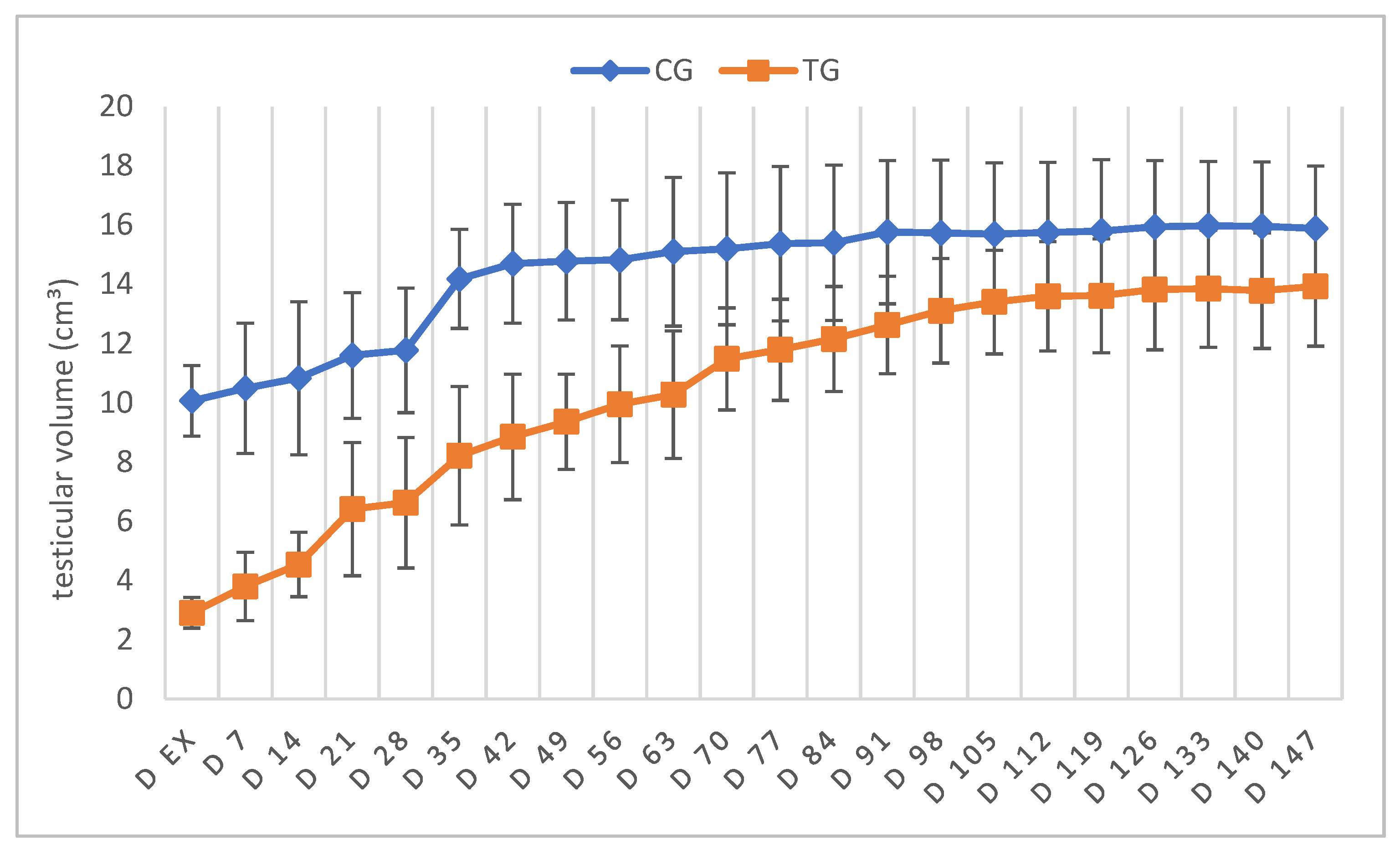
3.1. Physical and Andrological Examination
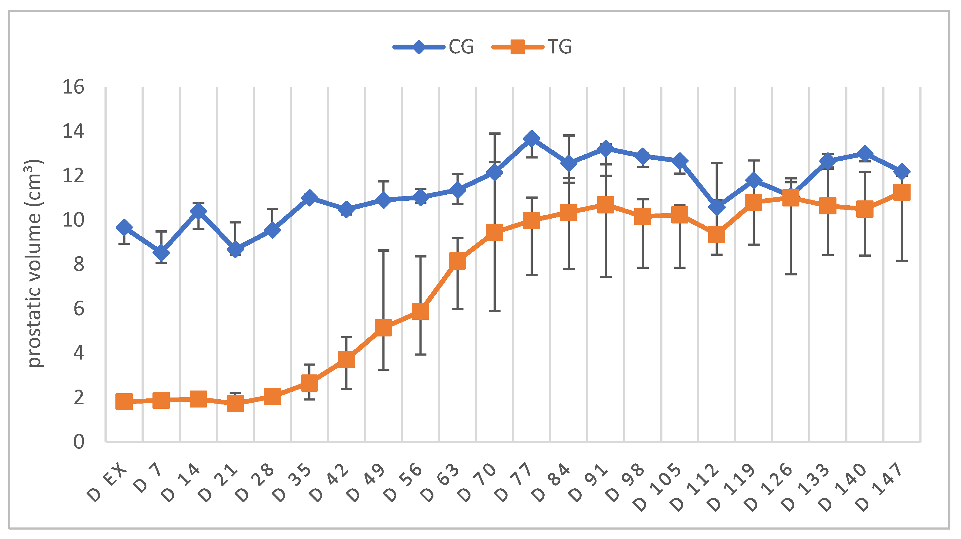
3.2. Ultrasound of the Prostate Gland
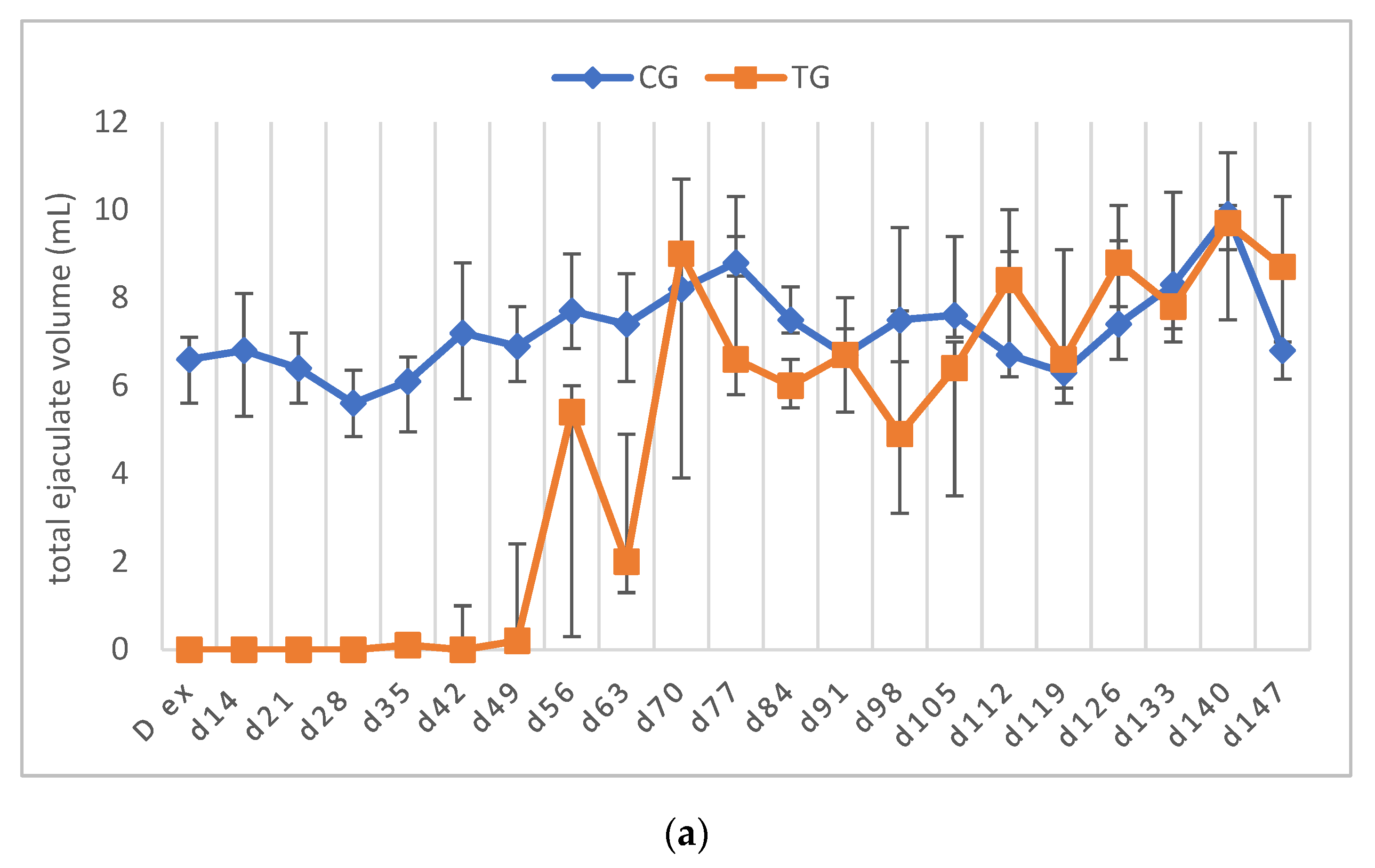
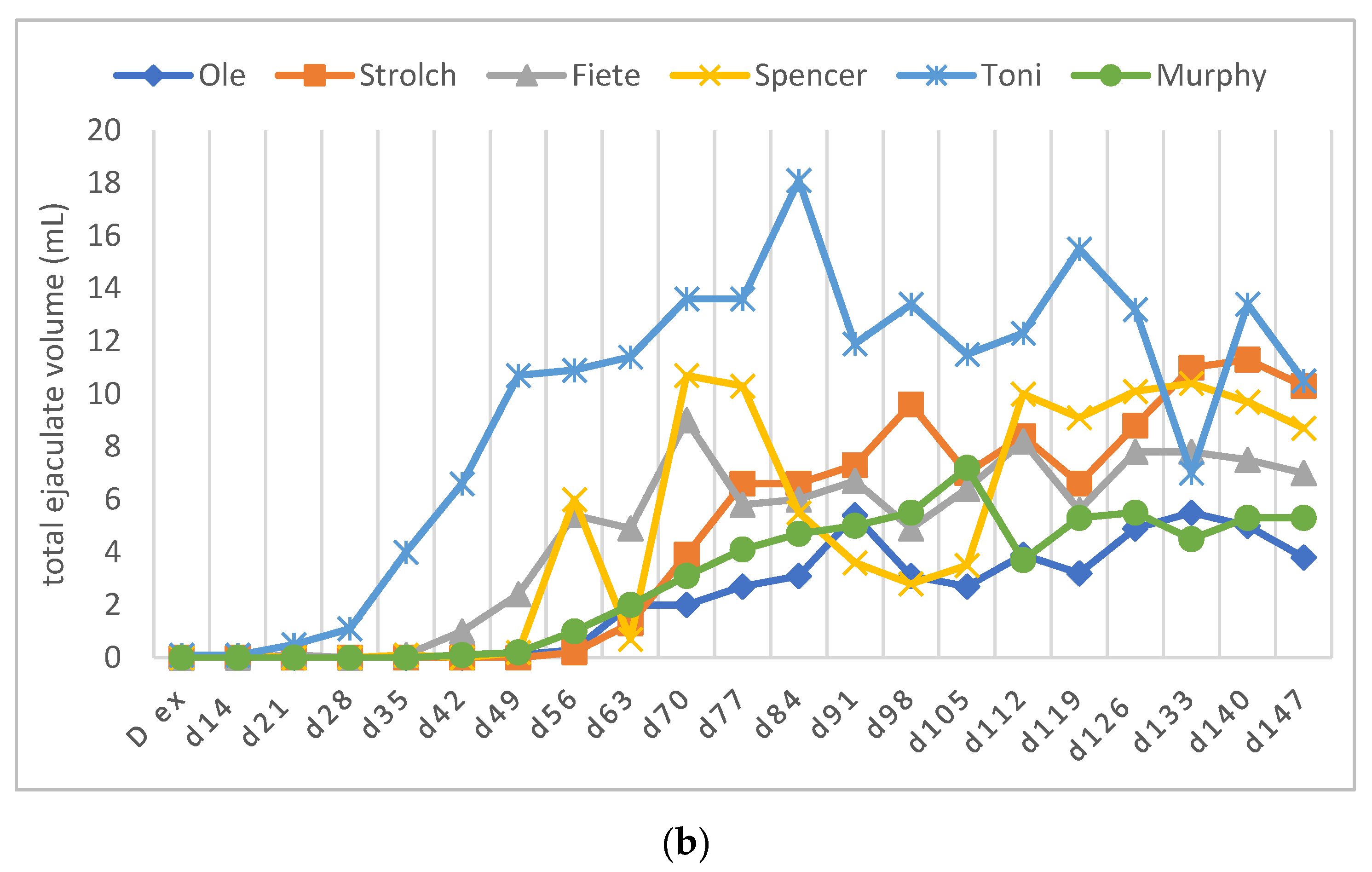
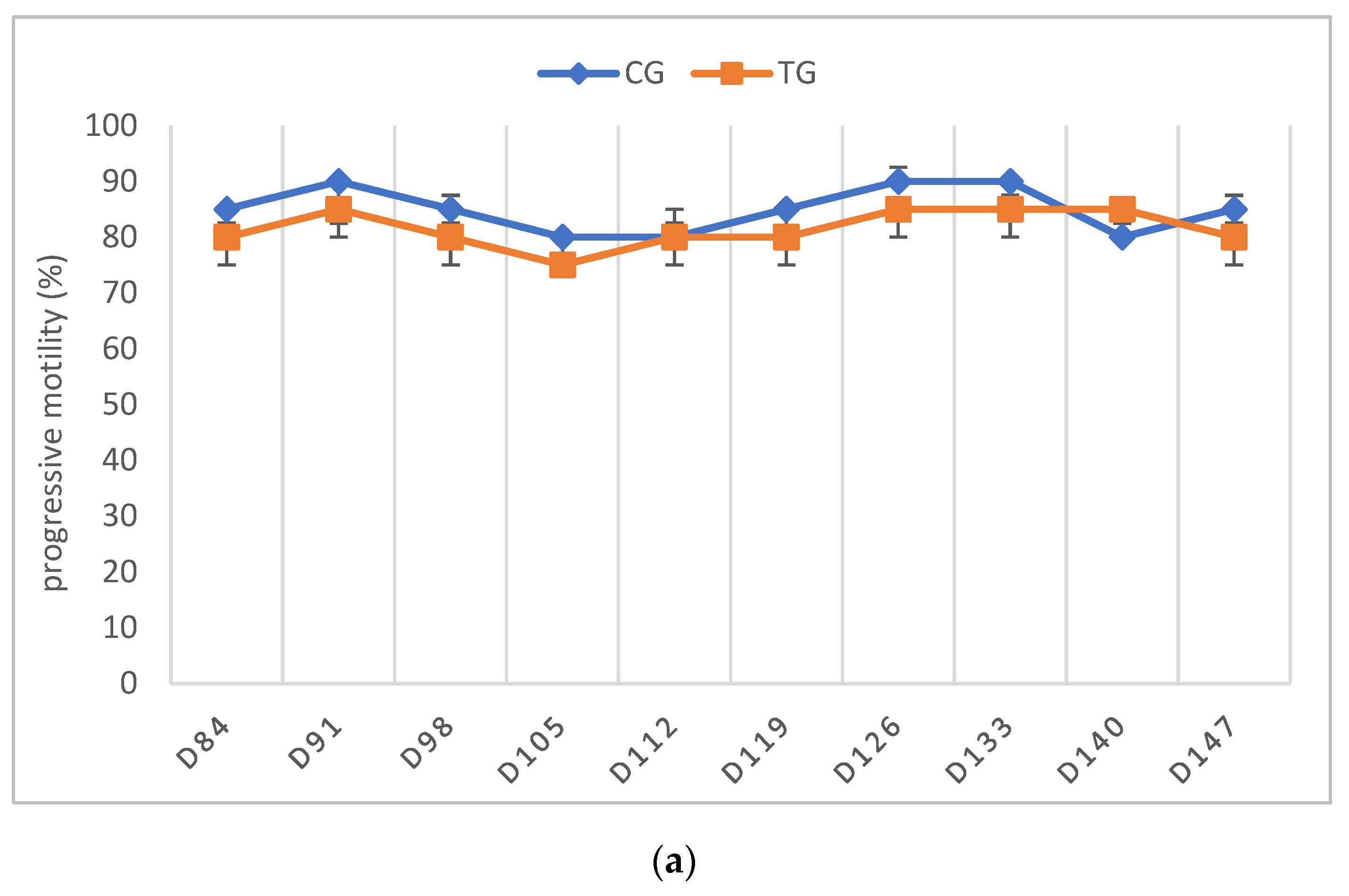
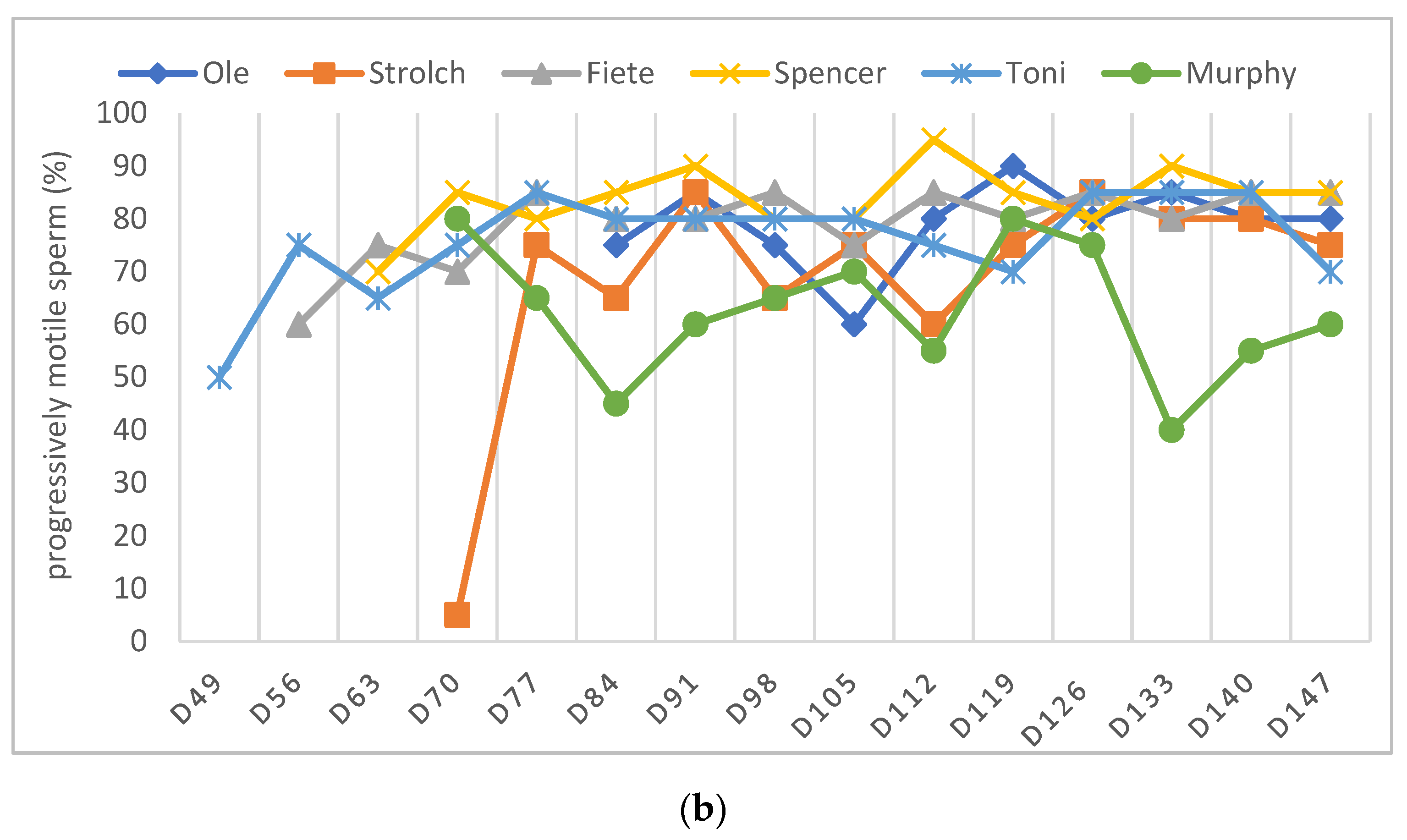
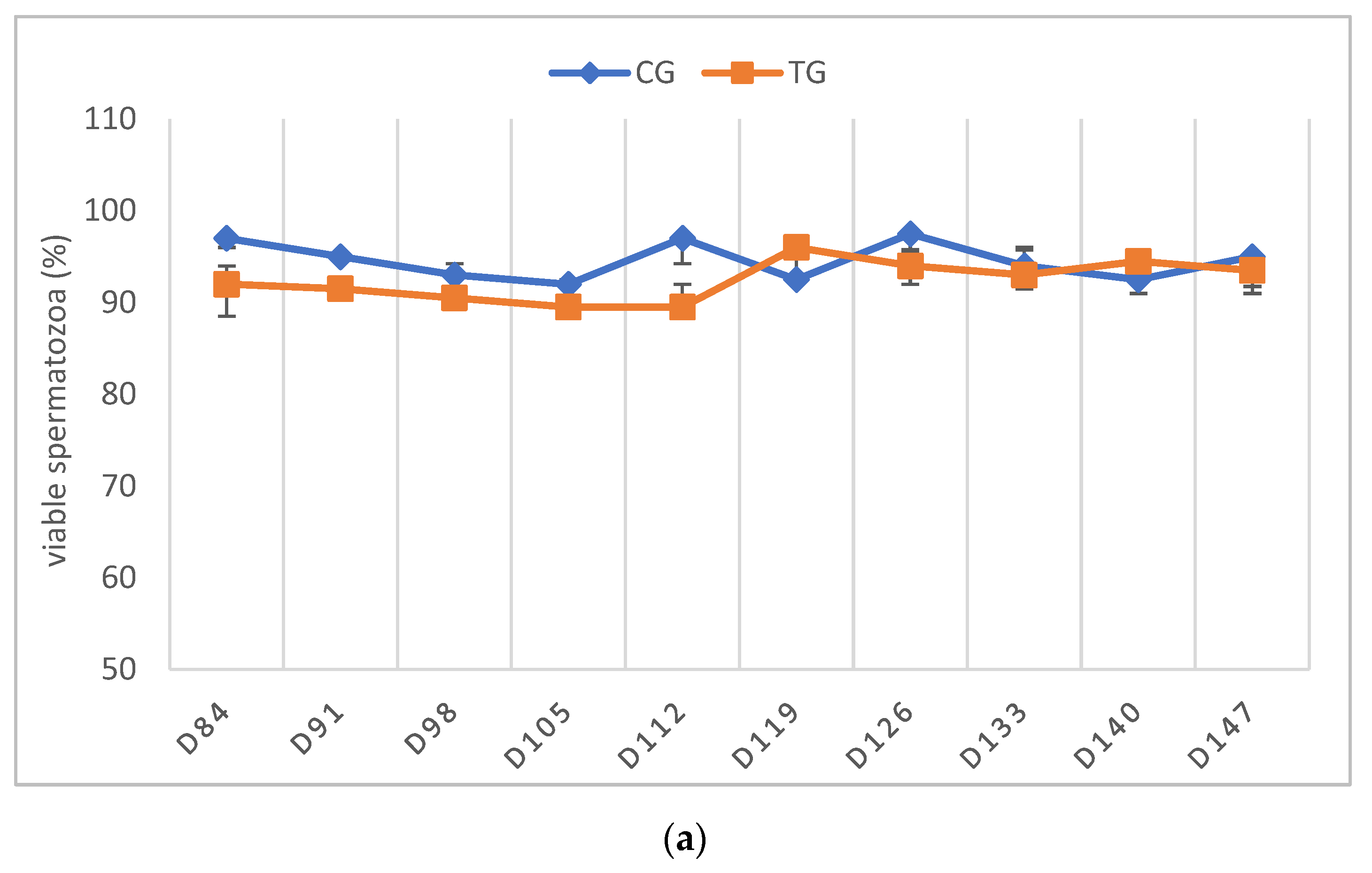
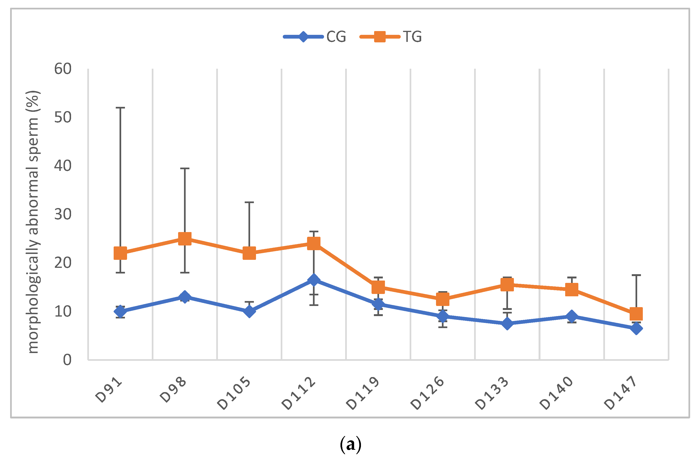
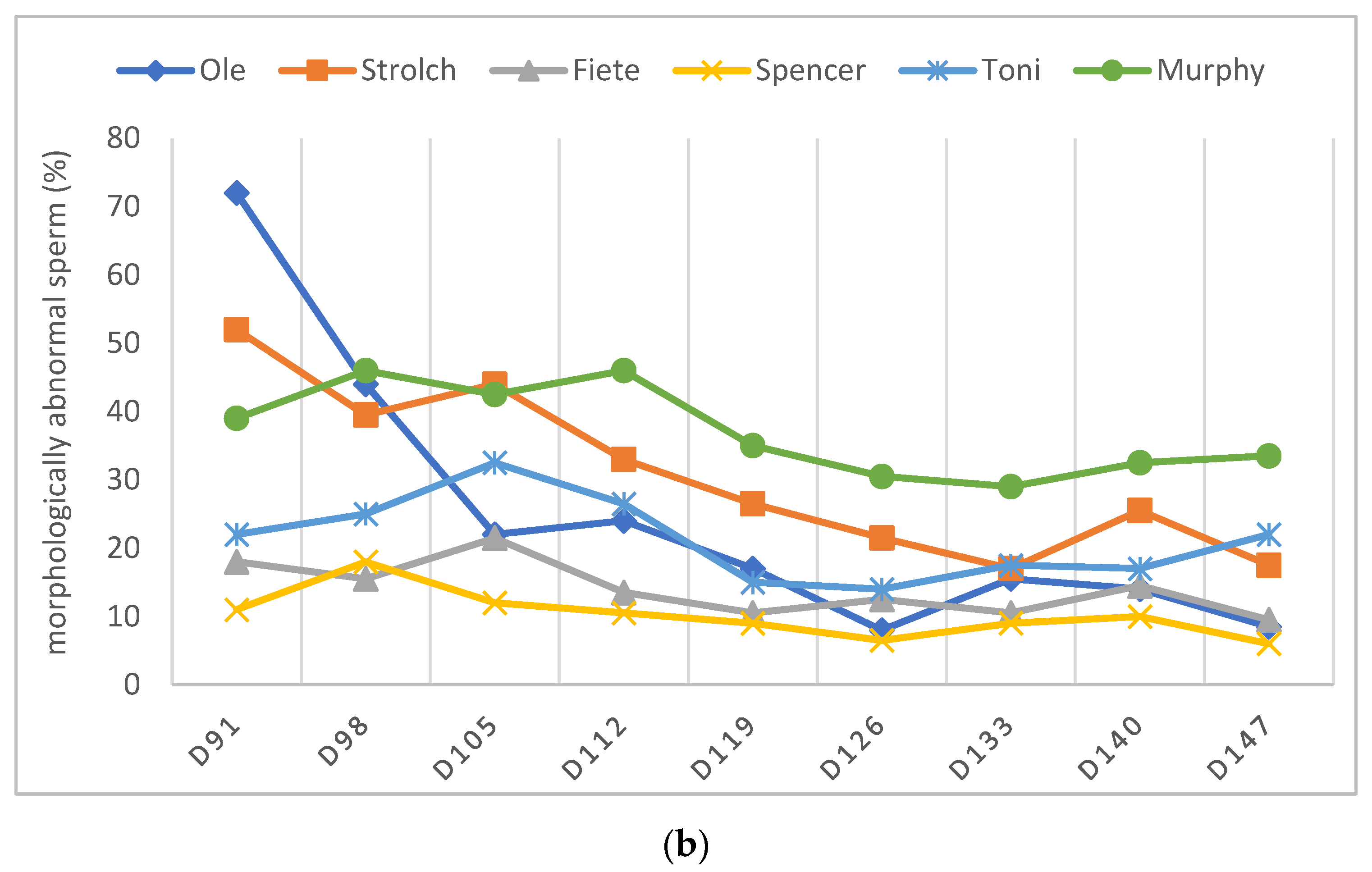
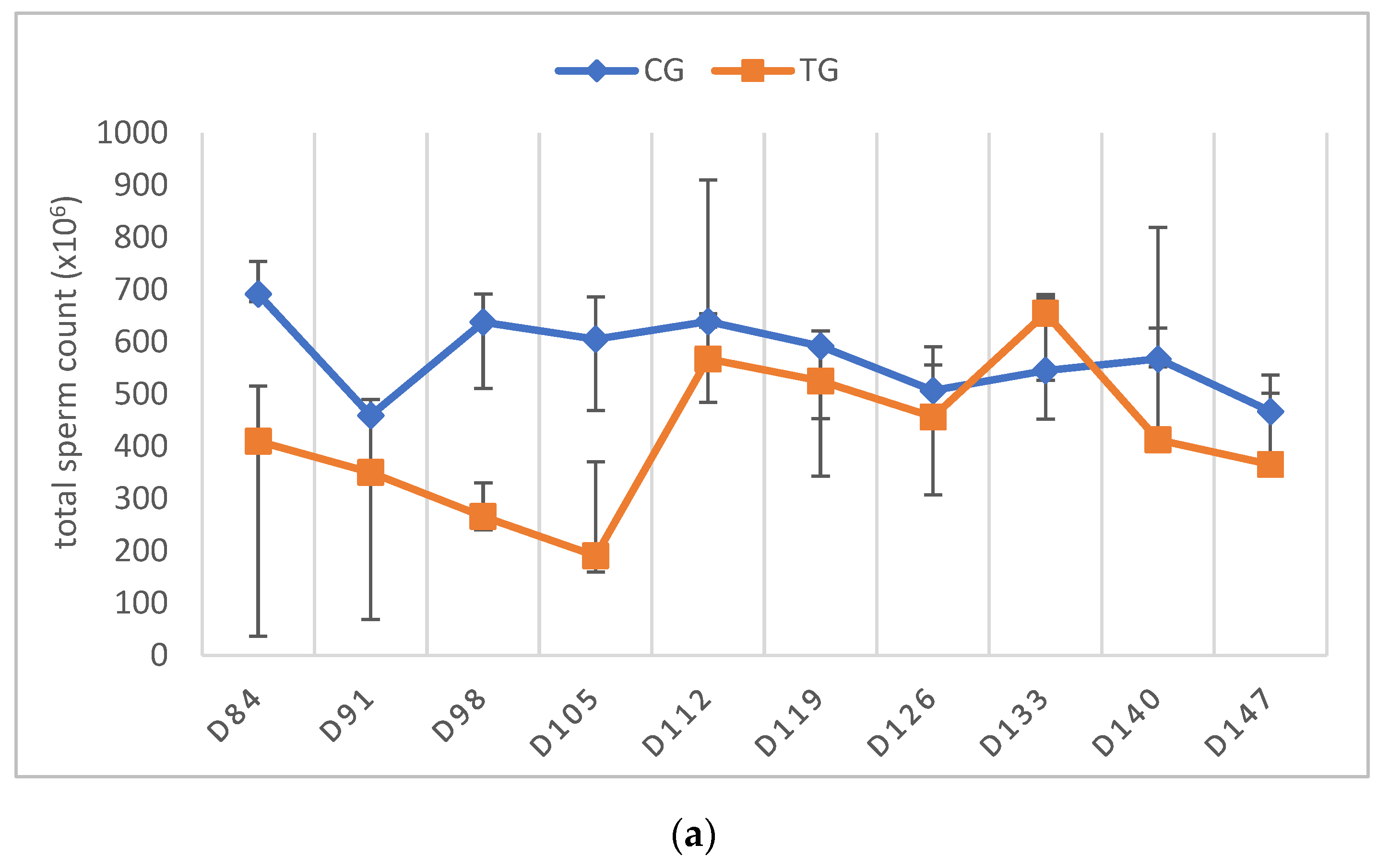
3.3. Semen Collection and Analysis
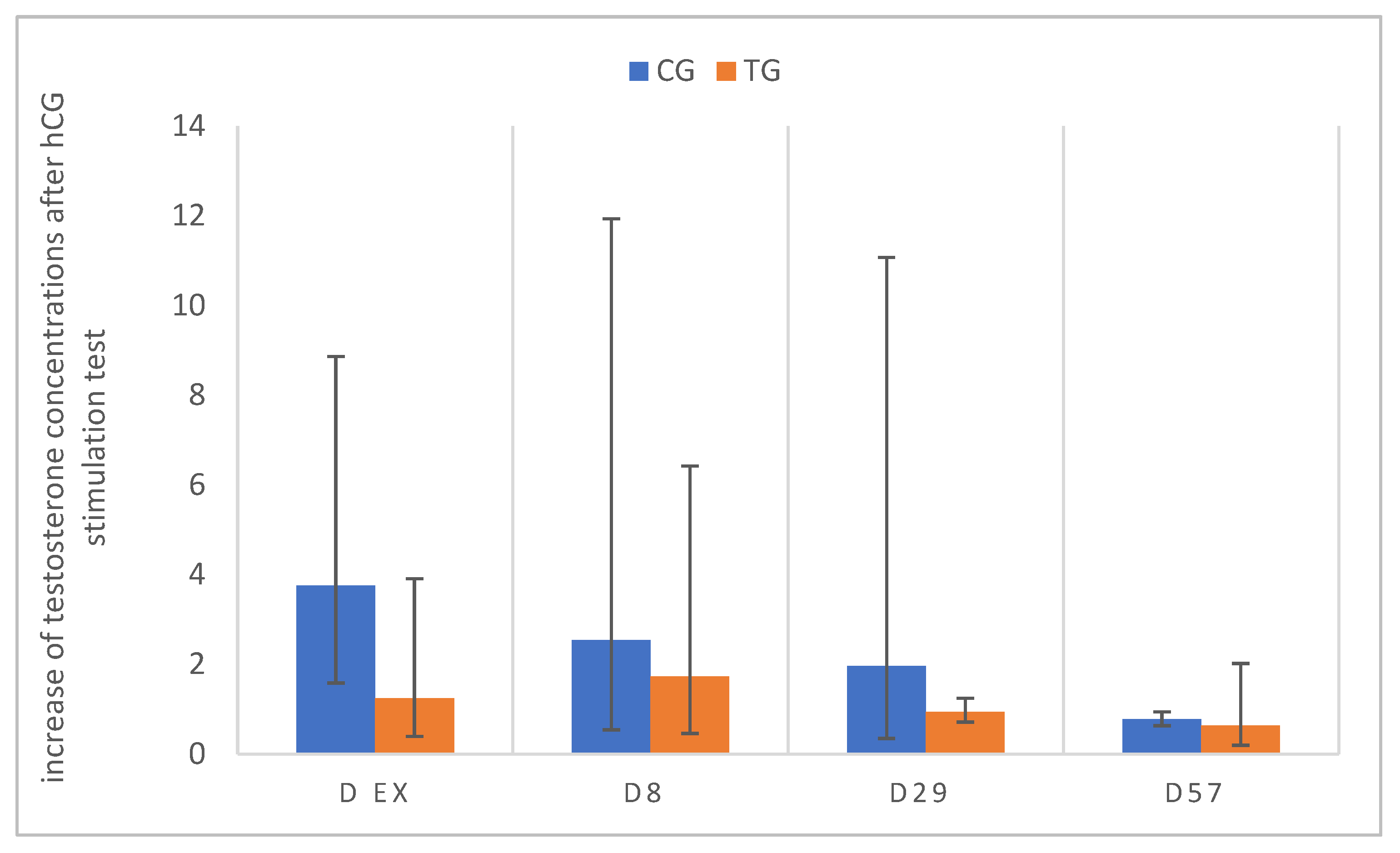
3.4. Testosterone Concentrations and Stimulation Tests
3.5. Histology of Testes
3.6. Effect of Treatment with a 4.7 mg Deslorelin Implant and Reversibility
4. Discussion
4.1. Testicular and Prostatic Volume
4.2. Testosterone
4.3. Semen Evaluation
5. Conclusions
Supplementary Materials
Author Contributions
Funding
Institutional Review Board Statement
Informed Consent Statement
Data Availability Statement
Acknowledgments
Conflicts of Interest
References
- Trigg, T.E.; Wright, P.J.; Armour, A.F.; Williamson, P.E.; Junaidi, A.; Martin, G.B.; Doyle, A.G.; Walsh, J. Use of a GnRH analogue implant to produce reversible long-term suppression of reproductive function in male and female domestic dogs. J. Reprod. Fertil. Suppl. 2001, 57, 255–261. [Google Scholar]
- Riesenbeck, A.; Klein, R.; Hoffmann, B. Downregulation, eine neue, reversible Möglichkeit zur Ausschaltung der Hodenfunktion beim Rüden. Prakt. Tierarzt 2002, 83, 512–520. [Google Scholar]
- Romagnoli, S.; Siminica, A.; Sontas, B.H.; Milani, C.; Mollo, A.; Stelletta, C. Semen quality and onset of sterility following administration of a 4.7-mg deslorelin implant in adult male dogs. Reprod. Domest. Anim. 2012, 47, 389–392. [Google Scholar] [CrossRef]
- Maenhoudt, C.; Santos, N.R.; Fontbonne, A. Suppression of fertility in adult dogs. Reprod. Domest. Anim. 2014, 49, 58–63. [Google Scholar] [CrossRef]
- Goericke-Pesch, S. Alternativen zur chirurgischen Kastration bei Hunden, Katzen und kleinen Heimtieren. Kleintierprax 2016, 61, 657–665. [Google Scholar]
- Arlt, S. Die hormonelle Kastration von Rüde und Hündin-ein Überblick. Veterinärspiegel 2010, 20, 155–160. [Google Scholar] [CrossRef]
- Palm, J.; Reichler, I.M. The use of deslorelin acetate (Suprelorin(R)) in companion animal medicine. Schweiz. Arch. Tierheilkd 2012, 154, 7–12. [Google Scholar] [CrossRef]
- Stempel, S.; Goericke-Pesch, S. GnRH agonist implants in small animal practice-What do we know 13 years following EU registration? Tierarztl. Prax. Ausg. K Kleintiere Heimtiere 2020, 48, 420–432. [Google Scholar]
- Trigg, T.E.; Doyle, A.G.; Walsh, J.D.; Swangchan-uthai, T. A review of advances in the use of the GnRH agonist deslorelin in control of reproduction. Theriogenology 2006, 66, 1507–1512. [Google Scholar] [CrossRef] [PubMed]
- Stempel, S.; Körber, H.; Reifarth, L.; Schuler, G.; Goericke-Pesch, S. What Happens in Male Dogs after Treatment with a 4.7 mg Deslorelin Implant? I. Flare up and Downregulation. Animals 2022, 12, 2379. [Google Scholar] [CrossRef]
- Junaidi, A.; Williamson, P.E.; Cummins, J.M.; Martin, G.B.; Blackberry, M.A.; Trigg, T.E. Use of a new drug delivery formulation of the gonadotrophin-releasing hormone analogue Deslorelin for reversible long-term contraception in male dogs. Reprod. Fertil. Dev. 2003, 15, 317–322. [Google Scholar] [CrossRef]
- Goericke-Pesch, S.; Wilhelm, E.; Hoffmann, B. Hormonelle Downregulation der Hodenfunktion bei Rüde und Kater; eine retrospektive Studie. Prakt. Tierarzt 2010, 91, 563–570. [Google Scholar]
- Goericke-Pesch, S.; Ludwig, C.; Hoffmann, B. Development of semen quality following reversible downregulation of testicular function in male dogs with a GnRH agonist implant. Reprod. Domest. Anim. 2012, 47, 625–628. [Google Scholar] [CrossRef]
- Goericke-Pesch, S.; Georgiev, P.; Atanasov, A.; Albouy, M.; Navarro, C.; Wehrend, A. Treatment of queens in estrus and after estrus with a GnRH-agonist implant containing 4.7 mg deslorelin; hormonal response, duration of efficacy, and reversibility. Theriogenology 2013, 79, 640–646. [Google Scholar] [CrossRef]
- Goericke-Pesch, S.; Georgiev, P.; Atanasov, A.; Wehrend, A. Treatment with Suprelorin in a pregnant cat. J. Feline Med. Surg. 2013, 15, 357–360. [Google Scholar] [CrossRef]
- Goericke-Pesch, S.; Georgiev, P.; Antonov, A.; Vodenicharov, A.; Navarro, C.; Wehrend, A. Reversibility of germinative and endocrine testicular function after long-term contraception with a GnRH-agonist implant in the tom-a follow-up study. Theriogenology 2014, 81, 941–946. [Google Scholar] [CrossRef]
- Ferre-Dolcet, L.; Carniello, L.; Ferro, S.; Cattai, A.; Romagnoli, S.; Mollo, A. Interval between Removal of a 4.7 mg Deslorelin Implant after a 3-, 6-, and 9-Month Treatment and Restoration of Testicular Function in Tomcats. Animals 2020, 10, 1559. [Google Scholar] [CrossRef]
- Ferre-Dolcet, L.; Ferro, S.; Contiero, B.; Andretta, F.; Cattai, A.; Fontaine, C.; Romagnoli, S. Resumption of ovarian activity following removal of a 4.7 mg deslorelin implant in queens. Reprod. Domest. Anim. 2021, 57, 3–9. [Google Scholar] [CrossRef]
- Novotny, R.; Cizek, P.; Vitasek, R.; Bartoskova, A.; Prinosilova, P.; Janosovska, M. Reversible suppression of sexual activity in tomcats with deslorelin implant. Theriogenology 2012, 78, 848–857. [Google Scholar] [CrossRef]
- Goericke-Pesch, S.; Spang, A.; Schulz, M.; Ozalp, G.; Bergmann, M.; Ludwig, C.; Hoffmann, B. Recrudescence of spermatogenesis in the dog following downregulation using a slow release GnRH agonist implant. Reprod. Domest. Anim. 2009, 44, 302–308. [Google Scholar] [CrossRef]
- Goericke-Pesch, S.; Gentil, M.; Spang, A.; Kowalewski, M.P.; Failing, K.; Hoffmann, B. Status of the down-regulated canine testis using two different GNRH agonist implants in comparison with the juvenile testis. Reproduction 2013, 146, 517–526. [Google Scholar] [CrossRef]
- Gouletsou, P.G.; Galatos, A.D.; Leontides, L.S. Comparison between ultrasonographic and caliper measurements of testicular volume in the dog. Anim. Reprod. Sci. 2008, 108, 1–12. [Google Scholar] [CrossRef]
- Mantziaras, G.; Alonge, S.; Faustini, M.; Luvoni, G.C. Assessment of the age for a preventive ultrasonographic examination of the prostate in the dog. Theriogenology 2017, 100, 114–119. [Google Scholar] [CrossRef]
- Moxon, R.; Bright, L.; Pritchard, B.; Bowen, I.M.; de Souza, M.B.; da Silva, L.D.; England, G.C. Digital image analysis of testicular and prostatic ultrasonographic echogencity and heterogeneity in dogs and the relation to semen quality. Anim. Reprod. Sci. 2015, 160, 112–119. [Google Scholar] [CrossRef]
- Ruel, Y.; Barthez, P.Y.; Mailles, A.; Begon, D. Ultrasonographic evaluation of the prostate in healthy intact dogs. Vet. Radiol. Ultrasound 1998, 39, 212–216. [Google Scholar] [CrossRef]
- Nizanski, W.; Ochota, M.; Fontaine, C.; Pasikowska, J. B-Mode and Doppler Ultrasonographic Findings of Prostate Gland and Testes in Dogs Receiving Deslorelin Acetate or Osaterone Acetate. Animals 2020, 10, 2379. [Google Scholar] [CrossRef]
- Günzel-Apel, A.R.; Terhaer, P.; Waberski, D. Hodendimensionen und Ejakulatbeschaffenheit fertiler Rüden unterschiedlicher Körpergewichte. Kleintierpraxis 1994, 39, 483–486. [Google Scholar]
- Günzel-Apel, A.R.; Bostedt, H. Reproduktionsmedizin und Neonatologie von Hund und Katze, 1st ed.; Schattauer GmbH: Stuttgart, Germany, 2016; pp. 3–738. [Google Scholar]
- Hoffmann, B.; Landeck, A. Testicular endocrine function, seasonality and semen quality of the stallion. Anim. Reprod. Sci. 1999, 57, 89–98. [Google Scholar] [CrossRef]
- Röcken, F.E.; Nothelfer, H.B.; Hoffmann, B. Testosteronkonzentrationen im peripheren Plasma sowie morphologische Hodenbefunde von Rüden mit einer Perinealhernie. Kleintierpraxis 1995, 40, 261–267. [Google Scholar]
- Ludwig, C.; Desmoulins, P.O.; Driancourt, M.A.; Goericke-Pesch, S.; Hoffmann, B. Reversible downregulation of endocrine and germinative testicular function (hormonal castration) in the dog with the GnRH-agonist azagly-nafarelin as a removable implant “Gonazon”; a preclinical trial. Theriogenology 2009, 71, 1037–1045. [Google Scholar] [CrossRef]
- Goericke-Pesch, S. Kryptorchismus bei Hund und Katze. Kleintierprax 2010, 55, 255–261. [Google Scholar]
- Polisca, A.; Orlandi, R.; Troisi, A.; Brecchia, G.; Zerani, M.; Boiti, C.; Zelli, R. Clinical efficacy of the GnRH agonist (deslorelin) in dogs affected by benign prostatic hyperplasia and evaluation of prostatic blood flow by Doppler ultrasound. Reprod. Domest. Anim. 2013, 48, 673–680. [Google Scholar] [CrossRef]
- Tsutsui, T.; Kurita, A.; Kirihara, N.; Hori, T.; Kawakami, E. Testicular compensatory hypertrophy related to hemicastration in prepubertal dogs. J. Vet. Med. Sci. 2004, 66, 1021–1025. [Google Scholar] [CrossRef]
- Günzel-Apel, A.R.; Brinckmann, H.G.; Hoppen, H.O. Dynamik der LH- und Testosteron-Sekretion bei Beagle-Rüden verschiedener Altersgruppen. Reprod. Domest. Anim. 1990, 25, 78–86. [Google Scholar] [CrossRef]
- De Palatis, L.; Moore, J.; Falvo, R.E. Plasma concentrations of testosterone and LH in the male dog. J. Reprod. Fertil. 1978, 52, 201–207. [Google Scholar] [CrossRef]
- Taha, M.B.; Noakes, D.E. The effect of age and season of the year on testicular function in the dog, as determined by histological examination of the seminiferous tubules and the estimation of peripheral plasma testosterone concentrations. J. Small Anim. Pract. 1982, 23, 352–357. [Google Scholar] [CrossRef]
- Sullivan, J.J.; Parker, W.G.; Larson, L.L. Duration of Estrus and Ovulation Time in Nonlactating Mares Given Human Chorionic Gonadotropin During Three Successive Estrous Periods. J. Am. Vet. Med. Assoc. 1973, 162, 895–898. [Google Scholar]
- Wilson, C.G.; Downie, C.R.; Hughes, J.P.; Roser, J.F. Effects of repeated hCG injections on reproductive efficiency in mares. J. Equine Vet. Sci. 1990, 10, 301–308. [Google Scholar] [CrossRef]
- Giordano, J.O.; Wiltbank, M.C.; Fricke, P.M. Humoral immune response in lactating dairy cows after repeated exposure to human chorionic gonadotropin. Theriogenology 2012, 78, 218–224. [Google Scholar] [CrossRef]
- Sundby, A.; Torjesen, P.A. Plasma levels of testosterone in bulls. Response to repeated HCG injections. Acta Endocrinol. 1978, 88, 787–792. [Google Scholar] [CrossRef]
- Swanson, W.F.; Horohov, D.W.; Godke, R.A. Production of exogenous gonadotrophin-neutralizing immunoglobulins in cats after repeated eCG-hCG treatment and relevance for assisted reproduction in felids. J. Reprod. Fertil. 1995, 105, 35–41. [Google Scholar] [CrossRef] [PubMed]
- Swanson, W.F.; Roth, T.L.; Graham, K.; Horohov, D.W.; Godke, R.A. Kinetics of the humoral immune response to multiple treatments with exogenous gonadotropins and relation to ovarian responsiveness in domestic cats. Am. J. Vet. Res. 1996, 57, 302–307. [Google Scholar]
- Al-Kafawi, A.A.; Hopwood, M.L.; Pineda, M.H.; Faulkner, L.C. Immunization of dogs against human chorionic gonadotropin. Am. J. Vet. Res. 1974, 35, 261–264. [Google Scholar] [PubMed]
- Foote, R.H.; Swierstra, E.E.; Hunt, W.L. Spermatogenesis in the dog. Anat. Rec. 1972, 173, 341–351. [Google Scholar] [CrossRef]
- Ibach, B.; Weissbach, L.; Hilscher, B. Stages of the cycle of the seminiferous epithelium in the dog. Andrologia 1976, 8, 297–307. [Google Scholar] [CrossRef]
- Soares, J.M.; Avelar, G.F.; Franca, L.R. The seminiferous epithelium cycle and its duration in different breeds of dog (Canis familiaris). J. Anat. 2009, 215, 462–471. [Google Scholar] [CrossRef]
- Greer, M.L. Canine Reproduction and Neonatology; CRC Press: Boca Raton, FL, USA, 2014. [Google Scholar]
- Ali Hassan, H.; Domain, G.; Luvoni, G.C.; Chaaya, R.; Van Soom, A.; Wydooghe, E. Canine and Feline Epididymal Semen-A Plentiful Source of Gametes. Animals 2021, 11, 2961. [Google Scholar] [CrossRef]
- Taha, M.B.; Noakes, D.E.; Allen, W.E. Hemicastration and castration in the beagle dog; the effects on libido, peripheral plasma testosterone concentrations, seminal characteristics and testicular function. J. Small Anim. Pract. 1982, 23, 279–285. [Google Scholar] [CrossRef]


 spermatogonia;
  spermatocytes;
  round spermatids;
  elongating spermatids;
  Sertoli cells.
spermatogonia; spermatocytes; round spermatids; elongating spermatids; Sertoli cells.
Sertoli cells.
spermatogonia; spermatocytes; round spermatids; elongating spermatids; Sertoli cells.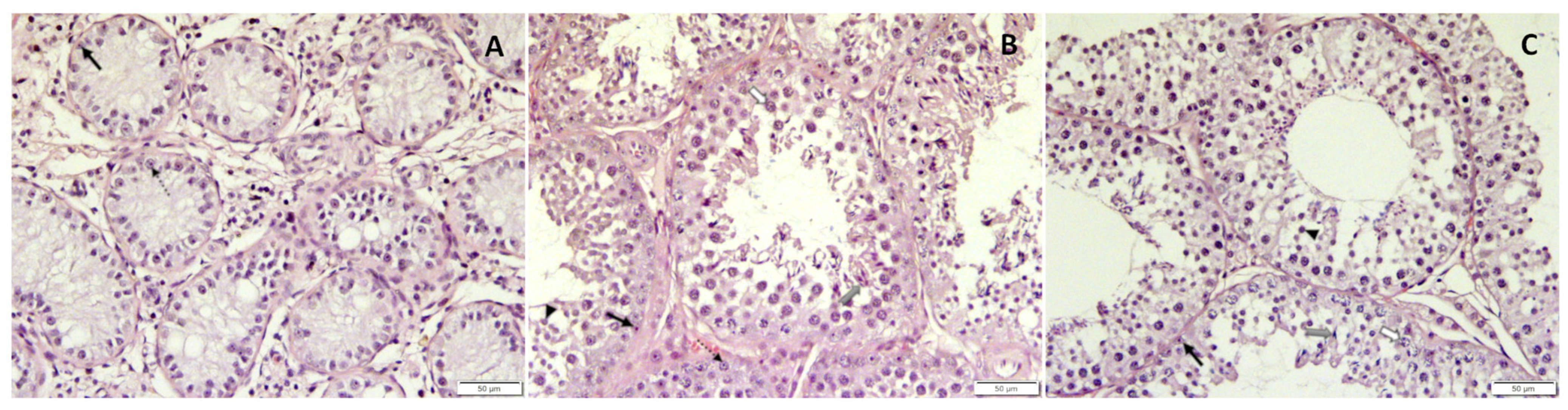

| I | II | III | |
|---|---|---|---|
| Parameter | D0 | D ex | Recovery |
| TV (cm3) | 8.6 ± 2.5 a | 2.9 ± 0.5 b | 13.9 ± 2.0 c |
| PV (cm3) | 9.5 (8.9, 11.6) a | 1.8 (1.7, 1.9) b | 11.3 (8.2, 12.0) a |
| T (ng/mL) | 3.8 (2.1) a | 0.2 (3.0) b,c | 2.0 (2.7) a,c |
| Parameter | I | II | III |
|---|---|---|---|
| VOL (mL) | 6.0 (2.3, 13.6) | 6.6 (5.4, 10.3) | 10.0 (6.0, 11.0) |
| % PM | 65.0 (65.0, 75.0) | 75.0 (75.0, 80.0) | 80.0 (80.0, 85.0) |
| % VS | 92.0 (91.5, 93.0) | 93.0 (92.5, 95.0) | 94.0 (93.0, 95.0) |
| % MAS | 14.5 (14.0, 15.0) a | 79.0 (72.0, 86.0) b,c | 17.0 (15.5, 26.5) a,c |
| TSC (×106) | 1091.6 (290.9, 1150.0) a,b | 56.3 (36.3, 62.5) a,b | 684.06 (567.19, 910.0) a |
| days | Before | D56–91 | D84–133 |
Publisher’s Note: MDPI stays neutral with regard to jurisdictional claims in published maps and institutional affiliations. |
© 2022 by the authors. Licensee MDPI, Basel, Switzerland. This article is an open access article distributed under the terms and conditions of the Creative Commons Attribution (CC BY) license (https://creativecommons.org/licenses/by/4.0/).
Share and Cite
Stempel, S.; Körber, H.; Reifarth, L.; Schuler, G.; Goericke-Pesch, S. What Happens in Male Dogs after Treatment with a 4.7 mg Deslorelin Implant? II. Recovery of Testicular Function after Implant Removal. Animals 2022, 12, 2545. https://doi.org/10.3390/ani12192545
Stempel S, Körber H, Reifarth L, Schuler G, Goericke-Pesch S. What Happens in Male Dogs after Treatment with a 4.7 mg Deslorelin Implant? II. Recovery of Testicular Function after Implant Removal. Animals. 2022; 12(19):2545. https://doi.org/10.3390/ani12192545
Chicago/Turabian StyleStempel, Sabrina, Hanna Körber, Larena Reifarth, Gerhard Schuler, and Sandra Goericke-Pesch. 2022. "What Happens in Male Dogs after Treatment with a 4.7 mg Deslorelin Implant? II. Recovery of Testicular Function after Implant Removal" Animals 12, no. 19: 2545. https://doi.org/10.3390/ani12192545
APA StyleStempel, S., Körber, H., Reifarth, L., Schuler, G., & Goericke-Pesch, S. (2022). What Happens in Male Dogs after Treatment with a 4.7 mg Deslorelin Implant? II. Recovery of Testicular Function after Implant Removal. Animals, 12(19), 2545. https://doi.org/10.3390/ani12192545