Article

Retrotransposon Insertion Polymorphisms (RIPs) in Pig Coat Color Candidate Genes

1
College of Animal Science and Technology, Yangzhou University, Yangzhou 225009, China
2
Department of Veterinary Sciences, University of Messina, Via Palatucci, 98168 Messina, Italy
*
Author to whom correspondence should be addressed.
These authors contributed equally to this work.
Animals 2022, 12(8), 969; https://doi.org/10.3390/ani12080969
Submission received: 10 February 2022 / Revised: 28 March 2022 / Accepted: 5 April 2022 / Published: 8 April 2022
(This article belongs to the Section Pigs)

Abstract

:

Simple Summary

Coat color is an important phenotypic trait of pig breeds, and a few genes are involved in its regulation. After long-term selection, pig breeds have formed diversified coat color phenotypes (e.g., black, red, white, spotted, brown, belted, two-end black, etc.). To date, several major genes affecting pig coat color phenotypes have been identified. In this view, recent studies suggested a new type of molecular marker known as retrotransposon insertion polymorphisms (RIPs). This marker is considered an ideal molecular marker, showing the potential to be used for population genetic analysis in pigs, with the possibility to be extended to other livestock animals as well. Here, we revealed 42 RIPs in 20 coat color genes in pigs, and some RIPs may be useful in distinguishing different breeds.

Abstract

The diversity of livestock coat color results from human positive selection and represents an indispensable part of breed identity. As an important biodiversity resource, pigs have many special characteristics, including the most visualized feature, coat color, and excellent adaptation, and the coat color represents an important phenotypic characteristic of the pig breed. Exploring the genetic mechanisms of phenotypic characteristics and the melanocortin system is of considerable interest in domestic animals because their energy metabolism and pigmentation have been under strong selection. In this study, 20 genes related to coat color in mammals were selected, and the structural variations (SVs) in these genic regions were identified by sequence alignment across 17 assembled pig genomes, from representing different types of pigs (miniature, lean, and fat type). A total of 167 large structural variations (>50 bp) of coat-color genes, which overlap with retrotransposon insertions (>50 bp), were obtained and designated as putative RIPs. Finally, 42 RIPs were confirmed by PCR detection. Additionally, eleven RIP sites were further evaluated for their genotypic distributions by PCR in more individuals of eleven domesticated breeds representing different coat color groups. Differential distributions of these RIPs were observed across populations, and some RIPs may be associated with breed differences.

1. Introduction

Animal pigmentation is one of the most visible and variable traits shaped by natural and/or artificial selection. As a visible phenotypic marker, pigmentation has played an important role in our inheritance understanding, development, and evolutionary theory [1]. In mammals, skin and hair pigmentation is a consequence of cellular activities and cooperation between two cell types, i.e., melanocytes that produce melanins and keratinocytes that distribute melanins throughout the skin [2]. Melanins are the principal pigments of the skin and hair, and their synthesis occurs within membrane-bound organelles of melanocytes called melanosomes [3]. It is commonly known that the synthesis of principal melanins occurs through the melanogenesis process (ko04916). This process is under the complex regulatory control of multiple genes, including MC1R, ASIP, MITF, DCT, TYR, TYRP1, and KIT. Besides these genes, the true melanogenesis operating mechanism should be started by forming melanosomes [4]. There are many key molecules participating in intracellular trafficking during melanosome biogenesis. Melanosome structure and function are determined by a cohort of resident transmembrane proteins, many of which are expressed only in pigment cells and localized specifically to melanosomes [5]. All melanosomal proteins are synthesized in the endoplasmic reticulum (ER) and transit through the Golgi complex and the trans-Golgi network (TGN) route to melanosomes [6]. Premelanosome protein (PMEL, also known as PMEL17, SILV, or gp100), an integral membrane protein, is targeted from the TGN to the plasma membrane, from where it is endocytosed and delivered to a Stage I melanosome as a melanosome fibroid structural framework [7]. The melanosome-associated G-protein-coupled receptor OA1 is involved as a regulator to assist the formation of PMEL [8]. Although the mature form of a Stage I melanosome is reached at Stage II and Stage III, a balanced environment inside of the Stage I melanosome is needed, which relies on SLC45A2 and OCA2 (also called P protein). These proteins act as ion pumps coupled with V-ATPase exchange cationic concentrations, such as Na+, K+, Ca2+, and H+, to regulate internal pH [9,10], while the protein of TYR, TYRP1, and DCT act as cargoes transported by Rab32/Rab38 and Rab27a from the TGN to the melanosome [11], along with the path constructed by the fibrous protein of KIF13A and Spire1/2 [12].
Almost 200 genes associated with color were identified in mice [13]. It has been reported that mutations in these genes may cause pathogenic variants, such as albinism disease in humans [14] (e.g., variable hypopigmentation of the skin, hair, and eyes), ranging from a total lack of melanin to residual or even near-normal melanin production.
In mammals, the similar mutation phenotype with humans also appears in tigers [15], horses [16,17], dogs [18], cattle [19,20], rabbits [21], mink [22], foxes [23], and raccoon dogs [24]. In pigs, the coat color is an important phenotypic characteristic in breeding work. However, one major challenge faced by pig researchers and breeders is merging different breeds’ advantages into one breed. The Sushan pig breeds were developed by Chinese experts using Meishan, Erhualian, and Yorkshire pig, resulted in a white body coat color with significant advantage in growth and reproduction. Similarly, the uniform black coat color with improved meat quality and reproduction observed in the Sutai pigs breed, is a crossbreed between the blood of Duroc and China local breeds (Jiangsu province) [25]. Kijas et al. [26] found that several SNPs in MC1R may be a strong candidate marker in different pig breeds. Wu et al. [27] conducted a genome-wide association study and RNA sequencing to demonstrate that KIT variants are responsible for diversifying coat color phenotypes, segregating in a Duroc (Landrace & Large White) hybrid pig population.
Retrotransposons are defined as retroelements that replicate by transcription of an RNA intermediate, subsequent reverse transcription, and insertion of a new copy elsewhere in the genome [28]. Retrotransposons, as an important component of plant and animal genomes, can be classified into non-LTR families (including Long Interspersed Nuclear Elements (LINE) and Short Interspersed Nuclear Elements (SINE)) and Long Terminal Repeat elements (LTR, including endogenous retrovirus (ERV)) [29,30]. Retrotransposons occupy one-third to half of the mammal genomes, which are dominated by LINEs and SINEs, followed by LTR retrotransposons [31]. Chen et al. [32] reported that retrotransposons accounted for 37.13% (929.09 MB) of the pig genome, while LINEs, LTRs, and SINEs accounted for 18.52%, 7.56%, and 11.05%, respectively.
Since transposable elements (TEs) were first discovered in maize in the 1940s [33], an increasing number of TEs and retrotransposons have shown more relevant functions in plants and vertebrates’ genomes. However, retrotransposon insertion polymorphisms (RIPs) as DNA markers in a gene are helpful for agricultural production in flax [34], strawberry [35], sweet potato [36], and grape [37]. Rebeca et al. [38] found that detecting RIPs in the intron and exon of particular genes for humans can act as a utility of developing bioinformatic tools to find autism spectrum disorder. Abundant polymorphic retrotransposon insertions were already observed in animals such as dogs [39,40] and Anolis carolinensis [41]. Three LINE RIP markers were strongly associated with economic traits in 462 Yorkshire pigs [42]. Two RIPs in the VRTN (vertebrae development-associated) gene were found in pigs, which showed Hardy-Weinberg equilibrium distributions in most pig populations [43]. Four RIPs in the GHR (growth hormone receptor) gene and one RIP in the IGF1 (insulin-like growth factor 1) gene was identified in pigs. Further analysis revealed that one RIP in the first intron of GHR might play a role in the regulation of GHR expression by acting as a repressor [44].
In this study, 20 genes related to coat color in mammals were selected. Then, the structural variations (SVs) of these gene sequences derived from retrotransposons were mined by comparative genomic analysis across 17 assembled pig genomes, combined with Repeat Masker annotation. The SVs annotated as retrotransposon insertions were further confirmed by pool-PCR, and their genotypic distributions were detected in eleven pig populations. Therefore, this particular polymorphic molecular marker was explored and intended to provide a theoretical basis for genetic improvement in pigs.

2. Materials and Methods

2.1. Ethical Statement

The collection of biological samples and experimental procedures involved in this study were approved by the Animal Experiment Ethics Committee of Yangzhou University (No. NSFC2020-dkxy-02, 27 March 2020).

2.2. Sequence Acquisition for Coat Color Genes

The detailed information of the genic sequences ASIP, KIT, MITF, PMEL, GPR143, TYR, EDNRB, TYRP1, MLPH, DCT, OCA2, SLC24A5, SLC45A2, Rab27a, Rab32, Rab38, KIF13A, Spire2, Spire1, and MC1R areshown in Table S1. These genes with their flanking regions (5-kb 5′upstream and 5-kb 3′downstream) from the reference (Duroc) genome (Sscrofa11.1) were downloaded from NCBI Genbank (https://www.ncbi.nlm.nih.gov/genbank/, accessed on 10 September 2021).
Then, 1000 bp upstream and 1000 bp downstream boundary sequences of each genic region were used to Blast the WGS to define the same genomic positions for the 16 assembled nonreference genomes. These genomes represented different types of pigs, including miniature pigs (Tibetan pig, AORO00000000; Wuzhishan, AJKK00000000; Bama, SIDA00000000), lean type pigs (Duroc, Hampshire, LUXS00000000; Berkshire, LUXW00000000; Pietrain, LUXU00000000; Landrace, LUXT00000000; Yorkshire, LUXX00000000), two crossbred pigs (Yorkshire & Landrace & Duroc, NPJO00000000), and fat type pigs (Bamei, LUXV00000000; Jinhua, LUXY00000000; Rongchang, LUXR00000000; Meishan). Two versions of assembled genomes from the same breeds of Meishan (named as MeishanBeijing, JABTCQ000000000; Meishan, LUXQ00000000) and Duroc (Durocref, AEMK00000000 and DurocNinghe, JACDOW000000000) submitted by different labs were used for SVs identification. The information of the assembled references and nonreference genomes are summarized in Table S1.

2.3. Structural Variation Prediction, Retrotransposon Annotation, and Insertion Polymorphic Prediction

The structural variations were visually inspected based on the multiple alignments of these genes by the ClustalX program (version 2.0, default parameters) [45]. Only the large structural variations (>50 bp) were retained for further analysis. Retrotransposon annotation of these coat color genes and their flanking sequences were performed using RepeatMasker [46] (version-4.1.2, -nolow). Only sequence segments displaying a cutoff score of more than 1000 and longer than 100 bp for the masking repeats were retained for further analysis, and only the structural variations overlapping at least 50 bp with retrotransposons were designated as retrotransposon insertion polymorphic sites (RIPs). The sequence was viewed by the software Jalview (version-2.11.1.5), Bioedit (version-7.0.9.0), and the result of RepeatMasker by the software Sublime Text 4 (version-build 4126).

2.4. Animals for RIPs Verification and Genotyping

Six wild pigs and eleven domestic pig breeds (Duroc, Meishan, Erhualian, Fengjing, Landrace, LargeWhite, Sushan, Bama, Wuzhishan, Banna, and Tibetan) were used for the first round of RIP detection by pool-PCR. Three individuals in one pool-PCR and two pools per domestic pig breed and wild breed were applied for RIP detection. Sushan from Jiangsu province is a synthetic breed that includes Meishan, Erhualian, and Yorkshire genetics. Meishan, Fengjing, and Erhualian originating from Jiangsu, Bama from Guangxi, Banan from Guizhou province, Wuzhishan from Hainan province, and Tibetan from Sichuan province are Chinese native pigs. Landrace, Yorkshire, and Duroc pigs are international commercial breeds collected from a breeding farm of Anhui Province. Three wild pig samples were collected from Anhui and Heilong Jiang provinces, respectively. For the second round of RIP PCR detection in population genetic analysis, 11 breeds (Duroc, Meishan, Erhualian, Fengjing, Landrace, LargeWhite, Sushan, Bama, Wuzhishan, Banna, and Tibetan) were used, and 24 individuals were used for each breed. The phenotypic characteristics of the samples we used were highly standardized in the coat color per breeds.

2.5. Samples Collection and PCR Analysis

Ear tissue was collected in parallel to agricultural procedures (i.e., pulling in ear tags). Total DNA was isolated from ear tissue with MiniBEST Universal Genomic DNA Extraction Kit by following the manufacturer’s instructions (TaKaRa, Dalian, China). Primer pairs used for RIPs detection were designed based on the flanking sequences of each insertion site in the Sscrofa11.1. We designed primers around 200bp of the insertion or deletion by the software of Oligo (version-7.0). PCR amplifications were carried out in a total volume of 20 µL, containing 50 ng of genomic DNA, 2 × Taq Master Mix buffer (Vazyme, Nanjing, China), and 10 pmol of each primer. PCR amplification was performed with the following cycling conditions: initial denaturation at 95 °C for 5 min, followed by 30 cycles of 95 °C for 30 s, 58 °C for 20 s, and 72 °C for 30 s, and a final extension of 5 min at 72 °C.
Marker: DL2000 (TaKaRa, Dalian, China). PCR products were analyzed by electrophoresis on a 1.5% agarose gel in 1 × TAE buffer. Gels were stained by ethidium bromide and visualized with UV fluorescence. In addition, the PCR amplification products for transposon insertion and deletion alleles of selected RIPs were further verified by sequencing.

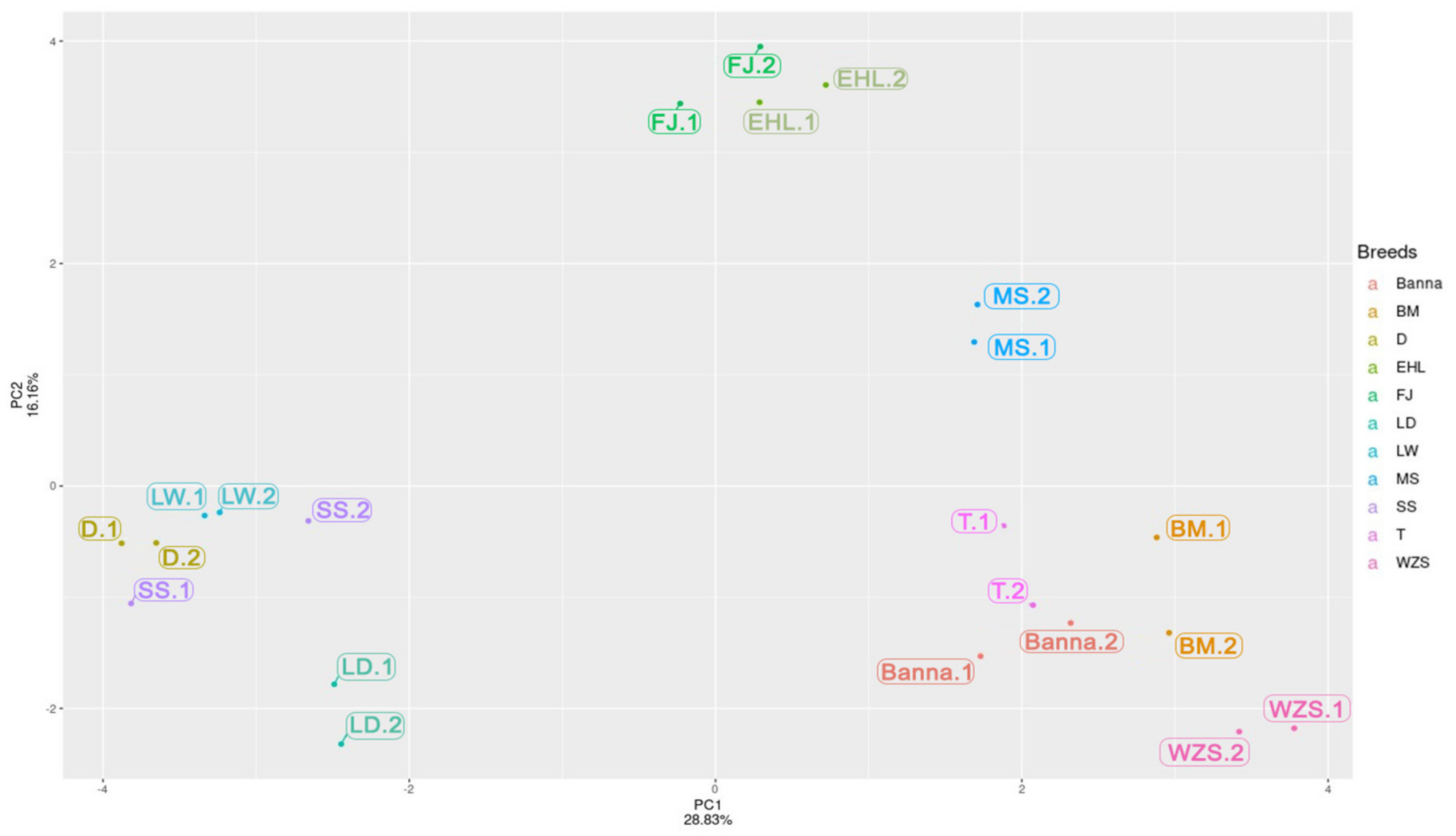
2.6. PCA and Cluster Analysis of the SINE RIPs
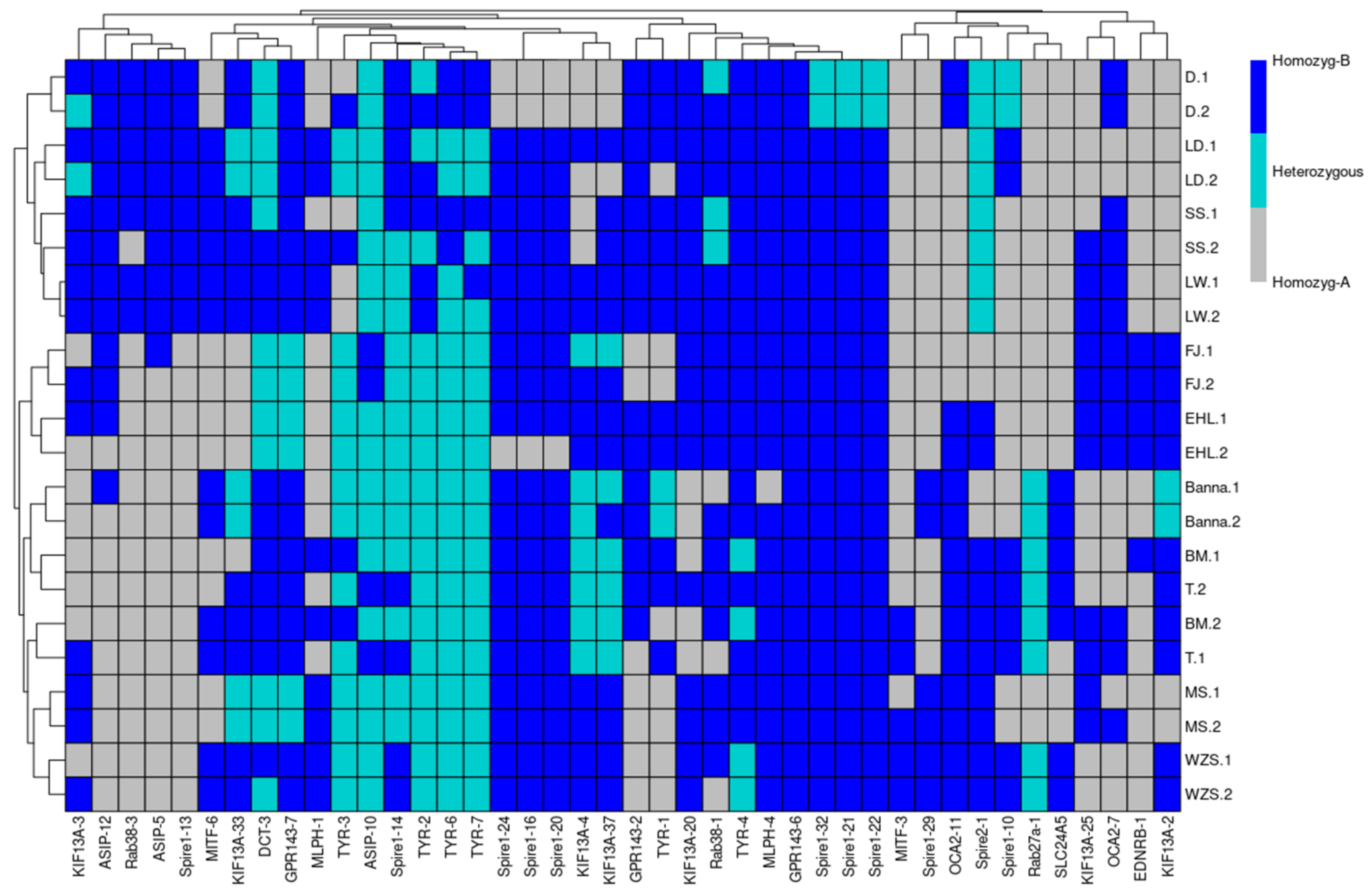
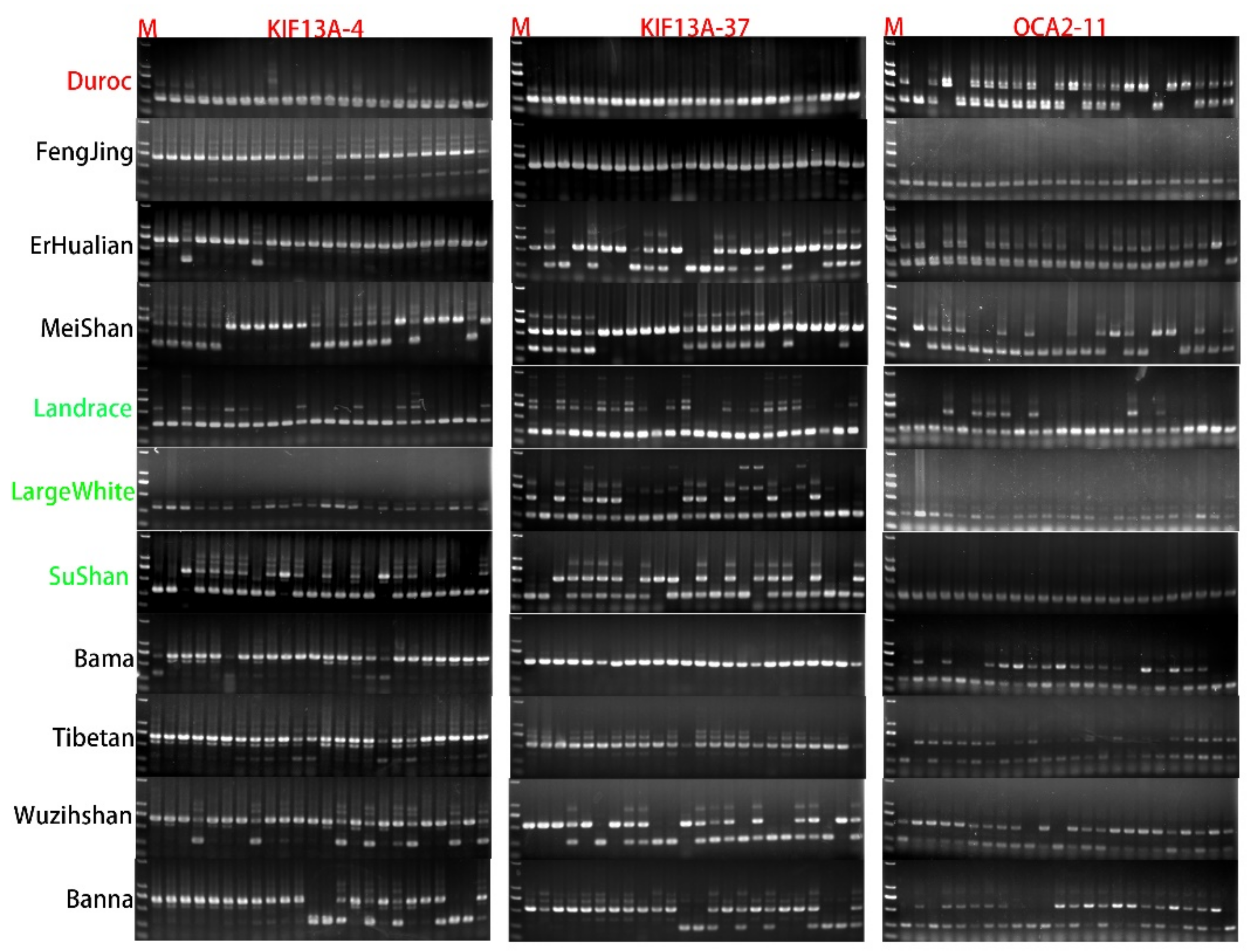
Based on the SINE RIPs identified in this study, the R statistics package (version 3.6.3) was used to generate a presence/absence matrix and perform the PCA analysis. On the same dataset, heatmaps and cluster analysis were computed by the use of the R package heatmap tool (version 1.0.12) [47], using the “Euclidean” distance method for clustering.

3. Results

Coat-Color Genes’ SVs Revelation and Detection by Pool-PCR in Different Pig Breeds

The sequences of 20 coat-color genes and their flanking regions (5 kb-5′upstream and 5 kb-3′downstream, respectively), which tend to contain most regulatory elements [48], were downloaded or reassembled based on the 16 genome sequences deposited in NCBI as described in the methods section. The genomic coordinates of the 20 analyzed coat-color genes and their flanking sequences are summarized in Table S1. Structural variations were identified by multiple alignments, using the ClustalX program for each gene. A total of 167 large structural variations (>50 bp) of coat-color genes, which overlapped with retrotransposon insertions (>50 bp), were designated as putative RIPs and obtained; they are summarized in Table 1 and Table S2.
Then, these predicted RIPs were investigated by the first round pool-PCR using genomic DNA samples from wild pigs (coat color not clear) and eleven domesticated pig breeds, which represented different types of pigs, including definite coat color white breeds: Landrace, LargeWhite and SuShan [49]; black breeds: Fengjing, Erhualian, and Meishan; mini-pig breeds: black (Tibetan and Banna), two-end-black (Bama), and black with white belly and feet (Wuzhishan); red coat color: Duroc. All these domestic breeds are reported in Figure 1. A total of 42 RIPs were confirmed by pool-PCR, with clear polymorphic PCR products across the analyzed DNA samples. The results are shown in Figure 2, and the electrophoresis results and the number format are illustrated in Figures S1 and S2 and Table S3. SVs were not encountered in the gene of MC1R and TYRP1, and among these RIPs’ predictions, 10 and 7 confirmed RIPs of Spire1 and KIF13A respectively, which most results detected by pool-PCR.
Relatively on the PCA results (Figure 3), we selected a light coat color breed of Sushan, Landrace, and LargeWhite as Group 1, selected a full dark coat color breed of Meishan, Fengjing, and Erhualian as Group 2, and selected a mini-size but two-end-black of Bama [50], dark coat color breed of Tibetan and Banna as Group 3 (Figure 1). We compared these three groups together to explore 11 RIP-sites’ polymorphism (the predicted RIP sites information are shown in Table 2) between different breeds for further genotyping distinction work carried out via the second round of PCR detecting in 24 individuals per breed.
The expected three genotypes of retrotransposon insertion polymorphisms were named RIP+/+, RIP+/−, and RIP−/−, respectively. GPR143-7 special presents RIP+/+ in all 24 individuals of Group 2, but other groups present three gene types in different frequencies. SLC24a5-1 and Rab27a-1 special present RIP−/− in all 24 individuals of Group 3, but other groups present three gene types in different frequencies. Spire1-13 and Rab38-3 special present RIP−/− in all 24 individuals of Group 2 and Group 3 but present RIP+/− in Group 1 (Table 3). We also found one or two breed-particular situations, such as OCA2-11 special presents RIP−/− in Fengjing and Sushan but other breeds present RIP+/−; Spire2-1 special presents RIP−/− in Fengjing and Meishan but other breeds present RIP+/−; ASIP-12 special presents RIP−/− in Erhualian, Meishan, Bama, Tibetan, and Wuzhishan but others present RIP+/−; MITF-6 special presents RIP−/− in Duroc, Erhualian, and Meishan but others present RIP+/− (Table 3).
KIF13A-37 special presents RIP+/+ in Fengjing and Bama but presents RIP−/− in others. KIF13A-4 special presents RIP−/− in Duroc and LargeWhite but presents RIP+/− in others. For the gene of KIF13A, we found the RIP site of KIF13A-4 and KIF13A-37 have the same RIP+/− gene type in all 24 individuals of Meishan, Sushan, and Banna, and similarly have a related expression in the genome (Figure 4).

4. Discussion

Nowadays, more researchers turn sight to SV exploration work from plant [36] to animal [51] phenotype and even in human healthy [52]. Particularly, RIPs, as important sources of SVs, play important roles in genomic shaping and phenotype variations [53]. In animals, several cases of the coat color phenotype variations derived from RIPs have been reported. Dong et al. [54] reported a case study in Water Buffaloes (Bubalus bubalis) in which their genomic analysis results revealed the 165 bp of 50 UTR transcribed from the LINE-1 was spliced into the first coding exon of ASIP, resulting in a chimeric transcript. The increased expression of ASIP prevents melanocyte maturation, leading to the absence of pigment in white buffalo skin and hairs. Li et al. [55] reported characterization of the endogenous retrovirus insertion in CYP19A1, associated with henny feathering in chicken. Finally, David et al. [56] found endogenous retrovirus insertion in the kit oncogene which determined the white and white spotting in domestic cats. Here, RIPs in the twenty coat-color genes (Table 1) were mined by comparative genomic protocol and PCR detection in Sus scrofa. Finally, 42 RIPs, which were generated by SINE or ERV insertions, were confirmed by PCR amplification (Table 1). We found that the number of the confirmed RIPs are much less than predicted. Similar result were found in our previous studies [44,57]. We believe that the number of differences between the predicted RIPs and the verified RIPs are due to the assembled levels and quality of genomes, although some genomes were not well-assembled, resulting in false positive RIP prediction. Furthermore, we found most of the confirmed SVs related to SINE insertion or deletion, while ERV only represented one RIP site (Table 2), which is generally agreed with our previous studies, where SINEs are deduced as the major sources of retrotransposon-derived SVs in the pig genome [58], although LINE, LTR, and SINE accounted for 18.52%, 7.56%, and 11.05% of the pig genome, respectively [32].
Based on the pool-PCR detection of RIPs’ result (Table S3) and the Principal Component Analysis (Figure 3), the RIPs that have at least two polymorphic genotypes contribute more to each PC; for example, pool-PCR results revealed that RIP-ASIP-5, RIP-Spire1-13, RIP-ASIP-12, and RIP-Rab38-3 were not inserted in Bama, Wuzhishan, Tibetan, and Banna, which were separated by PC1, but were inserted in Duroc, Landrace, LargeWhite, and SuShan, which were separated by PC2 from FengJing, Erhualian, and Meishan. The PCA result revealed that these breeds can be generally classified into three distinct clusters, which generally agrees with the known genetic background and histories of these populations. One cluster includes the breeds of Fengjing, Erhualian, and Meishan, which are distributed in the Taihu region (a lake in Jiangsu Province) and share the same origin; thus, they are also called as Taihu pigs. Fengjing and Erhualian are pure grey black pigs, while Meishan pigs display grey black in the main body with four white points (foot ends) [59]. The second cluster represents the lean type breeds, including SuShan, Duroc, Landrace, and LargeWhite. The coat color of introduced breeds, such as Landrace and LargeWhite have whole white body, and Duroc is in red or dark red, while the breed of Sushan is a newly developed Chinese breed that contains a 25% genetic component of Chinese Taihu pigs, 25% Landrace pigs, and 50% LargeWhite pigs, which also has a white coat color [48]. The third cluster contains the Chinese minipig breeds of Bama, Tibetan, Wuzhishan, and Banna, with three distinct coat color patterns: Tibetan and Banna have a pure black coat color, Bama has a white main body with the black head and tail, and the coat color of Wuzhishan dominates by black with a white belly bottom and feet.
To identify the potential relationships between breed differences (such as coat color and body size) and these RIPs, eleven typical polymorphic RIP-sites were selected to further detect their genotypic distributions in at least 24 individuals per breed for 11 pig populations. We found each RIP-sites with at least two polymorphic genotypes among 12 pig breeds, and the SVs related to retrotransposon insertion and deletion were consistent between varieties of breeds after we rechecked our 24 individuals’ detection results per breed compared to the predicted RIP-sites information. It supports that our PCR results and predicted strategy are credible. The RIP-sites detection results, illustrated from Figure 4 and Figure S3, revealed that some SVs in those target coat-color genes may be related to the differences between breeds. Then, we speculated that Rab27a-1 and SLC24A5-1 may be related to pig body size due to these two RIP-sites presenting the SVs deletion genotype in Group 1 and Group 2 only but heterozygous genotype in Group 3, which have a smaller body size than others. However, both the RIP-sites of Rab38-3 and Spire1-13 may be related to pig breed coat color due to these two RIP-sites presenting the SVs deletion genotype in Group 2 and Group 3 only but heterozygous genotype in Group 1 for which the coat color is lighter than others. GPR143-7 may be both related to body size and coat color due to this RIP-site presenting the SVs insertion genotype in Group 2 only but heterozygous genotype in Group 1, which has a light coat color, and also a heterozygous genotype in Group 3, which has a smaller body size but dark coat color. In terms of single breed characteristics, we found Spire2-1 and KIF13A-4 present the deletion genotype in the pig breeds of FengJing, Meishan, and LargeWhite, respectively, but other breeds present the heterozygous genotype.
Another interesting finding from the present studies indicated that the RIP-sites of KIF13A-4 and KIF13A-37 present similar polymorphic genotypes in the breed of Meishan and SuShan, for which we have introduced that the SuShan breed is a hybrid from the Taihu breed, Landrace, and Large White. By the sight of pig evolution, we can speculate that this correlation expression in SuShan may come from Meishan.

5. Conclusions

All of the coat-color genes that we chose for this study present an important function in pigmentation. We acquired almost 167 large SVs (>50 bp) target to these genes, which are related to retrotransposon insertion/deletion, and 42 RIPs were really detected in 12 pig breeds. Eleven RIPs that have more than two polymorphic genotypes were chosen to be verified in 24 individuals per 11 breeds to find if these RIPs are special in some breeds.
The findings obtained in this study reveal that all RIPs’ detection results are consistent with our predicted information, and some RIPs, such as Rab27a-1, SLC24A5-1, Rab38-3, Spire1-13, and GPR143-7, may be good molecular markers to distinguish different breeds on pig breeding and reproduction.

Supplementary Materials

The following supporting information can be downloaded at: https://www.mdpi.com/article/10.3390/ani12080969/s1, Figure S1: DNA screening results of color genes SVs in pigs_1_; Figure S2: DNA screening results of color genes SVs in pigs_picture_2_; Figure S3: Small group of detection; Table S1: Assemble information for 20 coat color genes from the 16 pig genomes; Table S2: Hair color genes SVs prediction; Table S3: PCR results convert to numbers.

Author Contributions

Conceptualization, Z.D. and E.D.; methodology, M.W. and Y.Z.; software, X.W.; validation, C.S. and X.W.; formal analysis Z.D. and E.D.; investigation, C.C.; resources, C.S.; data curation, Z.D. and E.D.; writing—original draft preparation, Z.D. and E.D.; writing—review and editing, Z.D. and E.D.; visualization, C.S.; supervision, C.S.; project administration, C.S.; funding acquisition, C.S. All authors have read and agreed to the published version of the manuscript.

Funding

This work was supported through grants from the NSFC (31872977 and 32002146), the Revitalization of Seed Industry (JBGS) in Jiangsu province [JBGS(2021)028)], the Independent Innovation Fund Project of Agricultural Science and Technology in Jiangsu Province [CX(19)2016], the Priority Academic Program Development of Jiangsu Higher Education Institutions, China Postdoctoral Science Foundation [2020M671630] to Cai Chen, and the High-end Talent Support Program of Yangzhou University to Chengyi Song.

Institutional Review Board Statement

The collection of biological samples and experimental procedures involved in this study were approved by the Animal Experiment Ethics Committee of Yangzhou University (No. NSFC2020-dkxy-02, 27 March 2020).

Informed Consent Statement

Not applicable.

Data Availability Statement

All data needed to evaluate the conclusions in this paper are present either in the main text or the Supplementary Materials.

Conflicts of Interest

The authors declare no conflict of interest.

References

  1. Cuthill, I.C.; Allen, W.L.; Arbuckle, K.; Caspers, B.; Chaplin, G.; Hauber, M.E.; Hill, G.E.; Jablonski, N.G.; Jiggins, C.D.; Kelber, A. The biology of color. Science 2017, 357, 6350. [Google Scholar] [CrossRef] [PubMed] [Green Version]
  2. Le, L.; Sirés-Campos, J.; Raposo, G.; Delevoye, C.; Marks, M.S. Melanosome biogenesis in the pigmentation of mammalian skin. Integr. Comp. Biol. 2021, 61, 1517–1545. [Google Scholar] [CrossRef] [PubMed]
  3. Raposo, G.; Marks, M.S. Melanosomes—Dark organelles enlighten endosomal membrane transport. Nat. Rev. Mol. Cell. Biol. 2007, 8, 786–797. [Google Scholar] [CrossRef] [PubMed] [Green Version]
  4. Moreiras, H.; Seabra, M.C.; Barral, D.C. Melanin Transfer in the Epidermis: The Pursuit of Skin Pigmentation Control Mechanisms. Int. J. Mol. Sci. 2021, 22, 4466. [Google Scholar] [CrossRef] [PubMed]
  5. Benito-Martinez, S.; Salavessa, L.; Raposo, G.; Marks, M.S.; Delevoye, C. Melanin transfer and fate within keratinocytes in human skin pigmentation. Integr. Comp. Biol. 2021, 61, 1546–1555. [Google Scholar] [CrossRef]
  6. Wiriyasermkul, P.; Moriyama, S.; Nagamori, S. Membrane transport proteins in melanosomes: Regulation of ions for pigmentation. Biochim. Biophys. Acta (BBA)—Biomembr. 2020, 1862, 183318. [Google Scholar] [CrossRef]
  7. Graham, M.; Tzika, A.C.; Mitchell, S.M.; Liu, X.; Leonhardt, R.M. Repeat domain-associated O-glycans govern PMEL fibrillar sheet architecture. Sci. Rep. 2019, 9, 6101. [Google Scholar] [CrossRef] [Green Version]
  8. Falletta, P.; Bagnato, P.; Bono, M.; Monticone, M.; Schiaffino, M.V.; Bennett, D.C.; Goding, C.R.; Tacchetti, C.; Valetti, C. Melanosome-autonomous regulation of size and number: The OA 1 receptor sustains PMEL expression. Pigment Cell Melanoma Res. 2014, 27, 565–579. [Google Scholar] [CrossRef]
  9. Le, L.; Escobar, I.E.; Ho, T.; Lefkovith, A.J.; Latteri, E.; Haltaufderhyde, K.D.; Dennis, M.K.; Plowright, L.; Sviderskaya, E.V.; Bennett, D.C. SLC45A2 protein stability and regulation of melanosome pH determine melanocyte pigmentation. Mol. Biol. Cell. 2020, 31, 2687–2702. [Google Scholar] [CrossRef]
  10. Yousaf, S.; Sethna, S.; Chaudhary, M.A.; Shaikh, R.S.; Riazuddin, S.; Ahmed, Z.M. Molecular characterization of SLC24A5 variants and evaluation of Nitisinone treatment efficacy in a zebrafish model of OCA6. Pigment Cell Melanoma Res. 2020, 33, 556–565. [Google Scholar] [CrossRef]
  11. Fukuda, M. Rab GTPases: Key players in melanosome biogenesis, transport, and transfer. Pigment Cell Melanoma Res. 2021, 34, 222–235. [Google Scholar] [CrossRef]
  12. Alzahofi, N.; Welz, T.; Robinson, C.L.; Page, E.L.; Briggs, D.A.; Stainthorp, A.K.; Reekes, J.; Elbe, D.A.; Straub, F.; Kallemeijn, W.W. Rab27a co-ordinates actin-dependent transport by controlling organelle-associated motors and track assembly proteins. Nat. Commun. 2020, 11, 3495. [Google Scholar] [CrossRef]
  13. Mort, R.L.; Jackson, I.J.; Patton, E.E. The melanocyte lineage in development and disease. Development 2015, 142, 620–632. [Google Scholar] [CrossRef] [Green Version]
  14. Pennamen, P.; Le, L.; Tingaud-Sequeira, A.; Fiore, M.; Bauters, A.; Béatrice, N.V.D.; Coste, V.; Bordet, J.; Plaisant, C.; Diallo, M. BLOC1S5 pathogenic variants cause a new type of Hermansky–Pudlak syndrome. Genet. Med. 2020, 22, 1613–1622. [Google Scholar] [CrossRef]
  15. Xu, X.; Dong, G.; Schmidt-Küntzel, A.; Zhang, X.; Zhuang, Y.; Fang, R.; Sun, X.; Hu, X.; Zhang, T.; Yang, H. The genetics of tiger pelage color variations. Cell Res. 2017, 27, 954–957. [Google Scholar] [CrossRef] [Green Version]
  16. Ludwig, A.; Pruvost, M.; Reissmann, M.; Benecke, N.; Brockmann, G.A.; Castaños, P.; Cieslak, M.; Lippold, S.; Llorente, L.; Malaspinas, A. Coat color variation at the beginning of horse domestication. Science 2009, 324, 485. [Google Scholar] [CrossRef] [Green Version]
  17. Corbin, L.J.; Pope, J.; Sanson, J.; Antczak, D.F.; Miller, D.; Sadeghi, R.; Brooks, S.A. An Independent Locus Upstream of ASIP Controls Variation in the Shade of the Bay Coat Colour in Horses. Genes 2020, 11, 606. [Google Scholar] [CrossRef]
  18. Hédan, B.; Cadieu, E.; Botherel, N.; Dufaure de Citres, C.; Letko, A.; Rimbault, M.; Drögemüller, C.; Jagannathan, V.; Derrien, T.; Schmutz, S. Identification of a missense variant in MFSD12 involved in dilution of Phaeomelanin leading to white or cream coat color in dogs. Genes 2019, 10, 386. [Google Scholar] [CrossRef] [Green Version]
  19. Schmutz, S.M. Genetics of coat color in cattle. In Bovine Genomics; Wiley Publication: Hoboken, NJ, USA, 2012; pp. 20–33. [Google Scholar]
  20. Trigo, B.B.; Utsunomiya, A.T.H.; Fortunato, A.A.; Milanesi, M.; Torrecilha, R.B.P.; Lamb, H.; Nguyen, L.; Ross, E.M.; Hayes, B.; Padula, R.C.M. Variants at the ASIP locus contribute to coat color darkening in Nellore cattle. Genet. Sel. Evol. 2021, 53, 40. [Google Scholar] [CrossRef]
  21. Demars, J.; Iannuccelli, N.; Utzeri, V.J.; Auvinet, G.; Riquet, J.; Fontanesi, L.; Allain, D. New insights into the melanophilin (MLPH) gene affecting coat color dilution in rabbits. Genes 2018, 9, 430. [Google Scholar] [CrossRef] [Green Version]
  22. Manakhov, A.D.; Andreeva, T.V.; Trapezov, O.V.; Kolchanov, N.A.; Rogaev, E.I. Genome analysis identifies the mutant genes for common industrial Silverblue and Hedlund white coat colours in American mink. Sci. Rep. 2019, 9, 4581. [Google Scholar] [CrossRef] [Green Version]
  23. Johnson, J.L.; Kozysa, A.; Kharlamova, A.V.; Gulevich, R.G.; Perelman, P.L.; Fong, H.; Vladimirova, A.V.; Oskina, I.N.; Trut, L.N.; Kukekova, A.V. Platinum coat color in red fox (Vulpes vulpes) is caused by a mutation in an autosomal copy of KIT. Anim. Genet. 2015, 46, 190–199. [Google Scholar] [CrossRef]
  24. Du, Z.; Huang, K.; Zhao, J.; Song, X.; Xing, X.; Wu, Q.; Zhang, L.; Xu, C. Comparative transcriptome analysis of raccoon dog skin to determine melanin content in hair and melanin distribution in skin. Sci. Rep. 2017, 7, 40903. [Google Scholar] [CrossRef]
  25. Kijas, J.; Wales, R.; Törnsten, A.; Chardon, P.; Moller, M.; Andersson, L. Melanocortin receptor 1 (MC1R) mutations and coat color in pigs. Genetics 1998, 150, 1177–1185. [Google Scholar] [CrossRef] [PubMed]
  26. Wu, Z.; Deng, Z.; Huang, M.; Hou, Y.; Zhang, H.; Chen, H.; Ren, J. Whole-genome resequencing identifies KIT new alleles that affect coat color phenotypes in pigs. Front. Genet. 2019, 10, 218. [Google Scholar] [CrossRef] [PubMed]
  27. Liu, Y.; Li, F.; Kong, X.; Tan, B.; Li, Y.; Duan, Y.; François, B.; Chien-An, A.; Yin, Y. Signaling pathways related to protein synthesis and amino acid concentration in pig skeletal muscles depend on the dietary protein level, genotype and developmental stages. PLoS ONE 2015, 10, e0138277. [Google Scholar] [CrossRef] [PubMed]
  28. Whitelaw, E.; Martin, D.I.K. Retrotransposons as epigenetic mediators of phenotypic variation in mammals. Nat. Genet. 2001, 27, 361–365. [Google Scholar] [CrossRef] [PubMed]
  29. Chuong, E.B.; Elde, N.C.; Feschotte, C. Regulatory activities of transposable elements: From conflicts to benefits. Nat. Rev. Genet. 2017, 18, 71–86. [Google Scholar] [CrossRef] [Green Version]
  30. Blumenstiel, J.P. Birth, school, work, death, and resurrection: The life stages and dynamics of transposable element proliferation. Genes 2019, 10, 336. [Google Scholar] [CrossRef] [Green Version]
  31. Platt, R.N.; Vandewege, M.W.; Ray, D.A. Mammalian transposable elements and their impacts on genome evolution. Chromosom. Res. 2018, 26, 25–43. [Google Scholar] [CrossRef] [Green Version]
  32. Chen, C.; Wang, W.; Wang, X.; Shen, D.; Wang, S.; Wang, Y.; Gao, B.; Wimmers, K.; Mao, J.; Li, K. Retrotransposons evolution and impact on lncRNA and protein coding genes in pigs. Mob. DNA 2019, 10, 19. [Google Scholar] [CrossRef]
  33. McClintock, B. The origin and behavior of mutable loci in maize. Proc. Natl. Acad. Sci. USA 1950, 36, 344–355. [Google Scholar] [CrossRef] [Green Version]
  34. Lancíková, V.; Žiarovská, J. Inter-retrotransposon amplified polymorphism markers revealed long terminal repeat retrotransposon insertion polymorphism in flax cultivated on the experimental fields around Chernobyl. J. Environ. Sci. Health Part. A 2020, 55, 957–963. [Google Scholar] [CrossRef]
  35. Hirata, C.; Waki, T.; Shimomura, K.; Wada, T.; Tanaka, S.; Ikegami, H.; Uchimura, Y.; Hirashima, K.; Nakazawa, Y.; Okada, K.; et al. DNA markers based on retrotransposon insertion polymorphisms can detect short DNA fragments for strawberry cultivar identification. Breed. Sci. 2020, 70, 19116. [Google Scholar] [CrossRef] [Green Version]
  36. Meng, Y.; Su, W.; Ma, Y.; Liu, L.; Gu, X.; Wu, D.; Shu, X.; Lai, Q.; Tang, Y.; Wu, L.; et al. Assessment of genetic diversity and variety identification based on developed retrotransposon-based insertion polymorphism (RBIP) markers in sweet potato (Ipomoea batatas (L.) Lam.). Sci. Rep. 2021, 11, 17116. [Google Scholar] [CrossRef]
  37. Pelsy, F.; Bevilacqua, L.; Blanc, S.; Merdinoglu, D. A molecular marker set combining a retrotransposon insertion and SSR polymorphisms is useful for assessing diversity in Vitis. OENO One 2021, 55, 403–414. [Google Scholar] [CrossRef]
  38. Borges-Monroy, R.; Chu, C.; Dias, C.; Choi, J.; Lee, S.; Gao, Y.; Shin, T.; Park, P.J.; Walsh, C.A.; Eunjung Alice Lee, E.A. Whole-genome analysis of de novo and polymorphic retrotransposon insertions in Autism Spectrum Disorder. bioRxiv 2021. [Google Scholar] [CrossRef]
  39. Kalla, S.E.; Moghadam, H.K.; Tomlinson, M.; Seebald, A.; Allen, J.J.; Whitney, J.; Choi, J.D.; Sutter, N.B. Polymorphic SINEC_Cf Retrotransposons in the Genome of the Dog (Canis familiaris). bioRxiv 2020. [Google Scholar] [CrossRef]
  40. Choi, J.D.; Del Pinto, L.A.; Sutter, N.B. SINE Retrotransposons Import Polyadenylation Signals to 3′UTRs in Dog (Canis familiaris). bioRxiv 2020. [Google Scholar] [CrossRef]
  41. Ruggiero, R.P.; Bourgeois, Y.; Boissinot, S. LINE insertion polymorphisms are abundant but at low frequencies across populations of Anolis carolinensis. Front. Genet. 2017, 8, 44. [Google Scholar] [CrossRef] [Green Version]
  42. Wang, W.; Chen, C.; Wang, X.; Zhang, L.; Shen, D.; Wang, S.; Gao, B.; Mao, J.; Song, C. Development of Molecular Markers Based on the L1 Retrotransposon Insertion Polymorphisms in Pigs (Sus scrofa) and Their Association with Economic Traits. Russ. J. Genet. 2020, 56, 183–191. [Google Scholar] [CrossRef]
  43. Zheng, Y.; Cai, C.; Wei, C.; Wang, X.; Wei, W.; Bo, G.; Wimmers, K.; Mao, J.; Song, C. Two new SINE insertion polymorphisms in pig Vertnin (VRTN) gene revealed by comparative genomic alignment. J. Integ. Agri. 2020, 19, 2514–2522. [Google Scholar] [CrossRef]
  44. Chen, C.; Zheng, Y.; Wang, M.; Murani, E.; D’Alessandro, E.; Moawad, A.S.; Wang, X.; Wimmers, K.; Song, C. SINE Insertion in the Intron of Pig GHR May Decrease Its Expression by Acting as a Repressor. Animals 2021, 11, 1871. [Google Scholar] [CrossRef] [PubMed]
  45. Larkin, M.A.; Blackshields, G.; Brown, N.P.; Chenna, R.; McGettigan, P.A.; McWilliam, H.; Valentin, F.; Wallace, I.M.; Wilm, A.; Lopez, R. Clustal W and Clustal X version 2.0. Bioinformatics 2007, 23, 2947–2948. [Google Scholar] [CrossRef] [PubMed] [Green Version]
  46. Nishimura, D. Repeat Masker. Biotech. Softw. Internet Rep. 2000, 1, 36–39. [Google Scholar]
  47. Kolde, R. Pheatmap: Pretty Heatmaps, R Packag v. 16; R Foundation for Statistical Computing: Nashville, TN, USA, 2012. [Google Scholar]
  48. Zhang, Z.D.; Paccanaro, A.; Fu, Y.; Weussnab, S.; Weng, Z.; Chang, J.; Snyder, M.; Merk, B. Statistical analysis of the genomic distribution and correlation of regulatory elements in the ENCODE regions. Genome Res. 2007, 17, 787–797. [Google Scholar] [CrossRef] [Green Version]
  49. Zhou, L.; Zhao, W.; Fu, Y.; Fang, X.; Ren, S.; Ren, J. Genome-wide detection of genetic loci and candidate genes for teat number and body conformation traits at birth in Chinese Sushan pigs. Anim. Genet. 2019, 50, 753–756. [Google Scholar] [CrossRef]
  50. Jia, Q.; Cao, C.; Tang, H.; Ying, Z.; Qian, Z.; Xiao, W.; Rui, Z.; Xiao, W.; Ai, L.; Hong, W.; et al. A 2-bp insertion (c. 67_68insCC) in MC1R causes recessive white coat color in Bama miniature pigs. J. Genet. Genom. 2017, 44, 215–217. [Google Scholar] [CrossRef]
  51. Weissensteiner, M.H.; Bunikis, I.; Catalán, A.; Francoijs, K.J.; Knief, U.; Heim, W.; Peona, V.; Pophaly, S.D.; Sedlazeck, F.J.; Suh, A.; et al. Discovery and population genomics of structural variation in a songbird genus. Nat. Commun. 2020, 11, 3403. [Google Scholar] [CrossRef]
  52. Chiang, C.; Scott, A.J.; Davis, J.R.; Tsang, E.K.; Li, X.; Kim, Y.; Hadzic, T.; Damani, F.N.; Ganel, L.; GTEx Consortium; et al. The impact of structural variation on human gene expression. Nat. Genet. 2017, 49, 692–699. [Google Scholar] [CrossRef] [Green Version]
  53. Yang, N.; Zhao, B.; Chen, Y.; D’Alessandro, E.; Chen, C.; Ji, T.; Wu, X.; Song, C. Distinct retrotransposon evolution profile in the genome of rabbit (Oryctolagus cuniculus). Genome Biol. Evol. 2021, 13, evab168. [Google Scholar]
  54. Liang, D.; Zhao, P.; Si, J.; Fang, L.; Pairo-Castineira, E.; Hu, X.; Xu, Q.; Hou, Y.; Gong, Y.; Liang, Z.; et al. Genomic analysis revealed a convergent evolution of LINE-1 in coat color: A case study in water buffaloes (Bubalus bubalis). Mol. Biol. Evol. 2021, 38, 1122–1136. [Google Scholar] [CrossRef]
  55. Li, J.; Davis, B.W.; Jern, P.; Dorshorst, B.J.; Siegel, P.B.; Andersson, L. Characterization of the endogenous retrovirus insertion in CYP19A1 associated with henny feathering in chicken. Mob. DNA 2019, 10, 38. [Google Scholar] [CrossRef]
  56. David, V.A.; Menotti-Raymond, M.; Wallace, A.C.; Roelke, M.; Kehler, J.; Leighty, R.; Eizirik, E.; Hannah, S.S.; Nelson, G.; Schäffer, A.A.; et al. Endogenous retrovirus insertion in the KIT oncogene determines white and white spotting in domestic cats. G3 Genes. Genomes Genet. 2014, 4, 1881–1891. [Google Scholar] [CrossRef] [Green Version]
  57. Wang, X.Y.; Chen, Z.X.; Murani, E.; D’Alessamdro, E.; An, Y.L.; Chen, C.; Li, K.; Galeano, G.; Wimmers, K.; Song, C.Y. A 192 bp ERV fragment insertion in the first intron of porcine TLR6 may act as an enhancer associated with the increased expressions of TLR6 and TLR1. Mob. DNA 2021, 12, 20. [Google Scholar] [CrossRef]
  58. Chen, C.; D’Alessandro, E.; Murani, E.; Zheng, Y.; Domenico, G.; Yang, N.S.; Wang, X.Y.; Gao, B.; Li, K.; Wimmer, K.; et al. SINE jumping contributes to large-scale polymorphisms in the pig genomes. Mob. DNA 2021, 12, 17. [Google Scholar] [CrossRef]
  59. Margeta, V.; Budimir, K.; Kralik, G.; Radišić, Ž. Genetic basis of pig coat color. Krmiva Časopis Hranidbi Zivotinj. Proizv. Tehnol. Krme 2011, 53, 85–92. [Google Scholar]
Figure 1. Different coat-color pig breeds. In Group-1, from top to bottom is Duroc, Landrace, Large White, and SuShan, respectively. In Group-2, from the top down is FengJing, Erhualian, and Meishan, respectively. In Group-3, from top to bottom is Bama, Tibetan, Wuzhishan, and Banna, respectively.
Animals 12 00969 g001
Figure 2. The pool-PCR results shown by heatmap. D (Duroc), LD (Landrace), LW (LargeWhite), SS (SuShan), EHL (Erhualian), FJ (Fengjing), Banna (Banna), BM (Bama), T (Tibetan), MS (Meishan), and WZS (Wuzhishan).
Animals 12 00969 g002
Figure 3. Principal Component Analysis (PCA) based on the results of the pool-PCR result.
Animals 12 00969 g003
Figure 4. RIP-sites of KIF13A-4, KIF13A-37, and OCA2-11 detection in different small group pig breeds by PCR. M: Marker DL2000.
Animals 12 00969 g004
Table 1. Predicted large structural variations (SVs) by alignment in coat-color genes and their flanking regions.
Gene NamePredicted RIPsConfirmed RIPs
ASIP142
KIT80
MITF122
PMEL10
GPR14373
TYR76
EDNRB21
TYRP100
MLPH113
DCT31
OCA2102
SLC24A511
SLC45A290
Rab27a61
Rab3220
Rab3842
KIF13A377
Spire231
Spire13010
MC1R00
Total Numbers16742
Table 2. The predicted RIP-sites’ information, which can be detected in different small group pig breeds.
Rip-SitesInsertion BreedsDeletion BreedsChrBeginEndGene Struture TE-Type Length (bp)
ASIP-12Duroc, BerkshireThe rest of the species1737,614,91437,615,246Intron-1SINEA332
GPR143-7The rest of the speciesLandrace, LargeWhite, Crossbred, Wuzhishan, BamaX6,248,7836,249,085Intron-9SINEA300
MITF-6Largewhite, BerkshireThe rest of the species1351,175,50451,175,505Intron-1SINEB94
KIF13A-4The rest of the speciesBerkshire, Crossbred, LargeWhite,
D-Ninghe, Hampshire, Landrace, Pietrain
713,691,83613,692,190Intron-1SINEA354
KIF13A-37Bamei, Wuzhishan, Bama, Tibetan, RongchangThe rest of the species713,497,15513,497,1563′flankERV III282
OCA2-11Wuzhishan, Jinhua, Rongchang, HampshireThe rest of the species1556,806,80456,806,805Intron-2SINEA339
Rab27a-1Bama, Tibetan, BameiThe rest of the species1116,538,879116,538,880Intron-1SINEA326
Rab38-3LargeWhite, Landrace, Hampshire, BerkshireThe rest of the species921,583,69021,583,691Intron-2SINEA293
SLC24A5-1BamaThe rest of the species1123,633,504123,633,5053′flankSINEA310
Spire1-13The rest of the speciesBamei, MS, Landrace696,966,83896,967,753Intron-2SINEA370
Spire2-1Duroc, Tibetan, Largewhite, D-Ninghe, Crossbred, PietrainThe rest of the species6248,403248,9805′flankSINEA577
Table 3. The result of eleven RIPs detected in eleven breeds, transformed based on the gel picture.
Asia-12GPR143-7MITF-6KIF13A-4KIF13A-37OCA2-11Rab27a-1Rab38SLC24A5-1Spire1-13Spire2-1
Duro+/+65000605000
+/−12170501401302324
−/−6224192442462410
Fengjing+/+1240024000000
+/−14024240000000
−/−90000242424242424
ErHualian+/+0240229100005
+/−00021120000017
−/−24024043242424242
Meishan+/+02401111400000
+/−00013131000000
−/−2402400102424242424
Landrace+/+79700003000
+/−1515101216701702024
−/−207128172442440
LargeWhite+/+361600008000
+/−6080162401602224
−/−1518024802402420
SuShan+/+4516330011000
+/−111381212001101424
−/−96099242422470
Bama+/+00319240130400
+/−0845017110707
−/−241617007024132417
Tibetan+/+040000130001
+/−01822241823407015
−/−242206172414248
Wuzhishan+/+0421570180001
+/−0143910223016012
−/−2461907232482411
Banna+/+06116150180100
+/−41274518508010
−/−20616446124152414
Note: N = 24; “+/+” means Homozygous insert in pig genome; “+/−” means heterozygous insert in pig genome; “−/−” means no insert in pig genome.
Publisher’s Note: MDPI stays neutral with regard to jurisdictional claims in published maps and institutional affiliations.

