Article

Impact of a Blend of Microencapsulated Organic Acids and Botanicals on the Microbiome of Commercial Broiler Breeders under Clinical Necrotic Enteritis

1
Animal Science Department, University of Wyoming, 1000 E University Ave., Laramie, WY 82071, USA
2
U.S. Department of Agriculture, Agricultural Research Service, Southern Plains Agricultural Research Center, College Station, TX 77845, USA
3
College of Agricultural and Life Sciences, University of Wisconsin, 1933 Observatory Dr., Madison, WI 53706, USA
4
DIMEVET, Department of Veterinary Medical Sciences, University of Bologna, Via Tolara di Sopra 50, Ozzano Emilia, 40064 Bologna, Italy
5
Vetagro S.p.A., Via Porro 2, 42124 Reggio Emilia, Italy
6
Vetagro Inc., 17 East Monroe St. Suite 179, Chicago, IL 60603, USA
*
Author to whom correspondence should be addressed.
Animals 2023, 13(10), 1627; https://doi.org/10.3390/ani13101627
Submission received: 29 March 2023 / Revised: 3 May 2023 / Accepted: 10 May 2023 / Published: 12 May 2023
(This article belongs to the Section Poultry)

Abstract

:

Simple Summary

Necrotic enteritis, a secondary gastrointestinal (GIT) infection in poultry, is a costly burden to the commercialized poultry industry, impacting the welfare of broiler chickens as well as production gains. Many feed supplements have been proposed to alleviate the burden of necrotic enteritis in poultry with varied success. A promising supplement, a microencapsulated blend of organic acids and botanicals, has been demonstrated to modulate the immune system and the gastrointestinal microbial ecology in healthy birds. Therefore, we aimed to test this supplement in the feed of broiler breeder chickens infected with necrotic enteritis. To determine if the supplement improved the microbial ecology of the GIT, where necrotic enteritis resides, the GIT microbiota were elucidated using microbiome sequencing. There were minimal differences in the microbiota diversity; however, the core microorganisms of birds fed the supplement consisted of Lactobacillus and Clostridiaceae. The supplemented-fed birds also had a higher abundance of normal microbiome inhabitants, namely, Actinobacteriota, Bacteroidota, and Verrucomicrobiota, than those not fed the supplement. Therefore, the supplementation of a microencapsulated blend supported beneficial and core microorganisms, potentially improving the bird’s response to NE.

Abstract

Previously, the supplementation of a microencapsulated blend of organic acids and botanicals improved the health and performance of broiler breeders under non-challenged conditions. This study aimed to determine if the microencapsulated blend impacted dysbiosis and necrotic enteritis (NE) in broiler breeders. Day-of-hatch chicks were assigned to non-challenge and challenge groups, provided a basal diet supplemented with 0 or 500 g/MT of the blend, and subjected to a laboratory model for NE. On d 20–21, jejunum/ileum content were collected for microbiome sequencing (n = 10; V4 region of 16S rRNA gene). The experiment was repeated (n = 3), and data were analyzed in QIIME2 and R. Alpha and beta diversity, core microbiome, and compositional differences were determined (significance at p ≤ 0.05; Q ≤ 0.05). There was no difference between richness and evenness of those fed diets containing 0 and 500 g/MT microencapsulated blend, but differences were seen between the non-challenged and challenged groups. Beta diversity of the 0 and 500 g/MT non-challenged groups differed, but no differences existed between the NE-challenged groups. The core microbiome of those fed 500 g/MT similarly consisted of Lactobacillus and Clostridiaceae. Furthermore, challenged birds fed diets containing 500 g/MT had a higher abundance of significantly different phyla, namely, Actinobacteriota, Bacteroidota, and Verrucomicrobiota, than the 0 g/MT challenged group. Dietary supplementation of a microencapsulated blend shifted the microbiome by supporting beneficial and core taxa.

1. Introduction

Necrotic enteritis (NE), often a secondary infection to coccidiosis (Eimeria spp.) caused by the opportunistic pathogen Clostridium perfringens [1], was estimated to globally cost the poultry industry USD 2 billion annually [2]. However, with the removal of antibiotic growth promoters (AGPs) from commercial poultry diets [3], there has been an associated increase in the incidence of NE in broiler flocks [4,5]. As such, more recent estimations (2015) of the economic burden of NE are closer to USD 6 billion annually across the globe [6].
The use of AGPs effectively reduced the incidence of NE by limiting the prevalence of coccidiosis and manipulating the intestinal microbiome [7,8]. Effective AGP alternatives would need to elicit similar responses. Alternative feed additives such as probiotics, phytogenics (essential oils, plant-derived compounds), and organic acids are promising candidates [1]. However, these alternatives’ efficacy in mitigating NE is inconsistent and merits further investigation [9]. These inconsistencies could result from numerous factors such as inclusion level, mode of action, or the location where the alternative is available to the host [1].
Microencapsulation using a lipid matrix is a promising technology that could allow for the improved efficacy of feed supplements. In recent years, with the advancement in technology, the microencapsulation of feed additives has allowed additives to bypass the crop and gizzard, reaching the lower small intestines [10], where nutrient digestion occurs [11,12]. As such, recent research investigating the efficacy of AviPlus® P (Vetagro S.p.A., Reggio Emilia, Italy), a microencapsulated blend of organic acids (25% citric and 16.7% sorbic) and botanicals (1.7% thymol and 1% vanillin), has demonstrated promising results on modulating the gastrointestinal tract (GIT) of broilers [13] and improving intestinal integrity and the inflammation of swine [14]. In addition, during a clinical infection model of NE, supplementation with the microencapsulated blend of organic acids and botanicals resulted in a decrease in mortality (p = 0.004), lower lesion scores (p = 0.006), and tended to improve body weight at d 20–21 (p = 0.19) compared to the control-fed birds by modulating the T cell receptor, TNF, and NF-kB signaling pathways and subsequent cytokine responses [15].
In a continuation with the efforts to determine the efficacy of a microencapsulated blend of organic acids and botanicals (AviPlus® P) as an alternative feed amendment [13,14,15,16,17], the objective of the current study was to: (1) elucidate the microbiome response to a microencapsulated blend of organic acids and botanicals in the lower small intestines during a clinical manifestation of NE; (2) describe the relationship of NE severity with the microbiome diversity and richness in the lower GIT, with and without the supplementation of a microencapsulated blend of organic acids and botanicals in the diet; and (3) determine the core microbiome of the lower GIT in response to the supplementation of a microencapsulated blend of organic acids and botanicals. Therefore, broiler breeders were infected with Eimeria spp. (d 14) and, subsequently, with Clostridium perfringens (d 17–19) to induce NE. On d 20 to 21, the digesta from the lower small intestines was collected for microbiome analyses via sequencing the V4 region of the 16S rRNA gene.

2. Materials and Methods

2.1. Husbandry

The current experiment was repeated three independent times (trials = 3), with chicks being sourced from different flocks for each trial to ensure the reproducibility of results. At the onset of each trial, on day-of-hatch, by-product male broiler breeder chicks (Gallus gallus) were sourced from a commercial hatchery, placed in floor pens (3 × 3 m) with fresh pine shavings, and provided feed, water, and supplemental heat ad libitum. Male chicks were randomly assigned to pens (n = 23–26; one pen/treatment/experimental replicate) and provided a control-basal diet or a supplemented basal diet. The basal diet, an unmedicated corn and soybean meal starter diet, was formulated according to industry standards to meet or exceed the established nutrient requirements [18]. The supplemented diet contained 500 g/MT of a microencapsulated blend of citric (25%) and sorbic (16.7%) acids, thymol (1.7%), and vanillin (1.0%) (AviPlus® P, Vetagro S.p.A., Reggio Emilia, Italy). From the onset of the experiments, no medication or other therapeutic interventions were administered. In accordance with the Guide for the Care and Use of Agricultural Animals in Research and Training [19], the care and handling of poultry was ethically conducted in the presence of an on-site veterinarian. All bird studies were under the approved experimental procedures outlined in protocol number 2019-002 and were approved by the USDA/ARS Institutional Animal Care and Use Committee operating under the Animal and Plant Health Inspection Service establishment number 334299.

2.2. Clostridium Perfringens Preparation

In all independent trials, a total of four field isolates of wild-type C. perfringens (type A) were obtained from active and confirmed outbreaks of NE in Georgia (two isolates), Texas (one isolate), and Virginia (one isolate). Prior to the onset of the experiment, frozen-stock isolates were cultured separately in thioglycollate broth (Becton Dickinson Co., Sparks, MD, USA) for 12 h anaerobically. Fresh cultures were then combined to yield the challenge stock, as previously described [20].

2.3. Necrotic Enteritis Model

On d 14, chicks were orally administered a 2× dose of a commercially available coccidiosis vaccine (1 mL, Coccivac®-B52; Merck Animal Health, Kenilworth, NJ, USA). On d 17–19, birds were additionally challenged orally (3 mL) with a stock culture containing the cocktail of the four C. perfringens strains (107 colony forming units (CFU)/mL) to induce NE. For all three experimental replicates, on d 20 or 21, chickens were euthanized by cervical dislocation and necropsied, where birds were scored for intestinal lesions, and the digesta of the jejunum/ileum was collected for microbiome sequencing (see next section for additional details). The aseptically collected digesta samples (6–10/group) were then shipped on dry ice to the Center for Food Safety at the University of Arkansas (Fayetteville, AR, USA) to prepare a DNA library for microbiome sequencing. Upon arrival at the Center for Food Safety, samples were stored at −20 °C until DNA extraction could be conducted.

2.4. Scoring Necrotic Enteritis Lesions

As described by Prescott, NE severity was evaluated by scoring gross lesions of the jejunum and ileum of the small intestines [21]. Regardless of the trial (n = 3), one person blindly scored the intestines to reduce bias across the experiment, and these data are published [15], but, for clarity, the scale is also provided herein. Lesions were scored on a scale ranging from 0 to 4: (0) normal healthy tissue with no gross lesions; (1) thin-walled or fragile tissue with ashen appearance; (2) thin-walled, focal necrosis, grey in appearance with small gas production; (3) thin-walled, considerable patches of necrosis, prolific gas production within intestines, small areas of blood; (4) acute widespread necrosis, pronounced hemorrhages and vast gas production.

2.5. DNA Extraction

The digesta samples were allowed to thaw on wet ice, and 200 mg of the digesta was aliquoted to a sterile microcentrifuge tube. The DNA of the 200 mg was then extracted according to the standard protocol of the QIAamp Fast DNA Stool Mini Kit and eluted in 100 μL of AE Buffer (Qiagen, Hilden, Germany). The DNA concentration was determined using a Nanodrop™ 1000 spectrophotometer (Thermo Fisher Scientific, Waltham, MA, USA). Samples with DNA concentrations under 15 ng/μL were not diluted; however, those exceeding 15 ng/μL were diluted to 10 ng/μL in Buffer AE (Qiagen, Hilden, Germany).

2.6. Library Preparation

Using dual-indexed primers, including unique eight nucleotide barcode sequences, developed by Kozich et al. [22], the V4 region of the 16S region was amplified using a high-fidelity polymerase (Accuprime Pfx DNA polymerase, Thermo Fisher Scientific, Waltham, MA, USA). Amplified products were verified using gel electrophoresis. Verified amplified products were normalized to equimolar and equal volumes (20 μL) using a SequalPrep™ Normalization kit (Life Technologies, Carlsbad, CA, USA). Following this, 5 μL of the normalized samples was aliquoted to one microcentrifuge tube (pool) to create the final library. Library concentrations were determined using both a KAPA library quantification kit for Illumina platforms (Kapa Biosystems, Inc., Wilmington, MA, USA) and with a Qubit 4 fluorometer using a 1× High Specificity Assay Kit (Invitrogen, Carlsbad, CA, USA). Amplicon product size was verified and assessed using an Agilent 2100 Bioanalyzer (Agilent, Santa Clara, CA, USA). The library and PhiX Control v3 (Illumina, San Diego, CA, USA) were diluted to 20 nM in HyB buffer and denatured in 0.2 N of fresh NaOH for a final concentration of 6 pM. The diluted library was then combined with PhiX for a final PhiX concentration of 20% (wt/vol), loaded into a MiSeq V2 500 cycle sequencing cartridge, and sequenced on an Illumina MiSeq (Illumina, San Diego, CA, USA). Resulting sequences (fastq files) were downloaded from Illumina BaseSpace and uploaded to the NCBI Sequence Read Archive (PRJNA899529) and GitHub https://github.com/RickeLab-UW/Microencapsulated-Blend-on-Broiler-Intestinal-Microbiota (accessed on 3 May 2023).

2.7. Bioinformatic Analyses

Raw sequencing data were demultiplexed and downloaded from Illumina BaseSpace (Illumina, San Diego, CA, USA). Demultiplexed sequences were uploaded into QIIME2 (quantitative insights into microbial ecology; 2021.11) using the paired-end demultiplexed version of Casava 1.8 (via QIIME 2 tools import) [23]. Imported demultiplexed sequences were then filtered and denoised using DADA2 (divisive amplicon denoising algorithm) (via q2-dada2) [24]. The ASVs were assigned using mafft with a rooted phylogenetic tree being produced using fasttee2 (via q2-phylogeny) [25]. Prior to taxonomic identification, the ASVs identified as mitochondria and chloroplast were filtered from the tables and sequences using taxonomy-based filtering via q2-taxa. They were, subsequently, excluded from all statistical analyses. Due to the evolution of mitochondria and chloroplast from bacterial origins, these eukaryotes contained 16S rRNA, and the subsequent sequences must have been removed prior to the interpretation of bacterial populations.
The treatments (NE infection and dietary supplementation) were applied as a 2 × 2 factorial designs; however, due to the limitations of advanced statistical models and post hoc analyses, the main effect of overall treatment (0 g/MT CON, 500 g/MT CON, 0 g/MT NE, 500 g/MT NE) was evaluated. As well as the interaction of treatment and trial (block effect) using ANOVA and ADONIS for alpha and beta diversity metrics via q2-longitudinal were determined [26,27]. The interaction of treatment and trial was performed to determine if the random variable, namely, trial (block effect), had a significant effect on the diversity results. Pairwise comparisons between treatment groups for alpha diversity metrics, namely, Shannon’s Entropy and Pielou’s Evenness, were completed using Kruskal–Wallis [28,29]. Pairwise comparisons between the beta diversity metrics, namely, Jaccard Distance, and Weighted Unifrac, of treatment groups were performed using ANOSIM (analysis of similarities) [30,31,32]. Using ANCOM, the differentially abundant taxa were determined between treatment groups (via q2-composition) [33].
Additionally, the lesion score data were incorporated into the metadata file. A linear mixed effect model was utilized to determine if a relationship existed between the richness and evenness of the microbiome and the severity of NE as determined by lesion scores (via q2-longitudinal). In the linear mixed model, trial was treated as the random effect. The main effects were considered significant at p ≤ 0.05 and pairwise differences at Q ≤ 0.05.
Taxonomic identification of the ASVs was performed using the sk-learn Bayesian algorithm (95% confidence interval) against SILVA full-length sequences (MD5: b8609f23e9b17bd4a1321a8971303310) [34,35], via q2-feature-classifier [36]. The feature table, taxonomy, and rooted-tree phylogenetic were imported into R Studio (R Studio 1.4.1103; R 4.2.1), where a heatmap (ggplot2) at the genus level was generated, and the core microbiome was determined (phyloseq; microbiome utilities) [37,38]. Core microbiome members were determined with a detection setting of 0.001 and a prevalence of more than 50%. All figures, with the exception of PCOA plots (QIIME2) and heatmaps (R Studio), were generated in Microsoft Excel (Microsoft, Redmond, WA, USA).

3. Results

3.1. Alpha Diversity and Its Relation to Lesion Scores

There was no interaction between treatment and trial on the richness of the microbiome, but there was a main effect of treatment on the richness (p < 0.05; Supplemental Table S1). Within the main effect of treatment, the richness of the digesta from birds infected with NE was significantly lower than those not infected (Q < 0.05; Figure 1A; Supplemental Table S2). However, there was no difference in richness between the supplemented diets within non-infected and infected birds (Q > 0.05). Although not statistically different, the mean richness was higher among the infected birds supplemented with 500 g/MT of a microencapsulated blend of organic acids and botanicals (Q > 0.05).
There was an interaction between treatment and trial (p = 0.045) but not a main effect of trial (p = 0.594) on the evenness of the community (Supplemental Table S1). Therefore, there was some interaction between treatment and trial but most likely due to the strong main effect of treatment (p < 0.001). The microbiome of the birds not infected with NE (CON) had a greater evenness than those infected (NE) (Q < 0.05; Figure 1B; Supplemental Table S2). As with richness, there was no difference in the addition of a microencapsulated blend of organic acids and botanicals on the evenness with either the non-infected or infected groups (Q > 0.05). However, those fed diets supplemented with a microencapsulated blend of organic acids and botanicals tended to have a greater evenness than those not supplemented when NE was induced (Q = 0.079).
When investigating the relationship between the severity of NE as determined by the lesion scores, a linear mixed model was explored. The model produced a significant effect of treatment on both the richness and the evenness of the microbiome (Supplemental Table S3). However, there was no interaction between the treatments and lesion severity or a main effect of lesions on the richness and evenness of the microbiome composition (p > 0.05). Although there was no significant relationship, there were parallel relationships between richness and evenness and lesion scores of those with induced NE (Figure 2). The relationship of those with induced NE was almost parallel, with no change in richness and evenness as lesion scores increased. However, those without NE appeared to share a relationship (p > 0.05) with those fed diets containing 0 g/MT of a microencapsulated blend of organic acids and botanicals having a negative relationship and those fed diets containing 500 g/MT of a microencapsulated blend of organic acids and botanicals having a positive relationship between lesion scores and alpha diversity (Figure 2).

3.2. Beta Diversity Strongly Related to Supplement and Necrotic Enteritis Infection

There was a main effect of treatment and trial as well as their interaction on the abundance, and phylogenetic diversity of the microbiome of broiler breeders fed diets containing a microencapsulated blend of organic acids and botanicals (p <0.05; Supplemental Table S3). Within the treatment effect, there was a difference in both abundances and weighted phylogenetic distances between all treatments except those within the NE-induced group (0 g/MT and 500 g/MT NE; Q > 0.05; Figure 3; Supplemental Table S4). There was also a clear separation in abundances and weighted phylogenetic distances between those not infected (CON) and those infected (NE; Figure 3B; Supplemental Table S5).

3.3. Core Communities Shifted by Feed Additive and Infection

To demonstrate the impact clinical NE-induced infection and supplementation of diets with 500 g/MT of a blend of microencapsulated organic acids and botanicals had on the prevalence of the microbiome members, a heatmap was produced (Figure 4). To note, there was a higher prevalence of genera belonging to the phyla Firmicutes, Actinobacteriota, and Bacteroidota among the digesta of the control birds compared to the NE induced populations. Regardless of infection, Enterobacteriaceae was highly prevalent in both populations. In the NE-induced population, the family of Clostridiaceae was highly prevalent. Looking at prevalence within the NE-induced chicks fed diets supplemented with 500 g/MT of a blend of microencapsulated organic acids and botanicals, the prevalence of genera within the phyla Firmicutes, Actinobacteriota, and Bacteroidota does appear to be restored compared to the non-supplemented NE group.
Although the heatmap allows the visualization of the impact of treatment on the members of the microbiome, it does not definitively determine the core members of the treatment groups. To determine the core members of the microbiome, core microbiome analyses were utilized, with core members being defined as being prevalent in 50% of samples within respective treatments. Of the non-supplemented control birds, the core microbiome consisted of 10 different amplicon sequence variants (ASV) belonging to the families: Lachnospiraceae, Erysipelotrichaceae, Enterobacteriaceae, Sutterellaceae, Akkermansiaceae, and Peptostreptococcaceae (Table 1). Six unique ASVs belonging to the families of Lachnospiraceae, Clostridiaceae, Lactobacillaceae, Enterobacteriaceae, and Peptostreptococcaceae were present in more than 50% of the control birds fed diets supplemented with 500 g/MT of microencapsulated organic acids and botanicals. In the NE-induced birds fed the control diets, only five unique ASVs were present in more than 50% of the birds. The core microbiome of the control-fed NE-induced birds consisted of members of the Clostridiaceae (4) and Enterobacteriaceae (1) families. Those fed diets containing the blend of microencapsulated organic acids and botanicals had seven unique ASVs identified as the core microbiome. Of those core members, six unique ASVs belonged to the family Clostridiaceae and one to Enterobacteriaceae.
Not many unique ASVs were considered core members across all four treatment groups. All groups shared one unique ASV belonging to the family of Enterobacteriaceae. Of the control, non-NE-induced birds, four unique ASVs were shared, belonging to the families Peptostreptococcaceae, Lachnospiraceae, and Enterobacteriaceae. In the NE-induced birds, the three ASVs belonged to the family Clostridiaceae, which were considered core members.

3.4. Relative Abundance of Taxa Impacted by Diet Supplementation and NE Infection

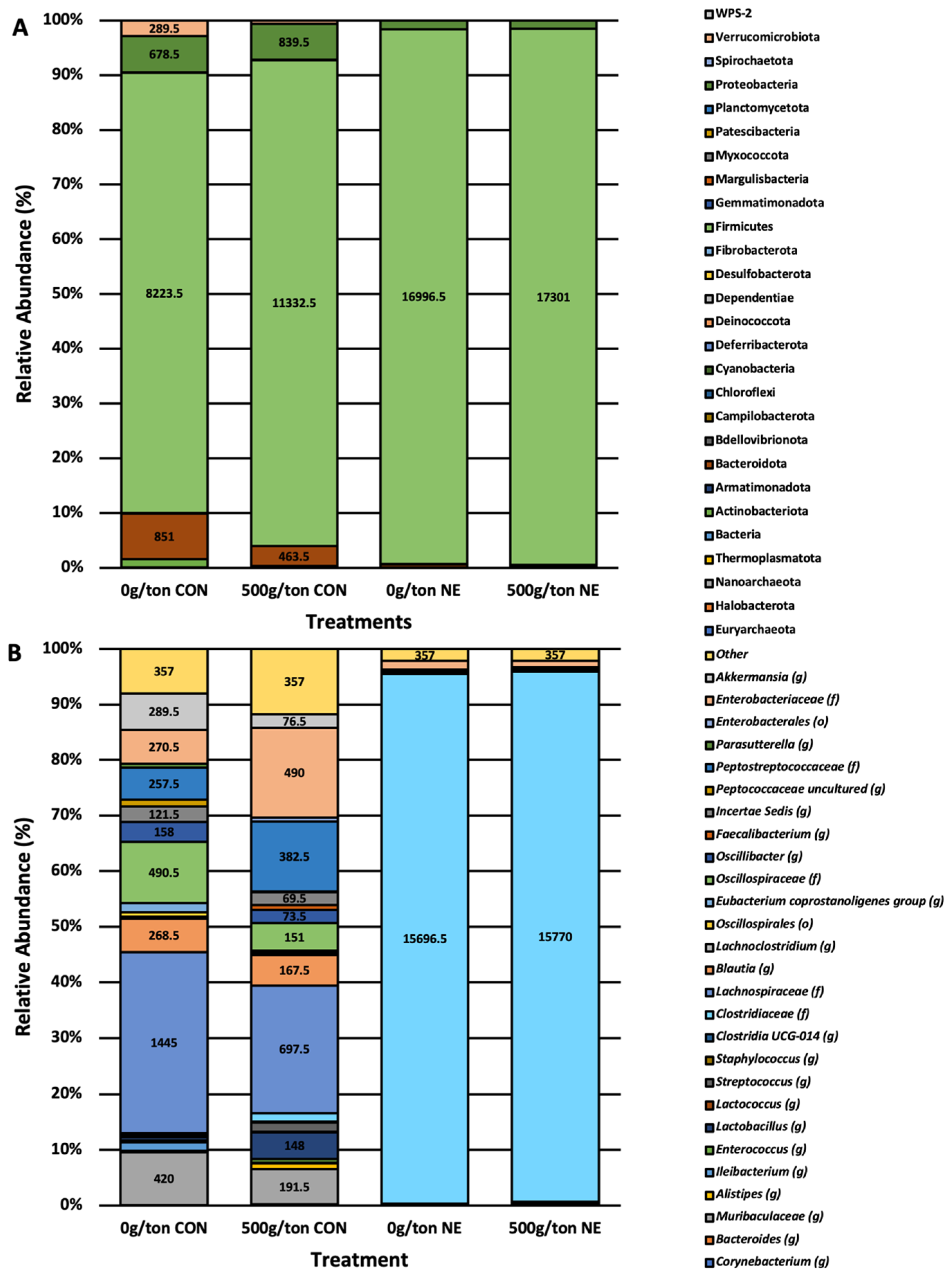
At the phyla level, the taxa most abundant were Firmicutes, Proteobacteria, and Bacteroidota, which comprised over 90% of the relative abundance of the microbiome (Figure 5A). Using analysis of communities of the microbiome (ANCOM), it was determined that Actinobacteriota, Bacteroidota, Firmicutes, and Verrucomicrobiota were impacted by the supplementation of a blend of microencapsulated organic acids and botanicals (W = 24, 24, 26, 22; p < 0.05; Supplemental Figure S1). The abundance of Actinobacteriota, Bacteroidota, and Verrucomicrobiota was decreased when NE was induced; however, the supplementation of 500 g/MT of a microencapsulated blend of organic acids and botanicals did appear to numerically increase these phyla compared to the non-supplemented groups. Likewise, those infected with Eimeria spp. and C. perfringens had higher levels of Firmicutes (Supplemental Figure S1).
At the genus level, there was a clear separation between the groups not infected (CON) and those with induced NE (NE; Figure 5B). Those not infected had a diverse taxonomic profile primarily comprised Enterobacteriaceae, Peptostrptococcaceae, Incertia sedis, Oscillospiraceae, Blautia, Lachnospiraceae, Lactobacillus, and Muribaculaceae when examined at the genus level. However, within those infected and NE was induced (NE), the microbiome composition was predominantly composed of Clostridiaceae and Enterobacteriaceae. The significantly different taxa at the genus level, as determined by ANCOM, were Clostridiaceae and Peptostreptococcaceae (W = 385, 369; p < 0.05; Supplemental Figure S2). The levels of Clostridiaceae were elevated in those with clinical NE-induced, while the levels of Peptostreptococcaceae were decreased in the same group. However, those supplemented with 500 g/MT of a microencapsulated blend of organic acids and botanicals (median of 509 features) had a higher level of Peptostreptococcaceae compared to those not supplemented with a microencapsulated blend of organic acids and botanicals (median of 1 feature).

4. Discussion

Understanding the impact of feed supplements on the microbiome of poultry is an important component in delineating the benefits of these additives, as the microbiome of the poultry GIT is directly impacted by feed supplementation and, subsequently, improves performance and disease resistance [39]. Therefore, many of the feed supplements utilized in the poultry industry that serve as antibiotic alternatives do so by directly impacting the microbiome of poultry. Recently, we have investigated the effect of a microencapsulated blend of organic acids and botanicals on broiler chickens on the performance, health, and microbiome under non-challenged conditions [13,16,17]. However, to determine the efficacy of the supplement in industry settings, we induced clinical NE in broiler breeder chickens fed diets supplemented with a microencapsulated blend of organic acids and botanicals. Supplementing the microencapsulated blend of organic acids and botanicals at 500 g/MT could reduce clinical signs of NE by modulating key specific immune-related pathways [15]. In continuation with these efforts, the current study aimed to determine if clinical signs of NE were improved by manipulating the microbiome of the lower small intestines (jejunum/ileum).

4.1. Lesion Scores Impact on Richness and Evenness

Previous research investigating the effect of supplementing broiler diets with a microencapsulated blend of organic acids and botanicals [13] showed no effect of diets on the alpha diversity of the jejunum and ileum (Shannon’s Diversity and Pielou’s Evenness). Instead, differences were driven by compartmental differences in the small intestines (jejunum and ileum). Pham et al. [40] also reported the lack of differences in the richness and evenness of the cecal microbiome when NE-challenged (sub-clinical infection) broiler chickens were fed diets supplemented with a microencapsulated blend of 4% thyme, 4% carvacrol, 0.5% hexanoic acid, 3.5% benzoic acid, and 0.5% butyric acid. The current study induced NE among broiler breeders fed 0 and 500 g/MT of the microencapsulated blend of organic acids and botanicals and demonstrated differences between treatment groups. However, as seen in the previous study, richness and evenness were less driven by the dietary treatments. The induction of clinical NE highly drove the differences. Therefore, as seen previously by Feye et al. [13], the dietary inclusion of a microencapsulated blend of organic acids and botanicals supplement did not appear to alter the richness and evenness of the microbiome of the small intestines.
In addition, the current study demonstrated the relationship of NE severity to the richness and evenness of the microbiome. Although there was no interaction between treatment and lesion score, the treatment had a significant effect. There was a parallel negative relationship between lesion score severity and richness and evenness of those induced with clinical NE. Those treated as controls (non-infected) did demonstrate relationships with the lesion scores (0 to 1). In contrast, those fed 0 g/MT had a negative association with lesion scores as the richness and evenness narrowed as the lesion scores reached 1. In comparison, those fed 500 g/MT and uninfected exhibited a positive relationship where the richness and evenness increased or stabilized as the lesion score increased. The current study presented evidence that the microbiome was correlated to the severity of lesion scores and that the supplementation of a microencapsulated blend of organic acids and botanicals had the potential to maintain or stabilize the richness and evenness of the community. However, it is important to note that the dietary treatments 0 and 500 g/MT microencapsulated blend of organic acids and botanicals behaved similarly when under clinical NE conditions.

4.2. Treatment and Infection on Abundance and Phylogenetic Diversity

As previously mentioned, Feye et al. [13] investigated the microbiome response of the jejunum and ileum to the dietary inclusion of the microencapsulated blend of organic acids and botanicals. In that research, Feye et al. [13] demonstrated beta diversity differences in the jejunum between those fed diets supplemented with 0 and 500 g/MT. Similarly, the current study demonstrated these differences within the control treatments (non-infected). However, there were no differences between the supplementation 0 and 500 g/MT of the microencapsulated blend of organic acids and botanicals when under clinical NE infection. The lack of significance between the NE-treated groups was most likely due to the severely diseased state of the birds.
In the industry, NE will often present itself as a sub-clinical infection where antibiotics and alternatives have demonstrated a level of protection [41]. Under sub-clinical conditions, Emami et al. [42] demonstrated improved health (reduced lesion scores) and maintained growth performance by modifying the gut microbiome, tight junctions, immune response, and cell metabolism. In a companion study, Swaggerty [15] made it evident that the supplementation of a microencapsulated blend of organic acids and botanicals could potentially reduce clinical manifestations of NE by altering specific immune-related pathways under NE clinical conditions, as revealed by kinome analysis. Although immune-related pathways were changed [15], the clinical presentation of NE may have suppressed the abundance and phylogenetic response by reducing the overall abundance of the microbiome.

4.3. Necrotic Enteritis Narrows Core Microbiome

In the past, numerous efforts have been placed on determining the microbiome of poultry during infection with sub-clinical and clinical NE; however, much of the research has focused narrowly on the cecal microbiome [43,44,45,46]. As the primary site of infection is the small intestines, typically the ileum [47], it may be more imperative to delineate the microbial changes occurring there rather than the hindgut, the ceca. More recent work has focused on the ileum, where clinical damage occurs [48,49]. The current study identified the core microbiome under non-challenged and NE clinical conditions. Clinical NE narrows the core communities within the small intestines. The non-challenged birds fed 0 and 500 g/MT had 10 and 6 different ASVs prevalent in more than 50% of the samples, whereas those challenged narrowed to 5 and 7 core ASVs. The narrowing of the microbiome is further evident by the taxonomic profiles (Figure 5) and the significantly different phyla and genera. As Clostridiaceae increases among the NE-challenged broiler breeders, the taxa belonging to Actinobacteriota, Bacteroidota, and Verrucomicrobiota decrease (Figure S1). These communities are typically present among healthy broilers and are a sign of a healthy microbiome [50]. It is important to note that the treatment of 500 g/MT of the microencapsulated blend of organic acids and botanicals demonstrated a slight rebound in the abundance of these communities compared to those challenged with NE and fed 0 g/MT diets. The slight rebound could be attributed to the nature of the supplementation of the microencapsulated blend of organic acids and botanicals to select for Lactobacillus and Clostridiaceae, as seen by Feye et al. [13] and the current study.

4.4. Lactobacillus and Clostridiaceae Core ASVs of Microencapsulated Blend

In the current study, Lactobacillus and Clostridiaceae were identified as the core members of the microbiome of the unchallenged birds fed diets containing 500 g/MT but not in those fed diets containing 0 g/MT of the microencapsulated blend of organic acids and botanicals. The same core Clostridiaceae ASVs were also present in the 500 g/MT of the challenged group as in the 500 g/MT unchallenged group (dc85940d84ddbd7315db16b14390cb8d). The challenged group fed diets containing 500 g/MT also had an additional unique ASV belonging to Clostridiaceae (9dece060ffcff93473a7023dcee0defd). Similarly, past research investigating the effects of the same microencapsulated blend of organic acids and botanicals led to an increase in Lactobacillus and Clostridiaceae in the jejunum of birds fed 300 and 500 g/MT compared to those fed diets containing no supplements [13]. Similar products have also shown increased specific taxa belonging to Lactobacillus [40]. Pham et al. demonstrated increased Lactobacillus abundance in the ceca when non-challenged NE birds were fed diets containing 500 mg/kg of a microencapsulated blend of organic acids and botanicals compared to those provided the basal control diets.
Specific Clostridial species have been identified as probiotic candidates, as not all Clostridium are created equal [51]. Additionally, taxa belonging to the family of Clostridiaceae are recognized as members of a normal poultry microbiome [52]. As such, the taxa belonging to the family Clostridiaceae identified as the core microbiome members of those fed diets supplemented with 500 g/MT of the microencapsulated blend of organic acids and botanicals in the current study could be members of a healthy or normal microbiome even when infected with other Clostridium spp. to induce NE. Stanley et al. [53] correlated the feed conversion ratio (FCR) of broilers with the microbiome composition. They determined the presence of taxa belonging to the family Clostridiaceae was positively related to improved FCR [53]. Future studies would need to be conducted to narrow in on what specific species belonging to the family of Clostridiaceae is being selected by supplementing a microencapsulated blend of organic acids and botanicals.

5. Conclusions

The current results demonstrated the modulation of the small intestine microbiome by supplementing a microencapsulated blend of organic acids and botanicals. Specifically, this research demonstrated that the supplementation of the microencapsulated blend did not alter the richness and evenness of the microbiome when under a clinical challenge of NE but did impact the core microbiome and the differentially abundant taxa. The supplementation of the microencapsulated blend selected for Lactobacillus and Clostridiaceae among the core microbiome regardless of challenge conditions. As the specific ASVs belonging to the family Clostridiaceae were present regardless of NE infection, the supplementation of the microencapsulated blend could select for beneficial members of Clostridiaceae that were indicative of a more stable microbiome. Future research would be needed to better identify the specific Clostridiaceae to verify its functionality within the poultry GIT.
Under clinical NE conditions, the effects of the supplementation of the microencapsulated blend of organic acids and botanicals were masked to a certain degree. The NE infection dominated the small intestines, as indicated by the abundance of Clostridiaceae in the NE-challenged broiler breeders. As subclinical infection of NE is very common in the poultry industry, it would be beneficial to delineate the impacts of this microencapsulated blend on maintaining performance and intestinal microbiome balance.

Supplementary Materials

The following supporting information can be downloaded at: https://www.mdpi.com/article/10.3390/ani13101627/s1, Table S1: Main effect and interaction of treatment and trial on alpha diversity using ANOVA; Table S2: Effect of treatment and trial on the alpha diversity of broiler digesta; Table S3: Relating lesions scores to evenness and richness of the microbiome of commercial broilers; Table S4: Main effect and interaction of treatment and trial on the beta diversity of broiler digesta when using ADONIS; Table S5: Effect of treatment and trial on the beta diversity of broiler digesta; Figure S1: Different phyla impacted by the supplementation of a blend of microencapsulated organic acids and botanicals; Figure S2: Different taxa at the genus level, impacted by the supplementation of a blend of microencapsulated organic acids and botanicals.

Author Contributions

C.L.S., S.C.R., A.P., and E.G. conceived the study. C.N.J., J.A.B.II, and C.L.S. conducted the animal trials; D.K.D. performed the microbiome analytics and prepared the manuscript for submittal. All authors have read and agreed to the published version of the manuscript.

Funding

This research was supported, in part, by Vetagro ((CLS) Agreement number 58-3091-8-005; https://www.vetagro.com, accessed on 3 May 2023) and the USDA/ARS (3091-32000-035-00D; https://www.ars.usda.gov/research/project/?accnNo=430283, accessed on 3 May 2023). There was no additional external funding received for this study. Vetagro provided support in the form of salaries for AP and EG, and supplied the AviPlus feed amendment, but did not have any additional role in data collection and analysis, decision to publish, or preparation of the manuscript. USDA/ARS provided support in the form of salary for CLS, CNJ, and JAB but did not have any additional role in the study design, data collection and analysis, decision to publish, or preparation of the manuscript.

Institutional Review Board Statement

All bird studies were under the approved experimental procedures outlined in protocol number 2019-002 and were approved by the USDA/ARS Institutional Animal Care and Use Committee operating under the Animal and Plant Health Inspection Service establishment number 334299. Experiments and bird welfare was overseen by Roger B. Harvey, attending veterinarian.

Informed Consent Statement

Not applicable.

Data Availability Statement

Demultiplexed sequencing reads are available on NCBI Sequence Read Archive (PRJNA899529) and GitHub (https://github.com/RickeLab-UW/Microencapsulated-Blend-on-Broiler-Intestinal-Microbiota, accessed on 3 May 2023). As well, the QIIME2 metadata file is available on Git Hub. Please contact S.C.R. if you require additional information.

Acknowledgments

The authors would like to acknowledge Denise Caldwell, Jackie Kotzur, and M. Reiley Street, Jr. (U.S. Department of Agriculture, Agricultural Research Service [USDA/ARS], College Station, TX) for their exceptional technical support and assistance with daily animal care. The current research was supported, in part, by Vetagro S.p.A (Agreement number 58-3091-8-005) and the USDA/ARS. The USDA is an equal opportunity provider and employer. Mention of trade names or commercial products in this publication is solely for the purpose of providing specific information and does not imply recommendation or endorsement by the USDA. AP and EG are Vetagro employees but did not participate in the analysis of this study.

Conflicts of Interest

A.P. and E.G. are Vetagro employees but did not participate in the analysis of the study. All other authors declare no conflict of interest.

References

  1. Paiva, D.; McElroy, A. Necrotic enteritis: Applications for the poultry industry. J. Appl. Poult. Res. 2014, 23, 557–566. [Google Scholar] [CrossRef]
  2. Van der Sluis, W. Clostridial enteritis is an often underestimated problem. World Poult. 2000, 16, 42–43. [Google Scholar]
  3. Castanon, J.I.R. History of the Use of Antibiotic as Growth Promoters in European Poultry Feeds. Poult. Sci. 2007, 86, 2466–2471. [Google Scholar] [CrossRef]
  4. Broom, L.J.; Kogut, M.H. Deciphering desirable immune responses from disease models with resistant and susceptible chickens. Poult. Sci. 2019, 98, 1634–1642. [Google Scholar] [CrossRef]
  5. Adhikari, P.; Kiess, A.; Jha, R. An approach to alternative strategies to control avian coccidiosis and necrotic enteritis. J. Appl. Poult. Res. 2020, 29, 515–534. [Google Scholar] [CrossRef]
  6. Wade, B.; Keyburn, A. The True Cost of Necrotic Enteritis. Available online: https://www.poultryworld.net/Meat/Articles/2015/10/The-true-cost-of-necrotic-enteritis-2699819W/ (accessed on 3 May 2023).
  7. Van Immerseel, F.; De Buck, J.; Pasmans, F.; Huyghebaert, G.; Haesebrouck, F.; Ducatelle, R. Clostridium perfringens in poultry: An emerging threat for animal and public health. Avian Pathol. 2004, 33, 537–549. [Google Scholar] [CrossRef]
  8. Cooper, K.K.; Songer, J.G. Necrotic enteritis in chickens: A paradigm of enteric infection by Clostridium perfringens type A. Anaerobe 2009, 15, 55–60. [Google Scholar] [CrossRef]
  9. Geier, M.; Mikkelsen, L.; Torok, V.; Allison, G.; Olnood, C.; Boulianne, M.; Hughes, R.; Choct, M. Comparison of alternatives to in-feed antimicrobials for the prevention of clinical necrotic enteritis. J. Appl. Microbiol. 2010, 109, 1329–1338. [Google Scholar] [CrossRef]
  10. Piva, A.; Pizzamiglio, V.; Morlacchini, M.; Tedeschi, M.; Piva, G. Lipid microencapsulation allows slow release of organic acids and natural identical flavors along the swine intestine 1, 2. J. Anim. Sci. 2007, 85, 486–493. [Google Scholar] [CrossRef] [PubMed]
  11. Svihus, B. Function of the digestive system. J. Appl. Poult. Res. 2014, 23, 306–314. [Google Scholar] [CrossRef]
  12. Svihus, B. Starch digestion capacity of poultry. Poult. Sci. 2014, 93, 2394–2399. [Google Scholar] [CrossRef] [PubMed]
  13. Feye, K.M.; Swaggerty, C.L.; Kogut, M.H.; Ricke, S.C.; Piva, A.; Grilli, E. The biological effects of microencapsulated organic acids and botanicals induces tissue-specific and dose-dependent changes to the Gallus gallus microbiota. BMC Microbiol. 2020, 20, 332. [Google Scholar] [CrossRef] [PubMed]
  14. Grilli, E.; Tugnoli, B.; Passey, J.L.; Stahl, C.H.; Piva, A.; Moeser, A.J. Impact of dietary organic acids and botanicals on intestinal integrity and inflammation in weaned pigs. BMC Vet. Res. 2015, 11, 96. [Google Scholar] [CrossRef]
  15. Swaggerty, C.L.; Byrd, J.A.; Arsenault, R.J.; Perry, F.; Johnson, C.N.; Genovese, K.J.; He, H.; Kogut, M.H.; Piva, A.; Grilli, E. A blend of microencapsulated organic acids and botanicals reduces necrotic enteritis via specific signaling pathways in broilers. Poult. Sci. 2022, 101, 101753. [Google Scholar] [CrossRef]
  16. Swaggerty, C.L.; Arsenault, R.J.; Johnson, C.; Piva, A.; Grilli, E. Dietary supplementation with a microencapsulated blend of organic acids and botanicals alters the kinome in the ileum and jejunum of Gallus gallus. PLoS ONE 2020, 15, e0236950. [Google Scholar] [CrossRef] [PubMed]
  17. Swaggerty, C.L.; He, H.; Genovese, K.J.; Callaway, T.R.; Kogut, M.H.; Piva, A.; Grilli, E. A microencapsulated feed additive containing organic acids, thymol, and vanillin increases in vitro functional activity of peripheral blood leukocytes from broiler chicks. Poult. Sci. 2020, 99, 3428–3436. [Google Scholar] [CrossRef]
  18. National Research Council. Nutrient Requirements of Poultry, 9th ed.; The National Academies Press: Washington, DC, USA, 1994. [Google Scholar]
  19. AgGuide. Guide for the Care and Use of Agricultural Animals in Research and Teaching. Available online: https://www.aaalac.org/pub/?id=E900BDB6-CCCF-AB13-89B6-DA98A4B52218 (accessed on 3 May 2023).
  20. McReynolds, J.L.; Byrd, J.A.; Genovese, K.J.; Poole, T.L.; Duke, S.E.; Farnell, M.B.; Nisbet, D.J. Dietary Lactose and its Effect on the Disease Condition of Necrotic Enteritis. Poult. Sci. 2007, 86, 1656–1661. [Google Scholar] [CrossRef]
  21. Prescott, J.F. The Prevention of Experimentally Induced Necrotic Enteritis in Chickens by Avoparcin. Avian Dis. 1979, 23, 1072. [Google Scholar] [CrossRef]
  22. Kozich, J.J.; Westcott, S.L.; Baxter, N.T.; Highlander, S.K.; Schloss, P.D. Development of a dual-index sequencing strategy and curation pipeline for analyzing amplicon sequence data on the MiSeq Illumina sequencing platform. Appl. Environ. Microbiol. 2013, 79, 5112–5120. [Google Scholar] [CrossRef]
  23. Bolyen, E.; Rideout, J.R.; Dillon, M.R.; Bokulich, N.A.; Abnet, C.C.; Al-Ghalith, G.A.; Alexander, H.; Alm, E.J.; Arumugam, M.; Asnicar, F.; et al. Reproducible, Interactive, Scalable and Extensible Microbiome Data Science using QIIME 2. Nat. Biotechnol. 2019, 37, 852–857. [Google Scholar] [CrossRef]
  24. Callahan, B.J.; Mcmurdie, P.J.; Rosen, M.J.; Han, A.W.; Johnson, A.J.A.; Holmes, S.P. DADA2: High-resolution sample inference from Illumina amplicon data. Nat. Methods 2016, 13, 581–583. [Google Scholar] [CrossRef] [PubMed]
  25. Price, M.N.; Dehal, P.S.; Arkin, A.P. FastTree 2—Approximately Maximum-Likelihood Trees for Large Alignments. PLoS ONE 2010, 5, e9490. [Google Scholar] [CrossRef] [PubMed]
  26. Bokulich, N.A.; Dillon, M.R.; Zhang, Y.; Rideout, J.R.; Bolyen, E.; Li, H.; Albert, P.S.; Caporaso, J.G. q2-longitudinal: Longitudinal and Paired-Sample Analyses of Microbiome Data. Msystems 2018, 3, e00219-18. [Google Scholar] [CrossRef] [PubMed]
  27. Anderson, M.J. A new method for non-parametric multivariate analysis of variance. Austral Ecol. 2001, 26, 32–46. [Google Scholar] [CrossRef]
  28. Shannon, C.A. A mathematical theory of communication. Bell Syst. Tech. J. 1948, 27, 379–423. [Google Scholar] [CrossRef]
  29. Pielou, E.C. The measurement of diversity in different types of biological collections. J. Theor. Biol. 1966, 13, 131–144. [Google Scholar] [CrossRef]
  30. Lozupone, C.A.; Hamady, M.; Kelley, S.T.; Knight, R. Quantitative and Qualitative β Diversity Measures Lead to Different Insights into Factors That Structure Microbial Communities. Appl. Environ. Microbiol. 2007, 73, 1576–1585. [Google Scholar] [CrossRef] [PubMed]
  31. Lozupone, C.; Knight, R. UniFrac: A New Phylogenetic Method for Comparing Microbial Communities. Appl. Environ. Microbiol. 2005, 71, 8228–8235. [Google Scholar] [CrossRef]
  32. Jaccard, P. The Distribution of the Flora in the Alpine Zone.1. New Phytol. 1912, 11, 37–50. [Google Scholar] [CrossRef]
  33. Mandal, S.; Van Treuren, W.; White, R.A.; Eggesbø, M.Å.; Knight, R.T.; Peddada, S.D. Analysis of composition of microbiomes: A novel method for studying microbial composition. Microb. Ecol. Health Dis. 2015, 26, 27663. [Google Scholar] [CrossRef]
  34. Quast, C.; Pruesse, E.; Yilmaz, P.; Gerken, J.; Schweer, T.; Yarza, P.; Peplies, J.; Glöckner, F.O. The SILVA Ribosomal RNA Gene Database Project: Improved Data Processing and Web-Based Tools. Nucleic Acids Res. 2013, 41, D590–D596. [Google Scholar] [CrossRef]
  35. Yilmaz, P.; Parfrey, L.W.; Yarza, P.; Gerken, J.; Pruesse, E.; Quast, C.; Schweer, T.; Peplies, J.; Ludwig, W.; Glöckner, F.O. The SILVA and “All-species Living Tree Project (LTP)” taxonomic frameworks. Nucleic Acids Res. 2013, 42, D643–D648. [Google Scholar] [CrossRef]
  36. Bokulich, N.A.; Kaehler, B.D.; Rideout, J.R.; Dillon, M.; Bolyen, E.; Knight, R.; Huttley, G.A.; Gregory Caporaso, J. Optimizing taxonomic classification of marker-gene amplicon sequences with QIIME 2′s q2-feature-classifier plugin. Microbiome 2018, 6, 90. [Google Scholar] [CrossRef] [PubMed]
  37. McMurdie, P.J.; Holmes, S. phyloseq: An R package for reproducible interactive analysis and graphics of microbiome census data. PLoS ONE 2013, 8, e61217. [Google Scholar] [CrossRef] [PubMed]
  38. Sudarshan, A.S.; Leo, L. Microbiomeutilities: Utilities for Microbiome Analytics; R Package Version 1.00.16; GitHub: San Francisco, CA, USA, 2020. [Google Scholar]
  39. Kogut, M.H. The effect of microbiome modulation on the intestinal health of poultry. Anim. Feed. Sci. Technol. 2018, 250, 32–40. [Google Scholar] [CrossRef]
  40. Pham, V.H.; Kan, L.; Huang, J.; Geng, Y.; Zhen, W.; Guo, Y.; Abbas, W.; Wang, Z. Dietary encapsulated essential oils and organic acids mixture improves gut health in broiler chickens challenged with necrotic enteritis. J Anim Sci Biotechnol. 2020, 11, 18. [Google Scholar] [CrossRef]
  41. Caly, D.L.; D’Inca, R.; Auclair, E.; Drider, D. Alternatives to Antibiotics to Prevent Necrotic Enteritis in Broiler Chickens: A Microbiologist’s Perspective. Front. Microbiol. 2015, 6, 1336. [Google Scholar] [CrossRef] [PubMed]
  42. Emami, N.K.; Calik, A.; White, M.B.; Kimminau, E.A.; Dalloul, R.A. Effect of Probiotics and Multi-Component Feed Additives on Microbiota, Gut Barrier and Immune Responses in Broiler Chickens During Subclinical Necrotic Enteritis. Front. Veter. Sci. 2020, 7, 572142. [Google Scholar] [CrossRef] [PubMed]
  43. Stanley, D.; Keyburn, A.L.; Denman, S.E.; Moore, R.J. Changes in the caecal microflora of chickens following Clostridium perfringens challenge to induce necrotic enteritis. Veter. Microbiol. 2012, 159, 155–162. [Google Scholar] [CrossRef]
  44. Stanley, D.; Wu, S.-B.; Rodgers, N.; Swick, R.; Moore, R.J. Differential Responses of Cecal Microbiota to Fishmeal, Eimeria and Clostridium perfringens in a Necrotic Enteritis Challenge Model in Chickens. PLoS ONE 2014, 9, e104739. [Google Scholar] [CrossRef]
  45. Lacey, J.A.; Stanley, D.; Keyburn, A.L.; Ford, M.; Chen, H.; Johanesen, P.; Lyras, D.; Moore, R.J. Clostridium perfringens-mediated necrotic enteritis is not influenced by the pre-existing microbiota but is promoted by large changes in the post-challenge microbiota. Vet. Microbiol. 2018, 227, 119–126. [Google Scholar] [CrossRef] [PubMed]
  46. Kiu, R.; Brown, J.; Bedwell, H.; Leclaire, C.; Caim, S.; Pickard, D.; Dougan, G.; Dixon, R.A.; Hall, L.J. Genomic analysis on broiler-associated Clostridium perfringens strains and exploratory caecal microbiome investigation reveals key factors linked to poultry necrotic enteritis. Anim. Microbiome 2019, 1, 12. [Google Scholar] [CrossRef]
  47. Hargis, B. Overview of Necrotic Enteritis in Poultry. Available online: https://www.merckvetmanual.com/poultry/necrotic-enteritis/overview-of-necrotic-enteritis-in-poultry (accessed on 8 November 2022).
  48. Xu, S.; Lin, Y.; Zeng, D.; Zhou, M.; Zeng, Y.; Wang, H.; Zhou, Y.; Zhu, H.; Pan, K.; Jing, B.; et al. Bacillus licheniformis normalize the ileum microbiota of chickens infected with necrotic enteritis. Sci. Rep. 2018, 8, 1744. [Google Scholar] [CrossRef] [PubMed]
  49. Latorre, J.D.; Adhikari, B.; Park, S.H.; Teague, K.D.; Graham, L.E.; Mahaffey, B.D.; Baxter, M.F.A.; Hernandez-Velasco, X.; Kwon, Y.M.; Ricke, S.C.; et al. Evaluation of the Epithelial Barrier Function and Ileal Microbiome in an Established Necrotic Enteritis Challenge Model in Broiler Chickens. Front. Veter. Sci. 2018, 5, 199. [Google Scholar] [CrossRef] [PubMed]
  50. Dittoe, D.K.; Ricke, S.C.; Kiess, A.S. Organic Acids and Potential for Modifying the Avian Gastrointestinal Tract and Reducing Pathogens and Disease. Front. Veter. Sci. 2018, 5, 216. [Google Scholar] [CrossRef]
  51. Guo, P.; Zhang, K.; Ma, X.; He, P. Clostridium species as probiotics: Potentials and challenges. J. Anim. Sci. Biotechnol. 2020, 11, 24. [Google Scholar] [CrossRef]
  52. Oakley, B.B.; Lillehoj, H.S.; Kogut, M.H.; Kim, W.K.; Maurer, J.J.; Pedroso, A.; Lee, M.D.; Collett, S.R.; Johnson, T.J.; Cox, N.A. The chicken gastrointestinal microbiome. FEMS Microbiol. Lett. 2014, 360, 100–112. [Google Scholar] [CrossRef]
  53. Stanley, D.; Hughes, R.J.; Geier, M.S.; Moore, R.J. Bacteria within the Gastrointestinal Tract Microbiota Correlated with Improved Growth and Feed Conversion: Challenges Presented for the Identification of Performance Enhancing Probiotic Bacteria. Front. Microbiol. 2016, 7, 187. [Google Scholar] [CrossRef]
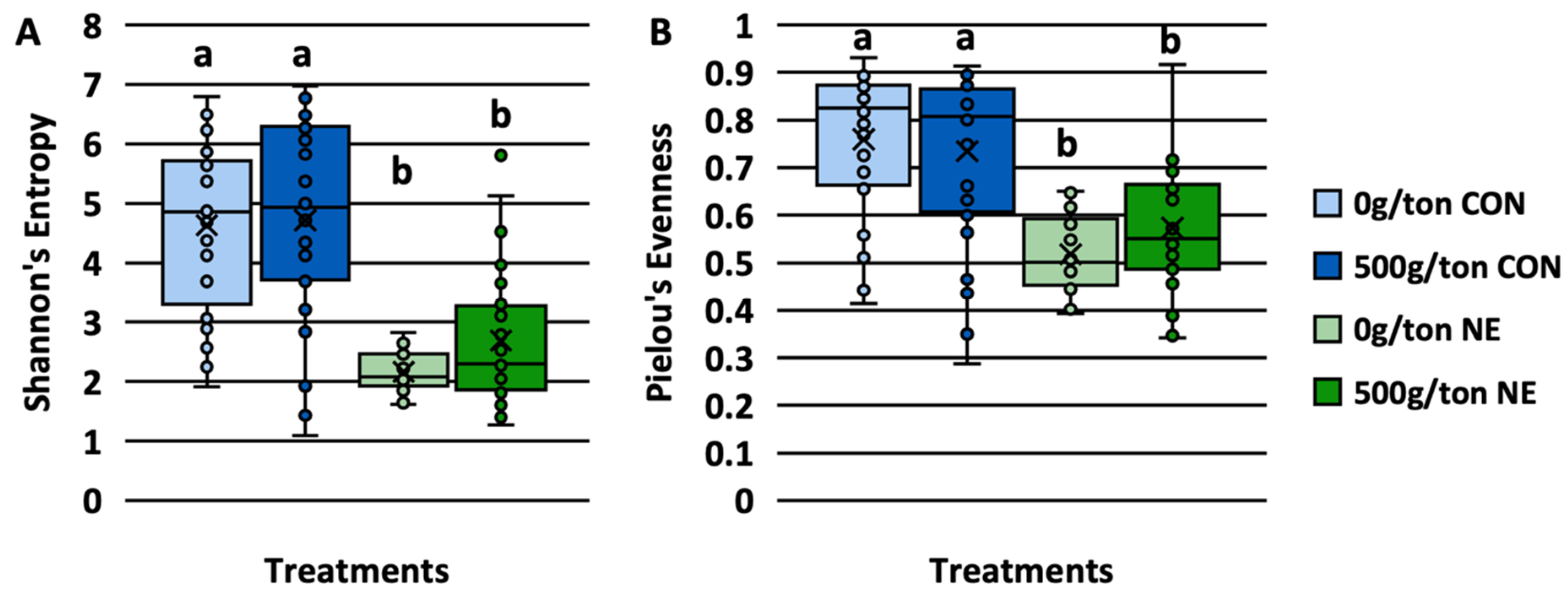
Figure 1. Shannon’s Entropy (A) and Pielou’s Evenness (B) of the microbiome of commercial broiler breeders fed diets containing a blend of microencapsulated organic acids and botanicals. Significant different means (×) are denoted with different connecting letters (a, b).
Animals 13 01627 g001
Figure 2. Relationship of lesions scores to the richness (A) and evenness (B) of the microbiome of commercial broiler breeders supplied supplemented diets containing a blend of microencapsulated organic acids and botanicals.
Animals 13 01627 g002
Figure 3. Abundance (A) and phylogenetic diversity (B) of the microbiome of commercial broiler breeders fed diets containing a blend of microencapsulated organic acids and botanicals.
Animals 13 01627 g003
Figure 4. Distribution of the microbiome within the four treatment groups, control and NE enteritis induced birds fed diets supplemented with or without 500 g/MT of a blend of microencapsulated organic acids and botanicals.
Animals 13 01627 g004
Figure 5. Taxonomic composition at the phylum (A) and genus (B) level of broiler breeders fed diets containing a blend of microencapsulated organic acids and botanicals. Significant different phyla, Actinobacteriota, Bacteroidota, Firmicutes, and Verrucomicrobiota, impacted by the supplementation of a blend of microencapsulated organic acids and botanicals (W = 24, 24, 26, 22; p < 0.05; Supplemental Figure S1). Significant different taxa at the genus level, Clostridiaceae and Peptostreptococcaceae, were impacted by the supplementation of a blend of microencapsulated organic acids and botanicals (W = 385, 369; p < 0.05; Supplemental Figure S2).
Animals 13 01627 g005
Table 1. Core microbiome (present in >50% of samples) of digesta of control and NE-induced birds.
InfectionDietOrganismsASV
Control0 g/MTLachnospiraceae (2 unique ASVs)10650515e35dcecde52015f8c34a4a06
Lachnospiraceae_Blautia0b6c85919018ef580a0e1111b794c86b
Lachnospiraceae_Blautia_Lachnospiraceae_bacterium74e7c89ab2da511c0003f650773f9c2f
Lachnospiraceae492f2e502cce64557efbfad55333f9ca
Erysipelotrichaceae_Ileibacterium_Ileibacterium_valensa922583a2cd922f6e2361c1df16702bb
Enterobacteriaceae20cfe7f61d18f6525cc71caae0ab28dc
Sutterellaceae_Parasutterellacf39b94723be8d7c4e5b41f4b20dab44
Akkermansiaceae_Akkermansiab6b05223adf86d071fd279f79dc2533c
Peptostreptococcaceaeee59a70b1b832493583e7bc2d6488ec7
Peptostreptococcaceae931dbe33cb8438cfd863ac9acfb5d17f
500 g/MTLachnospiraceae_Blautia0b6c85919018ef580a0e1111b794c86b
Lachnospiraceae_492f2e502cce64557efbfad55333f9ca
Clostridiaceae_dc85940d84ddbd7315db16b14390cb8d
Lactobacillaceae_Lactobacillus44d9fc4de8898b6c82c69654435a9f0b
Enterobacteriaceae_20cfe7f61d18f6525cc71caae0ab28dc
Peptostreptococcaceae_931dbe33cb8438cfd863ac9acfb5d17f
NE-Induced0 g/MTClostridiaceae3be4ebb35ff7f6217fae945cb0ad5413
Clostridiaceae2acc5f779e35ac658d8389859048ff61
Clostridiaceaedc85940d84ddbd7315db16b14390cb8d
Clostridiaceae3b55963ebca3b90a7dcf9a27ef40a76a
Enterobacteriaceae20cfe7f61d18f6525cc71caae0ab28dc
500 g/MTClostridiaceae3be4ebb35ff7f6217fae945cb0ad5413
Clostridiaceae2acc5f779e35ac658d8389859048ff61
Clostridiaceae9dece060ffcff93473a7023dcee0defd
Clostridiaceaedc85940d84ddbd7315db16b14390cb8d
Clostridiaceaee52b990e8b8ae704003a5ff38b791dbf
Clostridiaceae3b55963ebca3b90a7dcf9a27ef40a76a
Enterobacteriaceae20cfe7f61d18f6525cc71caae0ab28dc
Disclaimer/Publisher’s Note: The statements, opinions and data contained in all publications are solely those of the individual author(s) and contributor(s) and not of MDPI and/or the editor(s). MDPI and/or the editor(s) disclaim responsibility for any injury to people or property resulting from any ideas, methods, instructions or products referred to in the content.