Article

Molecular Characteristics of Chicken Infectious Anemia Virus in Central and Eastern China from 2020 to 2022

1
Henan Provincial Engineering and Technology Center of Health Products for Livestock and Poultry, Nanyang Normal University, Nanyang 473061, China
2
Henan Key Laboratory of Insect Biology in Funiu Mountain, Nanyang Normal University, Nanyang 473061, China
3
College of Animal Science, South China Agricultural University, Guangzhou 510642, China
*
Author to whom correspondence should be addressed.
Animals 2023, 13(17), 2709; https://doi.org/10.3390/ani13172709
Submission received: 30 June 2023 / Revised: 23 August 2023 / Accepted: 24 August 2023 / Published: 25 August 2023

Abstract

:

Simple Summary

An immune-suppressive disease known as chicken infectious anemia (CIA) develops after infection with chicken infectious anemia virus (CIAV). This study involved a systematic analysis of the epidemiology and genomics of CIAV in the provinces of Henan, Anhui, Jiangsu, and Hubei in China. The positive rates of the samples from each province flock ranged from 50% to 80%. Meanwhile, coinfections of CIAV with Marek’s disease virus, avian leukosis virus, infectious bursal disease virus, and fowl adenovirus were also identified. This study revealed the diversity of CIAV genomes as well as key mutation sites and intricate recombinants. The study findings underscore the importance of CIAV surveillance and provide a basis for further investigation into the evolution and molecular characteristics of CIAV strains.

Abstract

To evaluate the recent evolution of CIAV in China, 43 flocks of chickens from the provinces of Henan, Jiangsu, Hubei, and Anhui were screened via polymerase chain reaction during 2020–2022. Of these, 27 flocks tested positive for CIAV nucleic acids, including 12 which were positive for other immunosuppression viruses. Additionally, 27 CIAV strains were isolated, and their whole genomes were sequenced. The AH2001 and JS2002 strains shared the highest identity at 99.56%, and the HB2102 and HB2101 strains shared the lowest identity at 95.34%. Based on the genome sequences of these strains and reference strains, a phylogenetic tree was constructed and divided into eight main branches. Most of the strains were grouped with the East Asian strains, whereas the HB2101 strain belonged to the Brazil and Argentina cluster. A recombination event was detected in multiple strains, in which AH2002 recombined from KJ728827/China/2014 (from Taiwan Province) and HN2203, and AH2202 recombined from KX811526/China/2017 (from Shandong Province) and HN2203. All the obtained strains had a highly pathogenic Gln amino acid site at position 394 of the VP1. Overall, our findings demonstrate the importance of CIAV monitoring and provide data that aid in understanding the evolution of CIAV.

1. Introduction

The viral disease known as chicken infectious anemia (CIA) is caused by the chicken infectious anemia virus (CIAV) and leads to significant economic loss to the poultry industry worldwide [1,2]. Young birds without maternal antibodies are highly susceptible to develop primary signs of CIA, which include anemia, subcutaneous hemorrhages, growth retardation, aberrant feathers, limb paralysis, thymus atrophy, and bone marrow atrophy. Infected birds are often immunocompromised, which enhances the risk of secondary viral, bacterial, or fungal infections, even though CIA is frequently regarded as asymptomatic in adult chickens [3].
CIAV was first isolated in Japan in 1979 and has since been found to spread both vertically and horizontally [4]. Horizontal transmission throughout the flock occurs via excrement, dander, or feathers, primarily through oral infection. Notably, as vertical infection disseminates through eggs, it is quite likely that an outbreak of CIA in the progeny will occur [5]. Additionally, previous research has shown that vertical transmission of CIA can occur even when infected hens have developed an immune response [6].
CIAV belongs to the Anelloviridae family and the Gyrovirus genus [7]. Hematopoietic cells and T-lymphocytes of chickens are the primary targets of this nonenveloped virus, which has icosahedral single-stranded DNA [8]. One circular and single-stranded DNA molecule with a covalently closed end constitutes the whole CIAV genome. The genome with a length of 2.3 kb codes for peptides with molecular weights of 51 (VP1), 28 (VP2), and 13.6 (VP3) kDa [9]. VP1 is the only structural protein formed in the CIAV capsid, and VP2 functions as a scaffold protein, assisting VP1 in forming the proper shape and exposing its epitope [10]. Both VP1 and VP2 contain epitopes that elicit a neutralizing antibody response [11]. VP2 is also involved in reducing virulence and triggering apoptosis [12]. The nonstructural protein VP3 (13 kDa) mainly promotes apoptosis in infected chicken cells [13,14].
In numerous areas of China, CIAV has been detected in poultry farms, and its isolation from specific pathogen-free chickens may explain its widespread dissemination and vaccine contamination [15]. Reticuloendotheliosis virus (REV), fowl adenovirus (FAdV), and avian leukosis virus (ALV) are common viruses that cause immunosuppressive infections and have been linked to CIAV in a growing number of instances [16,17]. Continuous investigation of CIAV is necessary to understand its complex evolution process, which involves complex recombination and extensive mutations [5,18].
To evaluate the recent evolution of CIAV in China, molecular characteristics of 27 CIAV were systematically analyzed on the basis of their genome sequences, including phylogenic mutation and recombination analysis. Our findings demonstrate the importance of continuous CIAV monitoring and provide data that can help in understanding the recent evolution of CIAV.

2. Materials and Methods

2.1. Sample Collection and Pathogen Detection

During 2020–2022, 430 clinical samples (pooled samples of bone marrow, spleen, and thymus) were obtained from dead chickens which exhibited symptoms of depression, anorexia, emaciation, growth retardation, or pale skin. These birds were obtained from 43 chicken flocks (10 chickens per poultry flock) from the Chinese provinces of Henan, Anhui, Jiangsu, and Hubei. All the samples were stored at a temperature of −80 °C.

2.2. Detection, Polymerase Chain Reaction (PCR) Amplification, and Sequencing

Following the manufacturer’s instructions, viral DNAs and RNAs were separately extracted from the chicken samples using a commercial kit (TIANamp Virus DNA/RNA Kit, Tiangen, Beijing, China). Three primer pairs, as described previously, were used for the detection and genome amplification of CIAV [19]. The first primer pair consisted of a forward primer (P1: GCATTCCGAGTGGTTACTATTCC) and a reverse primer (P2: CGTCTTGCCATCTTACAGTCTTAT), with a projected target-band of 842 bp. The second pair of primers consisted of a forward primer (P3: CGAGTACAGGGTAAGCGAGCTAAC) and a reverse primer (P4: TGCTATTCATGCAGCGGACTT), with a projected target-band of 990 bp. The last pair of primers included a forward primer (P5: GAAAATGAGACCCGACGAGCAACAG) and a reverse primer (P6: GATTCGTCCACTTTGACTTTCTGTG), with a projected amplicon size of 736 bp. Primer pairs (P3 and P4) were used for CIAV detection, and all three primer pairs were used for complete genome amplification (P1–P6).
Meanwhile, ALV (F: GGATGAGGTGACTAAGAAAG; R: CGAACCAAAGGTAACACACG), IBDV (F: ATGTGGCTGGAAGAGAATGG; R: GCCCTTTGAGACTTGCTACCT), FAdV (F: AAGTTCAGGCAGACGGTCGTG; R: TAGTGATGCCGGGACATCATGTCG), REV (F: GCCTTAGCCGCCATTGTA; R: CCAGCCAACACCACGAACA), and Marek’s disease virus (MDV) (F: AGAAACATGGGGCATAGACG; R: CTTGCAGGTGTATACCAGGG) were also detected via PCR assay, as previously described [16,20,21,22].
To perform PCR, 100 ng of DNA template, 0.2 mM of each dNTP, 2 mM of MgCl2, 0.6 M of each primer, and 0.75 U of PlatinumTM Taq DNA Polymerase were prepared in 1× supplied PCR buffer (Thermo Fisher Scientific Baltics UAB, Vilnius, Lithuania). Amplification was performed using the following conditions: the predenaturation step at 95 °C for 5 min, 35 cycles of the denaturation step at 95 °C for 30 s, the annealing step at 60 °C (53 °C for ALV; 55 °C for IBDV; 55 °C for FAdV; 52 °C for REV; and 56 °C for MDV) for 30 s, the extension step at 72 °C for 30 s (30 s for ALV; 25 s for IBDV; 45 s for FAdV; 50 s for REV; and 1 min for MDV), and a final extension step at 72 °C for 10 min. Both positive and negative controls were included, and the amplified products were subsequently separated via 1% agarose gel electrophoresis. The purified amplicons were cloned into a pMD18-T simple vector (TaKaRa Biotechnology Co., Ltd., Dalian, China) and sequenced (Hongxun, Suzhou, China).

2.3. Virus Isolation

The CIAV present in the positive samples was cultured in MDCC–MSB1 lymphoblastoid cell lines following the previously described protocol [5]. Briefly, the homogenate suspension from the CIAV-positive samples was sterilized by filtration using a 0.22-μm filter and added onto the MDCC–MSB1 cells at 37 °C for 1 h. After virus absorption, the MSB1 cells were cultured in 5 mL of 1640 medium containing 2% fetal bovine serum (Gbico, New York, NY, USA) at 37 °C for 3 days. Five blind passages using MDCC-MSB1 cells were performed to enable viral growth. For each isolated CIAV, detection and genome amplification were performed using the PCR assays described above.

2.4. Phylogenetic and Recombination Analysis

Whole genome sequences of the 27 CIAV strains along with references were aligned and identified using the Lasergene 7.0 software (DNASTAR Inc., Madison, WI, USA). Next, an identity heatmap was created using an online program (https://www.chiplot.online/ (accessed on 26 May 2023)). In addition, the amino acid (aa) mutation sites of VP1 were summarized to assess genetic markers known to be associated with virulence.
The maximum likelihood method available in the molecular evolutionary genetics analysis (MEGA X) program was used with 1000 bootstrap replications to perform evolution analysis [23]. The online Evolview v3 program (http://www.evolgenius.info/evolview/ (accessed on 17 August 2023)) was used to visualize the generated phylogenetic tree [24].
To investigate the possibility of intergenic recombination, a total of 105 complete CIAV genomes from obtained and reference strains were analyzed. The Recombination Detection Program (RDP4), which includes the methods of GENECONV, BOOTSCAN, MaxChi, CHIMERA, SISCAN, and 3Seq, was used for the prediction of recombination events and the identification of potential parental sequences [25].

2.5. VP1 Structure Modeling

To visually reflect the VP1 structure change caused by the mutations in the CIAV isolates in this study, the tertiary structures of the VP1 of these isolates and the reference strain of SD24 were modeled using AlphaFold2.3.0 (https://wemol.wecomput.com/ui/#/ (accessed on 19 June 2023)).

3. Results

3.1. Sample Screening

Samples from 43 poultry farms were tested, and the results showed that 27 flocks were positive for CIAV, including 8 out of 13 (61.54%) from Jiangsu, 8 out of 12 (66.67%) from Henan Province, 6 out of 9 (66.67%) from Hubei, and 5 out of 9 (55.56%) from Anhui. The positive rates of the samples from each flock ranged from 50% to 80%, with an average of 69.3%. CIAV isolates were successfully obtained from positive samples in each flock. Among the 27 CIAV-positive flocks, 11 flocks of dual infections were caused by CIAV and the following coinfecting viruses: MDV (n = 3), REV (n = 2), ALV (n = 4), and IBDV (n = 2); additionally, one flock of quadruple infection was also identified (CIAV with FAdV + ALV + MDV). In CIAV-negative flocks, one flock each from Henan, Anhui, and Jiangsu was tested to be positive for IBDV, REV, and MDV, respectively. The infection and coinfection status of CIAV, ALV, IBDV, FAdV, REV, and MDV in various flocks was displayed via the UpSet map in Figure 1.

3.2. DNA Alignment and Identity Analysis

All 27 obtained CIAV strains had a complete genome length of 2298 bp, and the sequences were submitted to the GenBank database with accession numbers OQ869186–OQ869212. Viral sequences from positive samples and CIAV isolates from the same chicken flock shared 100% identical. Table 1 and Table 2 present detailed information on the 27 CIAV strains and 78 reference strains detected in chickens, humans, dogs, and cats. According to the sequence alignment of the entire genome, the nucleotide (nt) identity of the 27 CIAV strains ranged from 95.34% (HB2102 and HB2101) to 99.56% (AH2001 and JS2002). Between the 27 isolated and vaccine strains, HB2101 shared the lowest similarity with Cux-1 (95.39%) and Del-Ros (96.21%), whereas HN2101 had the highest similarity with Cux-1 (97.11%) and Del-Ros (98.3%). HB2102 and AF227982.1/Australia/2001 had the lowest similarity (91.97%), whereas HB1901 and KM496306.1/China/2013 had the highest similarity, (99.74%) compared with the reference strains. Supplementary Figure S1 illustrates the similarity comparison of all the strains.

3.3. Phylogenetic and Recombination Analysis

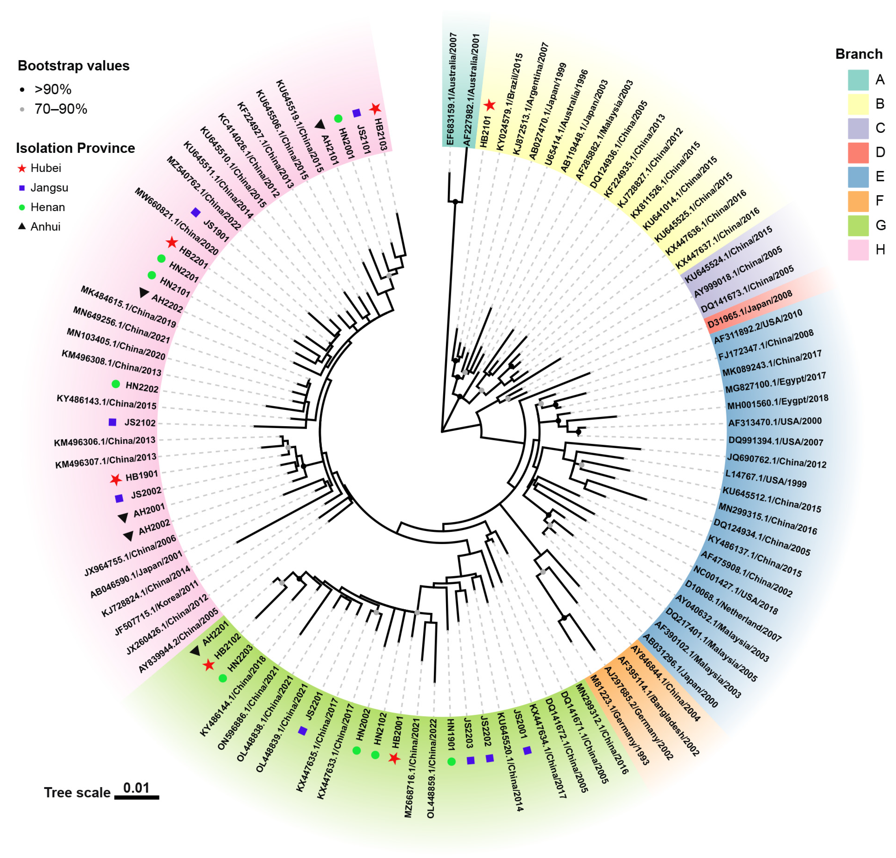
The phylogenetic tree of the entire genome of the 27 obtained strains and 78 reference CIAV strains is illustrated in Figure 2. The evolutionary tree was separated into eight branches (A to H). HB2101 (Branch B) was clustered with KY024579.1/Brazil/2015 and KJ872513.1/Argentina/2007 in South America, whereas the other strains (Branch G and Branch H) more closely resembled Asian strains. Both vaccine strains (Del-Ros and Cux-1) that were widely used abroad were clustered into Branch E. Eleven of the 27 obtained strains (Branch G) solely resembled the Chinese strain, whereas the remaining 15 strains (Branch H) resembled the Korean (JF507715.1/Korea/2011) and Japanese (AB046590.1/Japan/2001) strains.
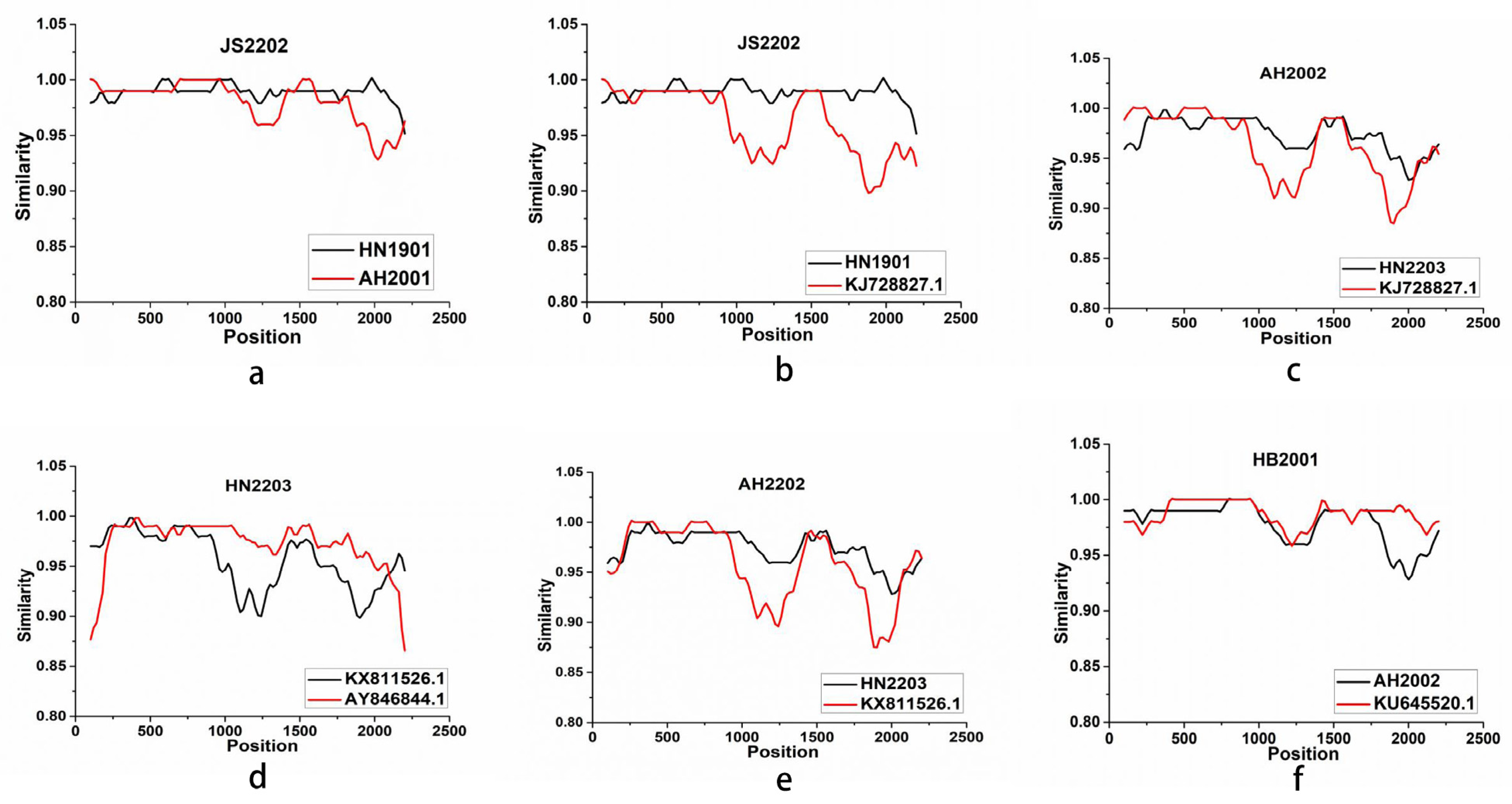
To assess the probability of genotype recombination, we analyzed the whole genomes of the 27 CIAV strains and the 78 reference strains using the RDP 4.83 and Simplot 3.51 software tools. The putative recombination event showed high confidence ratings and predicted five recombinant strains (JS2202, AH2002, HN2203, AH2202, and HB2001) on the basis of at least five independent detection methods (Table 3). Figure 3 illustrates the bootstrap analysis that we conducted to determine the breakpoint and identify each recombinant and its parent strain. Intriguingly, JS2202 may not only be formed by the recombination pair of major parent HN1901 and minor parent AH2001, but also by the recombination pair of major parent HN1901 and minor parent 18 (KJ728827.1/Taiwan/2014). Both the two recombinant events occurring in the JS2202 strain shared similar breakpoints from 89 to 1796 across the untranslated region (UTR, 2182 to 358), the VP2 coding region (359 to 1009), the VP3 coding region (465 to 830), and the VP1 coding region (832 to 2181). JS2202 shared similarity with HN1901 and AH2001 ranging from 0.93 to 1, and from 0.91 to 1 with KJ728827.1/China/2012.

3.4. Mutation Analysis of VP1

Previous studies have shown that aa position 394 is a major genetic determinant of pathogenesis, and sites 75, 89, 125, 139, 141, and 144 are related to viral replication and transmission [26,27,28]. In this study, Q394 was detected in all 27 strains that were analyzed, implying that these might be highly pathogenic strains [26]. T89 was found in all the analyzed strains; A89 was detected only in highly passaged CIAVs, but not sufficient to cause attenuation [27]. The 139 and 144 aa sites of HN2001, HB2101, JS2101, HB2103, and AH2101 contained the Q mutation, which is related to a reduced rate of cell-cultured spread [28]. Mutations at the important sites of VP1 are detailed in Table 4. Furthermore, the predicted tertiary structures of the SD24 and HB2103 strains indicated the structure change caused by mutations located at residues 22, 75, 139, and 144 (Figure 4).

4. Discussion

CIAV has been identified in chicken populations worldwide since it was originally discovered in China in 1996, posing a considerable threat to the poultry industry [29,30]. We screened 27 positive flocks in four provinces of China from 2020 to 2022, and the prevalence rates of CIAV were 61.54% (8/13) in Jiangsu province, 66.67% (8/12) in Henan Province, 66.67% (6/9) in Hubei Province, and 55.56% (5/9) in Anhui Province. These prevalence rates were higher than those reported in a survey of 13.30% of the average CIAV infection rate in 12 provinces from 2014 to 2015, indicating that CIAVs were more prevalent in recent years [31]. As previously reported, CIAV was mainly tested in a live attenuated vaccine against Newcastle disease, avian infectious bronchitis, and fowlpox [5]. Combined with vertical/horizontal transmission and contamination from live attenuated vaccine, CIAV has recently spread throughout the China and even globally, causing severe economic losses. Meanwhile, the coinfection of CIAV and other immunosuppressive viral agents such as ALV, REV, or IBDV might play a significant role in the infection course of CIAV. These results suggested the ongoing urgent need for continued screening tests for CIAV and other immunosuppression pathogens in clinical samples and vaccines.
The evolutionary tree of the 27 strains analyzed in this study and 78 reference strains revealed eight main branches, with the majority of the strains being closely related to the Chinese strains. However, the HB2101 strain showed a closer relationship to strains from Brazil and Argentina. The mechanisms and factors that contribute to the natural transmission of similar CIAV strains across borders remain to be determined. According to a previous study, local dissemination and long-distance migration are the main factors influencing CIAV evolution in South America. [32]. It is suspected that the transmission of the abroad-genotype CIAV is due to migratory bird movements or to the introduction of broiler breeders from abroad. These findings highlight the importance of detecting and monitoring the breeding chickens that are introduced as well as the migratory bird populations. Furthermore, the CIAV strains isolated in this study belonged mainly to Branch G and Branch H and were not locally determined, but distant from the internationally prevalent vaccine strains Cux-1 and Del-Ros, which belonged to Branch E. Although the genomic similarities between the 27 isolated and the two vaccine strains were almost similar, the different phylogenetic distribution suggested the distant-evolution trend.
Due to its genetic diversity, the VP1-enconding gene is commonly targeted for sequence analysis [33]. Several aa sites in VP1 are associated with viral pathogenicity and replication [9,26,27,28]. An important genetic predictor of virulence in CIAV is the aa at position 394, where pathogenic strains possessed glutamine (Q) whereas strains containing histidine (H) had a moderate virulence [26,28]. In this study, all 27 CIAV isolates carried Q394, indicating that they might be highly pathogenic. Cell culturing has demonstrated that aa located at 139 and 144 can influence viral growth and contagiousness and that CIAV strains harboring glutamine (Q) at these sites are less contagious [34]. Five (HN2001, HB2101, JS2101, HB2103, and AH21011) out of the 27 obtained strains had Q, indicating their weak contagiousness in cultured cells [34]. Meanwhile, amino acids at I75, L125, E141, and E144 of VP1 are related to reduced pathogenicity in chickens [9]. Further, A89 was detected in highly passaged CIAVs but was not sufficient to cause attenuation [27]. T89, I125, and Q141 were the major representative substitutions in VP1 of both genotype I and genotype II CIAV isolates in Egypt [35]. The CIAVs obtained in this study all harbored T89 and Q141, but the substitution 125 in VP1 was highly mutated. As shown in the predicted tertiary structures, mutations located at 22, 75, 139, and 144 may cause structure changes in VP1. These findings suggested the complicated relatedness between mutations sited in VP1 and CIAV pathogenicity. Given the genetic variability of VP1 and its crucial role in pathogenicity and transmissibility, the pathogenic differences between strains carrying extensive mutations require continuous and thorough evaluation.
Recombination is thought to be the main catalyst for viral evolution and the source of most viral mutations [36]. We discovered gene recombination events in five isolates (JS2202, AH2002, HN2203, AH2202, and HB2001). JS2202 may have resulted from a combination of the major parent HN1901 and the minor parent AH2001 as well as of the major parent HN1901 and the minor parent 18 (KJ728827.1/Taiwan/2014). The recombinant strain HN2003 and the strain 18 (KJ728827.1/Taiwan/2014) make up AH2002. Genetic recombination of CIAV has been observed to take place in the coding areas (nt 818–1295; nt 768–1286) [18,30], in the noncoding areas (nt 2108–2143; nt 2173–2231) [2,37], or in the regions that bridge the coding and noncoding regions (nt 1684–1757) [29]. The rearrangement breakpoint identified in this study spans both the coding and noncoding regions, extending from position 89 (the starting breakpoint) to position 1796 (the finishing breakpoint). Further investigations are necessary to trace the evolution of CIAV and establish the link between viral recombination and pathogenicity.

5. Conclusions

In conclusion, our study has revealed the complex infection status of CIAV in central and eastern China, the diversity of CIAV genomes, and key mutation sites and intricate recombinants. These findings provide a basis for further investigation into the evolution and molecular characterization of CIAV strains.

Supplementary Materials

The following supporting information can be downloaded at: https://www.mdpi.com/article/10.3390/ani13172709/s1, Figure S1: A heat map of the similarity analysis of the entire genome of the CIAV strain and other reference strains obtained in this study (lower left: different identity values are expressed in gradient colors ranging from 91% to 100%).

Author Contributions

Conceptualization, J.J. and L.Y.; methodology, S.X.; software, S.X., Z.Z. and X.X.; validation, X.X., J.J. and L.Y.; formal analysis, J.J. and Y.K.; investigation, S.X. and Z.Z.; resources, Q.X. and Y.B.; data curation, S.X. and Z.Z.; writing—original draft preparation, S.X.; writing—review and editing, J.J. and L.Y.; visualization, S.X. and Z.Z.; supervision, S.X. and Y.K.; project administration, L.Y. and Y.K.; funding acquisition L.Y. All authors have read and agreed to the published version of the manuscript.

Funding

This study was supported by the National Natural Science Foundation of China (Grant no. 31870917) and the Program for Science & Technology Innovation Talents in Universities of Henan Province (Grant no. 22HASTIT042).

Institutional Review Board Statement

The sampling protocols were approved by the South China Agricultural University Committee for Animal Experiments (approval ID: SYXK 2019-0136).

Informed Consent Statement

Sample collection protocols were approved by the flock owner.

Data Availability Statement

All the data generated or analyzed during this study are included in this article. Datasets are deposited in a publicly accessible repository; the datasets generated for this study can be found in GenBank: https://www.ncbi.nlm.nih.gov/Genbank/ (accessed on 18 August 2023). Genbank accession numbers are mentioned in the Materials and Methods of the article.

Acknowledgments

The authors would like to thank the members of the Nanyang Normal University for supporting this work.

Conflicts of Interest

The authors declare no conflict of interest.

References

  1. Kim, H.R.; Kwon, Y.K.; Bae, Y.C.; Oem, J.K.; Lee, O.S. Molecular characterization of chicken infectious anemia viruses detected from breeder and broiler chickens in South Korea. Poult. Sci. 2010, 89, 2426–2431. [Google Scholar] [CrossRef]
  2. Zhang, X.; Liu, Y.; Wu, B.; Sun, B.; Chen, F.; Ji, J.; Ma, J.; Xie, Q. Phylogenetic and molecular characterization of chicken anemia virus in southern China from 2011 to 2012. Sci. Rep. 2013, 3, 3519. [Google Scholar] [CrossRef]
  3. Hagood, L.T.; Kelly, T.F.; Wright, J.C.; Hoerr, F.J. Evaluation of chicken infectious anemia virus and associated risk factors with disease and production losses in broilers. Avian Dis. 2000, 44, 803–808. [Google Scholar] [CrossRef]
  4. Yuasa, N.; Taniguchi, T.; Yoshida, I. Isolation and Some Characteristics of an Agent Inducing Anemia in Chicks. Avian Dis. 1979, 23, 366–385. [Google Scholar] [CrossRef]
  5. Wang, X.W.; Feng, J.; Jin, J.X.; Zhu, X.J.; Sun, A.J.; Liu, H.Y.; Wang, J.J.; Wang, R.; Yang, X.; Chen, L.; et al. Molecular Epidemiology and Pathogenic Characterization of Novel Chicken Infectious Anemia Viruses in Henan Province of China. Front. Vet. Sci. 2022, 9, 871826. [Google Scholar] [CrossRef]
  6. Gimeno, I.M.; Schat, K.A. Virus-Induced Immunosuppression in Chickens. Avian Dis. 2018, 62, 272–285. [Google Scholar] [CrossRef]
  7. Rosario, K.; Breitbart, M.; Harrach, B.; Segalés, J.; Delwart, E.; Biagini, P.; Varsani, A. Revisiting the taxonomy of the family Circoviridae: Establishment of the genus Cyclovirus and removal of the genus Gyrovirus. Arch. Virol. 2017, 162, 1447–1463. [Google Scholar] [CrossRef]
  8. Jeurissen, S.H.; Wagenaar, F.; Pol, J.M.; van der Eb, A.J.; Noteborn, M.H. Chicken anemia virus causes apoptosis of thymocytes after in vivo infection and of cell lines after in vitro infection. J. Virol. 1992, 66, 7383–7388. [Google Scholar] [CrossRef]
  9. Ducatez, M.F.; Owoade, A.A.; Abiola, J.O.; Muller, C.P. Molecular epidemiology of chicken anemia virus in Nigeria. Arch. Virol. 2006, 151, 97–111. [Google Scholar] [CrossRef]
  10. Peters, M.A.; Jackson, D.C.; Crabb, B.S.; Browning, G.F. Chicken anemia virus VP2 is a novel dual specificity protein phosphatase. J. Biol. Chem. 2002, 277, 39566–39573. [Google Scholar] [CrossRef]
  11. Koch, G.; van Roozelaar, D.J.; Verschueren, C.A.; van der Eb, A.J.; Noteborn, M.H. Immunogenic and protective properties of chicken anaemia virus proteins expressed by baculovirus. Vaccine 1995, 13, 763–770. [Google Scholar] [CrossRef] [PubMed]
  12. Kaffashi, A.; Pagel, C.N.; Noormohammadi, A.H.; Browning, G.F. Evidence of apoptosis induced by viral protein 2 of chicken anaemia virus. Arch. Virol. 2015, 160, 2557–2563. [Google Scholar] [CrossRef] [PubMed]
  13. Zhuang, S.M.; Shvarts, A.; van Ormondt, H.; Jochemsen, A.G.; van der Eb, A.J.; Noteborn, M.H. Apoptin, a protein derived from chicken anemia virus, induces p53-independent apoptosis in human osteosarcoma cells. Cancer Res. 1995, 55, 486–489. [Google Scholar] [PubMed]
  14. Zhang, Y.; Zhang, X.; Cheng, A.; Wang, M.; Yin, Z.; Huang, J.; Jia, R. Apoptosis Triggered by ORF3 Proteins of the Circoviridae Family. Front. Cell Infect. Microbiol. 2020, 10, 609071. [Google Scholar] [CrossRef] [PubMed]
  15. Li, Y.; Wang, Y.; Fang, L.; Fu, J.; Cui, S.; Zhao, Y.; Cui, Z.; Chang, S.; Zhao, P. Genomic Analysis of the Chicken Infectious Anemia Virus in a Specific Pathogen-Free Chicken Population in China. BioMed Res. Int. 2016, 2016, 4275718. [Google Scholar] [CrossRef]
  16. Meng, F.; Dong, G.; Zhang, Y.; Tian, S.; Cui, Z.; Chang, S.; Zhao, P. Co-infection of fowl adenovirus with different immunosuppressive viruses in a chicken flock. Poult. Sci. 2018, 97, 1699–1705. [Google Scholar] [CrossRef]
  17. Su, Q.; Wang, T.; Meng, F.; Cui, Z.; Chang, S.; Zhao, P. Synergetic pathogenicity of Newcastle disease vaccines LaSota strain and contaminated chicken infectious anemia virus. Poult. Sci. 2019, 98, 1985–1992. [Google Scholar] [CrossRef]
  18. Eltahir, Y.M.; Qian, K.; Jin, W.; Qin, A. Analysis of chicken anemia virus genome: Evidence of intersubtype recombination. Virol. J. 2011, 8, 512. [Google Scholar] [CrossRef]
  19. Fang, L.; Li, Y.; Wang, Y.; Fu, J.; Cui, S.; Li, X.; Chang, S.; Zhao, P. Genetic Analysis of Two Chicken Infectious Anemia Virus Variants-Related Gyrovirus in Stray Mice and Dogs: The First Report in China, 2015. BioMed Res. Int. 2017, 2017, 6707868. [Google Scholar] [CrossRef]
  20. Gao, Q.Y.B.; Wang, Q.; Jiang, L.; Zhu, H.; Gao, Y.; Qin, L.; Wang, Y.; Qi, X.; Gao, H.; Wang, X.; et al. Development and application of a multiplex PCR method for rapid differential detection of subgroup A, B, and J avian leukosis viruses. J. Clin. Microbiol. 2014, 52, 37–44. [Google Scholar] [CrossRef]
  21. Fu, M.; Wang, B.; Chen, X.; He, Z.; Wang, Y.; Li, X.; Cao, H.; Zheng, S.J. MicroRNA gga-miR-130b Suppresses Infectious Bursal Disease Virus Replication via Targeting of the Viral Genome and Cellular Suppressors of Cytokine Signaling 5. J. Virol. 2017, 92, e01646-17. [Google Scholar] [CrossRef] [PubMed]
  22. Jiang, L.; Qi, X.; Gao, Y.; Hua, Y.; Li, K.; Deng, X.; Wang, Q.Z.L.; Chai, H.; Chen, Y.; Yin, C.; et al. Molecular characterization and phylogenetic analysis of the reticuloendotheliosis virus isolated from wild birds in Northeast China. Vet. Microbiol. 2013, 166, 68–75. [Google Scholar] [CrossRef] [PubMed]
  23. Kumar, S.; Stecher, G.; Li, M.; Knyaz, C.; Tamura, K. MEGA X: Molecular Evolutionary Genetics Analysis across Computing Platforms. Mol. Biol. Evol. 2018, 35, 1547–1549. [Google Scholar] [CrossRef] [PubMed]
  24. Subramanian, B.; Gao, S.; Lercher, M.J.; Hu, S.; Chen, W.H. Evolview v3: A webserver for visualization, annotation, and management of phylogenetic trees. Nucleic Acids Res. 2019, 47, W270–W275. [Google Scholar] [CrossRef]
  25. Martin, D.; Rybicki, E. RDP: Detection of recombination amongst aligned sequences. Bioinformatics 2000, 16, 562–563. [Google Scholar] [CrossRef]
  26. Yamaguchi, S.; Imada, T.; Kaji, N.; Mase, M.; Tsukamoto, K.; Tanimura, N.; Yuasa, N. Identification of a genetic determinant of pathogenicity in chicken anaemia virus. J. Gen. Virol. 2001, 82, 1233–1238. [Google Scholar] [CrossRef]
  27. Todd, D.; Scott, A.N.; Ball, N.W.; Borghmans, B.J.; Adair, B.M. Molecular basis of the attenuation exhibited by molecularly cloned highly passaged chicken anemia virus isolates. J. Virol. 2002, 76, 8472–8474. [Google Scholar] [CrossRef]
  28. Natesan, S.; Kataria, J.M.; Dhama, K.; Rahul, S.; Bhardwaj, N. Biological and molecular characterization of chicken anaemia virus isolates of Indian origin. Virus Res. 2006, 118, 78–86. [Google Scholar] [CrossRef]
  29. Li, Y.; Fang, L.; Cui, S.; Fu, J.; Li, X.; Zhang, H.; Cui, Z.; Chang, S.; Shi, W.; Zhao, P. Genomic Characterization of Recent Chicken Anemia Virus Isolates in China. Front. Microbiol. 2017, 8, 401. [Google Scholar] [CrossRef]
  30. Ou, S.C.; Lin, H.L.; Liu, P.C.; Huang, H.J.; Lee, M.S.; Lien, Y.Y.; Tsai, Y.L. Epidemiology and molecular characterization of chicken anaemia virus from commercial and native chickens in Taiwan. Transbound. Emerg. Dis. 2018, 65, 1493–1501. [Google Scholar] [CrossRef]
  31. Yao, S.; Tuo, T.; Gao, X.; Han, C.; Yan, N.; Liu, A.; Gao, H.; Gao, Y.; Cui, H.; Liu, C.; et al. Molecular epidemiology of chicken anaemia virus in sick chickens in China from 2014 to 2015. PLoS ONE 2019, 14, e0210696. [Google Scholar] [CrossRef] [PubMed]
  32. Techera, C.; Marandino, A.; Tomás, G.; Grecco, S.; Hernández, M.; Hernández, D.; Panzera, Y.; Pérez, R. Origin, spreading and genetic variability of chicken anaemia virus. Avian Pathol. 2021, 50, 311–320. [Google Scholar] [CrossRef] [PubMed]
  33. Islam, M.R.; Johne, R.; Raue, R.; Todd, D.; Müller, H. Sequence analysis of the full-length cloned DNA of a chicken anaemia virus (CAV) strain from Bangladesh: Evidence for genetic grouping of CAV strains based on the deduced VP1 amino acid sequences. J. Vet. Med. B Infect. Dis. Vet. Public Health 2002, 49, 332–337. [Google Scholar] [CrossRef] [PubMed]
  34. Renshaw, R.W.; Soiné, C.; Weinkle, T.; O’Connell, P.H.; Ohashi, K.; Watson, S.; Lucio, B.; Harrington, S.; Schat, K.A. A hypervariable region in VP1 of chicken infectious anemia virus mediates rate of spread and cell tropism in tissue culture. J. Virol. 1996, 70, 8872–8878. [Google Scholar] [CrossRef] [PubMed]
  35. Abdel-Mawgod, S.; Adel, A.; Arafa, A.S.; Hussein, H.A. Full genome sequences of chicken anemia virus demonstrate mutations associated with pathogenicity in two different field isolates in Egypt. Virusdisease 2018, 29, 333–341. [Google Scholar] [CrossRef] [PubMed]
  36. Tan, C.; Wang, Z.; Lei, X.; Lu, J.; Yan, Z.; Qin, J.; Chen, F.; Xie, Q.; Lin, W. Epidemiology, molecular characterization, and recombination analysis of chicken anemia virus in Guangdong province, China. Arch. Virol. 2020, 165, 1409–1417. [Google Scholar] [CrossRef]
  37. Erfan, A.M.; Selim, A.A.; Naguib, M.M. Characterization of full genome sequences of chicken anemia viruses circulating in Egypt reveals distinct genetic diversity and evidence of recombination. Virus Res. 2018, 251, 78–85. [Google Scholar] [CrossRef]
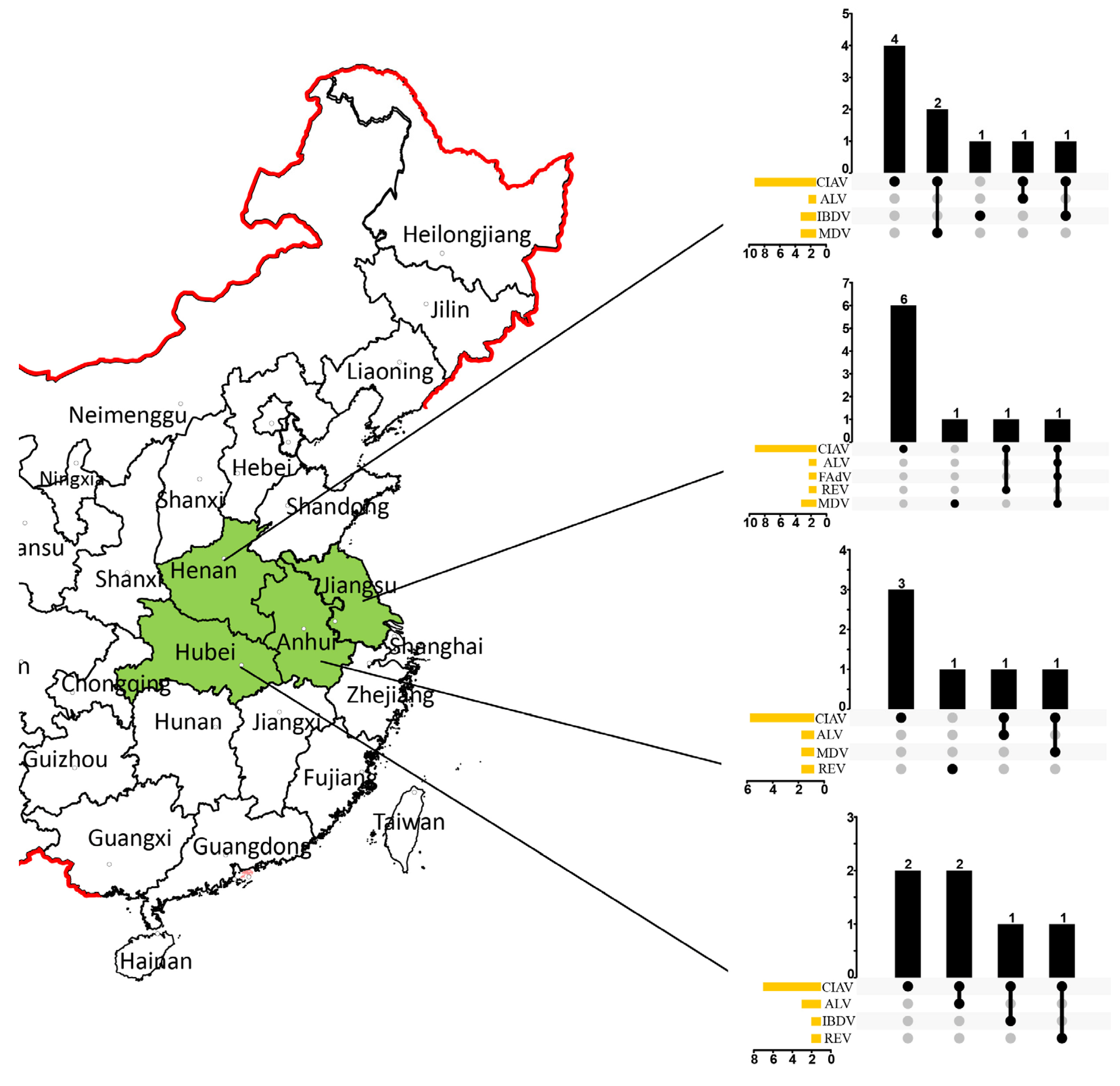
Figure 1. CIAV distribution in China and the coinfection status of ALV, IBDV, FAdV, REV, and MDV in various flocks. The chart marked with green represent the Province for CIAV investigation in this study. The UpSet plot presents the distribution of different viruses in the flocks. The bar chart above represents the number of positive flocks contained in each type of group, and the dotted line at the bottom right presents the infection status. The bar chart at the bottom left represents the number of positive numbers included in each type of virus.
Animals 13 02709 g001
Figure 2. The CIAV phylogenetic tree based on the entire genome. In the ML analysis using MEGA X, the filled circles along the branches represent bootstrap values. The black triangle represents the strains from Anhui Province, the blue square the strains from Jiangsu Province, the green circle the strains from Henan Province, and the red pentagram the strains from Hubei Province.
Animals 13 02709 g002
Figure 3. Recombination occurrence in JS2202, AH2002, HN2203, AH2202, and HB2001 strains analyzed using the Simplot software. (a) Recombination occurrence in JS2202. (b) Recombination occurrence in JS2202. (c) Recombination occurrence in AH2002. (d) Recombination occurrence in HN2203. (e) Recombination occurrence in AH2202. (f) Recombination occurrence in HB2001.
Animals 13 02709 g003
Figure 4. Cartoon scheme of the CIAV-VP1 protein structure. (A) Reference VP1 protein structure of strain SD24. (B) Mutant VP1 protein structure of strain HB2103. (C) Local protein structure of H22 of strain SD24. (D) Local protein structure of N22 of strain HB2103. (E) Local protein structure of K139 of strain SD24. (F) Local protein structure of Q139 of strain HB2103. (G) Local protein structure of E144 of strain SD24. (H) Local protein structure of Q144 of strain HB2103.
Animals 13 02709 g004
Table 1. Detailed information regarding the reference stains used in this study.
Accession No.Strain NameSpeciesSite of Isolation Genome Length (bp)Year
M81223M81223ChickenGermany22981993
U65414.1704ChickenAustralia22981996
L14767.1L14767.1ChickenUSA22981999
AB027470.1TR20ChickenJapan22981999
AF313470.1Del-Rosa aChickenUSA22942000
AB031296.1A2ChickenJapan22982000
AB046590.1C369ChickenJapan22982001
AF227982.1AF227982ChickenAustralia22862001
AJ297685.2clone34ChickenGermany22972002
AF475908.1AF475908ChickenChina22982002
AB119448.1G6ChickenJapan22982003
AY040632.13-IP60ChickenMalaysia22982003
AF285882.1SMSC-1ChickenMalaysia22982003
AF390102.1SMSC-IP60ChickenMalaysia22982003
AY846844.1TJBD40ChickenChina22982004
AF395114BD-3ChickenBangladesh22982004
AY999018SD24ChickenChina22982005
DQ124936.1AH4ChickenChina22982005
AY839944.2LF4ChickenChina22982005
DQ141673.1SD22ChickenChina22982005
DQ124934.1HA4ChickenChina22982005
DQ217401SMSC-1P123WTChickenMalaysia22982005
DQ141671.1SH16ChickenChina22982005
DQ141672HN9ChickenChina22982005
JX964755GXC060821ChickenChina22922006
EF6831593711ChickenAustralia22792007
DQ99139401-4201ChickenUSA22982007
KJ872513CIAV-10ChickenArgentina22982007
D10068.1D10068.1ChickenNetherland22982007
FJ172347.1SDLY08ChickenChina22982008
D31965.182-2ChickenJapan23192008
AF311892.298D02512ChickenUSA22982010
JF507715.1CIAVV89-69ChickenKorea22982011
KJ728827.118ChickenChina22982012
JX260426.1GD-1-12ChickenChina22982012
KC414026Cat-GyvCatChina22952012
JQ690762.1JQ690762.1HumanChina23162012
KM496308.1SC-NC1ChickenChina22982013
KF224935.1GD-K-12ChickenChina22982013
KM496307SC-MZ42AChickenChina22982013
KF224927.1GD-C-12ChickenChina22982013
KM496306.1SC-MZChickenChina22982013
KJ72882414ChickenChina22982014
KU645520.1HN1405ChickenChina22982014
KU645511.1LN1402ChickenChina22982014
KY024579.1RS-BR-15ChickenBrazil22982015
KU645512.1HN1504ChickenChina22982015
KU641014.1JN1503ChickenChina22982015
KX811526.1SD15ChickenChina22982015
KU645506.1SD1512ChickenChina22982015
KY486137.1HLJ15108ChickenChina22982015
KU645519SD1508ChickenChina22982015
KU645510.1SD1509ChickenChina22982015
KY486143.1HLJ15169ChickenChina22982015
KU645524CIAV-DogDogChina22982015
KU645525CIAV-MouseMouseChina22982015
KX447636.1LY-1ChickenChina22982016
KX447637.1LY-2ChickenChina22982016
MN299312.11716TWChickenChina22982016
MN299315.11535TWChickenChina22982016
KX447634.1BS-C2ChickenChina22982017
MG827100.1CAV-SK4-2017ChickenEgypt22982017
KX447633.1BS-C1ChickenChina22982017
MK089243.117SY0902ChickenChina22982017
KX447635HB160430ChickenChina22982017
NC001427Cux-1 aChickenUSA23192018
MH001560.1CAV-EG-13ChickenEgypt22982018
KY486144.1HLJ15170ChickenChina22982018
MK484615.1GX1804ChickenChina22982019
MN103405.1GX1805ChickenChina22982020
MW660821.1SDSPF2020ChickenChina22982020
MN649256.1GX1908L2ChickenChina22982021
MZ668716.1HN2021-1414ChickenChina22982021
ON596886.1Guangxi/2298/2021ChickenChina22982021
OL448839.1SD2008ChickenChina22982021
OL448838.1SD2007ChickenChina22982021
MZ540762.1YN04ChickenChina22982022
a Vaccine strain.
Table 2. Detailed information regarding the stains isolated in this study.
Accession No.Strain NameSpeciesSite of Isolation Genome Length (bp)Year
OQ869186HN1901ChickenChina22982019
OQ869187HB1901ChickenChina22982019
OQ869188JS1901ChickenChina22982019
OQ869189HN2001ChickenChina22982020
OQ869190HB2001ChickenChina22982020
OQ869191JS2001ChickenChina22982020
OQ869192AH2001ChickenChina22982020
OQ869193JS2002ChickenChina22982020
OQ869194HN2002ChickenChina22982020
OQ869195AH2002Chicken China22982020
OQ869196HB2101ChickenChina22982021
OQ869197HB2102ChickenChina22982021
OQ869198HN2101ChickenChina22982021
OQ869199JS2101ChickenChina22982021
OQ869200JS2102ChickenChina22982021
OQ869201HN2102ChickenChina22982021
OQ869202HB2103ChickenChina22982021
OQ869203AH2101ChickenChina22982021
OQ869204HB2201ChickenChina22982022
OQ869205JS2201ChickenChina22982022
OQ869206HN2201ChickenChina22982022
OQ869207HN2202ChickenChina22982022
OQ869208AH2201ChickenChina22982022
OQ869209HN2203ChickenChina22982022
OQ869210AH2202ChickenChina22982022
OQ869211JS2202ChickenChina22982022
OQ869212JS2203ChickenChina22982022
Table 3. Recombinant event I and related average p-values calculated using different recombination detection methods.
Methods RDPGENECONVBootscanMaxchiChimaeraSiSscan3Seq
p-Value8.26 × 10−77.35 × 10−69.20 × 10−62.27 × 10−94.03 × 10−101.46 × 10−157.56 × 10⁻¹⁷
Table 4. Representative mutation sites of VP1 compared with the CIAV reference strain SD24.
StrainSubstitution of the Amino Acid Residues in VP1
22758997125139141144157287290370376394413446
SD24HVTMLKQEVSASLQAG
HN1901---------NPA----
HB1901--------M--GI-S-
JS1901--------M--GI-S-
HN2001-I-LIQ-Q---GI-S-
HB2001---L----M-P-----
JS2001------------M---
AH2001-----------GI-S-
JS2002-----------GI-S-
HN2002Q--L----MTP-----
AH2002-----------GI-S-
HB2101-I-LIQ-Q-TPT----
HB2102---L----MTP-----
HN2101--------M--GI-S-
JS2101NI-LIQ-Q---GI-S-
JS2102-----------GI-S-
HN2102----P---MTP-----
HB2103NI-LIQ-Q-APGI-S-
AH2101NI-LIQ-Q---GI-S-
HB2201--------M--GI-S-
JS2201---L----M-P-----
HN2201--------M--GI-S-
HN2202-----------GI-S-
AH2201---L----MTP-----
HN2203---L----MTP-----
AH2202-----------GI-S-
JS2202--------MTP-----
JS2203--------MTPR----
Disclaimer/Publisher’s Note: The statements, opinions and data contained in all publications are solely those of the individual author(s) and contributor(s) and not of MDPI and/or the editor(s). MDPI and/or the editor(s) disclaim responsibility for any injury to people or property resulting from any ideas, methods, instructions or products referred to in the content.