Article

Occurrence and Genotypic Identification of Blastocystis spp., Enterocytozoon bieneusi, and Giardia duodenalis in Leizhou Black Goats in Zhanjiang City, Guangdong Province, China

1
School of Life Science and Engineering, Foshan University, Foshan 528231, China
2
Maccura Biotechnology Co., Ltd., Chengdu 510000, China
3
Guangdong Provincial Animal Husbandry Technology Promotion Station, Guangzhou 510500, China
4
National Institute of Parasitic Diseases, Chinese Center for Diseases Control and Prevention (Chinese Center for Tropical Diseases Research), Key Laboratory of Parasite and Vector Biology, National Health Commission of the People’s Republic of China (NHC), World Health Organization (WHO) Collaborating Center for Tropical Diseases, National Center for International Research on Tropical Diseases, Shanghai 200025, China
*
Authors to whom correspondence should be addressed.
Animals 2023, 13(17), 2777; https://doi.org/10.3390/ani13172777
Submission received: 4 August 2023 / Revised: 23 August 2023 / Accepted: 28 August 2023 / Published: 31 August 2023

Abstract

:

Simple Summary

We report the occurrence of Blastocystis spp., Enterocytozoon bieneusi, and Giardia duodenalis infections in Leizhou black goats in Zhanjiang City, Guangdong Province, China. The total prevalence rates of Blastocystis spp., E. bieneusi, and G. duodenalis were 33.63% (76/226), 17.7% (40/226), and 24.78% (56/226), respectively. Four Blastocystis spp. subtypes (ST5, ST10, ST14, and ST21), four E. bieneusi genotypes (CHG3, CM21, CHG1, and ET-L2) and two assemblages (A and E) of G. duodenalis were identified. The detection of zoonotic pathogen species, genotypes, and assemblages in Leizhou black goats implied their potential involvement in the transmission of zoonotic parasitic diseases.

Abstract

Blastocystis spp., Enterocytozoon bieneusi, and Giardia duodenalis are three common zoonotic intestinal parasites that cause severe diarrhea and enteric diseases. Leizhou black goats are characterized by a high reproductive rate, fast growth, and good meat quality, making them one of the pre-eminent goat breeds in China. Goats are reportedly common reservoirs of these three intestinal pathogens, but no information on their prevalence or genotypic distributions in black goats in Guangdong Province, China, is available. A total of 226 fecal samples were collected from goats in Zhanjiang city and genomic DNA was extracted from them. The presence of the three pathogens was detected using nested PCR targeting the sequences encoding SSU rRNA (Blastocystis spp.), the internal transcribed spacer of rRNA (ITS; E. bieneusi), as well as beta-giardin, glutamate dehydrogenase, and triosephosphate isomerase (G. duodenalis). All PCR products were sequenced to determine the species and genotypes of the organisms. The total prevalence rates of Blastocystis spp., E. bieneusi, and G. duodenalis were 33.63% (76/226), 17.70% (40/226), and 24.78% (56/226), respectively. Four subtypes of Blastocystis spp. were detected: ST5 (n = 6), ST10 (n = 50), ST14 (n = 14), and ST21 (n = 6). Among them, ST10 was the dominant genotype, accounting for 65.79% of strains, followed by the genotypes ST14 (18.42%), zoonotic ST5 (7.89%), and ST21 (7.89%). Four genotypes of E. bieneusi were detected: CHG3 (n = 32), CM21 (n = 4), CHG1 (n = 2), and ET-L2 (n = 2). Among these, CHG3 was the dominant genotype. Assemblage E (n = 54) and concurrent assemblages A and E (n = 2) were identified in the G. duodenalis-positive goats using multilocus genotyping. Blastocystis spp., E. bieneusi, and G. duodenalis infections were common in Leizhou black goats, all of which have zoonotic genotypes, indicating the potential risk of zoonotic transmission. Our results provide basic data for the prevention and control of these three intestinal pathogens. Further studies are required to better understand their genetic characteristics and zoonotic potential in Guangdong Province.

1. Introduction

Blastocystis spp., Enterocytozoon bieneusi, and Giardia duodenalis are three common opportunistic intestinal parasitic protozoa with wide host ranges, which include domestic animals, wildlife, and humans [1,2,3]. The oocysts or cysts of these three pathogens can survive in the environment for a long time. Human and animal infections often occur through fecal–oral contact or contact with contaminated water or food. These pathogens may cause severe abdominal pain, diarrhea, emaciation, and even death [2,4].
Among the approved subtypes (ST1–ST34) of Blastocystis species, ST1–ST10, ST12, ST14, ST16, and ST23 have been observed in humans [5]. In ASEAN countries, 11 subtypes of Blastocystis spp. have been identified in Artiodactyla: ST1–ST8, ST10, ST12, and ST14 [6]. In China, seven subtypes, ST1, ST3, ST4, ST5, ST6, ST10, and ST14, mainly infect ruminants, including cattle, sheep, and goats [7,8], and ST1, ST3, ST6, and ST7 have been detected in goats in Malaysia [9]. The occurrence of the zoonotic Blastocystis spp. subtypes ST1, ST3, ST4, ST5, ST7, and ST10 in goats suggests their potential transmission to humans.
Enterocytozoon bieneusi is a complex species, divided into 13 different genetic groups based on the sequence of the internal transcribed spacer (ITS) region of the rRNA genes [10]. Groups 1 and 2 contain the majority of zoonotic genotypes, whereas groups 3–13 contain host-adapted genotypes [11,12,13]. Enterocytozoon bieneusi has an estimated overall prevalence of 17.4% in sheep and 16.3% in goats worldwide [14]. Analyses of the ITS sequences have shown that the genotypes BEB6 and COS-1 are most common in sheep, whereas CHG3 and BEB6 are the predominant genotypes in goats [14,15].
Giardiasis has been prevalent or has occurred in outbreaks throughout the world since the 1970s, and is listed by the World Health Organization as one of the neglected diseases that endanger human health [16]. It is estimated that more than 280 million cases of human giardiasis occur annually throughout the world [17]. Giardia duodenalis can be divided into eight assemblages (A–H) according to the different genotypes. The zoonotic assemblages A and B and host-adapted assemblage E of G. duodenalis have been detected in goats, and previous reports have cited assemblage E as the dominant variant [3]. In goats, G. duodenalis infections can occur at any age, but are most commonly observed in young and immunocompromised animals. Genotyping G. duodenalis is a valuable way to identify zoonotic assemblages, and is achieved with sequence analyses of the PCR products of the glutamate dehydrogenase (gdh), β-giardin (bg), and triosephosphate isomerase genes (tpi) with multilocus genotyping (MLG) [18].
Goats are grain-saving types of livestock that provide high-quality meat, cashmere, and milk. These animals have an important role in animal husbandry in China. The Leizhou black goat (Capra hircus) and Chuanzhong black goat (C. hircus) are the two most widely farmed meat goat breeds in Guangdong, although the Leizhou black goat is the only landrace meat goat [19]. Zhanjiang city has the largest number of black goats in Guangdong Province. Black goats in local areas are usually free-range, so their excrement is usually deposited directly into the surrounding environment without further treatment. Three zoonotic parasites of goats, Blastocystis spp., E. bieneusi, and G. duodenalis, not only cause significant production losses in goats, but also pose a potential threat to public health through the cysts or oocysts excreted in goat feces. Understanding the sources and genetic diversity of Blastocystis spp., E. bieneusi, and G. duodenalis involved in these infections is essential to understanding their pathogenicity and the development of strategies for their control, because no effective vaccines or drugs are available. However, there is currently no information on Blastocystis spp., E. bieneusi, or G. duodenalis infections in Leizhou black goats.
The purpose of this study was to examine the occurrence and genotypic distributions of these three intestinal pathogens in Leizhou black goats in Zhanjiang city, in the southernmost region of Guangdong Province, China.

2. Materials and Methods

2.1. Sample Collection

Between October and December 2022, 226 fresh fecal samples from Leizhou black goats were collected on four farms scattered within Zhanjiang city, Guangdong Province (Figure 1). The samples were from 96 growing goats (aged 4–18 months), 64 reserve goats (aged 19–30 months), and 66 adult goats (aged >30 months). Each fresh fecal sample was placed in an individual disposable plastic pouch marked with the farm, goat age, and date of collection, transported quickly to the laboratory while packed in ice, and stored in 2.50% (w/v) potassium dichromate solution at 4 °C until processing (<1 week).

2.2. DNA Extraction

Each fecal sample was washed with distilled water to remove the potassium dichromate before DNA extraction. Approximately 200 mg of each fecal sample was used to extract DNA with an E.Z.N.A.® Stool DNA Kit (Omega Bio-tek Inc., Norcross, GA, USA), according to the instructions of the kit. The DNA samples were stored at −20 °C before PCR analysis.

2.3. PCR Amplification

The DNA from all the samples was amplified using nested PCR to identify those positive for any of the three pathogens of interest. Blastocystis spp. And E. bieneusi were identified based on the small subunit (SSU) rRNA gene [20] and its ITS region, respectively [21]. The detection and genotyping of G. duodenalis was based on the nested PCR amplification and sequence analysis of the bg, gdh, and tpi genes (Table 1).
The PCRs were performed in 25 μL reaction systems: 12.5 μL of 2 × rTaq mix (TaKaRa Co., Ltd., Beijing, China), 1 μL of each primer (10 μM each), 2 μL of DNA sample, and 8.5 μL of nuclease-free deionized water. The PCR products were separated with 1.20% agarose gel electrophoresis, stained with nucleic acid dye (Biosharp, Beijing, China), and observed and recorded with the Azure™ c200 Gel Image Analysis System (Dublin, CA, USA).

2.4. Sequencing and Phylogenetic Analysis

All of the final positive PCR amplicons were transferred to and sequenced by Sangon Biological Engineering Technology and Service Co., Ltd. (Songjiang, Shanghai, China). BLAST (https://blast.ncbi.nlm.nih.gov/Blast.cgi, accessed on 15 June 2023.) was used to compare each obtained sequence with the GenBank database at the National Center for Biotechnology Information (NCBI). SSU rRNA gene sequences of Blastocystis spp., ITS gene sequences of E. bieneusi, and bg gene sequences of G. duodenalis obtained (Supplementary Files S1–S3) were compared with reference sequences acquired from the GenBank database (Supplementary Tables S2–S4), respectively. Phylogenetic trees based on the amplicon sequences of Blastocystis spp., E. bieneusi, and G. duodenalis were constructed with the maximum likelihood (ML) method in MEGA 7.0 (http://www.megasoftware.net/, accessed on 19 June 2023) to assess their genetic relationships. Bootstrap values were calculated with 1000 replicates.

2.5. Statistical Analysis

A χ2 test was performed and 95% confidence intervals (CIs) were calculated using the Wald method in SPSS version 27.0 (SPSS Inc., Chicago, IL, USA) to evaluate the differences in the infection rates among different age groups. Differences were considered significant at p < 0.05.

3. Results

3.1. Occurrence of Blastocystis spp., E. bieneusi, and G. duodenalis

Of the 226 specimens analyzed, 76 (33.63%; 95% CI: 27.4–39.8) were positive for Blastocystis spp. The prevalence of Blastocystis spp. was significantly lower in growing goats (22.91%, 22/96; 95% CI: 14.4–31.5) than in reserve goats (46.88%, 30/64; 95% CI: 34.3–59.4; p = 0.03), but there was no significant difference between the growing and adult goats (36.36%, 24/66; 95% CI: 24.4–48.3; p = 0.09) (Table 2).
The overall detection rate of E. bieneusi was 17.70% (40/226; 95% CI: 12.7–22.7). The prevalence of E. bieneusi was significantly lower in adult goats (6.06%, 4/66; 95% CI: 0.1–12) than in growing goats (25.00%, 24/96; 95% CI: 16.2–33.8; p = 0.03), but did not differ significantly between adult goats and reserve goats (18.75%, 12/64; 95% CI: 8.9–28.6; p = 0.053).
Based on the PCR detection of any of the three genetic loci of G. duodenalis (bg, tpi, or gdh), G. duodenalis was detected in 24.78% of samples (56/226; 95% CI: 19.1–30.5). Unlike the E. bieneusi infection rate, which was highest in growing goats, the prevalence of G. duodenalis was significantly higher in reserve goats (40.63%, 26/64; 95% CI: 28.3–53; p = 0.000008) and growing goats (27.08%, 26/96; 95% CI: 18–36.1; p = 0.0015) than in adult goats (6.06%; 4/66; 95% CI: 0.1–12).
Viewed from the perspective of coinfection, the overall percentage of goats infected with the three pathogens was 7.08% (16/226). The percentages of goats infected with only Blastocystis spp., E. bieneusi, and G. duodenalis were 18.58% (42/226), 5.31% (12/226), and 7.96% (18/226), respectively. Furthermore, 11.50% (26/226) of the total goats were infected with two protozoa, with 3.54% (8/226) infected with G. duodenalis and E. bieneusi, 6.19% (14/226) with G. duodenalis and Blastocystis spp., and 1.77% (4/226) with Blastocystis spp. and E. bieneusi.

3.2. Distributions of Blastocystis spp. Subtypes

Based on an SSU rRNA gene sequence analysis, the 76 Blastocystis spp.-positive isolates were characterized into four subtypes: ST5 (n = 6), ST10 (n = 50), ST14 (n = 14), and ST21 (n = 6) (Table 2, Figure 2). ST5, a zoonotic genotype, accounted for 7.89% of the positive samples, and the sequences obtained in this study were 99% identical to the that of an isolate derived from sheep in China (ON809458.1). ST10 (n = 50) was the predominant subtype, and 30 of the SSU rRNA sequences shared 97–100% homology with that of an isolate derived from dairy cattle in Malaysia (MK240481), whereas the other 20 sequences shared 85–98% homology with a Blastocystis spp. from raccoon dogs in China (MT798805). Of the 14 Blastocystis spp. samples of subtype ST14, 6 shared >85% homology with a Blastocystis spp. (ON796560) from sheep in China, and the SSU rRNA sequences of the other 8 isolates shared >95% similarity with that of MW648930 from goats in Malaysia. ST21 (n = 6) was detected in growing goats (4–18 months) and reserve goats (19–30 months), and shared >85% SSU rRNA homology with ON796563 from goats in China.

3.3. Genotypes of E. bieneusi

In the present study, four E. bieneusi genotypes were identified in the goats based on their genetic divergence and their positions on the ITS-based phylogenetic tree (Table 2, Figure 3): CHG3 (n = 32), CM21 (n = 4), CHG1 (n = 2), and ET-L2 (n = 2). The predominant genotype of E. bieneusi in goats was CHG3 (32/40, 80.00%). Based on the phylogenetic analysis of the ITS sequences determined in this study and reference sequences downloaded from GenBank, all genotypes observed in the study were categorized as group 2. The CHG3 samples (n = 32) were distributed in goats of all ages and shared 95–100% ITS homology with MH822618 from goats in China. CM21 (n = 4) was detected in reserve goats (19–30 months) and shared 99% ITS homology with KU604931 from the golden monkey in China. CHG1 (n = 2) was only detected in adult goats (>30 months) and shared 95% ITS homology with MH822617 from goats in China. In contrast, ET-L2 (n = 2) was only observed in growing goats and shared 98.6% ITS homology with MT231509 from dairy cattle in Ethiopia.

3.4. Genotypes and Subtypes of G. duodenalis

Fifty-six DNA samples were positive for G. duodenalis according to the results for at least one gene locus (bg, gdh, or tpi). A total of 42, 42, and 22 samples were positive for G. duodenalis based on the bg, gdh, and tpi sequences, respectively. Two Giardia assemblages, E and A, were detected in these samples (Figure 4, Table 2). Two of the bg sequences were identified as assemblage A, and the other 40 were identified as assemblage E or subtype E12 (Table S1). Assemblage A (n = 2) shared 100% bg homology with KP687765 from humans in Spain. Thirty bg sequences of assemblage E shared 97–100% homology with that of MK452880 (n = 30) from sheep in Greece, whereas another ten bg sequences shared more than 99% homology with that of KY432834 (n = 10) from dairy cattle in China (Figure 4).
At the gdh locus, all positive samples were identified as assemblage E (n = 42), including subtypes E34 (n = 20) and E36 (n = 22), and shared 98% gdh homology with a G. duodenalis isolate derived from Tan sheep in China (MK645786, MK645797). Twenty-two isolates positive for G. duodenalis at the tpi locus were identified as assemblage E and shared 98–100% homology with KT922262 from calves in Ethiopia.

4. Discussion

Blastocystis spp. is a common intestinal parasite that infects various animals worldwide, including humans, sheep, goats, birds, and insects [6,25]. Because the host specificity of Blastocystis spp. is low, many animals act as storage hosts for its transmission. Nineteen blastocyst subtypes (ST1–ST10, ST12–ST14, ST17–ST22) have been identified in animal populations in China, and new subtypes are still being discovered in different animal populations [26]. Among these, ST3 is the most common subtype of human infections in China, whereas ST5, ST10, and ST1 are the dominant subtypes of infections in pigs, herbivorous animals (cattle, sheep), and carnivores, respectively. The prevalence of Blastocystis spp. in sheep and goats ranges from 0.3% to 94.7% worldwide (after studies with small sample sizes were excluded) [27]. In China, the Blastocystis spp.-positivity rates in sheep and goats range from 0.3% to 58.0%, and the dominant subtype is ST10. However, there are significant differences in the subtypes of Blastocystis spp. in sheep and goats from different provinces or regions in China [27]. The Blastocystis spp.-positive rate in Leizhou black goats was 33.63% in the present study, higher than in goats in most other provinces of China, except Shaanxi (58%), but including Jiangsu (24.0%), Shandong (16.7%), Inner Mongolia (10.70%), Qinghai (7.5%), Heilongjiang (5.5%), and Anhui (0.3%) [27,28]. The prevalence of Blastocystis spp. in Leizhou black goats in Zhanjiang was also higher than in many other countries, including Malaysia (30.9%) and Liberia (10.5%) [27,28]. In the present study, the infection rate in growing goats was 22.91%, significantly lower than the rate in reserve goats (19–30 months; 46.88%). However, there was no significant difference between the rates of Blastocystis spp. infection in growing and adult goats (36.36%).
Based on genetic typing and a phylogenetic analysis of the SSU rRNA gene sequence, four subtypes of Blastocystis spp. were identified: ST10 (n = 50), ST14 (n = 14), ST5 (n = 6), and ST21 (n = 6). Of these, ST10 was the predominant subtype, accounting for 65.79% of infections, consistent with the results of most studies in China [6,8,28]. Interestingly, a rarely detected subtype, ST21 (n = 6), was detected in the present study. The ST21 subtype has not been detected in sheep or goats in other regions of China, except recently in Tibetan sheep and Inner Mongolian goats [27,28]. We also detected the zoonotic ST5 subtype, which accounted for 7.9% (6/76) of the positive samples. ST5 is primarily found in Artiodactyla, such as pigs, goats, sheep, and cows, and in rodents, such as Rhizomys sinensis [29], Callosciurus erythraeus [30], Hydrochoerys hydrochaeris [31], and Clethrionomys glareolus [32]. In Australia, pig herds and workers in close contact with pigs showed a high prevalence of the ST5 subtype, indicating its potential transmission between animals and humans [33]. Blastocystis spp. has two life stages: the cyst and the trophozoite. The host often becomes infected by consuming food or drinking water contaminated with cysts. Some infected humans, especially those with mixed infections of multiple pathogens or an impaired immune system, often experience symptoms such as abdominal pain, diarrhea, and vomiting, which can be life-threatening in severe cases. Apart from zoonotic subtype ST5, it remains unclear whether any other zoonotic subtypes are present in Guangdong black goats. Because black goats potentially play a role in the transmission of Blastocystis spp. to humans [6], it is important that further epidemiological studies of Blastocystis spp. are conducted among animal husbandry workers, water sources, and black goat populations in nearby regions.
Enterocytozoon bieneusi is an emerging zoonotic intestinal pathogen that infects humans and many animal species. Many studies have identified and genotyped E. bieneusi in black goats throughout the world [34,35,36,37]. The infection rate of E. bieneusi in goats ranges from 0 to 100% worldwide, and the overall prevalence in goats is estimated to be 16.3% [14]. However, genotypes and prevalence rates probably also differ by region. This is the first report of E. bieneusi in black goats in Guangdong Province, with a prevalence of 17.7% (40/226), which is lower than in Egypt (100%, 11/11) [37], Thailand (19.2%, 14/73) [38], Henan (66.7%, 104/156; 50.7%, 73/144; 32.9%, 113/343) [15,39,40], Chongqing (62.5%, 5/8) [40], Shaanxi (47.8%, 22/46; 21.9%, 106/485) [15,40], the Ningxia Autonomous Region (29.7%, 89/300) [41], Yunnan (22.4%, 30/134) [40], Heilongjiang (21.8%, 12/55) [42], and Qinghai (18.6%, 11/59) [43], but higher than in Slovakia (0%, 0/20) [44], Shandong (0%, 0/24) [45], Jiangsu (2.7%, 2/74) [45], Anhui (5.2%, 30/574; 7.5%, 6/80) [40,45], Yunnan (8.93%, 30/336; 10.3%, 93/907) [34,36], and the Tibet Autonomous Region (9.6%, 25/260) [46]. These results indicate the importance of goats as hosts for E. bieneusi and their potential role in transmitting microsporidiosis caused by this parasite.
The molecular characterization identified four E. bieneusi genotypes: CHG3 (n = 32), CM21 (n = 4), CHG1 (n = 2), and ET-L2 (n = 2). CHG3 was the dominant genotype, accounting for 80% of the positive samples, consistent with the results of Wang et al. [39]. CHG3 is widely distributed globally, and is found extensively in domestic animals, such as goats and sheep, in different regions or provinces of China, including in ruminant animals in northwest China [47], sheep and goats in east–central China [45], black-boned sheep and goats in Yunnan Province in southwestern China [36], and black goats in the southernmost Hainan Province [35]. However, it is interesting to note that the BEB6 genotype, which was previously reported as prevalent or present in sheep and goats in the northwest region [47], east–central China [45], and Yunnan Province [39], was not detected in the present study. The BEB6 genotype was initially considered to be specific to cattle, but is now recognized as a dominant genotype with a wide geographic distribution, a broad host range, and zoonotic potential [45]. This discrepancy may be related to the fact that our study focused primarily on goats, and previous studies have suggested that CHG3 is the dominant genotype in goats, whereas the BEB6 genotype tends to be dominant in sheep [14,39,45].
G. duodenalis is widely distributed in ruminant populations, such as goats and sheep, worldwide. The total prevalence of giardiasis in sheep and goats in China is estimated to be 7.00%, although it ranges from 0.00% to 28.93% in different provinces [48]. The prevalence of G. duodenalis in Leizhou black goats in Guangdong in the present study was 24.78% (56/226), higher than in goats or sheep in Anhui (1.62%, 27/1847), Gansu (1.69%, 3/177), Qinghai (2.88%, 77/1321), Henan (3.4%, 192/4434), Shaanxi (4.72%, 227/2930), Xinjiang (7.55%, 24/318), Yunnan (8.09%, 125/1568), Hainan (11%, 11/100), Sichuan (13.14%, 65/465), Ningxia (14.5%, 147/1014), and Chongqing (23.59%, 71/301), but lower than Inner Mongolia (28.93%, 150/584) [48]. Many factors may contribute to these differences: the study sample size, local climate, detection method, study design, age of the tested hosts, and the management of the animals [48]. For example, the prevalence in reserve goats (19–30 months) in the present study was 40.63% (26/64), significantly higher than in adult sheep (6.06%, 4/66).
G. duodenalis strains infecting goats or sheep belong predominantly to assemblage E, but often occur in mixed infections of assemblages A and E, assemblages A and B, or assemblages A, B, and E [48,49,50,51]. Consistent with previous research [48], assemblage E was the predominant assemblage (96.43%, 54/56) in our Leizhou black goats. The co-occurrence of assemblages A and E accounted for 3.57% of infections. Assemblage A has a broad host range, infecting various mammals, including humans, domestic animals, and wildlife [52]. Assemblage E is commonly found in ruminant animals, such as sheep, goats, and cattle, but there have been increasing reports of human infections in recent years [53,54]. Preliminary investigations of the G. duodenalis infection status of pig and cattle herds in different regions of Guangdong Province were conducted by our laboratory [55,56]. These studies found that G. duodenalis infection was relatively common in pigs (18.04%, 94/521) and cattle (18.85%, 69/366) in different regions. Interestingly, the predominant assemblages of G. duodenalis found in cattle and pig herds are assemblage A, assemblage E, and their corresponding subtypes, whereas assemblage B was not detected in any of these livestock populations. Most parts of Guangdong Province have a subtropical monsoonal climate with abundant rainfall and a dense river network, and both assemblages (A and E), which are detected in pigs, dairy cattle and goats, possess the ability to transmit to humans and animals across species. This suggests a potential transmission cycle between workers who are in close contact with both goats and other mammals. More epidemiological research is required to better understand the spread of G. duodenalis among neighboring populations and water sources.

5. Conclusions

This study evaluated the prevalence and genotypic characteristics of Blastocystis spp., E. bieneusi, and G. duodenalis in Leizhou black goats in Zhanjiang City, Guangdong Province, China. The total prevalence rates of Blastocystis spp., E. bieneusi, and G. duodenalis were 33.63% (76/226), 17.7% (40/226), and 24.78% (56/226), respectively. Four Blastocystis spp. subtypes (ST5, ST10, ST14, and ST21), four E. bieneusi genotypes (CHG3, CM21, CHG1, and ET-L2), and two assemblages (A and E) of G. duodenalis were detected. Our findings provided valuable data for understanding the molecular epidemiology of Blastocystis spp., E. bieneusi, and G. duodenalis in Leizhou black goats. Further research is required to understand the epidemiology and genotypic characteristics of these pathogens in animal husbandry workers and water sources in nearby regions, and the possible repercussions for public health.

Supplementary Materials

The following supporting information can be downloaded at: https://www.mdpi.com/article/10.3390/ani13172777/s1. Table S1. Multilocus characterization of G. duodenalis isolates based on bg, tpi and gdh genes. Table S2. GenBank accession numbers of all SSU rRNA gene reference sequences of Blastocystis spp. used for phylogenetic analysis. Table S3. GenBank accession numbers of all ITS gene reference sequences of E. bieneusi used for phylogenetic analysis. Table S4. GenBank accession numbers of all bg gene reference sequences of G. duodenalis used for phylogenetic analysis. File S1. The raw sequences of the SSU rRNA gene of Blastocystis spp. used for phylogenetic analysis. File S2. The raw sequences of the ITS gene of E. bieneusi used for phylogenetic analysis. File S3. The raw sequences of the bg gene of G. duodenalis used for phylogenetic analysis.

Author Contributions

Conceptualization, X.Y., Y.H. and H.Z.; methodology, X.Y., H.W., X.M. and K.Y.; writing—original draft preparation, X.Y. and H.W.; writing—review and editing, X.Y., Y.H. and H.W.; visualization, H.W. and A.W.; statistical analysis, H.W. and Y.L.; sampling, H.W., X.M., K.Y., A.W., J.G. and Y.L.; funding acquisition, X.Y. and H.Z. All authors have read and agreed to the published version of the manuscript.

Funding

This research was funded by the Guangdong Basic and Applied Basic Research Foundation (2022A1515110474, 2021B1515120006).

Institutional Review Board Statement

This study was conducted under the approval and instructions of the ethics committee of Foshan University and the animal ethics requirements of the People’s Republic of China [No. FS2022107].

Informed Consent Statement

Not applicable.

Data Availability Statement

All datasets are contained within manuscript.

Conflicts of Interest

The authors declare no conflict of interest. The company had no role in the design of the study; in the collection, analyses, or interpretation of data; in the writing of the manuscript, or in the decision to publish the results.

References

  1. Muadica, A.S.; Köster, P.C.; Dashti, A.; Bailo, B.; Hernández-de-Mingo, M.; Reh, L. Molecular diversity of Giardia duodenalis, Cryptosporidium spp. and Blastocystis sp. in asymptomatic school children in Leganés, Madrid (Spain). Microorganisms 2020, 8, 466. [Google Scholar] [CrossRef]
  2. Zhang, K.; Zheng, S.; Wang, Y.; Wang, K.; Wang, Y.; Gazizova, A. Occurrence and molecular characterization of Cryptosporidium spp., Giardia duodenalis, Enterocytozoon bieneusi, and Blastocystis sp. in captive wild animals in zoos in Henan. BMC Vet. Res. 2021, 17, 332–341. [Google Scholar] [CrossRef]
  3. Feng, Y.; Xiao, L. Zoonotic potential and molecular epidemiology of Giardia species and giardiasis. Clin. Microbiol. Rev. 2011, 24, 110–140. [Google Scholar] [CrossRef]
  4. Santín, M.; Fayer, R. Microsporidiosis: Enterocytozoon bieneusi in domesticated and wild animals. Res. Vet. Sci. 2011, 90, 363–371. [Google Scholar] [CrossRef]
  5. Zhang, J.; Fu, Y.; Bian, X.; Han, H.; Dong, H.; Zhao, G. Molecular characterization and prevalence of Cryptosporidium spp. in sheep and goats in western Inner Mongolia, China. Parasitol. Int. 2023, 122, 537–545. [Google Scholar] [CrossRef]
  6. Rauff-Adedotun, A.A.; Zain, S.N.M.; Haziqah, M.T.F. Current status of Blastocystis sp. in animals from Southeast Asia: A review. Parasitol. Res. 2020, 119, 3559–3570. [Google Scholar] [CrossRef]
  7. Zhou, T.Z.; Ju, R.C.; Huang, S.X.; Qin, J.J.; Xiao, H.C.; Li, J.C.; Li, F.M.; Peng, J.B. Progress in the Blastocystis research of bovine, sheep and goats. Chin. J. Vet. Med. 2022, 58, 92–95. [Google Scholar]
  8. Song, J.K.; Yin, Y.L.; Yuan, Y.J.; Tang, H.; Ren, G.J.; Zhang, H.J. First genotyping of Blastocystis sp. in dairy, meat, and cashmere goats in northwestern China. Acta Trop. 2017, 176, 277–282. [Google Scholar] [CrossRef] [PubMed]
  9. Tan, T.C.; Tan, P.C.; Sharma, R.; Sugnaseelan, S.; Suresh, K.G. Genetic diversity of rodent Blastocystis sp. from Peninsular Malaysia. Parasitol. Res. 2018, 35, 85–89. [Google Scholar] [CrossRef]
  10. Jiang, Y.; Liu, L.; Yuan, Z.; Liu, A.; Cao, J.; Shen, Y. Molecular identification and genetic characteristics of Cryptosporidium spp., Giardia duodenalis, and Enterocytozoon bieneusi in human immunodeficiency virus/acquired immunodeficiency syndrome patients in Shanghai, China. Parasites Vectors 2023, 16, 53–62. [Google Scholar] [CrossRef]
  11. Li, W.; Feng, Y.; Santin, M. Host Specificity of Enterocytozoon bieneusi and Public Health Implications. Trends Parasitol. 2019, 35, 436–451. [Google Scholar] [CrossRef]
  12. Liu, Y.Y.; Qin, R.L.; Mei, J.J.; Zou, Y.; Zhang, Z.H.; Zheng, W.B. Molecular detection and genotyping of Enterocytozoon bieneusi in beef cattle in Shanxi province, North China. Animals 2022, 12, 2961. [Google Scholar] [CrossRef] [PubMed]
  13. Zhang, T.; Ren, G.; Zhou, H.; Qiang, Y.; Li, J.; Zhang, Y. Molecular prevalence and genetic diversity analysis of Enterocytozoon bieneusi in humans in Hainan province, China: High diversity and unique endemic genetic characteristics. Front. Public Health 2022, 27, 1007130. [Google Scholar] [CrossRef] [PubMed]
  14. Taghipour, A.; Bahadory, S.; Javanmard, E. The global molecular epidemiology of microsporidia infection in sheep and goats with focus on Enterocytozoon bieneusi: A systematic review and meta-analysis. Trop. Med. Health 2021, 49, 66–78. [Google Scholar] [CrossRef]
  15. Peng, X.Q.; Tian, G.R.; Ren, G.J.; Yu, Z.-Q.; Lok, J.B.; Zhang, L.X. Infection rate of Giardia duodenalis, Cryptosporidium spp. and Enterocytozoon bieneusi in cashmere, dairy and meat goats in China. Infect. Genet. Evol. 2016, 41, 26–31. [Google Scholar] [CrossRef]
  16. Savioli, L.; Smith, H.; Thompson, A. Giardia and Cryptosporidium join the ‘Neglected Diseases Initiative’. Trends Parasitol. 2006, 22, 203–208. [Google Scholar] [CrossRef] [PubMed]
  17. Cai, W.; Ryan, U.; Xiao, L.; Feng, Y. Zoonotic giardiasis: An update. Parasitol. Res. 2021, 120, 4199–4218. [Google Scholar] [CrossRef] [PubMed]
  18. Zou, Y.; Li, X.D.; Meng, Y.M.; Wang, X.L.; Wang, H.N.; Zhu, X.Q. Prevalence and multilocus genotyping of Giardia duodenalis in zoo animals in three cities in China. Parasitol. Res. 2022, 121, 2359–2366. [Google Scholar] [CrossRef] [PubMed]
  19. Tian, H.C.; Deng, M.; Guo, Y.Q.; Liu, G.B.; Wu, L.F.; Sun, B.L. Analysis of differences in serum physiological and biochemical indicators and meat quality between Leizhou black goats and Chuanzhong black goats. Heilongjiang Anim. Sci. Vet. Med. 2020, 22, 55–58. [Google Scholar]
  20. Santín, M.; Trout, J.M.; Fayer, R. Prevalence of Enterocytozoon bieneusi in post-weaned dairy calves in the eastern United States. Parasitol. Res. 2004, 93, 287–289. [Google Scholar] [CrossRef]
  21. Feng, Y.; Li, N.; Dearen, T.; Lobo, M.L.; Matos, O.; Cama, V. Development of a multilocus sequence typing tool for high-resolution genotyping of Enterocytozoon bieneusi. Appl. Environ. Microbiol. 2011, 77, 4822–4828. [Google Scholar] [CrossRef]
  22. Lalle, M.; Pozio, E.; Capelli, G.; Bruschi, F.; Crotti, D.; Cacciò, S.M. Genetic heterogeneity at the β-giardin locus among human and animal isolates of Giardia duodenalis and identification of potentially zoonotic subgenotypes. Int. J. Parasitol. 2005, 35, 207–213. [Google Scholar] [CrossRef] [PubMed]
  23. Sulaiman, I.M.; Fayer, R.; Bern, C.; Gilman, R.H.; Trout, J.M.; Schantz, P.M. Triosephosphate isomerase gene characterization and potential zoonotic transmission of Giardia duodenalis. Emerg. Infect. Dis. 2003, 9, 1444–1452. [Google Scholar] [CrossRef] [PubMed]
  24. Cacciò, S.M.; Beck, R.; Lalle, M.; Marinculic, A.; Pozio, E. Multilocus genotyping of Giardia duodenalis reveals striking differences between assemblages A and B. Int. J. Parasitol. 2008, 38, 1523–1531. [Google Scholar] [CrossRef] [PubMed]
  25. Karimi, P.; Shafaghi-Sisi, S.; Meamar, A.R.; Razmjou, E. Molecular identification of Cryptosporidium, Giardia, and Blastocystis from stray and household cats and cat owners in Tehran, Iran. Sci. Rep. 2023, 13, 1554–1566. [Google Scholar] [CrossRef]
  26. Liu, L.K.; Wang, P.L.; Han, H.; Wang, R.J.; Jian, F.C. Current status of Blastocystis infection in China. Chin. J. Zoonoses 2021, 37, 548–562. [Google Scholar]
  27. Yang, F.; Gou, J.M.; Yang, B.K.; Du, J.Y.; Yao, H.Z.; Ren, M. Prevalence and subtype distribution of Blastocystis in Tibetan sheep in Qinghai province, Northwestern China. Protist 2020, 174, 125948. [Google Scholar] [CrossRef]
  28. Zhang, J.; Fu, Y.; Bian, X.; Han, H.; Dong, H.; Zhao, G. Molecular identification and genotyping of Blastocystis sp. in sheep and goats from some areas in Inner Mongolia, Northern China. Parasitol. Int. 2023, 94, 102739. [Google Scholar] [CrossRef]
  29. Song, J.; Yang, X.; Ma, X.; Wu, X.; Wang, Y.; Li, Z. Molecular characterization of Blastocystis sp. in Chinese bamboo rats (Rhizomys sinensis). Parasite 2021, 28, 81–87. [Google Scholar] [CrossRef]
  30. Liu, X.; Ge, Y.; Wang, R.; Dong, H.; Yang, X.; Zhang, L. First report of Blastocystis infection in Pallas’s squirrels (Callosciurus erythraeus) in China. Vet. Res. Commun. 2021, 45, 441–445. [Google Scholar] [CrossRef]
  31. Cian, A.; Safadi, D.E.; Osman, M.; Moriniere, R.; Gantois, N.; Benamrouz-Vanneste, S. Molecular epidemiology of Blastocystis sp. in various animal groups from two French zoos and Evaluation of Potential Zoonotic Risk. PLoS ONE 2017, 12, e0169659. [Google Scholar] [CrossRef]
  32. Alfellani, M.A.; Taner-Mulla, D.; Jacob, A.S.; Imeede, C.A.; Yoshikawa, H.; Stensvold, C.R. Genetic diversity of blastocystis in livestock and zoo animals. Protist 2013, 164, 497–509. [Google Scholar] [CrossRef] [PubMed]
  33. Wang, W.; Owen, H.; Traub, R.J.; Cuttell, L.; Inpankaew, T.; Bielefeldt-Ohmann, H. Molecular epidemiology of Blastocystis in pigs and their in-contact humans in Southeast Queensland, Australia, and Cambodia. Vet. Parasitol. 2014, 203, 264–269. [Google Scholar] [CrossRef] [PubMed]
  34. Xie, S.C.; Zou, Y.; Li, Z.; Yang, J.F.; Zhu, X.Q.; Zou, F.C. Molecular detection and genotyping of Enterocytozoon bieneusi in black goats (Capra hircus) in Yunnan province, Southwestern China. Animals 2021, 11, 3387. [Google Scholar] [CrossRef] [PubMed]
  35. Zhou, H.H.; Zheng, X.L.; Ma, T.M.; Qi, M.; Cao, Z.X.; Chao, Z. Genotype identification and phylogenetic analysis of Enterocytozoon bieneusi in farmed black goats (Capra hircus) from China’s Hainan province. Parasite 2019, 26, 62–69. [Google Scholar] [CrossRef]
  36. Chen, D.; Wang, S.S.; Zou, Y.; Li, Z.; Xie, S.C.; Shi, L.Q. Prevalence and multi-locus genotypes of Enterocytozoon bieneusi in black-boned sheep and goats in Yunnan province, southwestern China. Infect. Genet. Evol. 2018, 65, 385–391. [Google Scholar] [CrossRef]
  37. Al-Herrawy, A.Z.; Gad, M.A. Microsporidial Spores in Fecal Samples of Some Domesticated Animals Living in Giza, Egypt. Iran. J. Parasitol. 2016, 11, 195–203. [Google Scholar]
  38. Udonsom, R.; Prasertbun, R.; Mahittikorn, A.; Chiabchalard, R.; Sutthikornchai, C.; Palasuwan, A. Identification of Enterocytozoon bieneusi in goats and cattle in Thailand. BMC Vet. Res. 2019, 15, 308–315. [Google Scholar] [CrossRef]
  39. Wang, P.; Zheng, L.; Liu, L.; Yu, F.; Jian, Y.; Wang, R. Genotyping of Cryptosporidium spp, Giardia duodenalis and Enterocytozoon bieneusi from sheep and goats in China. BMC Vet. Res. 2022, 18, 361–372. [Google Scholar] [CrossRef]
  40. Shi, K.; Li, M.; Wang, X.; Li, J.; Karim, M.R.; Wang, R. Molecular survey of Enterocytozoon bieneusi in sheep and goats in China. Parasites Vectors 2016, 9, 23–31. [Google Scholar] [CrossRef]
  41. Zhang, Y.; Mi, R.; Yang, J.; Wang, J.; Gong, H.; Huang, Y. Enterocytozoon bieneusi genotypes in farmed goats and sheep in Ningxia, China. Infect. Genet. Evol. 2020, 85, 104559. [Google Scholar] [CrossRef] [PubMed]
  42. Zhao, W.; Zhang, W.; Yang, D.; Zhang, L.; Wang, R.; Liu, A. Prevalence of Enterocytozoon bieneusi and genetic diversity of ITS genotypes in sheep and goats in China. Infect. Genet. Evol. 2015, 32, 265–270. [Google Scholar] [CrossRef]
  43. Zhang, Q.; Zhang, Z.; Ai, S.; Wang, X.; Zhang, R.; Duan, Z. Cryptosporidium spp., Enterocytozoon bieneusi, and Giardia duodenalis from animal sources in the Qinghai-Tibetan Plateau Area (QTPA) in China. Comp. Immunol. Microbiol. Infect. Dis. 2019, 67, 101346. [Google Scholar] [CrossRef] [PubMed]
  44. Valenčáková, A.; Danišová, O. Molecular characterization of new genotypes Enterocytozoon bieneusi in Slovakia. Acta Trop. 2018, 191, 217–220. [Google Scholar] [CrossRef] [PubMed]
  45. Li, W.C.; Wang, K.; Gu, Y.F. Detection and genotyping study of Enterocytozoon bieneusi in sheep and goats in East-central China. Acta Parasitol. 2019, 64, 44–50. [Google Scholar] [CrossRef]
  46. Chang, Y.; Wang, Y.; Wu, Y.; Niu, Z.; Li, J.; Zhang, S. Molecular characterization of Giardia duodenalis and Enterocytozoon bieneusi isolated from Tibetan sheep and Tibetan goats under natural grazing conditions in Tibet. J. Eukaryot. Microbiol. 2020, 67, 100–106. [Google Scholar] [CrossRef]
  47. Dong, H.; Zhao, Z.; Zhao, J.; Fu, Y.; Lang, J.; Zhang, J. Molecular characterization and zoonotic potential of Enterocytozoon bieneusi in ruminants in northwest China. Acta Trop. 2020, 234, 106622. [Google Scholar] [CrossRef]
  48. Geng, H.L.; Yan, W.L.; Wang, J.M.; Meng, J.X.; Zhang, M.; Zhao, J.X. Meta-analysis of the prevalence of Giardia duodenalis in sheep and goats in China. Microb. Pathog. 2023, 179, 106097. [Google Scholar] [CrossRef]
  49. Jafari, H.; Jalali, M.H.R.; Shapouri, M.S.A.; Hajikolaii, M.R.H. Determination of Giardia duodenalis genotypes in sheep and goat from Iran. J. Parasit. Dis. 2014, 38, 81–84. [Google Scholar] [CrossRef]
  50. Zhang, W.; Zhang, X.; Wang, R.; Liu, A.; Shen, Y.; Ling, H. Genetic characterizations of Giardia duodenalis in sheep and goats in Heilongjiang province, China and possibility of zoonotic transmission. PLoS Negl. Trop. Dis. 2012, 6, e1826. [Google Scholar] [CrossRef]
  51. Yin, Y.L.; Zhang, H.J.; Yuan, Y.J.; Tang, H.; Chen, D.; Jing, S. Prevalence and multi-locus genotyping of Giardia duodenalis from goats in Shaanxi province, northwestern China. Acta Trop. 2018, 182, 202–206. [Google Scholar] [CrossRef] [PubMed]
  52. Li, J.; Wang, H.; Wang, R.; Zhang, L. Giardia duodenalis infections in humans and other animals in China. Front. Microbiol. 2017, 8, 2004. [Google Scholar] [CrossRef]
  53. Garcia-R, J.C.; Ogbuigwe, P.; Pita, A.B.; Velathanthiri, N.; Knox, M.A.; Biggs, P.J. First report of novel assemblages and mixed infections of Giardia duodenalis in human isolates from New Zealand. Acta Trop. 2021, 220, 105969. [Google Scholar] [CrossRef] [PubMed]
  54. Helmy, Y.A.; Klotz, C.; Wilking, H.; Krücken, J.; Nöckler, K.; Samson-Himmelstjerna, G.V. Epidemiology of Giardia duodenalis infection in ruminant livestock and children in the Ismailia province of Egypt: Insights by genetic characterization. Parasites Vectors 2014, 7, 321. [Google Scholar] [CrossRef]
  55. Zhong, M.L.; Zhang, H.J.; Chen, X.J.; Li, F.K.; Qiu, X.X.; Li, Y.; Huang, F.Q.; Wang, N.N.; Yu, X.G. Infection status and molecular characterization of Giardia dunodinalis in pigs in Guangdong province. Chin. Anim. Husb. Vet. Med. 2022, 49, 1592–1598. [Google Scholar]
  56. Wang, H.C.; Mu, X.R.; Zhong, M.L.; Li, B.; Yuan, K.J.; Wu, A.F.; Li, J.N.; Yu, X.G.; Zhang, H.J. Prevalence and molecular characterization of Giardia duodenalis in dairy cattle in parts of Guangdong province. Prog. Vet. Med. 2023, 44, 16–22. [Google Scholar]
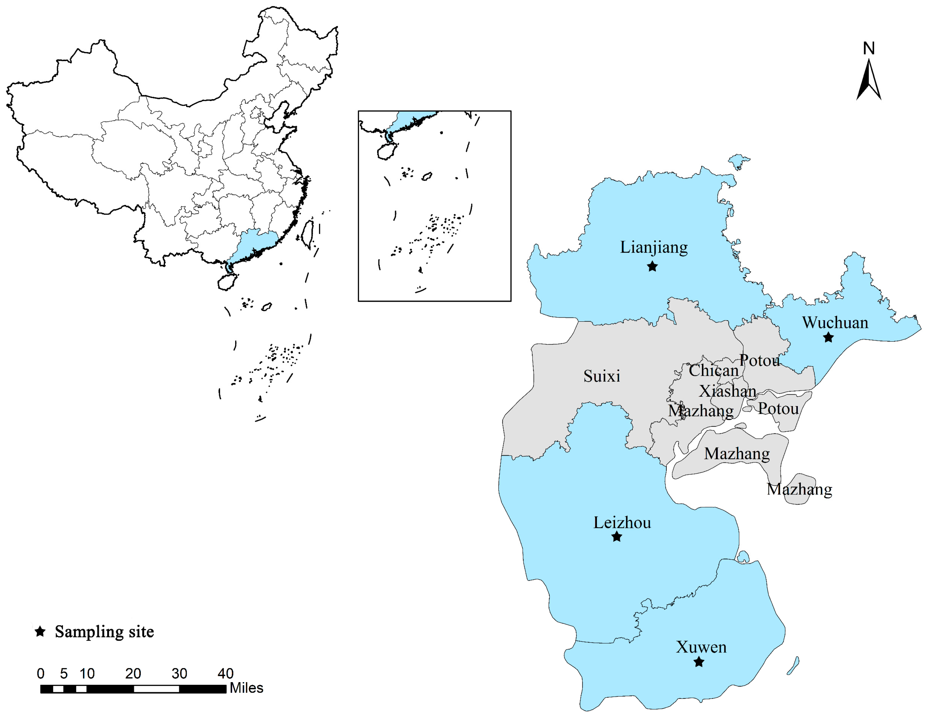
Figure 1. Sampling sites in the study area in Zhanjiang City, Guangdong Province, China. Black solid pentagrams indicate sampling sites.
Animals 13 02777 g001
Figure 2. Phylogenetic tree of Blastocystis spp. in black goats based on SSU rRNA gene sequences. The Tamura–Nei model method was used with bootstrap evaluation of 1000 replicates. All the genotypes identified in this study are marked by red solid triangles. Bootstrap values are shown when >70%.
Animals 13 02777 g002
Figure 3. Phylogenetic tree of E. bieneusi in black goats based on ITS gene sequences. The Tamura–Nei model method was used with bootstrap evaluation of 1000 replicates. All the genotypes identified in this study are marked by red solid triangles. Bootstrap values are shown when >70%.
Animals 13 02777 g003
Figure 4. Phylogenetic tree of G. duodenalis in black goats based on bg gene sequences. The Tamura–Nei model method was used with bootstrap evaluation of 1000 replicates. All the known genotypes identified in this study are marked by red solid triangles. Bootstrap values are shown when >70%.
Animals 13 02777 g004
Table 1. Primers used in the characterization of the G. duodenalis, E. bieneusi, and Blastocystis spp.
GeneNucleotide Sequences of Primer (5′-3′)Expected Product Size (bp)Annealing Temperature (°C)Reference
bg gene of G. duodenalisBG1: AAGCCCGACGACCTCACCCGCAGTGC 51555[22]
BG2: GAGGCCGCCCTGGATCTTCGAGACGAC
BG3: GAACGAACGAGATCGAGGTCCG55
BG4: CTCGACGAGCTTCGTGTT
tpi gene of G. duodenalisTPI1:AAATYATGCCTGCTCGTCG53057[23]
TPI2:CAAACCTTYTCCGCAAACC
TPI3:CCCTTCATCGGYGGTAACTT57
TPI4:GTGGCCACCACYCCCGTGCC
gdh gene of G. duodenalisGdh1: TTCCGTRTYCAGTACAACTC53059[24]
Gdh2: ACCTCGTTCTGRGTGGCGCA
Gdh3: ATGACYGAGCTYCAGAGGCACGT59
Gdh4: GTGGCGCARGGCATGATGCA
ITS gene of E. bieneusiITS1: GATGGTCATAGGGATGAAGAGCTT 39257[21]
ITS2: TATGCTTAAGTCCAGGGAG
ITS3: AGGGATGAAGAGCTTCGGCTCTG55
ITS4: AGTGATCCTGTATTAGGGATATT
SSU rRNA of Blastocystis spp.SSU rRNAF: ATCTGGTTGATCCTGCCAGT60055[20]
SSU rRNAR: GAGCTTTTTAACTGCAACAACG
Table 2. Colonization frequency and genotypes of Blastocystis spp., E. bieneusi and G. duodenalis in different age groups.
Age
(Months)
Sample Size
(n)
BlastocystisE. bieneusiG. duodenalis
No.
Positive
Prevalence %
(95% CI)
Subtypes
(n)
No.
Positive
Prevalence %
(95% CI)
Genotype
(n)
No.
Positive
Prevalence%
(95% CI)
Assemblage
(n)
Growing goats (4–18 months)962222.91%
(14.4–31.5)
ST10 (12)
ST14 (8)
ST21 (2)
2425.00%
(16.2–33.8)
CHG3 (22)
ET-L2 (2)
2627.08%
(18–36.1)
E (26)
Reserve goats (19–30 months)643046.88%
(34.3–59.4)
ST10 (20)
ST14 (2)
ST21 (4)
ST5 (4)
1218.75%
(8.9–28.6)
CHG3 (8)
CM21 (4)
2640.63%
(28.3–53)
E (24)
A + E (2)
Adult goats
(>30 months)
662436.36%
(24.4–48.3)
ST10 (18)
ST14 (4)
ST5 (2)
46.06%
(0.1–12)
CHG3 (2)
CHG1 (2)
46.06%
(0.1–12)
E (4)
Total2267633.63%
(27.4–39.8)
ST5 (6)
ST10 (50)
ST14 (14)
ST21 (6)
4017.70%
(12.7–22.7)
CHG3 (32)
CHG1 (2)
CM21 (4)
ET-L2 (2)
5624.78%
(19.1–30.5)
E (54)
A + E (2)
Disclaimer/Publisher’s Note: The statements, opinions and data contained in all publications are solely those of the individual author(s) and contributor(s) and not of MDPI and/or the editor(s). MDPI and/or the editor(s) disclaim responsibility for any injury to people or property resulting from any ideas, methods, instructions or products referred to in the content.