Article

Thyreophagus tauricus sp. n., a New Subcortical Mite Species (Acari: Acaridae), with a COX1 DNA Sequence Analysis of Several Economically Important Species of Thyreophagus †

by
Pavel B. Klimov
1,2,*,
Vasiliy B. Kolesnikov
1,3,
Vladimir A. Khaustov
1 and
Alexander A. Khaustov
1
1
X-Bio Institute, Tyumen State University, 10 Semakova Str., 625003 Tyumen, Russia
2
Lilly Hall of Life Sciences, Purdue University, G-225, 915 W State St., West Lafayette, IN 47907, USA
3
Federal Public Budgetary Scientific Institution, All-Russian Research Institute of Plant Protection, 396030 Voronezh, Russia
*
Author to whom correspondence should be addressed.
urn:lsid:zoobank.org:pub:37D3DEB8-69F6-4EC6-9BDB-338F4A987BB9.
Animals 2023, 13(22), 3546; https://doi.org/10.3390/ani13223546
Submission received: 16 October 2023 / Revised: 2 November 2023 / Accepted: 10 November 2023 / Published: 16 November 2023
(This article belongs to the Section Animal Genetics and Genomics)

Abstract

:

Simple Summary

In recent years, there has been a growing interest in finding sustainable and environmentally friendly solutions to combat agricultural pests while minimizing the adverse impacts of chemical pesticides. Species of the genus Thyreophagus have emerged as a valuable asset in this pursuit. These mites are utilized as factitious prey for the mass rearing of predatory mites. Predatory mites, in turn, play a crucial role in biological pest control—they feed on a variety of agricultural pests such as spider mites, thrips and other small arthropods that harm crops. Despite their significance, we do not know much about Thyreophagus mites because many species live in hidden habitats and are difficult to study. As part of our survey, we discovered a new species, Thyreophagus tauricus, and provide detailed descriptions of its different life stages. Genetic sequencing was also performed to identify this new species and compare it with others: Thyreophagus corticalis (broadly distributed Palearcic species), Th. calusorum, Th. entomophagus (economically important factitious prey mites). We also correct some mistakes in mite identification, particularly the economically important species Th. entomophagus, which can be crucial for future studies and biocontrol applications.

Abstract

As part of a survey of the subcortical astigmatic mites of Crimea, we discovered a new sexual acarid species, Thyreophagus tauricus sp. n. This species was cultured in the laboratory to correlate the adult and deutonymphal stages. Using specimens obtained by these rearing experiments, we provide a detailed description of Th. tauricus (light microscopy, SEM) based on females, males and heteromorphic deutonymphs. Furthermore, to facilitate molecular identification, the entire COX1 gene was also sequenced for this and three other Palearctic species reared in the lab as pure cultures. Adults of Th. tauricus sp. n. are distinct among all other species of the genus by the presence of flattened, button-shaped or minute spiniform setae s III and IV, which are well-developed spiniform in all other known species of Thyreophagus. Heteromorphic deutonymphs of Th. tauricus are distinct from all other species of Thyreophagus by the presence of well-developed setae cm on the dorsal part of the subcapitular remnant (absent all other species). Th. tauricus is morphologically very similar to Th. corticalis; however, COX1 K2P distances between these two species were large, 19.8%. COX1 K2P distances between Th. tauricus and other species (Th. entomophagus, Th.entomophagus” NC 066986.1, Th. calusorum, Th. corticalis) ranged between 20.1 and 24.3%. We show that the GenBank sequence of Th. “entomophagus” from China (NC 066986.1) was probably misidentified.

1. Introduction

The genus Thyreophagus Rondani, 1874 (Acari: Acaridae) is distributed worldwide, except Antarctica [1,2,3,4]. Various species of Thyreophagus occur in subcortical habitats, stored food, in association with scale insects, and nests of wasps and bees [1,2,3,4,5,6,7,8]. Some Thyreophagus species are beneficial or economically and medically important [9,10,11,12,13,14]. For example, Thyreophagus entomophagus (Laboulbène and Robin, 1862) is widely used as factitious prey for mass rearing of phytoseiid predatory mites for biocontrol applications [9]; additional beneficial species have been recently identified, tested and used in industrial settings, such as Thyreophagus calusorum Klimov, Demard, Stinson, Duarte, Wäckers et Vangansbeke, 2022 [15] and Thyreophagus cracentiseta Barbosa, OConnor and Moraes, 2016 [7]. In the genus Thyreophagus, there are 34 nominal species and one subspecies; however, the actual diversity of the genus Thyreophagus may be underappreciated, as most species prefer hidden habitats, and can be easily overlooked [1]. Living in habitats that have a limited number of natural predators is also a key biological feature that makes these mites well suited as factitious prey in industrial settings. Mites of the genus Thyreophagus, known for their slow movement and a lack of certain natural defenses (like long setae), are especially useful as prey for mass produced phytoseiid mites.
Most commonly, mite taxonomists collect and describe females, while other taxonomically important stages, males (absent in asexual species) and heteromorphic deutonymphs are omitted. These ontogenetic stages can be obtained through rearing in the lab, but this is rarely performed. As a result, only four species are currently recognizable from both adult and deutonymphal stages: Th. australis Clark, 2009, Th. corticalis (Michael, 1885), Th. entomophagus and Th. calusorum [1,16,17,18].
Because of its economic importance and the presence of potentially interesting biological features related to asexuality, a comprehensive study of the Thyreophagus species morphological and molecular diversity is needed. Such a study should be based on different stages (females, males, heteromorphic deutonymphs) obtained through rearing experiments of pure cultures and/or through the correlation of ontogenetic stages in the wild populations. Rearing in the lab is also important to confirm if a species is indeed asexual.
As part of a survey of the subcortical astigmatic mites, we discovered a new sexual species, Thyreophagus tauricus sp. n., from Crimea. This new species was cultured in the laboratory to correlate the adult and deutonymphal stages. Based on specimens obtained by these rearing experiments, we provide a detailed description (light microscopy, SEM) based on females, males and heteromorphic deutonymphs. To facilitate molecular identification and species delimitation, we sequenced the entire COX1 gene (a DNA barcoding gene) of four Palearctic species, including Th. tauricus its closely related species, Th. corticalis. We used these and GenBank sequence data to compare the genetic distances among different species of Thyreophagus.

2. Materials and Methods

Fallen twigs of different species of deciduous trees were collected, transferred into a laboratory and examined for the mites under a dissecting microscope. Mites were collected using a camel brush and preserved in 96% ethanol, cleared in lactic acid 80% for 1–2 days and mounted in Hoyer’s medium, followed by 7-day drying at 60 °C.
For rearing in the lab, live mite specimens were transferred into rearing units and maintained on a mixture of yeast and bran as a food source. The purity of a culture was confirmed via morphological identification of a large of number of mites (n = 50) harvested from the same culture.
Cultures were established for following species: Thyreophagus calusorum—USA: Florida, Fort Pierce, branch on ground, stick2, 12 October 2020, Emilie Demard, 27°25′34.5″ N 80°24′22.7″ W, PBK 20-0101-007); Thyreophagus entomophagus—Russia: Tyumenskaya oblast’, Tyumen, culture from a Russian biocontrol company, 20 November 2021, Vladimir Khaustov, PBK 20-0101-199; Thyreophagus corticalis—Russia: Voronezhskaya oblast’, Voronezh, mixed forest, under bark of Acer platanoides, 14 November 2021, Vasiliy Kolesnikov, PBK 20-0101-061; Thyreophagus tauricus sp. n. (see below).
Images were taken from multiple focal planes and assembled in Helicon Focus 7.6.4 Pro (algorithm B, rarely A) with subsequent manual editing (retouching) of misassembled regions. Individual, partially overlapping images were merged into a full panorama in Adobe Photoshop 22.2.0. Line drawings were made in Photoshop 22.2.0 using microphotographs as the background. Background images were taken using a Euromex Color HD-Ultra camera and a Bioptic C-400 (Bioptic, Moscow, Russian Federation) microscope equipped with bright field and differential interference contrast optics (DIC). Publication-quality microphotographs were taken using an Axio Imager A2 (Carl Zeiss, Oberkochen, Germany) compound microscope equipped with DIC and phase contrast optics and an Axiocam 506 color (Carl Zeiss, Oberkochen, Germany) digital camera. For scanning electron microscope imaging, alcohol-preserved mites were dried in a JFD 320 freeze dryer (JEOL, Tokyo, Japan), dusted with gold, and scanned using a JEOL-JSM-6510LV SEM microscope. Specimens used for SEM were not preserved.
In descriptions, idiosomal chaetotaxy follows [19]; the terminology of coxisternal setae follows [20]; for appendages, the chaetotaxy and solenidiotaxy follow Grandjean for palps [21] and legs [22]. Designations of tarsal dorsoapical setae of legs III–IV follow [8]. All measurements are given in micrometers (μm).
For each cultured species, genomic DNA was extracted from 200 females obtained from a pure culture (see above), using a QIAamp DNA Micro kit (Qiagen, Venlo, The Netherlands) with modifications as described here [23]. Illumina sequencing libraries were generated and sequenced commercially on an Illumina NovaSeq 6000 sequencing system. Short Illumina reads were assembled in SPAdes v.3.15.5 [24] as follows: metaspades.py -t 24 -m 240 -1 ${name}_R1_001.fastq -2 ${name}_R2_001.fastq -o ${name}. Full-length COX1 sequences were found using a local NCBI BLAST search and then deposited into the GenBank database, accession IDs: OR640973-OR640976. Genetic distances were calculated in PAUP v.4a168 [25] as follows: begin paup; dset distance = p; savedist format = tabtext undefined = asterisk file = 1_p_distances.tab; dset distance = k2p; savedist format = tabtext undefined = asterisk file = 2_k2p_distances.tab; end.

3. Results

3.1. Molecular Identification

For the COX1 gene of Th. tauricus, the top blastx hit (translated nucleotide to protein analysis, genetic code = invertebrate mitochondrial) was Thyreophagus entomophagus from China (NC_066986.1), with a 90.31% amino acid sequence similarity. Our sequence of Th. tauricus was therefore classified in the genus Thyreophagus correctly. However, the Th. entomophagus GenBank entry did not match our sequence of Th. entomophagus, with K2P COX1 nucleotide distance = 0.212 (21.2%) (Table 1). Since our sequence was obtained from specimens from a pure culture and given the careful identification of our morphological co-vouchers (Fain, 1982), we believe that the GenBank sequence of Th. entomophagus (NC_066986.1) may be misidentified.
Given our dataset, Th. tauricus has the closets match to Th. corticalis, COX1 K2P nucleotide distance = 0.198 (19.8%) vs. 0.201–0.243 for other species (Table 1). This result makes sense because these two species have only minor morphological differences (see below).

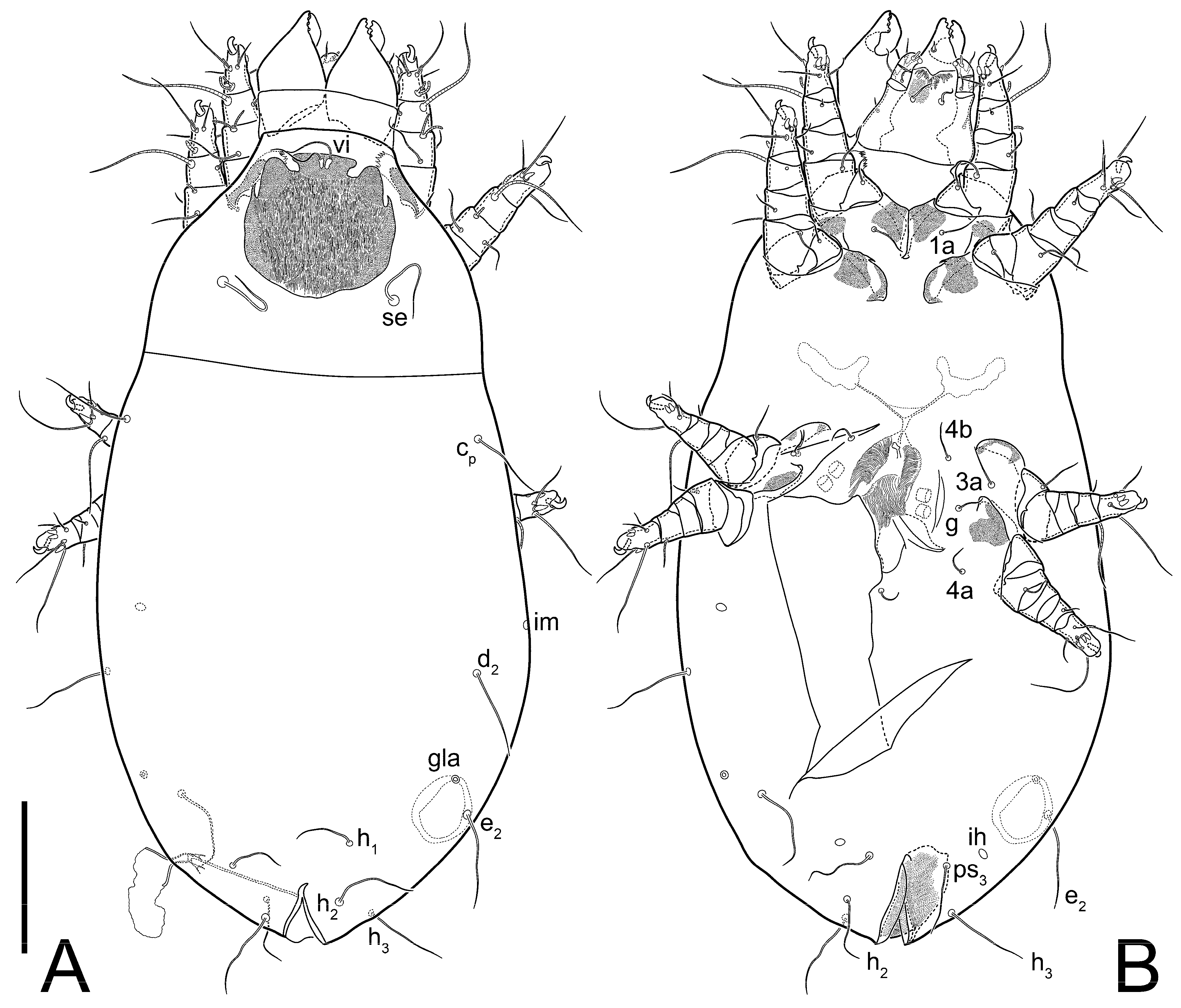
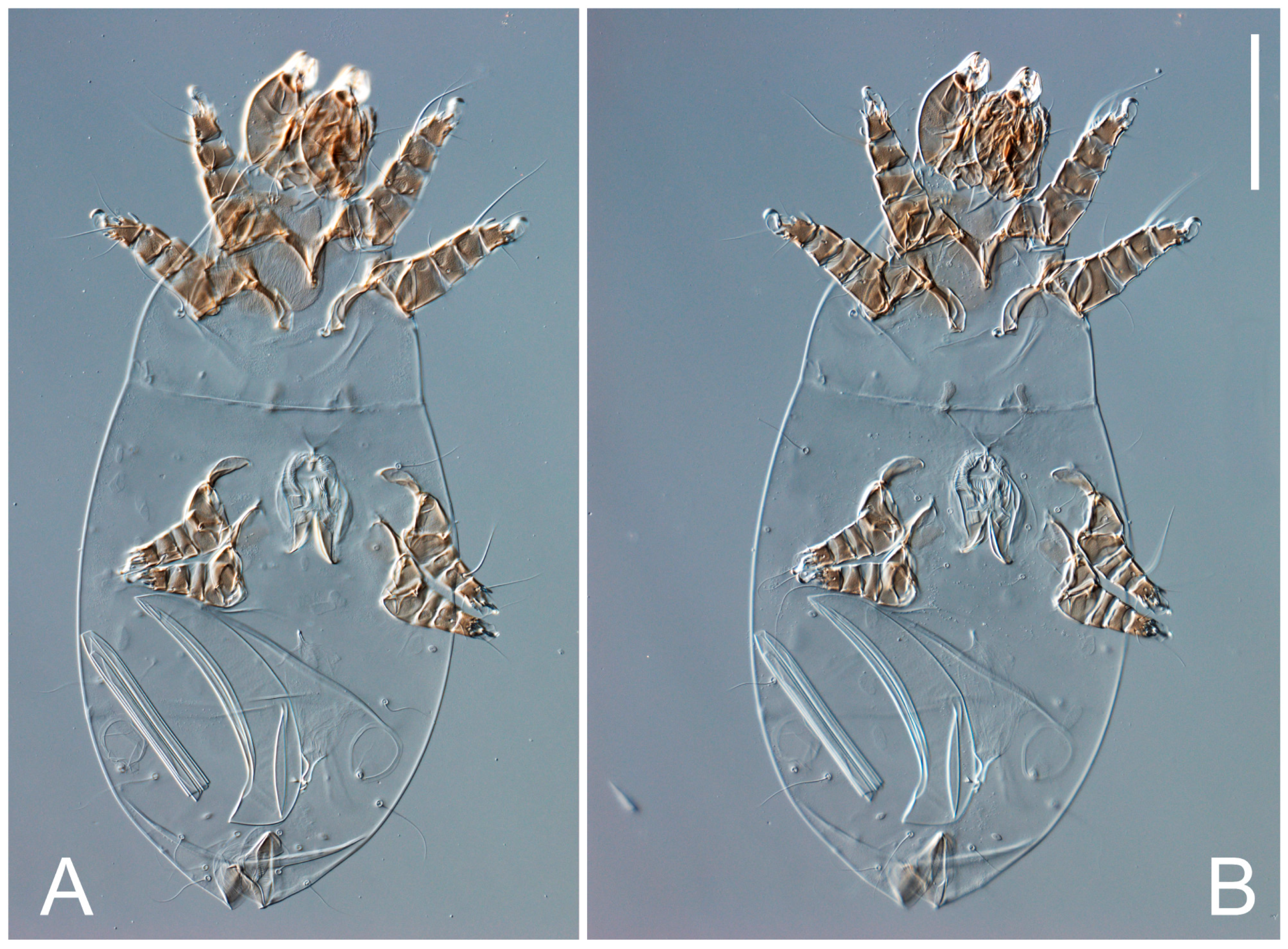
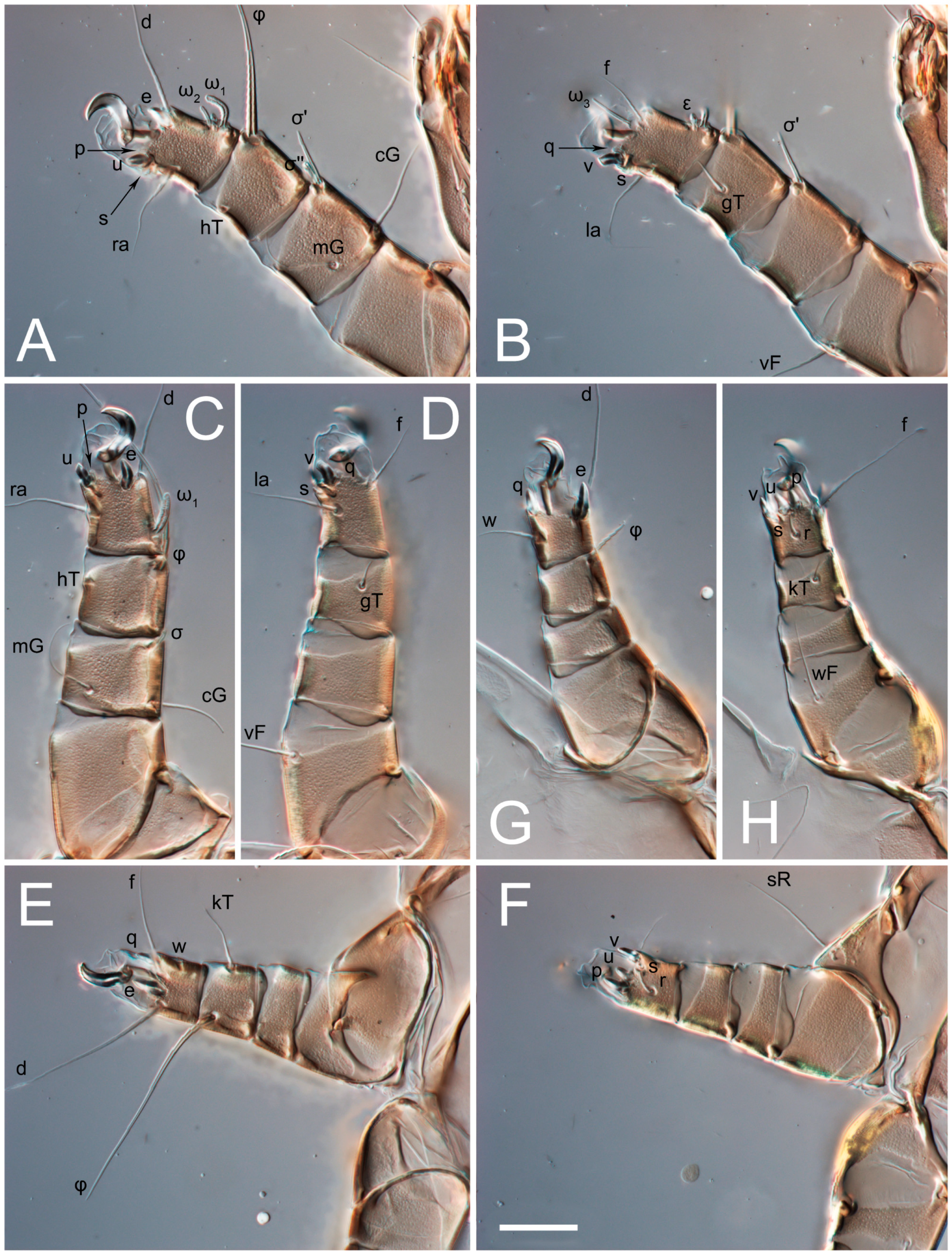
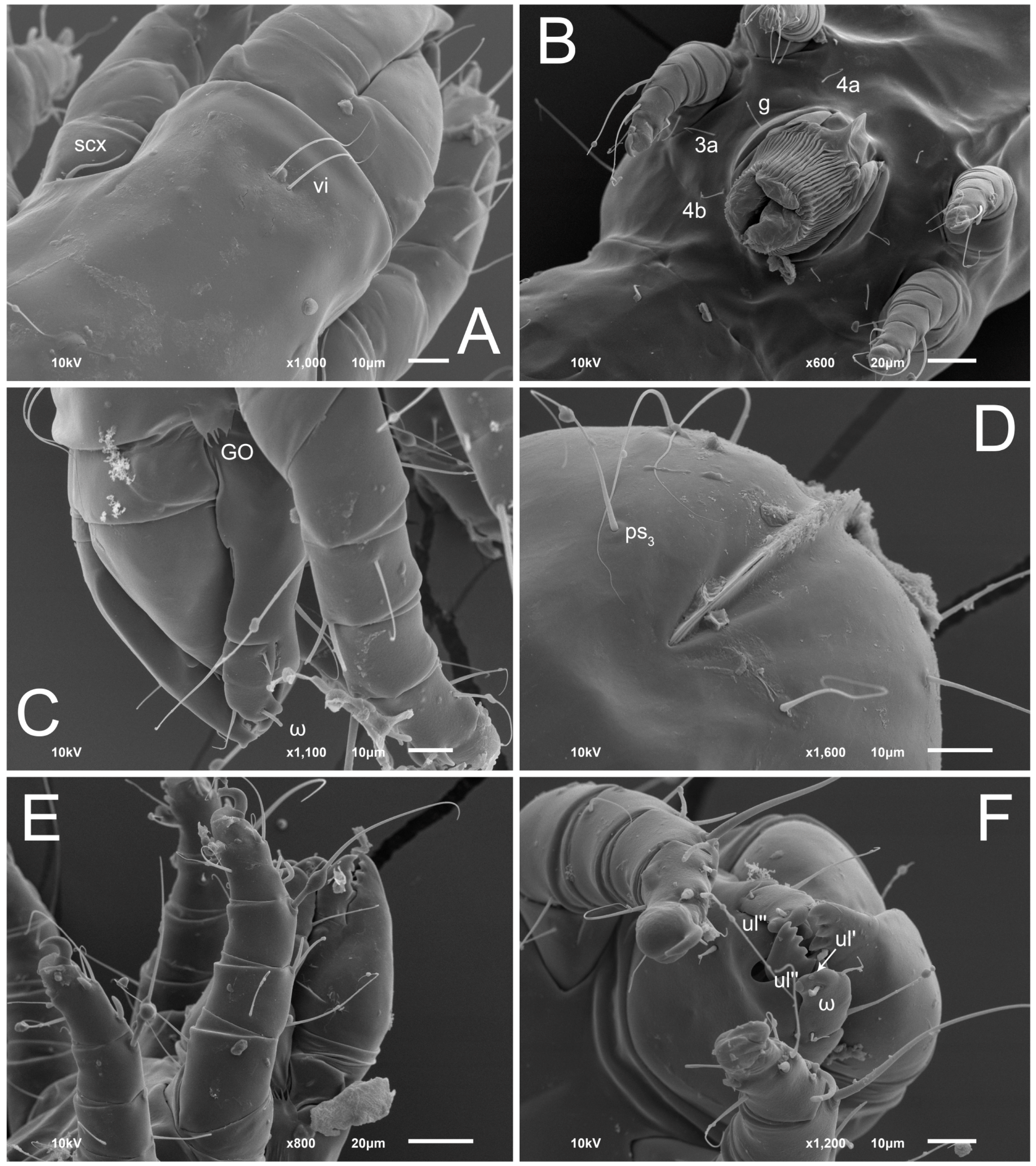
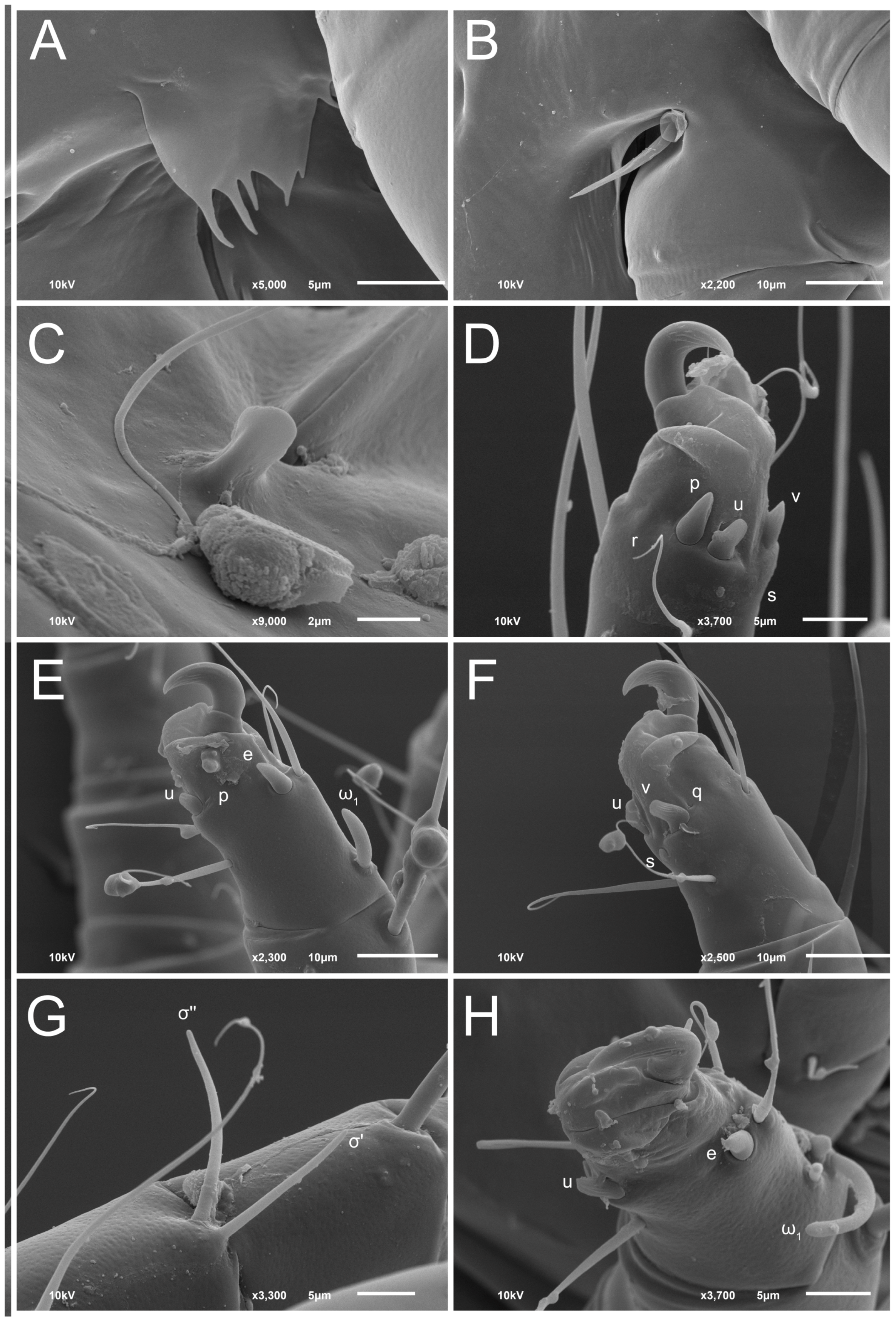
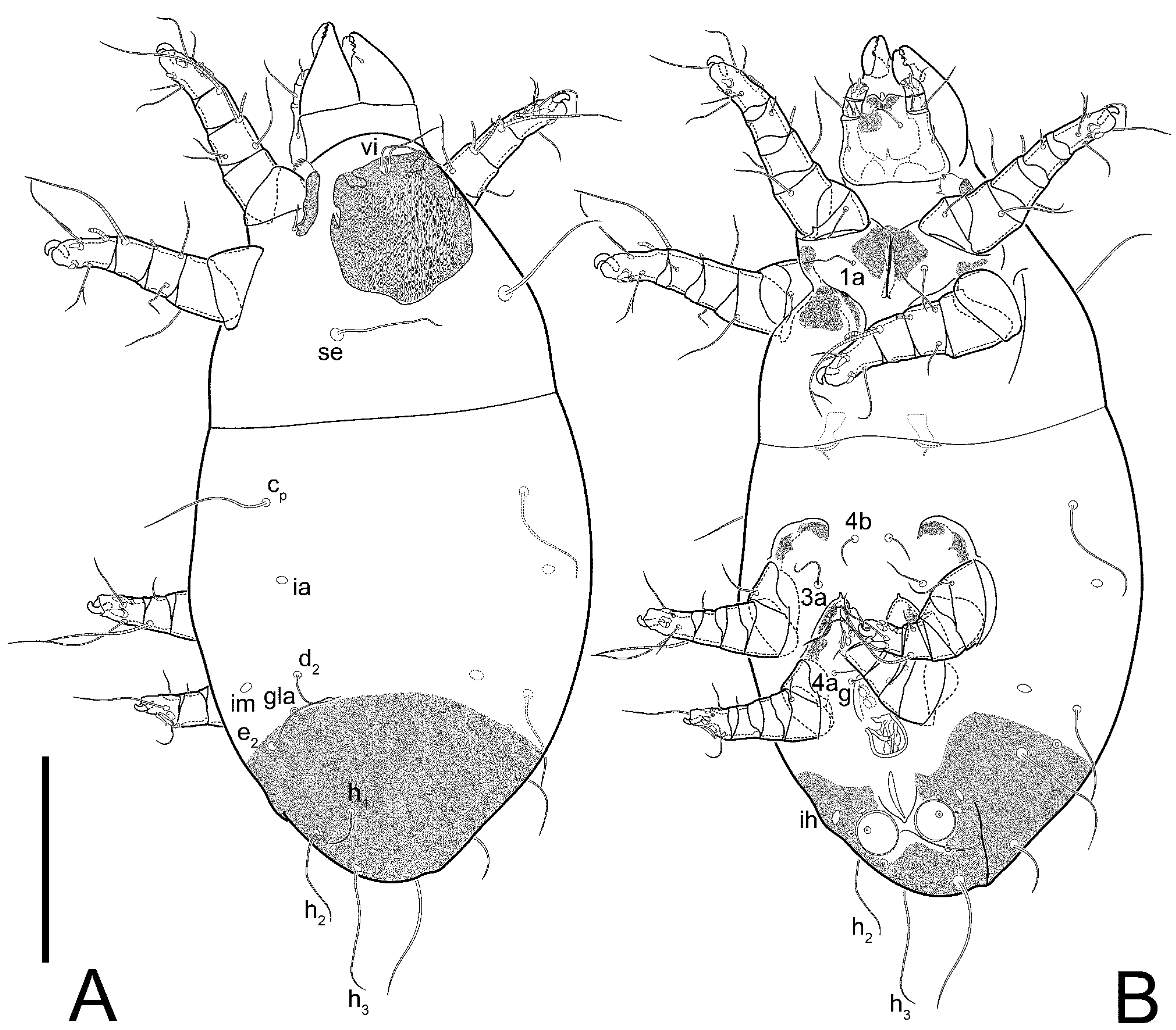
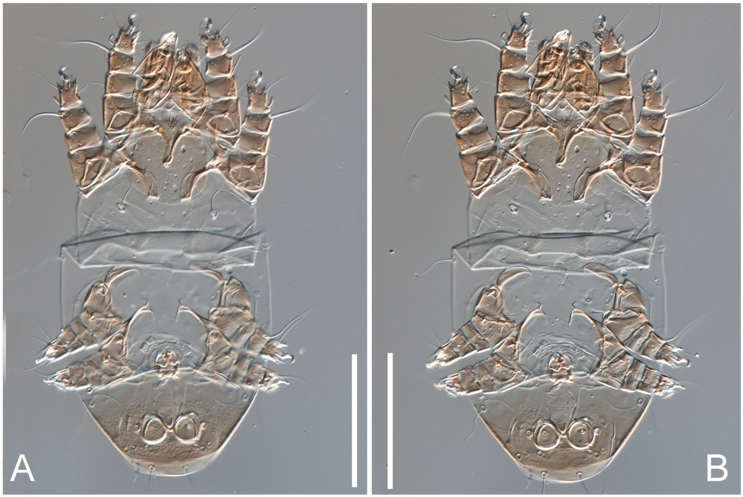
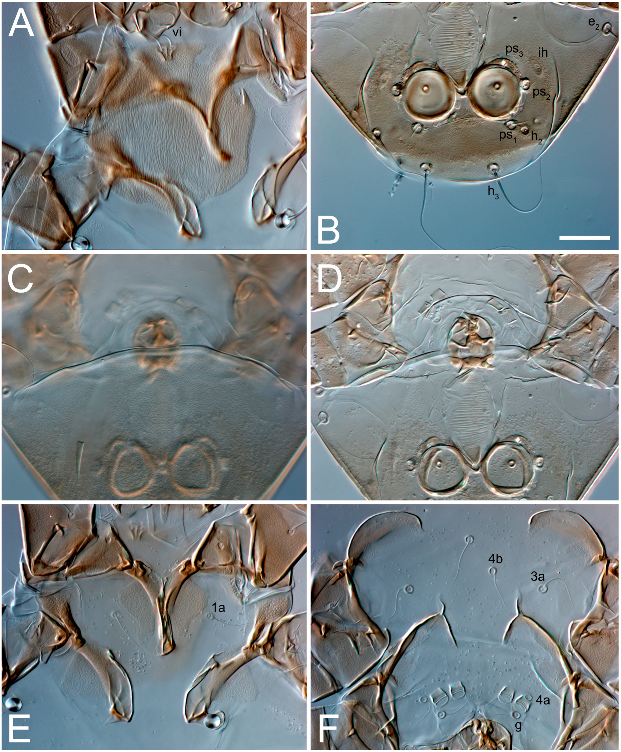
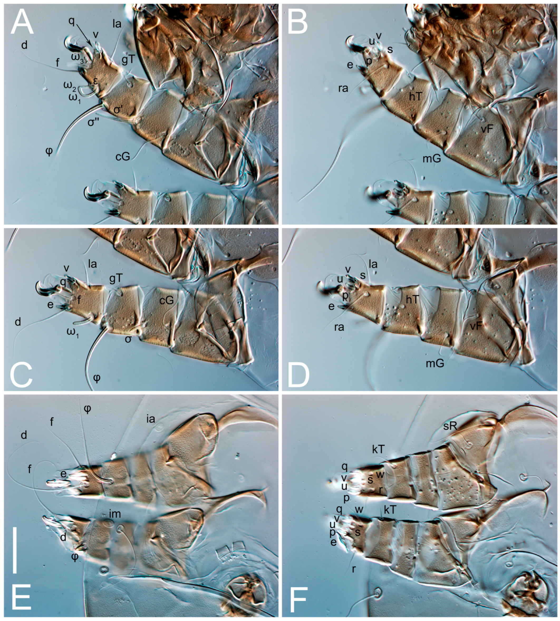
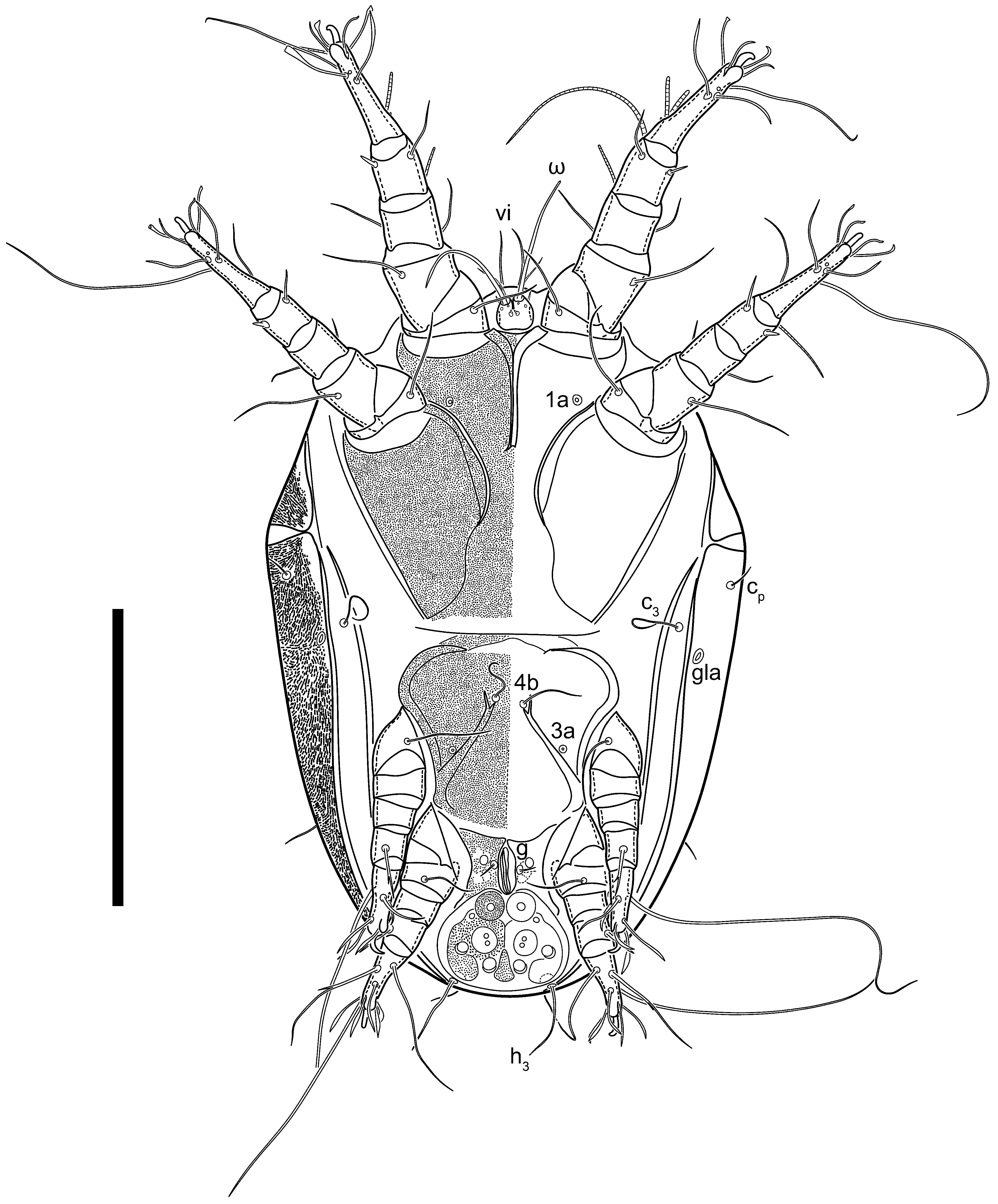
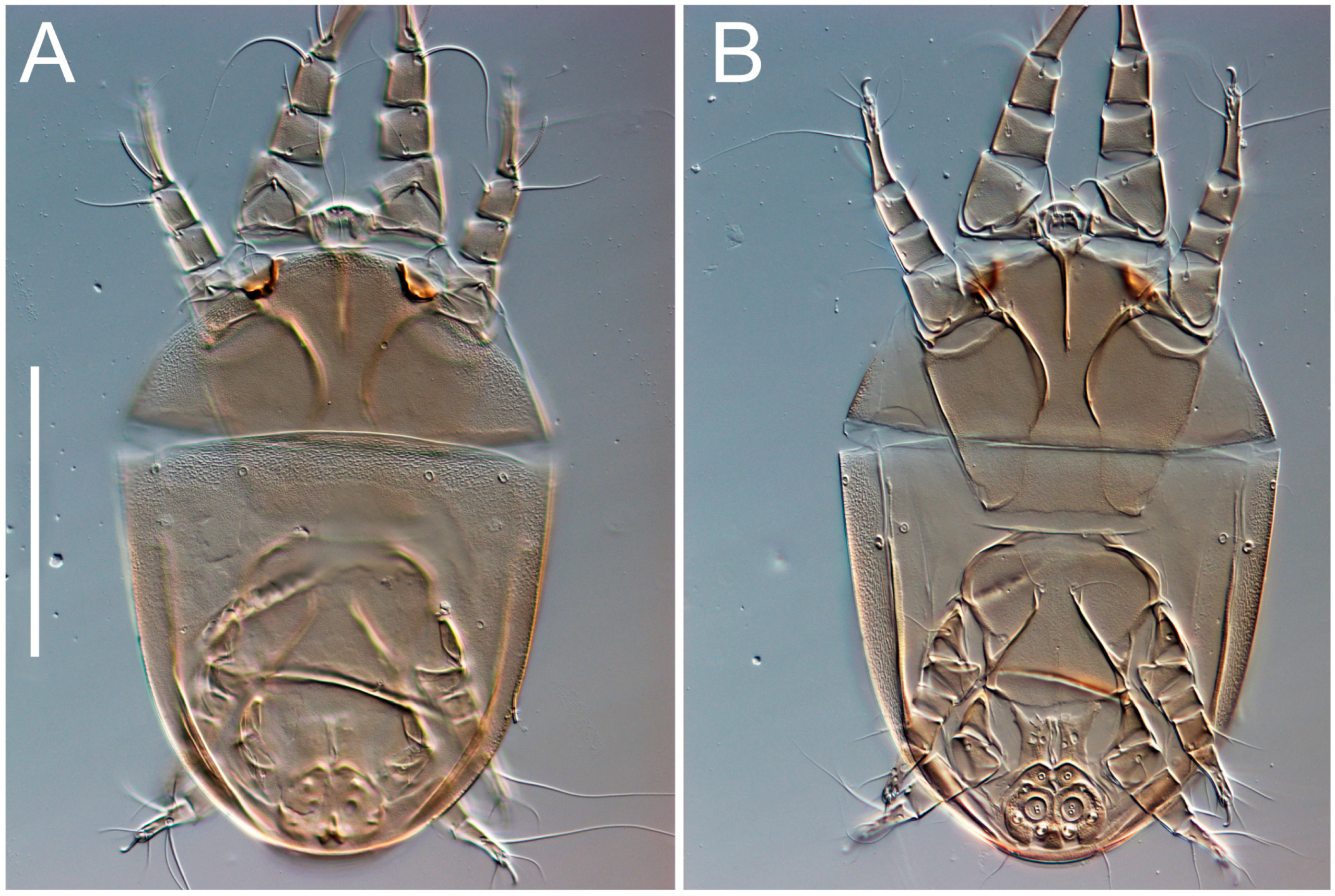
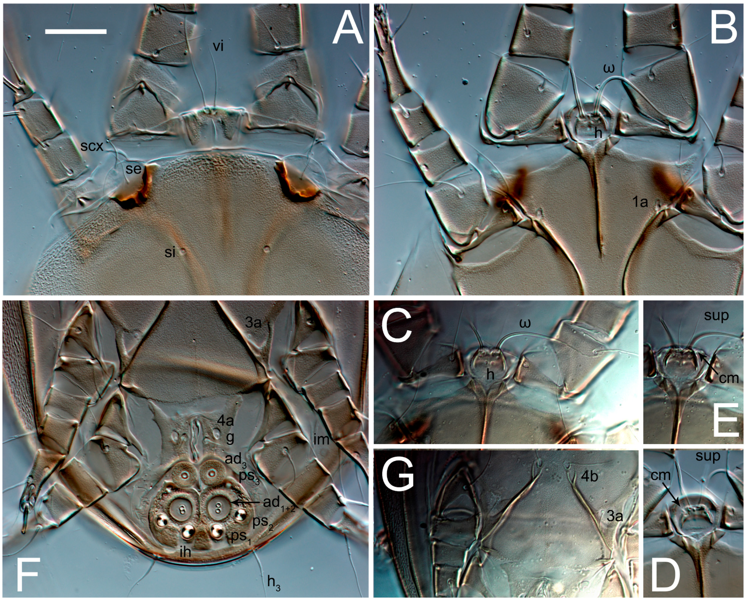
3.2. Morphological Description

3.2.1. Genus Thyreophagus Rondani, 1874

Thyreophagus Rondani, 1874: 67 (=Moneziella Berlese, 1897; Monetiella Berlese, 1897; Monieziella Berlese, 1897; Fumouzea Zachvatkin, 1953; Michaelopus Fain and Johnston, 1974).
Type species Thyreophagus entomophagus (Laboulbène and Robin, 1862) (=Acarus entomophaus Laboulbène, 1852 (nomen nudum)), by monotypy.

3.2.2. Thyreophagus tauricus sp. n.

Description
Female (Figure 1, Figure 2, Figure 3, Figure 4, Figure 5 and Figure 6, Figure 7A–C, Figure 8 and Figure 9). Idiosoma elongate, 500 × 280 (holotype), 400–530 × 190–270 (paratypes, n = 9), 1.8 (2.0–2.1) times longer than wide. Idiosomal cuticle smooth. Subcapitular setae (h) long, widened basally; palp tibial setae (a), lateral dorsal palp tibial setae (sup), dorsal palp tarsal seta (cm) filiform; supracoxal seta elcp present; terminal palp tarsal solenidion ω short; external part of terminal eupathidium ul’’ dome-shaped; terminal eupathidium ul’ small, rounded. Prodorsal sclerite 100 (84–102) long, 97 (75–90) wide, 1.0 (1.1) times longer than wide, with setae vi (situated at anterior part of shield, alveoli separated), rounded anterolateral incisions, and elongate midlateral incisions (insertion points of setae ve). Prodorsal sclerite smoothly punctate except large lineate central region; posterior end of sclerite with lineate pattern. Grandjean’s organ (GO) with seven membranous finger-shaped processes. Supracoxal setae (scx) smooth, sword-shaped, widened and flattened, tapering at tip. Idiosomal setae (vi, se, cp, d2, e2, h1, h2, h3, ps3) smooth, filiform and short; opisthosomal gland openings slightly anteriad setal bases e2. Three pairs of fundamental cupules (ia, im and ih) present, ip not observed. Ventral idiosoma with four pairs of coxal setae (1a, 3a, 4a and 4b) and 1 pair of genital setae (g). Shape of coxal sclerites as in Figure 1B and Figure 4E,F. Genital region situated between coxal fields III and IV; genital valves form an inverted Y; epigynal and medial apodemes well-developed. Diameter of genital papillae approximately 0.3–0.4 the length of coxal and genital setae. Anal opening terminal. Copulatory tube present, situated anterodorsally to anus, with developed opening. Canal of spermatheca long, slender tube-like, leading from copulatory opening to spermatheca, uniformly wide, wider at entrance to spermatheca. Sclerotized vase-shaped atrium of spermatheca with length greater than width, base 3–4 times wider than end of atrium at junction with sclerites of oviducts. Paired Y-shaped sclerites of oviducts, small, elongated.
Legs short, all segments free. Trochanters I–III each with long, filiform seta, pR I–II, sR III; trochanter IV without setae. Femoral setation 1-1-0-1; setae vF I–II and wF IV long, filiform. Genual setation 2-2-0-0; setae mG and cG I–II long, filiform; seta nG III absent. Tibial setation 2-2-1-1; setae hT I-II represented by alveoli or minute setae; setae gT I–II and kT III–IV elongate, somewhat spiniform. Tarsal setation 10-10-10-10; pretarsi consists of hooked empodial claws attached to short paired condylophores. Tarsus I and II with setae ra, la, f and d filiform, e, u, v spiniform, p and q represented by small triangular rudiments, s flattened, button-shaped or minute spiniform; setae wa absent. Tarsus III with setae f, d, r filiform, e, u, v, p, q spiniform, s flattened, button-shaped or minute spiniform, w flattened, button-shaped. Tarsus IV similar to tarsus III, except w filiform. Solenidion ω1 on tarsus I cylindrical, with clavate apex, curved; solenidion ω1 on tarsus II simple, cylindrical, with clavate apex, not bent, shorter and wider than ω1 on tarsus I. Solenidion ω2 on tarsus I shorter than ω1, cylindrical, with rounded apex, slightly widened distally, situated slightly distal to ω1. Solenidion ω3 on tarsus I cylindrical, with rounded tip, subequal to ω1, longer than ω2. Famulus (ε) of tarsus I wide, spiniform, with broadly rounded apex, widest at middle. Solenidia φ of tibiae I–III elongate, tapering, well extending beyond apices of respective tarsi with ambulacra; solenidion φ IV shorter, shorter than tarsus IV (with ambulacra). Solenidia σ′ and σ″ on genu I elongate, tapering, subequal in length, slightly not reaching bases of φ I. Solenidion σ on genu II more than 6–7 times longer than its width) with rounded tip. Solenidion σ of genu III absent.
Male (n = 4, paratypes). (Figure 7D–F and Figure 10, Figure 11, Figure 12, Figure 13, Figure 14 and Figure 15). Idiosoma elongate, 300–400 × 160–210, 1.9–2.1 times longer than wide. Idiosomal cuticle smooth. Gnathosoma as in female. Prodorsal sclerite 63–78 long, 58–73 wide, 1.1–1.2 times longer than wide, with setae vi, incisions and ornamented as in female. Grandjean’s organ (GO) with 5–7 membranous processes. Supracoxal seta (scx) as in female. Idiosomal setae (vi, se, cp, d2, e2, h1, h2, h3) smooth, filiform and short; opisthosomal gland openings slightly anteriad setal bases e2. Three pairs of fundamental cupules (ia, im and ih) present, ip not observed. Opisthonotal shield smoothly punctate; ventral part extends to anal suckers. Ventral idiosoma with four pairs of coxal setae (1a, 3a, 4a and 4b) and one pair of genital setae (g). Shape of coxal sclerites on Figure 10B and Figure 13E,F. Genital region between coxisternal fields IV; arms of genital capsule rounded; aedeagus short, not protruding beyond anterior edge of genital capsule. Diameter of genital papillae approximately 0.3–0.4 the length of coxal and genital setae. Anal suckers rounded in outline. Setae ps1–3 very short.
Legs I-III as female, except solenidion ω3 on tarsus I very short, truncated; and solenidion σ″ about two-times longer than σ′. Trochanter and genu IV without setae, femur IV with wF IV long, filiform, tibia IV with kT IV elongate, somewhat spiniform. Tarsus IV with 10 setae, of them, f, r, w filiform, d and e represented by suckers, u, v, p, q spiniform, s flattened, button-shaped or minute spiniform. Solenidion φ on tibia IV short and wide.
Phoretic deutonymph (n = 4, paratypes) (Figure 16, Figure 17, Figure 18, Figure 19, Figure 20 and Figure 21). Body elongate, 1.33–1.44 times longer than wide, widest in sejugal region; idiosomal length 220–240 width 153–180. Gnathosoma short, subcapitulum and palps fused, bearing palpal solenidia (ω) apically and filiform apicodorsal setae (sup); setae h present, minute (Figure 20C) or absent (their positions marked by somewhat refractile spots), setae cm present.
Dorsum. Idiosoma smoothly punctate; distinct linear pattern present on anterior and lateral sides of prodorsal sclerite and hysterosomal shield. Apex of propodosoma anterior to anterior border of prodorsal sclerite, with apical internal vertical setae (vi) (bases separated) and a pair of band-like sclerites coalescing anteriorly. A pair of lateral, widely separated ocelli (distance 40–49) present on prodorsum; lenses and pigmented spots present; maximal diameter of lenses 18–20. External vertical setae (ve) absent; external scapular setae se situated below lenses; internal scapular setae (si) distinctly posterior and medial to external scapulars (se). Supracoxal setae of legs I (scx) filiform, with extended base, positioned below si and anterolaterad to ocelli. Sejugal furrow well developed. Prodorsal sclerite 67–73, hysterosomal shield 150–160, ratio hysterosoma shield/prodorsal sclerite length = 2.2–2.3. Hysterosoma with 11 pairs of simple, filiform setae on hysterosomal shield (c1, c2, cp, d1, d2, e1, e2, f2, h1, h2, h3), setae h3 distinctly longer than others. Opisthonotal gland openings (gla) situated ventrally on hysterosomal shield, slightly posterior to setae c3. Of four fundamental pairs of cupules, three pairs were observed: ia posteriomediad setae c2, im ventral, laterad of trochanters IV and ih ventral, laterad posterior sides of attachment organ.
Venter. Coxal fields sclerotized, smoothly punctate. Anterior apodemes of coxal fields I fused forming sternum; sternum not reaching posterior border of sternal shield by distance exceeding its length. Posterior border of sternal shield not sclerotized. Anterior apodemes of coxal fields II curved medially. Posterior apodemes of coxal fields II weakly developed, thin, curved medially. Sternal and ventral shield contiguous. Anterior apodemes of coxisternal fields III free, connected by thin transverse sclerotization. Posterior medial apodeme in area of coxisternal fields IV weakly developed. Posterior apodemes IV absent. Subhumeral setae (c3) filiform, situated on ventral surface between legs II–III, adjacent to region separating sternal and ventral shields. Coxal setae 1a, 3a reduced, represented by minute structures or filiform, situated in alveoli (Figure 20G). Setae 4b, g filiform; 4a in form of small, rounded conoids. Genital region in posterior portion of coxisternal fields IV; opening elongate, with two pairs of genital papillae within genital atrium; papillae two-segmented, with rounded apices. Coxal setae (4b) situated at tips anterior coxal apodemes IV; genital setae (g) laterad of genital opening. Attachment organ posterior to coxisternal fields IV. Anterior suckers (ad3) round, median suckers (ad1+2) distinctly larger, with paired vestigial alveoli (not situated on common sclerite); pair of small refractile spots anterolateral to median suckers (ps3); lateral conoidal setae of attachment organ (ps2) situated distinctly posterior to line joining centers of median suckers, slightly anterior to conoidal setae (ps1); anterior and posterior lateral and posterior median cuticular conoids well developed; anus situated between anterior suckers (ad3).
Legs. Legs elongate, all segments free. Trochanters I–III each with long, filiform seta, pR I–II, sR III. Femoral setation 1-1-0-1; setae vF I–II and wF IV long, filiform. Genual setation 2-2-0-0; setae mG and cG I–II filiform, seta nG III absent. Tibial setation 2-2-1-1; setae hT I somewhat spiniform; setae gT I filiform; setae gT and hT II spiniform, setae gT II longer than hT II; setae kT III spiniform and with distinct prong; setae kT IV somewhat spiniform, shorter than kT III, with short prong. Tarsal setation 8-9-8-8. All pretarsi consisting of hooked empodial claws attached to short paired condylophores. Tarsus I with setae ra, la, p, q, d, f narrowly lineolate; e slightly spoon-shaped; seta d elongate, its base at level of ra and la; seta s represented by alveolus; setae wa, aa and ba I absent; tarsus II similar to tarsus I except seta ba present, filiform, situated close to ω1. Tarsus III with setae w, r, s, p, q, e, f and d smooth; all setae, except for d III, more or less foliate; seta d longer than leg. Tarsus IV similar to tarsus III, except seta r longer, filiform; seta w filiform and with distinct prong. Solenidia ω1 on tarsi I–II cylindrical, with slightly clavate apices; ω3 on tarsus I slightly shorter than ω1, with rounded apex, situated slightly distal to ω1; ω1 and ω3 separated by bulbous famulus (ε); solenidion ω2 of tarsus I expanding slightly apically, situated somewhat more basal and posterior to ω1 + ε + ω3 group; solenidia φ of tibiae I–III elongate, tapering; φ I longer than tarsus I; φ II shorter than tarsus II; φ III reaching tip of tarsus III without ambulacrum; φ IV short; σ of genu I elongate, slightly tapering, nearly reaching tip of tibia I; σ of genu II much shorter, cylindrical, not reaching midlength of tibia II; σ of genu III absent.
Type material. Holotype (female) and paratypes (14 females, 9 males and 4 heteromorphic deutonymphs) from lab culture; culture started from specimens collected in Crimea, vicinity of Yalta, under the bark of fallen twigs of Tilia sp., 6 April 2022, 44.483333 N, 34.083333 E, coll. Khaustov V.A, PBK 20-0101-065.
Depository. The holotype and paratypes (11 females, 6 males and 1 heteromorphic deutonymphs) are deposited in the Museum of Zoology, Tyumen State University, Russia. The remaining paratypes (4 females, 3 males and 3 heteromorphic deutonymphs) are deposited in the Zoological Institute, Russian Academy of Sciences, Saint Petersburg, Russia.
Etymology. Tauricus (of Taurica, Lat. adjective). Taurica is a historical name of the Crimean Peninsula used by the Greeks and Romans. This is a nomen in supposition.
Diagnosis. Adults of Thyreophagus tauricus are distinct among all other species of the genus by the presence of flattened, button-shaped or minute spiniform (much less than v and p) setae s III-IV (Figure 9F) (vs. well-developed spiniform (not much less than v and p) in all other species, the adults of which are known). The new species is close to Th. corticalis (patterns of prodorsal sclerite, length of dorsal setae and shaped of legs setae, except s III–IV), but differs from it in having the vase-shaped atrium of the spermatheca, which is 3–4 times wider at the basal part than at junction with sclerites of oviducts (vs. 1.5–2 times wider in Th. corticalis).
Heteromorphic deutonymphs of Th. tauricus are distinct from all other species of Thyreophagus by the presence of the well-developed setae cm on the dorsal part of the subcapitular remnant (vs. absent all other species). Thyreophagus tauricus has a prong on kT III (as in Th. australis Clark, 2009 and Th. sminthurus (Fain and Johnston, 1974)), but it differs from Th. australis in having the larger ocelli 20 (vs. 10 in Th. australis). Thyreophagus tauricus differs from Th. sminthurus in having the wider idiosoma, which is 1.33–1.44 times longer than wide (vs. 2.1 in T. sminthurus), and the presence of tibial setae hT I, II (vs. absent in Th. sminthurus).

4. Discussion

In recent years, there has been a growing interest in finding sustainable and environmentally friendly solutions to combat agricultural pests while minimizing the adverse impacts of chemical pesticides. Mite species of the genus Thyreophagus represent an exciting alternative to synthetic pesticides in the realm of pest control in agriculture. For example, Thyreophagus entomophagus and Thyreophagus calusorum are widely used as factitious prey for mass rearing of phytoseiid predatory mites for biocontrol applications [9,15], while Thyreophagus cracentiseta, has been proposed as such a species based on its performance in laboratory experiments [7]. As several species of Thyreophagus have proven to be valuable factitious prey for the mass rearing of predatory mites, these predatory mites, in turn, play a crucial role in biological pest control [26]. They feed on a variety of agricultural pests such as spider mites, thrips and other small arthropods that harm crops [27]. The significance of this approach lies in its potential to reduce our reliance on synthetic pesticides. Unlike chemical pesticides, Thyreophagus-based biocontrol methods are environmentally sustainable, as they do not introduce harmful chemicals into the ecosystem. This approach is highly targeted, focusing solely on the pests without affecting beneficial organisms or pollinators. It is also adaptable to various crops and integrated pest management systems [26]. Furthermore, the use of Thyreophagus mites to produce predatory mites for biocontrol can potentially lead to a reduction in pesticide residues on agricultural products, making them safer for consumption. As the demand for organic and environmentally friendly farming practices continues to grow, the role of Thyreophagus mites in replacing synthetic pesticides becomes increasingly significant, offering a promising and sustainable solution for pest management in agriculture.
Given the economic significance of Thyreophagus, it is important to know its biodiversity, habitat and species boundaries based both on morphology and DNA sequences. However, these aspects remain significantly underexplored. For instance, it is noteworthy that a substantial number of Thyreophagus species are thought to be yet undescribed [1,8] even in regions where extensive biodiversity research has been carried out, notably in Europe and North America [4]. In addition, there is only a single sequence of Thyreophagusentomophagus” in GenBank; however, this sequence is likely based on misidentification (see below). The prevalence of undescribed or poorly characterized species raises important questions about our understanding of the global Thyreophagus biodiversity. Numerous Thyreophagus mite species live in subcortical environments, alongside scale insects, or within the nests of bees and wasps, thus eluding their discovery due to their secretive lifestyles and cryptic habitats. Understanding and documenting these species is crucial for achieving a comprehensive picture of the global Thyreophagus biodiversity. Curiously, as Thyreophagus mites are adapted to live in concealed habitats, which have limited number of natural predators, this adaptation is one of the biological features that render Thyreophagus mites useful as factitious prey in industrial settings. These mites, characterized by slow movement and a lack of many natural defenses, such as long setae, are particularly suitable as prey for phytoseiid mites when reared industrially. Therefore, as Thyreophagus mites play an important role in biocontrol applications, a more thorough examination of their diversity and biology is also essential. Improved knowledge of these mites would not only facilitate their use in pest management but also potentially uncover new species suitable for local production of phytoseiid mites, thereby minimizing the risk of introducing non-native Thyreophagus species into new regions. In light of these considerations, it is clear that a more concerted effort is needed to study Thyreophagus mites comprehensively. This entails employing a range of research methods, from intensive fieldwork to laboratory-based studies. Additionally, molecular techniques can aid in precisely identifying and classifying these mites, shedding light on their genetic diversity and evolution.
Here, we report the discovery of a new sexual species, Thyreophagus tauricus sp. n., and provide thorough analyses of its morphology, key life stages, molecular characteristics, and its relationship with other species. As we established a pure culture of this new species in the laboratory, we were able to confidently correlate all its taxonomically important life stages, males, females and deutonymphs (a dispersal stage). To facilitate molecular identification and species delimitation, we sequenced the entire COX1 gene, a useful DNA barcoding gene [28] of four Palearctic species: Th. tauricus, Th. corticalis, Th. calusorum and Th. entomophagus. Of them, the two latter species are used for mass-rearing of phytoseiid mites [9,15]. We found that the new species is morphologically close to Th. corticalis, a widely distributed Palearctic species; however, it differs from Th. corticalis and other Thyreophagus species by the following character states: adults of Th. tauricus are distinct by the presence of flattened, button-shaped or minute spiniform setae s III-IV, which are well-developed and spiniform in all other known species of Thyreophagus; heteromorphic deutonymphs of Th. tauricus are distinct from all other species of Thyreophagus by the presence of well-developed setae cm on the dorsal part of the subcapitular remnant (absent in all other species). Despite being very close to Th. corticalis, genetic COX1 K2P distances were large, 19.8% (Table 1), suggesting the presence of a well-delimited species, Th. tauricus sp. n., which is distant by both morphology and DNA sequences. COX1 K2P distances between Th. tauricus and other species (Th. entomophagus, Th.entomophagus” NC 066986.1, Th. calusorum, Th. corticalis) ranged between 20.1 and 24.3% (Table 1).
As part of our study, we also verified sequences deposited into the GenBank database. One such sequence from China (NC 066986.1) was initially identified as ‘Th. entomophagus’. However, our sequence, derived from a pure, industrially produced European culture carefully identified by us, displayed a significant 21.16% COX1 K2P distance from this GenBank sequence (Table 1). This substantial genetic distance strongly suggests that the GenBank sequence NC 066986.1 was misidentified and does not belong to Th. entomophagus. This raises questions about the reliability of public databases and the importance of rigorous verification and validation in biological research.

5. Conclusions

The discovery and comprehensive study of Thyreophagus tauricus underscore the importance of biodiversity research, taxonomic methods, molecular techniques and the need for rigorous scientific practices. Understanding the diversity and characteristics of mite species like Thyreophagus can have broader implications for ecology, agriculture and biocontrol efforts.

Author Contributions

Conceptualization, P.B.K. and V.B.K.; methodology, P.B.K. and V.B.K.; validation, P.B.K., V.B.K., V.A.K. and A.A.K.; formal analysis, P.B.K.; investigation, P.B.K. and V.B.K.; resources, P.B.K. and V.A.K.; data curation, P.B.K. and V.B.K.; writing—original draft preparation, V.B.K.; writing—review and editing, P.B.K., V.B.K., V.A.K. and A.A.K.; visualization, V.B.K. and V.A.K.; supervision, P.B.K.; project administration, P.B.K.; funding acquisition, P.B.K., V.B.K., V.A.K. and A.A.K. All authors have read and agreed to the published version of the manuscript.

Funding

This research was funded by Ministry of Science and Higher Education of the Russian Federation within the framework of the Federal Scientific and Technical Program for the Development of Genetic Technologies for 2019–2027 (agreement № 075-15-2021-1345, unique identifier RF—193021X0012).

Institutional Review Board Statement

Not applicable.

Informed Consent Statement

Not applicable.

Data Availability Statement

Data are available in a publicly accessible repository.

Conflicts of Interest

The authors declare no conflict of interest.

References

  1. Klimov, P.B.; Demard, E.P.; Stinson, C.S.A.; Duarte, M.V.A.; Wäckers, F.L.; Vangansbeke, D. Thyreophagus calusorum sp. n. (Acari, Acaridae), a new parthenogenetic species from the USA, with a checklist of Thyreophagus species of the world. Syst. Appl. Acarol. 2022, 27, 1920–1956. [Google Scholar] [CrossRef]
  2. Fain, A. Revision des genres Thyreophagus Rondani, 1874 et Michaelopus Fain & Johnston, 1974 (Acari, Acaridae) avec description de neuf especes nouvelles. Bull. L’institut R. Sci. Nat. Belg. Entomol. 1982, 54, 1–47. [Google Scholar]
  3. Ostojá-Starzewski, J.C. Michaelopus spinitarsis Fain (Acari: Acaridae): A first record in the British Isles. Br. J. Entomol. Nat. Hist. 2001, 14, 217–221. [Google Scholar]
  4. Portus, M.; Gomez, M.S. Thyreophagus gallegoi a new mite from flour and house dust in Spain (Acaridae, Sarcoptiformes). Acarologia 1980, 21, 477–481. [Google Scholar] [PubMed]
  5. Chmielewski, W. Stored products mites (Acaroidea) in Polish bee hives. In Modern Acarology, Volume I: Proceedings of the 8 International Congress of Acarology, Ceske Budejovice, Czechoslovakia, 6–11 August 1990; SPB Academic Publishing: The Hague, The Netherlands, 1991; pp. 615–619. [Google Scholar]
  6. Okabe, K.; OConnor, B.M. Thelytokous reproduction in the family Acaridae (Astigmata). In Acarology: Proceedings of the 10th International Congress; Halliday, R.B., Walter, D.E., Proctor, H.C., Norton, R.A., Colloff, M.J., Eds.; CSIRO Publishing: Melbourne, Australia, 2001; pp. 170–175. [Google Scholar]
  7. Barbosa, M.F.D.C.; OConnor, B.M.; De Moraes, G.J. A new species of Thyreophagus (Acari: Acaridae) from Brazil, with notes on species associated with stored food and human habitats and a key to species of this genus. Zootaxa 2016, 4088, 279–291. [Google Scholar] [CrossRef] [PubMed]
  8. Klimov, P.B.; Mwangi, E.; Merckx, J.; Duarte, M.V.A.; Wäckers, F.L.; Vangansbeke, D. Thyreophagus plocepasseri sp. n., a new parthenogenetic species of acarid mites (Acariformes: Acaridae) from Kenya. Syst. Appl. Acarol. 2020, 25, 2250–2262. [Google Scholar] [CrossRef]
  9. Fidgett, M.J.; Stinson, C.S.A. A method of rearing Amblyseius predatory mites using Thyreophagus entomophagus as prey. Patent CA2658292A1, 7 February 2008. Available online: https://patentimages.storage.googleapis.com/e7/4f/61/612fa7977fb51a/CA2658292A1.pdf (accessed on 10 November 2023).
  10. Iglesias-Souto, J.; Sánchez-Machín, I.; Iraola, V.; Poza, P.; González1, R.; Matheu, V. Oral mite anaphylaxis by Thyreophagus entomophagus in a child: Acase report. Clin. Mol. Allergy 2009, 7, 1–3. [Google Scholar] [CrossRef]
  11. Simoni, S.; Nannelli, R.; Roversi, P.F.; Turchetti, T.; Bouneb, M. Thyreophagus corticalis as a vector of hypovirulence in Cryphonectria parasitica in chestnut stands. Exp. Appl. Acarol. 2014, 62, 363–375. [Google Scholar] [CrossRef]
  12. Barbosa, M.F.D.C.; de Moraes, G.J. Evaluation of astigmatid mites as factitious food for rearing four predaceous phytoseiid mites (Acari: Astigmatina, Phytoseiidae). Biol. Control 2015, 91, 22–26. [Google Scholar] [CrossRef]
  13. Bouneb, M.; Turchetti, T.; Nannelli, R.; Roversi, P.F.; Paoli, F.; Danti, R.; Simoni, S. Occurrence and transmission of mycovirus Cryphonectria hypovirus 1 from dejecta of Thyreophagus corticalis (Acari, Acaridae). Fungal Biol. 2016, 120, 351–357. [Google Scholar] [CrossRef] [PubMed]
  14. Knapp, M.; van Houten, Y.; van Baal, E.; Groot, T. Use of predatory mites in commercial biocontrol: Current status and future prospects. Acarologia 2018, 58, 72–82. [Google Scholar] [CrossRef]
  15. Stinson, C.S.A.; Klimov, P.B.; Demard, E.P.; Dunham, J.L.; Duarte, M.V.A.; Vangansbeke, D.; Bolckmans, K.J.F.; Wäckers, F.L. Mite Composition and Method for Feeding Predatory Mites. Patent WO/2023/170145, 8 March 2023. [Google Scholar]
  16. Clark, J.M. A new Thyreophagus mite from honeydew scale insects on black beech (Nothofagus). Rec. Canterb. Mus. 2009, 23, 1–9. [Google Scholar]
  17. Fain, A.; Johnston, D. Three new species of hypopi phoretic on springtails (Collembola) in England (Acari: Acarididae). J. Nat. Hist. 1974, 8, 411–420. [Google Scholar] [CrossRef]
  18. Fain, A.; Knülle, W.; Wurst, E. First description of the hypopial stage of Thyreophagus entomophagus (Laboulbène, 1852) (Acari Acaridae). Bull. Soc. R. Belg. Entomol. 2000, 136, 153–156. [Google Scholar]
  19. Griffiths, D.A.; Atyeo, W.T.; Norton, R.A.; Lynch, C.A. The idiosomal chaetotaxy of astigmatid mites. J. Zool. 1990, 220, 1–32. [Google Scholar] [CrossRef]
  20. Norton, R.A. Morphological evidence for the evolutionary origin of Astigmata (Acari: Acariformes). Exp. Appl. Acarol. 1998, 22, 559–594. [Google Scholar] [CrossRef]
  21. Grandjean, F. Au sujet de l’organe de Claparède, des eupathidies multiples et des taenidies mandibulaires chez les Acariens actinochitineux. Arch. Sci. Phys. Nat. 5ème Période 1946, 28, 63–87. [Google Scholar]
  22. Grandjean, F. La chaetotaxie des pattes chez les Acaridiae. Bull. Société Zool. Fr. 1939, 64, 50–60. [Google Scholar]
  23. Matthews, A.E.; Klimov, P.B.; Proctor, H.C.; Dowling, A.P.G.; Diener, L.; Hager, S.B.; Larkin, J.L.; Raybuck, D.W.; Fiss, C.J.; McNeil, D.J.; et al. Cophylogenetic assessment of New World warblers (Parulidae) and their symbiotic feather mites (Proctophyllodidae). J. Avian Biol. 2018, 49, 1–17. [Google Scholar] [CrossRef]
  24. Bankevich, A.; Nurk, S.; Antipov, D.; Gurevich, A.A.; Dvorkin, M.; Kulikov, A.S.; Lesin, V.M.; Nikolenko, S.I.; Pham, S.; Prjibelski, A.D.; et al. SPAdes: A new genome assembly algorithm and Its applications to single-cell sequencing. J. Comput. Biol. 2012, 19, 455–477. [Google Scholar] [CrossRef] [PubMed]
  25. Swofford, D.L. PAUP*. Phylogenetic Analysis Using Parsimony (*and other methods). Version 4.0a168, 2022. Distributed by the author. Available online: https://paup.phylosolutions.com (accessed on 10 November 2023).
  26. Mcmurtry, J.A.; De Moraes, G.J.; Sourassou, N.F. Revision of the lifestyles of phytoseiid mites (Acari: Phytoseiidae) and implications for biological control strategies. Syst. Appl. Acarol. 2013, 18, 297–320. [Google Scholar] [CrossRef]
  27. McMurtry, J.A.; Croft, B.A. Life-styles of phytoseiid mites and their role in biological control. Annu. Rev. Entomol. 1997, 42, 291–321. [Google Scholar] [CrossRef] [PubMed]
  28. Klimov, P.B.; Skoracki, M.; Bochkov, A.V. Cox1 barcoding versus multilocus species delimitation: Validation of two mite species with contrasting effective population sizes. Parasites Vectors 2019, 12, 8. [Google Scholar] [CrossRef] [PubMed]
Figure 1. Thyreophagus tauricus sp. n., female, holotype: (A)—dorsal view; (B)—ventral view. Scale bar 100 µm.
Animals 13 03546 g001
Figure 2. Thyreophagus tauricus sp. n., female, paratype: (A)—right leg I, posterior view; (B)—tarsus I, anterior view; (C)—right leg II, posterior view; (D)—tarsus II, anterior view; (E)—right leg III, anterior view; (F)—tarsus III, posterior view; (G)—right leg IV, anterior view; (H)—tarsus IV, posterior view; (I)—supracoxal sclerite and Grandjean’s organ; (J)—spermatheca. Scale bar 100 µm.
Animals 13 03546 g002
Figure 3. Thyreophagus tauricus sp. n., female, paratypes, DIC images: (A)—dorsal views; (B)—ventral views. Scale bar 100 µm.
Animals 13 03546 g003
Figure 4. Thyreophagus tauricus sp. n., female, paratypes, DIC images: (A,B)—prodorsal shields; (C,D)—gnathosoma, ventral views; (E)—coxisternal fields I–II; (F)—coxisternal fields III–IV. Scale bar 20 µm.
Animals 13 03546 g004
Figure 5. Thyreophagus tauricus sp. n., female, paratypes, DIC images: (A)—left leg I, posterior view; (B)—left leg I, anterior view; (C)—left leg II, posterior view; (D)—left leg II, anterior view; (E)—left leg III, anterior view; (F)—left leg III, posterior view; (G)—left leg IV, posterior view; (H)—left leg IV, anterior view. Scale bar 20 µm.
Animals 13 03546 g005
Figure 6. Thyreophagus tauricus sp. n., female, paratypes, DIC images: (AD)—spermatheca. Scale bar 20 µm.
Animals 13 03546 g006
Figure 7. Thyreophagus tauricus sp. n., SEM images: (A)—female, lateral view; (B)—female, dorsal view; (C)—female, ventral view; (D)—male, dorsal view; (E)—male, ventral view; (F)—male, lateral view.
Animals 13 03546 g007
Figure 8. Thyreophagus tauricus sp. n., female, SEM images: (A)—prodorsal shield, dorsal view; (B)—genital area and legs III–IV, ventral view; (C,E)—gnathosoma and legs I, lateral views; (D)—anus, ventral view; (F)—gnathosoma and legs I, ventral view.
Animals 13 03546 g008
Figure 9. Thyreophagus tauricus sp. n., female, SEM images: (A)—Grandjean’s organ; (B)—supracoxal seta; (C)—copulatory tube; (D)—tarsus III, posterior view; (E)—tarsus II, posterior view; (F)—tarsus II, anterior view; (G)—solenidion σ I; (H)—tarsus I, posterior view.
Animals 13 03546 g009
Figure 10. Thyreophagus tauricus sp. n., male, paratype: (A)—dorsal view; (B)—ventral view. Scale bar 100 µm.
Animals 13 03546 g010
Figure 11. Thyreophagus tauricus sp. n., male, paratype: (A)—right leg I, dorsal view; (B)—tarsus I, ventral view; (C)—left leg IV, anterior view; (D)—tarsus IV, posterior view; (E)—genitalia. Scale bar 100 µm.
Animals 13 03546 g011
Figure 12. Thyreophagus tauricus sp. n., male, paratypes, DIC images: (A)—dorsal views, (B)—ventral views. Scale bar 100 µm.
Animals 13 03546 g012
Figure 13. Thyreophagus tauricus sp. n., male, paratypes, DIC images: (A)—prodorsal shield; (B)—anal region, ventral view; (C)—opisthonotal shield, dorsal view; (D)—genital region, ventral view; (E)—coxisternal fields I–II; (F)—coxisternal fields III–IV. Scale bar 20 µm.
Animals 13 03546 g013
Figure 14. Thyreophagus tauricus sp. n., male, paratypes, DIC images: (A)—left leg I, anterior view; (B)—left leg I, posterior view; (C)—left leg II, anterior view; (D)—left leg II, posterior view; (E)—left legs III–IV, dorsal view; (F)—left legs III–IV, ventral view. Scale bar 20 µm.
Animals 13 03546 g014
Figure 15. Thyreophagus tauricus sp. n., male, SEM images: (A)—gnathosoma, legs I and coxisternal fields I, ventral view; (B)—anogenital area, ventral view; (C)—tibia and tarsus I, dorsal view; (D)—tarsus I, anterior view; (E)—leg IV, dorsal view; (F)—tarsus IV, anterior view.
Animals 13 03546 g015
Figure 16. Thyreophagus tauricus sp. n., phoretic deutonymph, paratype, dorsal view. Scale bar 100 µm.
Animals 13 03546 g016
Figure 17. Thyreophagus tauricus sp. n., phoretic deutonymph, paratype, ventral view. Scale bar 100 µm.
Animals 13 03546 g017
Figure 18. Thyreophagus tauricus sp. n., phoretic deutonymph, paratype: (A)—right leg I, posterior view; (B)—tarsus I, anterior view; (C)—right leg II, dorsal view; (D)—tarsus II, ventral view; (E)—right leg III, anterior view; (F)—right leg IV, ventral view; (G)—gnathosoma, ventral view. Scale bar 100 µm.
Animals 13 03546 g018
Figure 19. Thyreophagus tauricus sp. n., phoretic deutonymph, paratype, DIC images: (A)—dorsal view; (B)—ventral view. Scale bar 100 µm.
Animals 13 03546 g019
Figure 20. Thyreophagus tauricus sp. n., phoretic deutonymph, paratype, DIC images: (A)—prodorsum; (B)—propodosoma, ventral view; (C)—gnathosoma, ventral view; (D,E)—gnathosoma, dorsal views; (F,G)—hysterosoma, part, ventral view. Scale bar 20 µm.
Animals 13 03546 g020
Figure 21. Thyreophagus tauricus sp. n., phoretic deutonymph, paratype, DIC images: (A)—legs I, dorsal view; (B)—legs I, ventral view; (C)—left leg II, dorsal view; (D)—left leg II, ventral view; (E)—right legs III–IV, dorsal view; (F)—right legs III–IV, ventral view. Scale bar 20 µm.
Animals 13 03546 g021
Table 1. COX1 nucleotide distances of four species of the genus Thyreophagus. Uncorrected p-distances are in the upper diagonal, K2P distances are in the lower diagonal.
Th. “entomophagusTh. calusorumTh. entomophagusTh. corticalisTh. tauricus
Th. “entomophagus” China NC 066986.1-0.16860.18340.16990.2059
Th. calusorum OR6409730.1929-0.17050.17050.1905
Th. entomophagus OR6409740.21160.1942-0.15380.1750
Th. corticalis OR6409750.19340.19840.1732-0.1725
Th. tauricus OR6409760.24280.22160.20080.1980-
Disclaimer/Publisher’s Note: The statements, opinions and data contained in all publications are solely those of the individual author(s) and contributor(s) and not of MDPI and/or the editor(s). MDPI and/or the editor(s) disclaim responsibility for any injury to people or property resulting from any ideas, methods, instructions or products referred to in the content.

