Article

Molecular and Serological Detection of Anaplasma phagocytophilum in Dogs from Germany (2008–2020)

1
LABOKLIN GmbH and Co. KG., 97688 Bad Kissingen, Germany
2
Small Animal Clinic, Faculty of Veterinary Medicine, Freie Universität Berlin, 14163 Berlin, Germany
3
Institute of Infectology, Friedrich-Loeffler-Institute, 17493 Greifswald, Germany
4
Avia-GIS, 2980 Zoersel, Belgium
*
Author to whom correspondence should be addressed.
Animals 2023, 13(4), 720; https://doi.org/10.3390/ani13040720
Submission received: 28 December 2022 / Revised: 7 February 2023 / Accepted: 13 February 2023 / Published: 17 February 2023
(This article belongs to the Topic Zoonotic Vector-Borne Diseases of Companion Animals)

Abstract

:

Simple Summary

The bacterium Anaplasma phagocytophilum can cause granulocytic anaplasmosis in domestic animals, wildlife, and humans. The pathogen is transmitted by ticks and is predominantly endemic in Central and Northern Europe. We discuss the percentages of dogs that tested positive for A. phagocytophilum by direct detection of the pathogen (4.9%) and by detecting antibodies (23.3%). We found a statistically significant impact of seasonality and years of testing on PCR results as well as sex, age, years of testing and seasonality on antibody results. The dynamics of infections with A. phagocytophilum in dogs in Germany are consistent with peaks in vector activity. There is a notable increase in canine granulocytic anaplasmosis in Germany over the time of this study, which calls for increased attention and demonstrates its rising importance in clinical practice.

Abstract

Anaplasma phagocytophilum is an obligate intracellular bacterium that causes granulocytic anaplasmosis in domestic animals, wildlife, and humans and is primarily transmitted by ticks of the Ixodes persulcatus complex. This retrospective study aims to determine the percentages of dogs that tested positive for A. phagocytophilum in Germany. It included the results of direct (polymerase chain reaction [PCR]) and indirect (immunofluorescence antibody test [IFAT], antibody-enzyme-linked immunosorbent assay [ELISA]) detection methods performed in the laboratory LABOKLIN on canine samples provided by German veterinarians from 2008 to 2020. Out of a total of 27,368 dogs tested by PCR, 1332 (4.9%) tested positive, while 24,720 (27.4%) of the 90,376 dogs tested by IFAT/ELISA had positive serology. High rates of positive PCR results were observed in months with known peaks in vector activity, showing that the dynamics of A. phagocytophilum infections in dogs in Germany are consistent with vector activity. In dogs with a positive PCR result, peaks in serology could be observed four weeks after initial testing. Male and senior dogs had higher rates of positive serology. A possible impact of environmental factors such as changes in climate should be investigated further. Overall, the upward trend in positive test results over the years indicates that canine granulocytic anaplasmosis will continue to become increasingly important for veterinary medicine.

1. Introduction

Anaplasma phagocytophilum is a gram-negative obligate intracellular bacterium that causes granulocytic anaplasmosis in humans as well as in domestic and wild animals [1,2]. The distribution of A. phagocytophilum infections depends largely on vector occurrence and prevalence. Its primary vectors are ticks of the Ixodes persulcatus complex, including Ixodes ricinus, which can be found throughout Germany [3,4,5]. While much less frequent, blood transfusions are a second potential route of transmission in dogs [6,7] or humans [8].
A. phagocytophilum infection can be diagnosed by PCR testing or the detection of either antibodies (IFAT and/or ELISA) or morulae (microscopy of peripheral blood smears or buffy coats) [4]. A positive PCR result is indicative of acute infection [9,10], while a single positive antibody titer may be caused by past contact with the pathogen. Antibodies cannot be detected in the early stages of an acute infection prior to seroconversion [11]. A four-fold increase in antibody titer between two paired serum samples taken within four weeks is indicative of an acute infection with A. phagocytophilum [12]. Single positive antibody titers are of minimal diagnostic value but may be of epidemiological interest. Antibodies can persist for several months or even years following an active infection, pathogen contact, or resolution [13,14,15]. Cross-reactions with other Rickettsiales can further complicate the interpretation of serology results [4,16]. Considering these limitations of antibody testing, PCR is considered the gold standard for the diagnosis of acute infections [4]. The detection of morulae in neutrophilic granulocytes in peripheral blood smears is an alternative diagnostic method for acute infections only, but it is much less sensitive than PCR testing [6,17,18].
There are several previous reports on the rates of dogs in Germany with positive PCR or serology, with varying results (PCR: 4–6% [15,19,20], IFAT: 19–50% [15,19,21,22,23], ELISA: 19–21% [24,25]). The prevalence of A. phagocytophilum in dogs in Germany is largely unknown. Of the studies mentioned, only Krupka et al. [24] included all federal states in Germany. The aim of this study was to compare the frequency of dogs with positive serology and PCR in different federal states, the seasonal as well as annual distribution of positive test results, and the possible age and sex correlation of dogs that tested positive.

2. Materials and Methods

In this retrospective study, a total of 110,240 dogs were tested for A. phagocytophilum by direct and/or indirect detection methods between 2008 and 2020. Direct (PCR) and indirect (ELISA, IFAT) diagnostic assays for the detection of A. phagocytophilum in dogs were included. In cases with repeat samples for the same animal and/or detection method, only the results of either the first positive sample or the first sample overall were included. All diagnostic material used was surplus from samples provided to the commercial laboratory LABOKLIN (Bad Kissingen, Germany) by veterinarians in Germany between January 2008 and December 2020. According to the terms and conditions of the laboratory LABOKLIN as well as the decision of the government of Lower Franconia RUF-55.2.2.2532-1-86-5, no special permission from animal owners or the animal welfare commission was needed for additional testing on residual samples once diagnostics were complete.
The direct detection method used in this study was a qualitative TaqMan real-time PCR (Applied Biosystems/Life Technologies, target gene: 60-kDa heat shock protein [HSP60]) applied on 27,368 dogs out of EDTA or whole blood (n = 24,260), cerebrospinal fluid (n = 3034), synovial fluid (n = 67), or bone marrow samples (n = 7). Automated nucleic acid extraction was carried out on sample volumes of 200 μL using a commercially available kit (“MagNA Pure 96 DNA and Viral NA Small Volume Kit”, Roche Diagnostics GmbH, Mannheim, Germany) according to the manufacturer’s instructions. The resulting nucleic acid was eluted in a final volume of 100 μL. A TaqMan real-time PCR assay was performed to quantitatively examine for/the presence of A. phagocytophilum, targeting the 60-kDa heat shock protein (Gene Expression Assay, Applied Biosystems/Thermo Fisher Scientific, Waltham, MA, USA) on a LightCycler® 96 instrument (Roche Diagnostics GmbH, Mannheim, Germany). The reaction mixture was amplified as follows: denaturation at 95 °C for 30 s and 40 cycles at denaturation at 95 °C for five seconds each, and annealing/extension at 60 °C for 30 s. The PCR was applied as a qualitative assay (negative/positive). Ct-values below 35 were considered positive. Each PCR run included a negative and a positive control as well as an extraction control for each sample to check for nucleic acid extraction and PCR inhibition (“DNA Process Control Detection Kit”, Roche Diagnostics GmbH, Mannheim, Germany).
Serum samples of 90,367 dogs were tested for IgG antibodies by IFAT (MegaFLUO ANAPLASMA; MegaCor Diagnostik; >1:40 positive) and/or ELISA testing (Anaplasma-ELISA Dog; afosa GmbH; >11 LE positive), the latter having been added from 2017. The results of both methods were analyzed without discrimination due to their comparable sensitivity and specificity (sensitivity of 92.5% and specificity of up to 97% using 154 characterized sera from dogs) [26].
All dogs tested for A. phagocytophilum by PCR and/or serological testing were included in the study, independent of breed, age, or sex status. Dogs were subdivided by age and time of sample collection (by year and month). Descriptive statistical analysis was carried out using SPSS for Windows (version 28.0; International Business Machines Corporation). p < 0.05 was considered statistically significant. Data were checked for normal distribution by Kolmogorov–Smirnov testing and suitable tests were chosen for statistical analysis (Chi-square test, Mann–Whitney U test, Fisher’s exact test). Binomial logistic regression was performed to determine the effect of sex, age groups, and time of testing. The 95% CI for the proportion of dogs that tested positive by PCR and IFAT/ELISA was calculated by the Wilson procedure including correction for continuity.

3. Results

Of the 110,240 dogs tested for A. phagocytophilum by direct and/or indirect detection methods between 2008 and 2020, 25,724 tested positive by either or both methods: A. phagocytophilum was detected in 1332 of 27,368 (4.9%) dogs tested by PCR and 24,720 of 90,376 (27.4%) dogs tested by IFAT/ELISA. PCR testing was performed on whole blood/EDTA blood (1322 of 24,260 dogs tested positive; 5.4%), cerebrospinal fluid (10 of 3034 dogs tested positive, 0.3%), synovial fluid (67 dogs tested, no positive cases), and bone marrow (7 dogs tested, no positive cases). A total of 7504 dogs were tested by both PCR and antibody detection. Among these, 327 of 7504 (4.4%) dogs were double positive, while both tests were negative in 3427 of 7504 dogs (45.6%). Antibodies were detected in 3601 of 7504 dogs (48%) with a negative PCR result, while the remaining 149 of 7504 dogs (2%) had a positive PCR result but no detectable antibodies.
Of the total 110,240 dogs in this study, the breed was known in 101,846 dogs (92.4%, most commonly mixed breeds [42,251/101,846 dogs, 41.5%]). The age was known in 96,723 dogs (87.7%, mean 5.9 years, median 6.0 years, range 0.2 to 19.0 years with a standard derivation of 3.83 years). Dogs were subdivided by age into five groups (Table 1). Results were not normally distributed between different age groups (Kolmogorov–Smirnov p < 0.001) and statistically significant changes were detected in both PCR (Mann –Whitney U test, p < 0.001, 154,666,055.500) and antibody testing (Mann–Whitney U test, p < 0.001, 825,081,293.000) (Table 1).
The sex of 101,505 out of 110,240 dogs was documented (92.1%, 52,237 out of 101,505 males [51.5%], 49,268 out of 101,505 females [48.5%]). There appeared to be no statistically significant difference in PCR results between the sexes (χ2 = 4.665, df = 3, p = 0.196). Male dogs were however significantly more likely to have detectable antibodies than female dogs (Fisher’s exact test: p < 0.001).
The year of sample collection had a statistically significant impact on the results of both PCR and antibody testing (PCR: χ2 = 25.264, df = 12, p = 0.014, φ = 0.030; antibody testing: χ2 = 2845.303, df = 12, p < 0.001, φ = 0.177), as did the time of year (by month) a sample was taken (PCR: χ2 = 775.053, df = 11, p < 0.001, φ = 0.168; antibody testing: χ2 = 730.484, df = 11, p < 0.001, φ = 0.090) (Table 2).
More dogs tested positive in the summer (June to August, Figure 1), starting one month after the known peak activity for the potential vector I. ricinus in Germany [27]. There was a one-month lag between the peak of positive PCR results and that of antibody detection (Figure 1).
Binomial logistic regression was carried out to determine the effect of sex, age groups, and time of sample collection (by year and calendar month) on the results of PCR and antibody testing (Table 3). Correlations between predictor variables were low (r < 0.70), indicating that multicollinearity was not a confounding factor in this analysis. Of the four variables entered into the regression model, two contributed significantly to positive results of PCR testing (years of testing p = 0.001, months of testing p < 0.001), while all four had an impact on antibody testing (sex, age groups, years, and months of testing: p < 0.001 respectively). Male dogs were more likely to have detectable antibodies (OR = 1.269, 95%-CI [1.228, 1.312]), while sex had no statistically significant effect on PCR results (p = 0.995). Older age (>7 years) also increased the rates of antibody detection (OR = 2.392, 95%-CI [2.314, 2.472]) but did not affect PCR results (p = 0.122). The odds of testing positive by either PCR (OR = 1.029, 95%-CI [1.011, 1.047]) or antibody testing (OR = 1.056, 95%-CI [1.051, 1.060]) increased over time (Table 3).
Location (as determined by the federal state in Germany) significantly impacted the rate of dogs with positive PCR results (χ2 = 129.897; df = 12; p < 0.001; φ = 0.069) or detectable antibodies (χ2 = 1053.471, df = 12; p < 0.001; φ = 0.108) to A. phagocytophilum (Table 4). The highest rates of dogs with positive PCR results were found in Berlin-Brandenburg (7.6%), Saxony (7.6%), and Thuringia (7.4%). The highest rates of dogs with detectable antibodies were seen in Bavaria (34.8%), Lower Saxony/Bremen (33.6%), and the Saarland (30.3%). A Supplementary File S1 demonstrates percentages of dogs that tested positive for A. phagocytophilum by PCR and antibody testing per year for all individual federal states [see Supplementary File S1].

4. Discussion

To the best of the authors’ knowledge, this is the first study evaluating A. phagocytophilum detection by direct (PCR) and indirect (IFAT, ELISA) detection methods in dogs in Germany to span 13 years (2008–2020) and to include all federal states. Overall, 5.4% of dogs tested positive by PCR (on EDTA blood), and 27.4% of dogs had positive serology. These findings are consistent with previous studies reporting A. phagocytophilum detection rates of 4–6% by PCR [15,19,20] and antibody detection rates of 21–50% in individual federal states [15,19,21,22,23,24,25]. The range of I. ricinus appears to be expanding toward northern Europe, resulting in an increasing prevalence of A. phagocytophilum in these areas [28,29,30,31,32]. This is consistent with the apparent increase in affected dogs in the northeastern areas of Germany, as well as generally over time in our study. Fewer dogs had positive serology in southwestern federal states like Hesse (20%) and Baden Wuerttemberg (23.5%), as well as in Mecklenburg Western Pomerania (23.4%, Table 4). There is no centralized data on tick prevalence in Germany and so it was not currently possible to correlate the prevalence of A. phagocytophilum in ticks to these findings. Further research in this area would be of interest.
Changes in climate, land use, wildlife reservoirs, and population density can affect the ranges and population sizes of many tick vectors like I. ricinus, which may have impacted the changes in A. phagocytophilum detection in dogs in Germany over time.
The findings from a limited number of cerebrospinal fluids, synovial fluids, and bone marrow samples tested by PCR support the current literature that peripheral EDTA blood may be superior for the detection of acute A. phagocytophilum infections [33].
Since 2015, the percentage of dogs with positive PCR results has remained between 5.0 and 5.7%. Between 2017 and 2020, IgG antibodies were consistently detected in over 30% of dogs (Table 2). From 2008 to 2020, there was an overall increase in A. phagocytophilum detection by PCR (OR = 1.029) or antibody detection by ELISA/IFAT (OR = 1.056) (Table 3). This trend might be at least partly due to increased awareness of A. phagocytophilum among owners and veterinarians. Between 2013 and 2016, a decrease in the rates of positive PCR or serology results could be observed in the south of Germany, possibly due to a localized drop in tick population or activity.
The prevalence of A. phagocytophilum infections detected by PCR and antibody testing exhibited statistically significant seasonality, with peaks in summer (p < 0.001 each, Table 3, Figure 1). Dogs tested in the summer months of June to August were more likely to test positive by PCR (OR = 3.233) and antibody testing (OR = 1.403), though to a lesser degree (Table 3). This is consistent with previous reports of increased clinical presentation and PCR detection between May and August [34,35,36,37].
Gethmann et al. analyzed abiotic factors and their influence on I. ricinus activity over a two-year period at several tick collection sites in Germany [27]. Month and season as well as ground and air temperature had a statistically significant impact on the number of ticks caught. The highest activity of I. ricinus ticks was seen between 20 and 23 °C for air temperature and 13 and 15 °C for ground temperature, with most ticks caught between April and July in Germany [27]. This is in accordance with the peaks of the percentages of dogs tested positive by PCR and antibody testing in summer in this study (Figure 1) and demonstrates a correlation between I. ricinus tick activity and molecular as well as serological detection of the pathogen A. phagocytophilum in dogs.
The slight delay between peaks in vector activity and seasonally increased pathogen detection by PCR may be due to the 1–2-week incubation period of A. phagocytophilum [6]. Antibody detection also appears to lag behind PCR results, at a rate that is consistent with the time needed for seroconversion. While A. phagocytophilum can be detected by PCR as soon as 2–3 days after infection [17,38], antibodies were not detectable until 10–22 days post-infection in experimental studies [6,17,38]. The majority of dogs tested by both methods had negative PCR results in the presence of detectable antibodies (45.6%), which could be interpreted as persistent antibody titers after previous pathogen contact. Pathogen detection by PCR without detectable antibodies was rare (2%) and is most likely due to an acute infection prior to seroconversion, possibly between 2–3 days and 10–22 days post-infection [6,17,38]. In 4.4% of dogs, both A. phagocytophilum PCR and serology were positive. These cases are most likely due to an acute infection with seroconversion or persistent antibodies from past contact with the pathogen with a superimposed acute infection. Even though cross-reactions in antibody testing, especially with other Rickettsiales, may occur and complicate the interpretation of serological results [4,16], our study demonstrated a consistent similarity between the percentages of dogs tested positive by PCR and antibody testing (Figure 1). This highlights the complementary of both diagnostic tests, and therefore their value in epidemiological studies.
Both sex and age had a statistically significant impact on the results of PCR and/or antibody testing. Male dogs were more likely to have detectable antibodies than female dogs (OR = 1.269), indicating more frequent pathogen contact and/or higher susceptibility in male than female dogs. In this regard, sex-dependent behavior patterns that could result in a higher risk of vector contact in males may merit further discussion. Previous studies have found no effect of sex or age on dogs that tested positive but did report that older dogs were more frequently seropositive, possibly due to an increased risk of exposure to ticks and A. phagocytophilum over time or the prolonged persistence of antibodies after pathogen contact [15,39,40]. This is consistent with our finding of higher rates of antibody detection in older dogs (OR = 2.392). Similarly, the seroprevalence in dogs aged up to 4 years (51.3–55.6%) in an Austrian study was less than that of older age groups (59.1–69.2%) [41]. These effects could not be shown in the binominal logistic regression for PCR results (Table 3). In this study, dogs older than 10 years were significantly more likely to test positive by either detection method (Table 1). Age-related immunosuppression and comorbidities may be possible explanations for these trends, especially considering similar reports regarding A. phagocytophilum infections in humans older than 50 years [42].

Limitations of the Study

The limitations of this study include its retrospective design. Potentially important background history was unavailable, including ectoparasite prophylaxis, living conditions, travel history, or the indication for investigations (e.g., screening, clinically sick dogs suspicious for granulocytic anaplasmosis). Evaluation of the breeds of dogs tested for A. phagocytophilum by direct and indirect detection methods might be of interest, as, for example, hunting dogs or dogs living in rural areas may have a higher risk for tick infestation and therefore for pathogen contact and/or infection. These factors could all impact the rates of A. phagocytophilum in the dogs tested, though their effect is likely minimized by the large number of dogs included in the study. Antibody testing is complicated by potential cross-reactions with other Rickettsiales. In Europe, Anaplasma platys or Ehrlichia canis would pose the greatest risk, though no Rickettsiales are currently endemic in Germany [43]. Possible infections would therefore most likely be linked to stays abroad, which could not be assessed. Dogs were also not tested for possible coinfections with other pathogens.

5. Conclusions

This is the first nationwide study in Germany to report the detection rates of A. phagocytophilum in dogs by PCR and antibody testing over a period of 13 years. Both pathogen detection by PCR and antibody detection in dogs in Germany show peaks that correlate to vector activity, especially in the summer months. The overall upward trend of positive test results over the years may indicate that canine granulocytic anaplasmosis will continue to become increasingly important for veterinary medicine in Germany. Sex and age appear to have little or no effect on PCR results. Older dogs seem to be more likely to be seropositive. The rates of positive PCR or serology results differ between federal states in Germany. More research may be necessary into possible causes, such as local climate or the prevalence of A. phagocytophilum in local tick populations. Ectoparasite prophylaxis in dogs to avoid the development of reservoirs, as well as the screening of potential blood donors is highly recommended.

Supplementary Materials

The following supporting information can be downloaded at: https://www.mdpi.com/article/10.3390/ani13040720/s1, File S1: Amounts of dogs tested positive for Anaplasma phagocytophilum by PCR and/or IFAT/ELISA in the individual federal states in Germany.

Author Contributions

Conceptualization, I.S.; methodology, I.S., C.M. and G.H.; software, I.S.; validation, I.S. and E.M.; formal analysis, I.S.; investigation, I.S.; resources, I.S.; data curation, I.S.; writing—original draft preparation, I.S.; writing—review and editing, B.K., C.S., S.F., C.M., G.H. and E.M.; visualization, I.S.; supervision, B.K., C.S., S.F. and E.M.; project administration, I.S. and E.M.; All authors have read and agreed to the published version of the manuscript.

Funding

This research received no external funding.

Institutional Review Board Statement

Not applicable.

Informed Consent Statement

Informed consent was obtained from all subjects involved in the study.

Data Availability Statement

Not applicable.

Acknowledgments

All data generated or analyzed during this study are included in this published article. Parts of this study were presented as an oral talk at the Annual Meeting of the Society for Parasitology and Parasitic Diseases of the German Veterinary Association (DVG) in Berlin, Germany (23–25 May 2022) and as a poster at the 32nd ECVIM-CA annual congress in Göteborg, Sweden (1–3 September 2022) and at the 16th International Conference on Lyme Borreliosis and other Tick-borne diseases (ICLB) in Amsterdam, The Netherlands (4–7 September 2022). The authors would like to thank Anna Sophia Müller MB, BCh, BAO for language editing.

Conflicts of Interest

E.M. is the CEO of the commercial laboratory LABOKLIN (Bad Kissingen, Germany) and I.S. is an employee of the laboratory. G.H. is the CEO of Avia-GIS R&D Department (Zoersel, Belgium) and C.M. is an employee of the company. This has not influenced the results of our study in any way. There are no relevant financial and non-financial competing interests to report.

References

  1. Cohn, L.A. Ehrlichiosis and related infections. Vet. Clin. N. Am. Small Anim. Pract. 2003, 33, 863–884. [Google Scholar] [CrossRef] [PubMed]
  2. Dumler, J.S.; Barbet, A.F.; Bekker, C.P.; Dasch, G.A.; Palmer, G.H.; Ray, S.C.; Rikihisa, Y.; Rurangirwa, F.R. Reorganization of genera in the families Rickettsiaceae and Anaplasmataceae in the order Rickettsiales: Unification of some species of Ehrlichia with Anaplasma, Cowdria with Ehrlichia and Ehrlichia with Neorickettsia, descriptions of six new species combinations and designation of Ehrlichia equi and ‘HGE agent’ as subjective synonyms of Ehrlichia phagocytophila. Int. J. Syst. Evol. Microbiol. 2001, 51, 2145–2165. [Google Scholar] [CrossRef] [PubMed] [Green Version]
  3. Katavolos, P.; Armstrong, P.M.; Dawson, J.E.; Telford, S.R. Duration of tick attachment required for transmission of granulocytic ehrlichiosis. J. Infect. Dis. 1998, 177, 1422–1425. [Google Scholar] [CrossRef] [PubMed]
  4. ESCCAP. Control of Vector-Borne Diseases in Dogs and Cats; European Scientific Counsel Companion Animal Parasites. 2019. Available online: https://www.esccap.org/uploads/docs/yhwdhifq_0775_ESCCAP_Guideline_GL5_v10_1p.pdf (accessed on 20 December 2022).
  5. Rubel, F.; Brugger, K.; Chitimia-Dobler, L.; Dautel, H.; Meyer-Kayser, E.; Kahl, O. Atlas of ticks (Acari: Argasidae, Ixodidae) in Germany. Exp. Appl. Acarol. 2021, 84, 183–214. [Google Scholar] [CrossRef] [PubMed]
  6. Egenvall, A.; Bjoersdorff, A.; Lilliehook, I.; Olsson Engvall, E.; Karlstam, E.; Artursson, K.; Hedhammar, A.; Gunnarsson, A. Early manifestations of granulocytic ehrlichiosis in dogs inoculated experimentally with a Swedish Ehrlichia species isolate. Vet. Rec. 1998, 143, 412–417. [Google Scholar] [CrossRef] [PubMed]
  7. Kohn, B. Canine transfusion medicine. Kleintierprax 2010, 7, 387–395. [Google Scholar]
  8. Kemperman, M.; Neitzel, D.; Jensen, K.; Gorlin, J.; Perry, E.; Myers, T.; Miley, T.; McQuiston, J.; Eremeeva, M.E.; Nicholson, W.; et al. Anaplasma phagocytophilum transmitted through blood transfusion—Minnesota, 2007. JAMA-J. Am. Med. Assoc. 2008, 300, 2718–2720, reprinted from MMWR 2008, 57, 1145–1148. [Google Scholar]
  9. Bakken, J.S.; Dumler, J.S. Clinical diagnosis and treatment of human granulocytotropic anaplasmosis. Ann. N. Y. Acad. Sci 2006, 1078, 236–247. [Google Scholar] [CrossRef]
  10. Courtney, J.W.; Kostelnik, L.M.; Zeidner, N.S.; Massung, R.F. Multiplex real-time PCR for detection of Anaplasma phagocytophilum and Borrelia burgdorferi. J. Clin. Microbiol. 2004, 42, 3164–3168. [Google Scholar] [CrossRef] [Green Version]
  11. Lappin, M.R.; Breitschwerdt, E.B.; Jensen, W.A.; Dunnigan, B.; Rha, J.Y.; Williams, C.R.; Brewer, M.; Fall, M. Molecular and serologic evidence of Anaplasma phagocytophilum infection in cats in North America. JAVMA 2004, 225, 893–896. [Google Scholar] [CrossRef]
  12. Sainz, A.; Roura, X.; Miro, G.; Estrada-Pena, A.; Kohn, B.; Harrus, S.; Solano-Gallego, L. Guideline for veterinary practitioners on canine ehrlichiosis and anaplasmosis in Europe. Parasit. Vectors 2015, 8, 75. [Google Scholar] [CrossRef] [PubMed] [Green Version]
  13. Egenvall, A.; Lilliehook, I.; Bjoersdorff, A.; Engvall, E.O.; Karlstam, E.; Artursson, K.; Heldtander, M.; Gunnarsson, A. Detection of granulocytic Ehrlichia species DNA by PCR in persistently infected dogs. Vet. Rec. 2000, 146, 186–190. [Google Scholar] [CrossRef] [PubMed]
  14. Klein, M.B.; Nelson, C.M.; Goodman, J.L. Antibiotic susceptibility of the newly cultivated agent of human granulocytic ehrlichiosis: Promising activity of quinolones and rifamycins. Antimicrob. Agents Chemother. 1997, 41, 76–79. [Google Scholar] [CrossRef] [PubMed] [Green Version]
  15. Kohn, B.; Silaghi, C.; Galke, D.; Arndt, G.; Pfister, K. Infections with Anaplasma phagocytophilum in dogs in Germany. Res. Vet. Sci. 2011, 91, 71–76. [Google Scholar] [CrossRef] [PubMed]
  16. Inokuma, H.; Nane, G.; Uechi, T.; Yonahara, Y.; Brouqui, P.; Okuda, M.; Onishi, T. Survey of tick infestation and tick-borne ehrlichial infection of dogs in Ishigaki Island, Japan. J. Vet. Med. Sci. 2001, 63, 1225–1227. [Google Scholar] [CrossRef] [Green Version]
  17. Scorpio, D.G.; Dumler, J.S.; Barat, N.C.; Cook, J.A.; Barat, C.E.; Stillman, B.A.; DeBisceglie, K.C.; Beall, M.J.; Chandrashekar, R. Comparative strain analysis of Anaplasma phagocytophilum infection and clinical outcomes in a canine model of granulocytic anaplasmosis. Vector-Borne Zoonotic Dis. 2011, 11, 223–229. [Google Scholar] [CrossRef]
  18. Eberts, M.D.; Vissotto de Paiva Diniz, P.P.; Beall, M.J.; Stillman, B.A.; Chandrashekar, R.; Breitschwerdt, E.B. Typical and atypical manifestations of Anaplasma phagocytophilum infection in dogs. J. Am. Anim. Hosp. Assoc. 2011, 47, e86–e94. [Google Scholar] [CrossRef]
  19. Jensen, J.; Simon, D.; Murua Escobar, H.; Soller, J.T.; Bullerdiek, J.; Beelitz, P.; Pfister, K.; Nolte, I. Anaplasma phagocytophilum in dogs in Germany. Zoonoses Public Health 2007, 54, 94–101. [Google Scholar] [CrossRef]
  20. Chirek, A.; Silaghi, C.; Pfister, K.; Kohn, B. The occurrence of Anaplasma phagocytophilum in canine blood donors in Berlin/Brandenburg (2006–2012): Retrospective analysis of clinical data and relevance for transfusion medicine. Berl. Münchener Tierärztliche Wochenschr. 2017, 131, 124–130. [Google Scholar]
  21. Barutzki, D.; De Nicola, A.; Zeziola, M.; Reule, M. Seroprevalence of Anaplasma phagocytophilum infection in dogs in Germany. Berl. Münchener Tierärztliche Wochenschr. 2006, 119, 342–347. [Google Scholar]
  22. Schaarschmidt-Kiener, D.; Müller, W. Labordiagnostische und klinische Aspekte der kaninen Anaplasmose und Ehrlichiose. Tierärztliche Prax. 2007, 35, 129–136. [Google Scholar] [CrossRef]
  23. Preyss-Jageler, C.; Müller, E.; Straubinger, R.K.; Hartmann, K. Prevalence of antibodies against Borrelia burgdorferi, Anaplasma phagocytophilum, and Leptospira interrogans serovars in Bernese Mountain Dogs. Tierärztliche Prax. Ausg. K Kleintiere Heimtiere 2016, 44, 77–85. [Google Scholar] [CrossRef]
  24. Krupka, I.; Pantchev, N.; Weise, M.; Straubinger, R.K. Tick-transmitted, bacterial infections in dogs: Seroprevalences of Anaplasma phagocytophilum, Borrelia burgdorferi sensu lato and Ehrlichia canis in Germany. Prakt. Tierarzt 2007, 10, 776–787. [Google Scholar]
  25. Barth, C.; Straubinger, R.K.; Sauter-Louis, C.; Hartmann, K. Prevalence of antibodies against Borrelia burgdorferi sensu lato and Anaplasma phagocytophilum and their clinical relevance in dogs in Munich, Germany. Berl. Münchener Tierärztliche Wochenschr. 2012, 125, 337–344. [Google Scholar]
  26. afosa GmbH: Instructions for Use: Anaplasma-ELISA Dog. 2021. Available online: https://shop.indical.com/en/assays-and-reagents/companion-animals/anaplasma-elisa-dog-1-elisa-plate.html (accessed on 19 December 2022).
  27. Gethmann, J.; Hoffmann, B.; Kasbohm, E.; Süss, J.; Habedank, B.; Conraths, F.J.; Beer, M.; Klaus, C. Research paper on abiotic factors and their influence on Ixodes ricinus activity-observations over a two-year period at several tick collection sites in Germany. Parasitol. Res. 2020, 119, 1455–1466. [Google Scholar] [CrossRef] [PubMed] [Green Version]
  28. Pantchev, N.; Pluta, S.; Huisinga, E.; Nather, S.; Scheufelen, M.; Vrhovec, M.G.; Schweinitz, A.; Hampel, H.; Straubinger, R.K. Tick-borne diseases (Borreliosis, Anaplasmosis, Babesiosis) in German and Austrian dogs: Status quo and review of distribution, transmission, clinical findings, diagnostics and prophylaxis. Parasitol. Res. 2015, 114, S19–S54. [Google Scholar] [CrossRef] [PubMed]
  29. Jore, S.; Vanwambeke, S.O.; Viljugrein, H.; Isaksen, K.; Kristoffersen, A.B.; Woldehiwet, Z.; Johansen, B.; Brun, E.; Brun-Hansen, H.; Westermann, S.; et al. Climate and environmental change drives Ixodes ricinus geographical expansion at the northern range margin. Parasit. Vectors 2014, 7, 11. [Google Scholar] [CrossRef] [Green Version]
  30. Jaenson, T.G.; Jaenson, D.G.; Eisen, L.; Petersson, E.; Lindgren, E. Changes in the geographical distribution and abundance of the tick Ixodes ricinus during the past 30 years in Sweden. Parasit. Vectors 2012, 5, 8. [Google Scholar] [CrossRef] [Green Version]
  31. Martello, E.; Mannelli, A.; Ragagli, C.; Ambrogi, C.; Selmi, M.; Ceballos, L.A.; Tomassone, L. Range expansion of Ixodes ricinus to higher altitude, and co-infestation of small rodents with Dermacentor marginatus in the Northern Apennines, Italy. Ticks Tick Borne Dis. 2014, 5, 970–974. [Google Scholar] [CrossRef] [Green Version]
  32. Lindgren, E.; Talleklint, L.; Polfeldt, T. Impact of climatic change on the northern latitude limit and population density of the disease-transmitting European tick Ixodes ricinus. Environ. Health Perspect. 2000, 108, 119–123. [Google Scholar] [CrossRef]
  33. Diniz, P.P.; Breitschwerdt, E.B. Anaplasma phagocytophilum infection (canine granulocytotropic anaplasmosis). In Infectious Diseases of the Dog and Cat; Greene, C.E., Ed.; Elsevier: St. Louis, MI, USA, 2012; Volume 4, pp. 244–254. [Google Scholar]
  34. Greig, B.; Asanovich, K.M.; Armstrong, P.J.; Dumler, J.S. Geographic, clinical, serologic, and molecular evidence of granulocytic ehrlichiosis, a likely zoonotic disease, in Minnesota and Wisconsin dogs. J. Clin. Microbiol. 1996, 34, 44–48. [Google Scholar] [CrossRef] [Green Version]
  35. Poitout, F.M.; Shinozaki, J.K.; Stockwell, P.J.; Holland, C.J.; Shukla, S.K. Genetic variants of Anaplasma phagocytophilum infecting dogs in Western Washington State. J. Clin. Microbiol. 2005, 43, 796–801. [Google Scholar] [CrossRef] [PubMed] [Green Version]
  36. Kohn, B.; Galke, D.; Beelitz, P.; Pfister, K. Clinical features of canine granulocytic anaplasmosis in 18 naturally infected dogs. J. Vet. Intern. Med. 2008, 22, 1289–1295. [Google Scholar] [CrossRef] [PubMed]
  37. Granick, J.L.; Armstrong, P.J.; Bender, J.B. Anaplasma phagocytophilum infection in dogs: 34 cases (2000-2007). J. Am. Vet. Med. Assoc. 2009, 234, 1559–1565. [Google Scholar] [CrossRef] [PubMed]
  38. Moroff, S.; Sokolchik, I.; Woodring, T.; Woodruff, C.; Atkinson, B.; Lappin, M.R. Detection of antibodies against Anaplasma phagocytophilum in dogs using an automated fluorescence based system. Vet. J. 2014, 202, 348–352. [Google Scholar] [CrossRef] [PubMed]
  39. Egenvall, A.E.; Hedhammar, A.A.; Bjoersdorff, A.I. Clinical features and serology of 14 dogs affected by granulocytic ehrlichiosis in Sweden. Vet. Rec. 1997, 140, 222–226. [Google Scholar] [CrossRef]
  40. Egenvall, A.; Bonnett, B.N.; Gunnarsson, A.; Hedhammar, A.; Shoukri, M.; Bornstein, S.; Artursson, K. Sero-prevalence of granulocytic Ehrlichia spp. and Borrelia burgdorferi sensu lato in Swedish dogs 1991-94. Scand. J. Infect. Dis. 2000, 32, 19–25. [Google Scholar] [CrossRef]
  41. Kirtz, G.; Czettel, B.; Thum, D.; Leidinger, E. Anaplasma phagocytophilum in dogs in Austria: A serological prevalence study (2001–2006). Kleintierprax 2007, 52, 562–568. [Google Scholar]
  42. Nelder, M.P.; Russell, C.B.; Lindsay, L.R.; Dibernardo, A.; Brandon, N.C.; Pritchard, J.; Johnson, S.; Cronin, K.; Patel, S.N. Recent emergence of Anaplasma phagocytophilum in Ontario, Canada: Early serological and entomological indicators. Am. J. Trop. Med. Hyg. 2019, 101, 1249–1258. [Google Scholar] [CrossRef] [Green Version]
  43. Greig, B.; Armstrong, P.J. Canine granulocytotrophic anaplasmosis (A. phagocytophilum infection). In Infectious Diseases of the Dog and the Cat, 4th ed.; Greene, C.G., Ed.; Saunders Elsevier: St. Louis, MI, USA, 2012; pp. 244–254. [Google Scholar]
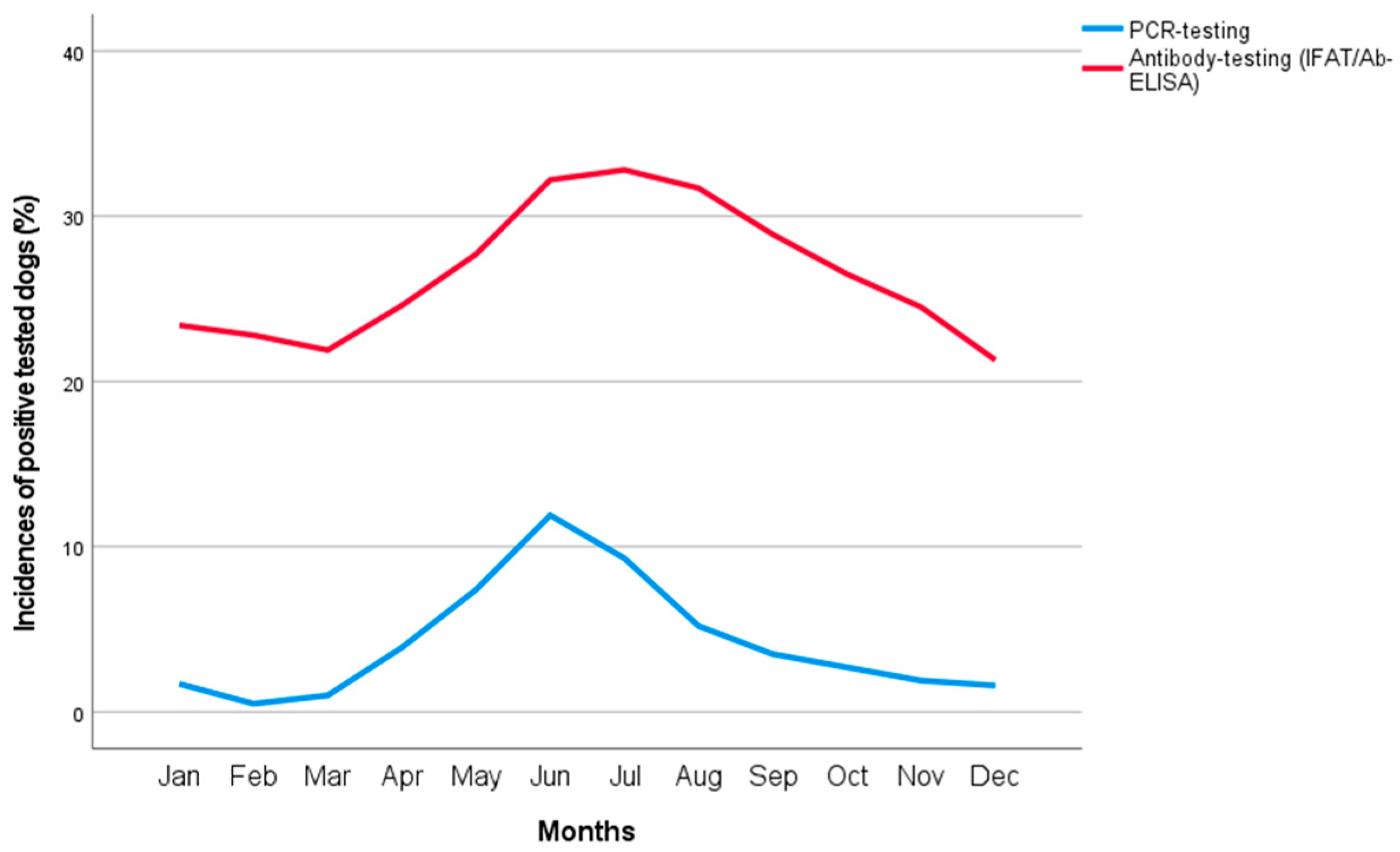
Figure 1. Monthly distribution of dogs that tested positive for Anaplasma phagocytophilum by direct (PCR; blue line) and indirect detection methods (IFAT, ELISA; red line) from 2008 to 2020.
Animals 13 00720 g001
Table 1. Number of positive tests in dogs including direct and indirect detection methods for Anaplasma phagocytophilum in dogs living in Germany from 2008–2020 sorted by age (n positive/N total (% [95% CI lower limit; 95% CI upper limit]).
Age-GroupPCRIFAT/ELISA
0–2 years (junior)206/4934 (4.2 [3.7; 4.8])2557/20,749 (12.3 [11.9; 12.8])
>2–7 years (adult)478/9529 (5.0 [4.6; 5.5])8924/31,198 (28.6 [28.1; 29.1])
>7–10 years (mature)285/6019 (4.7 [4.2; 5.3])6473/16,349 (39.6 [38.8; 40.4])
>10–13 years (senior)213/3809 (5.6 [4.9; 6.4])3704/8757 (42.3 [41.3; 43.3])
>13 years (geriatric)49/762 (6.4 [4.8; 8.5])600/1475 (40.7 [38.2; 43.2])
Total1231/25,053 (4.9 [4.7; 5.2])22,258/78,528 (28.3 [28.0; 28.7])
Mann–Whitney U testp < 0.001p < 0.001
CI: confidence interval; ELISA: Antibody-enzyme-linked immunosorbent assay; IFAT: immunofluorescence antibody test; PCR: Polymerase chain reaction; PCR: Mann–Whitney U test, p < 0.001, 154,666,055.500; IFAT/ELISA: Mann–Whitney U test, p < 0.001, 825,081,293.000.
Table 2. Number of positive tests in dogs including direct and indirect detection methods for Anaplasma phagocytophilum in dogs living in Germany from 2008–2020 sorted by years of testing (n positive/N total (% [95% CI lower limit; 95% CI upper limit]).
YearPCRIFAT/ELISAIFATELISA
200840/850 (4.7 [3.4; 6.4])531/2988 (17.8 [16.4; 19.2])531/2988 (17.8 [16.4; 19.2])
200953/1194 (4.4 [3.4; 5.8])1054/4082 (25.8 [24.5; 27.2])1054/4082 (25.8 [24.5; 27.2])
201042/1245 (3.4 [2.5; 4.6])1187/4160 (28.5 [27.2; 29.9])1187/4160 (28.5 [27.2; 29.9])
201155/1182 (4.7 [3.6; 6.1])1491/5174 (28.8 [27.6; 30.1])1491/5174 (28.8 [27.6; 30.1])
201258/1355 (4.3 [3.3; 5.5])1669/6187 (27.0 [25.9; 28.1])1669/6187 (27.0 [25.9; 28.1])
201346/1461 (3.1 [2.3; 4.2])1698/6539 (26.0 [24.9; 27.1])1698/6539 (26.0 [24.9; 27.1])
201484/1907 (4.4 [3.5; 5.4])1534/7875 (19.5 [18.6; 20.4])1534/7875 (19.5 [18.6; 20.4])
2015100/1887 (5.3 [4.4; 6.4])984/7616 (12.9 [12.2; 13.7])984/7616 (12.9 [12.2; 13.7])
2016124/2378 (5.2 [4.4; 6.2])1208/7487 (16.1 [15.3; 17.0])1208/7487 (16.1 [15.3; 17.0])
2017167/2955 (5.7 [4.9; 6.6])3383/9146 (37.0 [36.0; 38.0])451/1365 (33.0 [30.6; 35.6])3206/8949 (35.8 [34.8; 36.8])
2018157/3160 (5.0 [4.3; 5.8])3183/8892 (35.8 [34.8; 36.8])305/1085 (28.1 [25.5; 30.9])3032/8729 (34.7 [33.7; 35.7])
2019190/3598 (5.3 [4.6; 6.1])2998/9390 (31.9 [31.0; 32.9])248/964 (25.7 [23.0; 28.6])2848/9235 (30.8 [29.9; 31.8])
2020216/4196 (5.1 [4.5; 5.9])3800/10,840 (35.1 [34.2; 36.0])254/1092 (23.3 [20.8; 25.9])3639/10,668 (34.1 [33.2; 35.0])
Total1332/27,368 (4.9 [4.6; 5.1])24,720/90,376 (27.4 [27.1; 27.6])12,614/56,614 (22.3 [21.9; 22.6])12,725/37,581 (33.9 [33.4; 34.3])
CI: confidence interval; ELISA: Antibody-enzyme-linked immunosorbent assay; IFAT: Immunofluorescence antibody test; PCR: Polymerase chain reaction; Total: χ2 = 2370.973; df = 12; p < 0.001; φ = 0.147; PCR: χ2 = 25.264; df = 12; p = 0.014; φ = 0.03; IFAT/ELISA: χ2 = 2845.303, df = 12; p < 0.001; φ = 0.177; IFAT: χ2 = 1120.448, df = 12; p < 0.001; φ = 0.141; ELISA: χ2 = 56.346, df = 3; p < 0.001; φ = 0.039.
Table 3. Binominal logistic regression analysis in dogs tested for Anaplasma phagocytophilum by direct (PCR) and indirect detection methods (IFAT, ELISA) in 74973 dogs with known sex, age, years of testing, and months of testing from 2008 to 2020.
BSEWaldpOdds Ratio95%-CI for Odds Ratio
Lower BoundUpper Bound
PCR testing
 Sex0.0000.0600.0000.9951.0000.8881.125
 Age0.0940.0612.3880.1221.0990.9751.238
 Years0.0290.00910.5820.0011.0291.0111.047
 Season1.1730.061370.629<0.0013.2332.8693.643
 Constant−61.15817.72111.910<0.001
Antibody testing (IFAT/ELISA)
 Sex0.2380.017203.113<0.0011.2691.2281.312
 Age0.8720.0172656.929<0.0012.3922.3142.472
 Years0.0540.002623.234<0.0011.0561.0511.060
 Season0.3380.017379.519<0.0011.4031.3561.451
 Constant−117.7574.868585.175<0.001
B: unstandardized regression weight; CI: confidence interval; ELISA: Antibody-enzyme-linked immunosorbent assay; IFAT: Immunofluorescence antibody test; PCR: Polymerase chain reaction; SE: standard deviation to the mean; Wald: Wald chi-squared test; Variables entered: sex: male, age: > 7 years, year, season: summer; Degrees of freedom were 1 for all Wald statistics.
Table 4. Number of positive tests in dogs including direct and indirect detection methods of Anaplasma phagocytophilum in dogs living in Germany sorted by federal states of the submitting veterinarians (n positive/N total (% [95% CI lower limit; 95% CI upper limit]).
Federal StatePCRIFAT/ELISA
Baden-Wuerttemberg118/2887 (4.1 [3.4; 4.9])2111/8968 (23.5 [22.7; 24.4])
Bavaria139/2721 (5.1 [4.3; 6.0])2511/7215 (34.8 [33.7; 35.9])
Berlin-Brandenburg231/3038 (7.6 [6.7; 8.6])1062/3475 (30.6 [29.0; 32.1])
Hesse74/2496 (3.0 [2.4; 3.7])1943/9739 (20.0 [19.2; 20.8])
Lower Saxony/Bremen247/4341 (5.7 [5.0; 6.4])5733/17,056 (33.6 [32.9;34.3])
Mecklenburg Western Pomerania28/419 (6.7 [4.6; 9.6])158/676 (23.4 [20.3; 26.8])
North Rhine Westphalia237/6124 (3.9 [3.4; 4.4])6913/26,786 (25.8 [25.3;26.3])
Rhineland Palatinate55/1734 (3.2 [2.4; 4.1])1358/6286 (21.6 [20.6; 22.6])
Saarland28/748 (3.7 [2.5; 5.4])672/2216 (30.3 [28.4; 32.3])
Saxony76/1006 (7.6 [6.0; 9.4])471/1817 (25.9 [23.9; 28.0])
Saxony-Anhalt16/383 (4.2 [2.5; 6.8])273/930 (29.4 [26.5; 32.4])
Schleswig-Holstein/Hamburg52/1051 (4.9 [3.8; 6.5])1176/4046 (29.1 [27.7; 30.5])
Thuringia31/420 (7.4 [5.2; 10.4])339/1166 (29.1 [26.5; 31.8])
Total1332/27,368 (4.9 [4.6; 5.1])24,720/90,376 (27.4 [27.1; 27.6])
Chi-square testp < 0.001p < 0.001
CI: confidence interval; ELISA: Antibody-enzyme-linked immunosorbent assay; IFAT: Immunofluorescence antibody test; PCR: Polymerase chain reaction; Total: χ2 = 1048.245; df = 12; p < 0.001; φ = 0.098); PCR: χ2 = 129.897; df = 12; p < 0.001; φ = 0.069); IFAT/ELISA: χ2 = 1053.471, df = 12; p < 0.001; φ = 0.108).
Disclaimer/Publisher’s Note: The statements, opinions and data contained in all publications are solely those of the individual author(s) and contributor(s) and not of MDPI and/or the editor(s). MDPI and/or the editor(s) disclaim responsibility for any injury to people or property resulting from any ideas, methods, instructions or products referred to in the content.