Article

Fluorescence Microscopy and Flow-Cytometry Assessment of Substructures in European Red Deer Epididymal Spermatozoa after Cryopreservation

by
Anna Dziekońska
1,*,
Marek Lecewicz
1,
Agnieszka Partyka
2 and
Wojciech Niżański
2
1
Department of Animal Biochemistry and Biotechnology, University of Warmia and Mazury in Olsztyn, Oczapowskiego 5, 10-719 Olsztyn, Poland
2
Department of Reproduction and Clinic of Farm Animals, Wroclaw University of Environmental and Life Sciences, Pl. Grunwaldzki 49, 50-366 Wroclaw, Poland
*
Author to whom correspondence should be addressed.
Animals 2023, 13(6), 990; https://doi.org/10.3390/ani13060990
Submission received: 30 December 2022 / Revised: 9 February 2023 / Accepted: 6 March 2023 / Published: 8 March 2023
(This article belongs to the Special Issue Current Status and Advances in Semen Preservation)

Abstract

:

Simple Summary

Flow cytometry (FC) is the recommended technique for assessing sperm quality. In comparison with fluorescence microscopy (FM), FC supports analyses of much larger sperm populations and generates more reliable results. However, FC is not always accessible, and sperm populations are often evaluated with the use of FM. In the present study, FC and FM were used to assess the functionality of various organelles in European red deer epididymal spermatozoa stored in liquid nitrogen. Spermatozoa were collected from the epididymides of hunter-harvested European red deer stags. The epididymides were stored in a refrigerator (2–4 °C) for 12 h before analysis. The study demonstrated that the refrigerated storage of the epididymides for 12 h had no significant effect on the sperm quality before cryopreservation, but it significantly influenced the percentage of early necrotic sperm after thawing. The results of FM and FC assays differed significantly, excluding in the assessment of the plasma membrane integrity. However, the results of both assays revealed significant correlations between the examined variables, except for mitochondrial activity. The study demonstrated that the spermatozoa from epididymides chill-stored for 12 h can be used for cryopreservation. Fluorescence microscopy and FC are equally reliable techniques, but FM was more useful for evaluating mitochondrial activity.

Abstract

Thawed spermatozoa, sampled post mortem from the fresh epididymides of European red deer and epididymides stored for up to 12 h at 2–4 °C, were evaluated by fluorescence microscopy (FM) and flow cytometry (FC). The sperm samples were extended and cryopreserved. The sperm motility (CASA), sperm viability (SYBR+/PI-), acrosome integrity, mitochondrial activity, apoptotic changes, and chromatin stability were assessed. Sperm were analyzed by FM before cryopreservation, and by FM and FC after thawing. Epididymal storage time (for 12 h) had no significant effect (p > 0.05) on the examined variables before cryopreservation. After thawing, the storage variants differed (p ˂ 0.05) in the percentage of apoptotic sperm (FM and FC) and DNA integrity (FC). The results of FM and FC differed (p ˂ 0.05) in all the analyzed parameters, excluding SYBR+/PI. Significant correlations (p ˂ 0.01) were observed between the sperm viability, acrosome integrity, and the percentage of non-apoptotic spermatozoa, regardless of the applied technique. In FM, the above parameters were also significantly correlated with mitochondrial activity. The study demonstrated that European red deer spermatozoa stored in the epididymides at 2–4 °C for 12 h can be used for cryopreservation. Both techniques were equally reliable, but FM was better suited for evaluating mitochondrial activity whereas FC was more useful in the evaluation of DNA fragmentation.

1. Introduction

Sperm collected from hunter-harvested wild-living animals is a valuable source of genetic material that can be used for the conservation and genetic improvement of animal populations in breeding farms [1,2,3]. Sperm can be cryopreserved in liquid nitrogen to preserve its fertilizing potential for a long period of time (many years), which has important implications for species conservation [4,5,6].
Ejaculated sperm is the most recommended for reproductive purposes, but it is not easy to acquire, and epididymal sperm can also be used [7]. In wild-living animals, post-mortem sperm sampling from the epididymides is the easiest sperm-harvesting method, which, unlike electroejaculation, does not require specialist equipment or complex procedures [8,9]. The epididymal sperm for reproductive purposes can be stored in a liquid state or cryopreserved [4,10,11]. However, epididymal spermatozoa cannot always be collected directly from hunter-harvested animals because the hunting grounds are usually situated in remote locations. As a result, the sperm are harvested several hours after the hunt. The collected material is transported to a laboratory and stored in a refrigerator at a temperature of 0–6 °C and 80–85% humidity. The storage conditions can significantly affect the viability of the epididymal sperm, and further research is needed to address this aspect.
Previous studies have shown that the sperm of many animal species, including mice [12], rams [13], dogs [14], and bulls [15,16,17], is resistant to low storage temperatures in the range of 0–6 °C. Iberian red deer spermatozoa can be stored in the epididymides for up to several days at a temperature 4–5 °C [7,18] before cryopreservation [19,20]. These observations suggest that European red deer spermatozoa intended for cryopreservation should also tolerate temperatures in the range of 2–4 °C.
Sperm quality is evaluated with the use of various laboratory methods, where fluorescence techniques, motility analyses, and morphology assessments are most recommended. These assays are conducted with the use of a fluorescence microscope (FM) or a flow cytometer (FC). Each technique has its strengths and weaknesses [21]. Fluorescence microscopy is a time-consuming method that should be performed by a qualified and experienced observer, and it involves time limits and a small number of cells [22]. In turn, FC supports accurate and simultaneous assessments of various cell structures [23,24]. Flow cytometry is highly recommended in clinical trials because it supports rapid and objective analyses of large cell samples [22,25]. Flow cytometry is also recommended for fluorescence analyses of sperm quality [23]. However, flow cytometers are expensive and not readily accessible. Most laboratories are equipped with fluorescence microscopes, but only small sperm samples (200–300 cells) are analyzed under an FM. According to many researchers, these limitations undermine the reliability of the results.
In view of the above, the aim of this study was to evaluate different structures in cryopreserved epididymal spermatozoa of European red deer, collected directly from fresh epididymides and from epididymides stored for 12 h at a temperature of 2–4 °C, with the use of FM and FC. Sperm motility and motility parameters were determined, and fluorescence analyses of sperm viability, acrosomal membrane integrity, mitochondrial membrane potential, apoptotic changes, and DNA status were performed. Spermatozoa were subjected to FM only before cryopreservation, and to both FM and FC after thawing. The same fluorochromes were used in FM and FC.

2. Materials and Methods

2.1. Animals

Red deer stags were hunted in accordance with the harvest plan for each species of game animals, in the in the Forest District of Nowe Ramuki (Region of Warmia and Mazury, Poland), in September and October (during the rutting season), in accordance with the Polish Hunting Law. Experimental material was collected post mortem, and it consisted of the testes and epididymides that were stored in the scrotum until sperm collection. The sperm samples were collected from the epididymides of 16 European red deer stags (aged 4 to 11 years, with body weight of 140 kg to approx. 180 kg).

2.2. Sampling and Cryopreservation of Spermatozoa

Spermatozoa were collected from the tail of the epididymis (within 2 h post mortem, variant I) and from epididymides refrigerated for 12 h (2–4 °C, variant II). Spermatozoa were sampled according to a previously described procedure [26]. In the collected samples, the sperm motility was assessed using the computer-assisted semen analysis (CASA) system and the sperm concentration was evaluated using a Bürker counting chamber (Equimed-Medical Instruments, Cracow, Poland).
The spermatozoa were cryopreserved according to the procedure described by Martínez-Pastor et al. [27], with some modifications. In the first trial, the samples were diluted with a commercial freezing extender (Andromed, Minitub GmbH, Tiefenbach, Germany) to a concentration of 400 × 106 cells/mL and transferred to a refrigerator (4 °C) for 2 h to equalize the temperature. Then, the samples were diluted with the same extender to a concentration of 160–200 × 106 cells/mL and left to stand for 1 h. The samples were then packed in 0.25 mL straws (IMV, L’Aigle Cedex, France) and frozen in nitrogen vapor (4 cm above liquid nitrogen) for 10 min. The straws were transferred to liquid nitrogen, where they were kept for at least 1 year. The samples were thawed by placing the straws in water (65 °C) for 6 s.

2.3. Sperm Analysis

2.3.1. Sperm Motility

Sperm motility was evaluated using the CASA system (Hamilton Thorne Sperm Analyzer IVOS version 12.2l; Hamilton Thorne Biosciences, MA, USA). Sperm samples were diluted 1 : 100 (fresh) and 1 : 5 (fresh diluted sperm, frozen-thawed) in phosphate-buffered saline (PBS) to a concentration of around 30–50 × 106 cells/mL. A 5 μL aliquot of the sample was placed in a pre-warmed Makler counting chamber (Sefi-Medical Instruments Ltd., Haifa, Israel) and evaluated at 37 °C. In each sample, spermatozoa were analyzed in five fields of view selected randomly by the computer. The analyses included the determination of: percentage of total motile sperm (TMOT), percentage of progressive motile sperm (PMOT), and parameters characterizing sperm movement: VAP, VSL, VCL, ALH, BCF, LIN (VSL/VCL ratio × 100 %), and straightness (STR, VSL/VAP ratio × 100 %).
The CASA system settings recommended by Hamilton Thorne for gazelle/deer [26] were used in the analyses. Progressive motility was defined as the percentage of spermatozoa with a VAP > 75.0 μm/s and an STR > 80%.

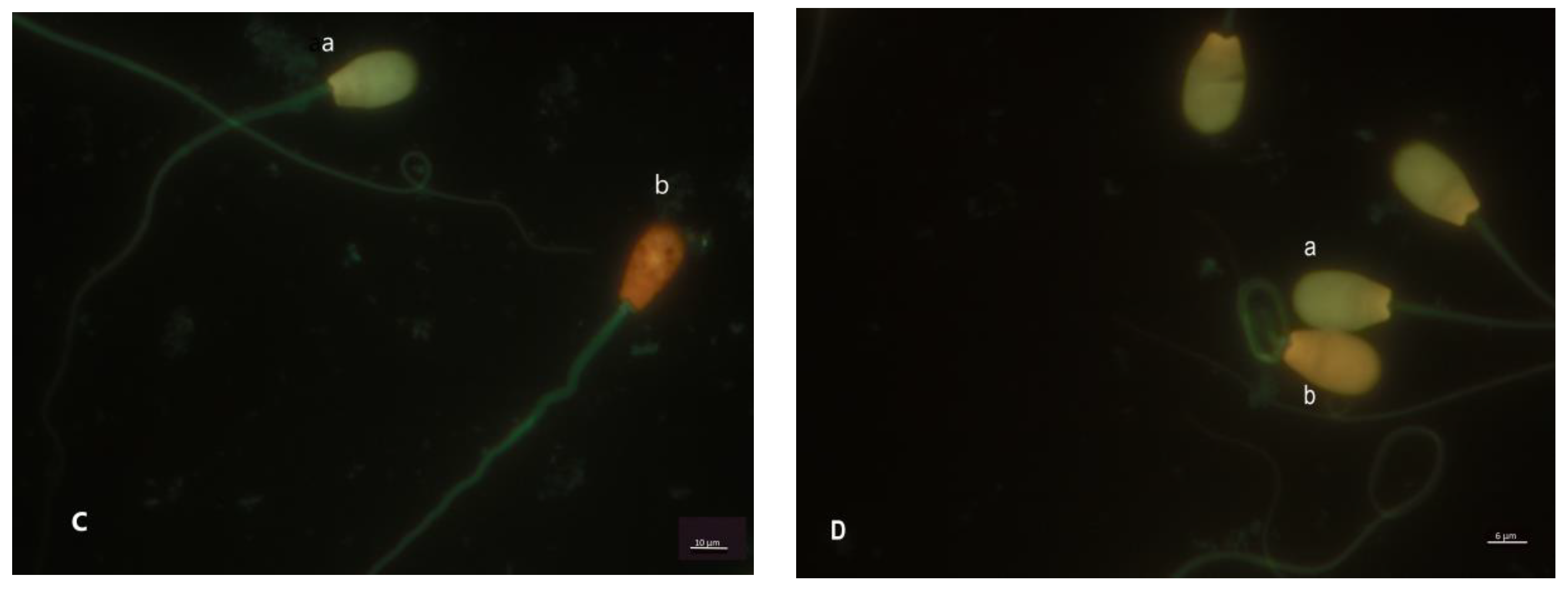
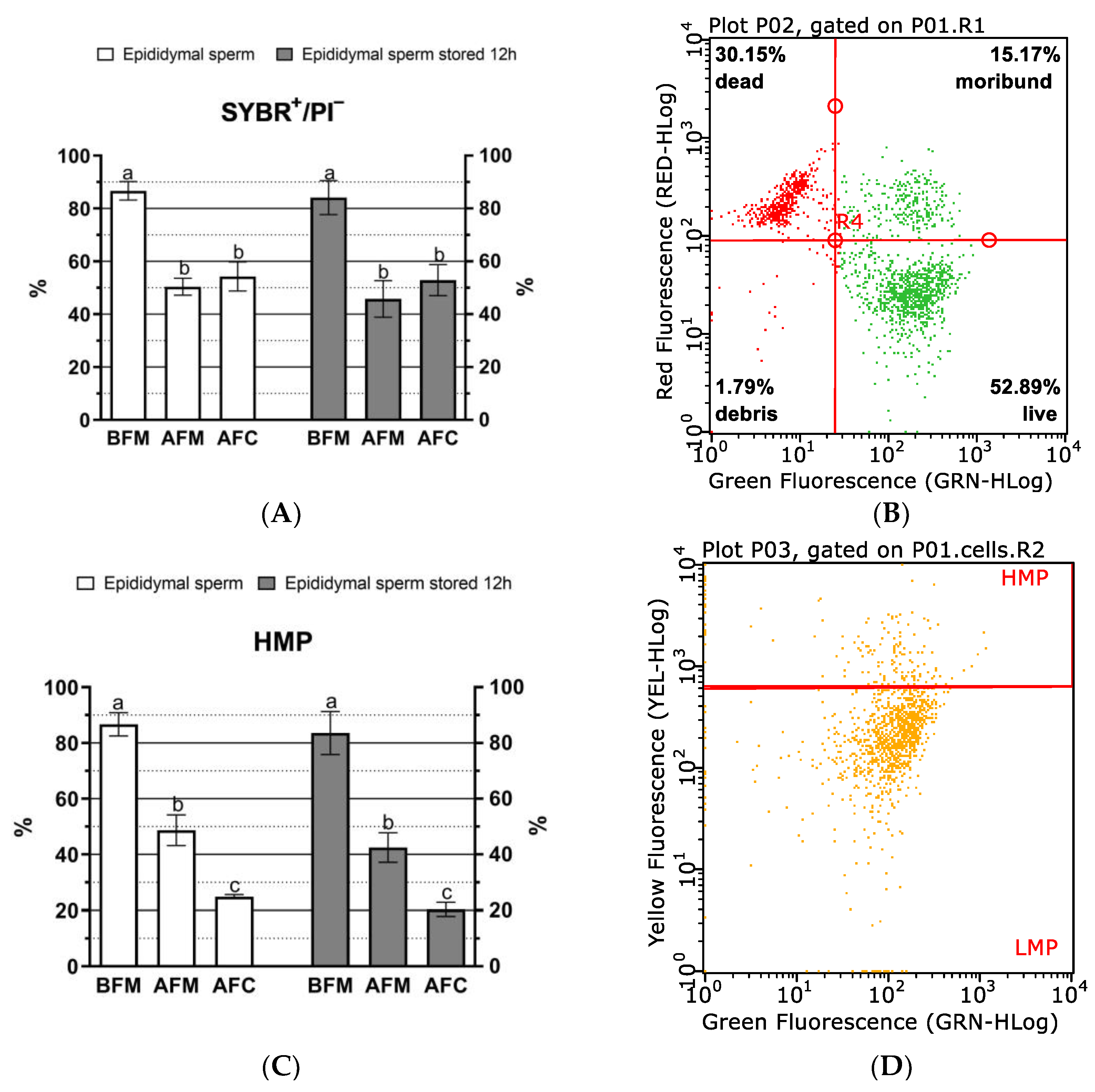
2.3.2. Fluorescence Assay

The sperm characteristics were evaluated by the fluorescence method before and after cryopreservation. The Olympus BX41 Fluorescence Microscope, which offers ultraviolet (330–385 nm), blue (460–490 nm), and green (510–550 nm) excitation wavelengths, was used for fluorescence analyses. Stained samples were analyzed at 600 × magnification. A minimum of 300 cells per slide were examined in each aliquot.
The viability (plasma membrane integrity) was assessed using SYBR-14 and propidium iodide (PI) fluorescent probes (Live/Dead Sperm Viability Kit; Life Technologies Ltd., Grand Island, NY, USA) according to a previously described method [26]. The diluted sperm samples (200 μL) were supplemented with 2 μL of 1 mM SYBR-14 solution and 2 μL of PI (2.4 μM in Tyrode’s salt solution), and incubated at 37 °C in the dark for 10 min. Sperm with green heads (SYBR-14+/PI, live sperm with integral membranes) and sperm with red heads (dead sperm) were identified.
The acrosomal status of spermatozoa was evaluated using fluorescein isothiocyanate-labeled peanut (Arachis hypogaea) agglutinin (FITC-PNA; Life Technologies Ltd., Grand Island, NY, USA) with a PI solution. The method was described in earlier reports [11,28]. Next, 1 µL of JC-1 (1 mg JC-1/mL anhydrous dimethyl sulfoxide, DMSO) was added to diluted sperm samples (200 μL), and they were incubated at 37 °C for 15 min (to identify all sperm cells under the fluorescence microscope). Then, 2 μL of FITC-PNA solution and 2 μL of PI were added to the samples, which were incubated again for 5 min at 37 °C. The FITC-PNA working solution was prepared by dissolving 2 mg of FITC-PNA in 1 mL of PBS. Four sperm populations were observed under the microscope: non-stained spermatozoa in the head region with fluorescence in the midpiece were classified as live sperm with intact acrosomes (FITC-PNA/PI); sperm with green-red fluorescence in the head (FITC-PNA+/PI+) were classified as early necrotic, acrosome-reacted sperm; sperm with a red head were considered as dead; and a small population of spermatozoa had only green staining of the acrosomal cap (acrosome-reacted sperm) (Figure 1A).
The mitochondrial activity was evaluated by examining the mitochondrial membrane potential (MMP) of sperm with the use of JC-1 fluorochromes (Life Technologies Ltd., Grand Island, NY, USA) with PI, according to a previously described method [11,28]. Diluted sperm samples (200 μL) were incubated with 1 μL of JC-1 solution for 15 min at 37 °C. Next, 1 μL of PI was added to the samples, which were incubated again for 5 min at 37 °C. The analysis revealed the presence of sperm with orange midpieces (with active mitochondria, high MMP) and sperm with green midpieces or unstained, often with red fluorescence in the head region (low MMP) (Figure 1B).
The DNA integrity was assessed using acridine orange, as previously described by Partyka et al. [29] with some modifications. Sperm samples (50 μL) were subjected to acid denaturation (30 s) by adding 200 μL of a lysis buffer (Triton X-100 0.1% (v/v), NaCl 0.15 M, HCl 0.08 M, pH 1.4). Subsequently, 600 µL of acridine orange solution was added to the samples, which were incubated for 3 min in the dark. The fluorescence analysis revealed the presence of sperm with green heads (with integral DNA) and sperm with orange or red heads (with DNA fragmentation; DFI) (Figure 1C,D).
The Vybrant Apoptosis Assay Kit #4 (Life Technologies Ltd., Grand Island, NY, USA) was used to assess apoptosis and membrane integrity according to a previously described method [11,28]. First, to visualize all cells under a fluorescence microscope, the diluted samples (200 µL) were stained with 1 µL JC-1 (as described above). Then, 2 μL of YO-PRO-1 and 2 μL of PI were added to the samples, which were incubated at 37 °C for 5 min. Four sperm populations were distinguished in the fluorescence analysis: viable sperm (YO-PRO-1/PI), sperm showing apoptotic-like changes (YO-PRO-1+/PI), sperm with early necrotic changes (YO-PRO-1+/PI+), and dead/necrotic sperm (YO-PRO-1/PI+), according to a previously described staining model [11].

2.3.3. Flow-Cytometry Analysis

Flow-cytometry analyses were performed in a Guava EasyCyte 5 (Merck KGaA, Darmstadt, Germany) cytometer. Fluorescence was induced by an argon ion 488 nm laser. Data were acquired using the GuavaSoft™ 3.1.1 software (Merck KGaA, Darmstadt, Germany). Non-sperm events were gated out based on scatter properties and excluded from analyses. A total of 10,000 events were analyzed for each sample. For the flow-cytometry analysis, the stained sperm samples were extended to a concentration of around 5–10 × 106 spermatozoa/1 mL. The same fluorochromes were used in fluorescence flow-cytometry analyses and in the microscopic analysis.
To assess the plasma membrane integrity, spermatozoa were stained with SYBR-14 and propidium iodide (PI) according to the protocol described by Partyka et al. [29]. The diluted samples (500 μL) were stained with 3 μL of SYBR-14 and 3 μL of PI. SYBR-14-positive sperm with green fluorescence and PI-negative sperm were classified as live sperm cells with intact plasma membranes.
Sperm acrosome status was assessed with lectin PNA from Arachis hypogaea conjugated with Alexa Fluor® 488 (Life Technologies Ltd., Grand Island, NY, USA). The diluted semen samples were combined with 10 μL of PNA working solution (1 μg/mL) and incubated for 5 min at room temperature in the dark. After incubation, the samples were washed, and 3 μL of PI was added before the fluorescence analysis [29].
Sperm mitochondrial activity was determined by JC-1 and PI staining. A 3 mM stock solution of JC-1 in DMSO was prepared. Diluted sperm samples (500 μL) with a concentration of 50 × 106 cells/mL were combined with 0.67 μL of JC-1. Before analysis, the probes were incubated for 20 min, in the dark, at 37 °C [30]. Sperm emitting orange fluorescence were categorized as cells with high mitochondrial potential (HMP), whereas sperm emitting green fluorescence were classified as cells with low mitochondrial potential (LMP).
Apoptosis and sperm viability were evaluated with YO-PRO-1 (25 μM solution in DMSO) and PI fluorochromes. Sperm were diluted with PBS to a concentration of 50 × 106 cells/mL. A diluted sample of 1 mL was stained with 1 μL of YO-PRO-1 (with a final concentration of 25 nM) and 1 μL of PI [31]. In the FC analysis, fluorescence was measured with a FL-2 sensor and a 575 nm band-pass filter to detect PI, and with a FL-1 sensor and a 525 nm band-pass filter to detect YO-PRO-1.
Acridine orange was used to assess DNA integrity, as described above, with minor changes. The sperm suspension (100 μL) was denatured with a lysing solution (200 μL) for 30 s, and then AO solution (600 μL) was added. The samples were analyzed after 3 min of incubation. Spermatozoa with normal double-stranded DNA displayed green fluorescence and were regarded as the main population. Spermatozoa with increased red fluorescence indicative of DNA fragmentation (DFI) were located to the right of the main population.

2.4. Statistical Analysis

The results were processed with the use of a general linear model (GLM) and ANOVA in the Statistica program (v. 13.3, StatSoft Incorporation, Tulsa, OK, USA). The assumption of normality was checked using the Shapiro–Wilk test, and the homogeneity of variance was assessed with Levene’s test. The data that were not normally distributed were transformed before statistical analysis. The percent data were arcsine transformed. The results were presented as means ± standard error of the mean (SEM). Tukey’s post-hoc test was used to compare the means, and the statistical significance was set at p < 0.05. The relationships between sperm motility and fluorescence parameters were examined by calculating the Pearson correlation coefficient.

3. Results

The sperm motility analysis revealed that the compared storage variants had no significant effect (p > 0.05) on sperm motility parameters both before cryopreservation and after thawing (Table 1). In both variants, cryopreservation induced a significant (p ˂ 0.05) decrease in the values of TMOT, PMOT, VAP, VSL, VCL, and ALH. No significant changes were found in the values of BCF, STR, and LIN before and after thawing.
The FM assay revealed that cryopreservation significantly influenced all examined sperm structures, excluding DNA status, in both storage variants (Figure 2 and Figure 3). The variant where sperm were stored in the epididymides for up to 12 h before cryopreservation did not induce significant changes in sperm cell structures, relative to the variant where sperm were sampled directly from fresh epididymides and cryopreserved.
A similar relationship was observed in thawed sperm, excluding the percentage of normal spermatozoa without apoptotic changes (FC), the percentage of apoptotic-like spermatozoa (FM and FC) (Figure 3A,B), and DNA fragmentation (DFI, FC) (Figure 3E,F).
In the thawed sperm, the percentage of spermatozoa with intact plasma membranes was the only parameter that was not significantly (p > 0.05) affected by the applied fluorescence technique. The results of FM and FC assays differed significantly (p ˂ 0.05) in the following parameters: the percentage of spermatozoa with high mitochondrial membrane potential (Figure 2C,D), acrosome integrity (Figure 2E,F), apoptotic changes (Figure 3A–C), and chromatin stability (Figure 3E,F). In the analysis of mitochondrial activity, the percentage of spermatozoa with a high mitochondrial membrane potential was significantly higher in FM than in FC, regardless of the storage variant (Figure 2C,D). The percentage of sperm with intact acrosomes was significantly higher in FC than in FM (Figure 2E,F). The percentage of non-apoptotic sperm was also significantly higher in FC than in FM (Figure 3A). However, the percentage of apoptotic-like and early necrotic spermatozoa was higher in FC than in FM, irrespective of the storage variant (Figure 3B,C). In the DNA status analysis, a significantly (p ˂ 0.05) smaller percentage of spermatozoa with fragmented DNA was identified in FC than in FM (Figure 3E,F).
The correlations between sperm motility and the analyzed fluorescence parameters in FM and FC are presented in Table 2 and Table 3, respectively.
The correlation analysis for FM revealed significant (p ˂ 0.05) correlations between the percentage of motile sperm (TMOT) versus acrosomal membrane integrity, the percentage of non-apoptotic sperm, and the percentage of sperm with high mitochondrial membrane potential (HMP). The parameter HMP was also correlated (p ˂ 0.01) with plasma membrane integrity, acrosome integrity, and the percentage of non-apoptotic sperm. Progressive motility was significantly (p ˂ 0.05) correlated with plasma membrane integrity.
The FC assay also revealed a positive correlation (p ˂ 0.05) between TMOT versus acrosome integrity, and between PMOT versus plasma membrane integrity and the percentage of non-apoptotic spermatozoa. Plasma membrane integrity was highly positively correlated (p ˂ 0.001) with acrosome integrity, and positively correlated (p ˂ 0.01) with the percentage of non-apoptotic sperm. In turn, the HMP was not significantly (p > 0.05) correlated with the remaining parameters in the FC assay. No significant correlations were found between the DNA status and the remaining parameters.

4. Discussion

The study demonstrated that the storage of European red deer spermatozoa in the epididymides at a temperature of 2–4 °C for up to 12 h did not significantly decrease sperm viability or functionality in comparison with the variant where spermatozoa were sampled directly from fresh epididymides. Sperm motility, motility parameters, plasma membrane integrity, mitochondrial activity, and apoptotic changes were similar in both storage variants. These results confirm the previous observations made in Iberian red deer, and indicate that red deer spermatozoa are relatively resistant to cold shock [7,18,19]. Interestingly, even small fluctuations in temperature in the range of 2–4 °C did not lead to a significant decrease in the quality of spermatozoa stored in the epididymides for 12 h. Possibly, the storage time was too short to induce significant changes in the sperm function. In other studies, significant changes in the motility and structure of spermatozoa stored in the epididymides at a temperature of 4–5 °C were reported only after 24 h [32] or even 48 h [19,20]. However, in the present study, the epididymides were stored in the scrotum, whereas in the cited studies, the epididymides and the testicles were removed from the scrotum, placed in plastic bags, and stored in beakers filled with water at a temperature of 4–5 °C. The above factor could have played a role in preserving high sperm quality before cryopreservation.
Despite considerable advances in freezing and thawing methods, cryopreservation always leads to damage in cell structures, which alters sperm motility, motility parameters, and decreases semen fertilizing ability [33,34]. In the current study, significant changes in all the evaluated parameters were also observed, including the plasma membrane integrity, acrosome integrity, and mitochondrial membrane potential, which decreased the TMOT and, in particular, PMOT values that are influenced by the VAP and VSL values.
After thawing, most of the analyzed variables did not differ significantly between storage variants, which could be attributed to similar sperm quality before cryopreservation. The quality of sperm cells before cryopreservation significantly influences their resistance to freezing damage [19]. The significant differences between the tested storage variants were observed only in the percentage of apoptotic spermatozoa (FM and FC) and DNA fragmentation (DFI, FC). This observation suggests that sperm stored in the epididymides at a temperature of 2–4 °C were more susceptible to oxidative damage and apoptotic changes during cryopreservation. In another study, cryopreservation induced apoptosis in bovine spermatozoa [35]. In boar spermatozoa, freezing and thawing stimulated an excessive generation of reactive oxygen species (ROS), exerted a negative effect on the cell membrane, mitochondrial activity, and chromatin stability, and accelerated apoptosis [36].
Fluorescent microscopy and FC are widely used to assess sperm structures and semen quality. In the present study, the applied fluorescence technique significantly affected the values of most variables, which could be attributed to numerous factors, including the number of sperm cells. A total of 10,000 sperm cells were evaluated by FC, but only 300 spermatozoa were assessed by FM. It should also be noted that sperm sampled from the tail of the epididymis can be contaminated with blood cells, epithelial cells, or tissue fragments, which can compromise the differentiation of stained spermatozoa by FC [22]. Forward-scatter (FSC) and side-scatter (SCC) gating strategies can be applied to eliminate these contaminants based on differences in the size and complexity of the debris and spermatozoa, but this approach does not always generate reliable results [23]. Flow cytometry is the most recommended technique for the fluorescence assessment of cells, but FM is also an accurate and reliable method, regardless of the origin of sperm cells (ejaculate, epididymides, or testicles) [22].
The compared fluorescence techniques produced similar results only with regard to the viability of sperm stained with SYBR-14/PI. Similar observations were made by Merkies et al. [37], where sperm viability evaluated with the same combination of fluorochromes was similar in both FC and FM.
The greatest differences in the results of FM and FC were noted in the percentage of sperm with high mitochondrial membrane potential (HMP) and DNA integrity in thawed spermatozoa. JC-1 staining supports the identification of two or three sperm populations with different mitochondrial membrane potential [23]. Sperm with a medium MMP (green-orange fluorescence) were most difficult to identify in both FM and FC, which could have affected the results. In the FM analysis, sperm with a medium MMP were difficult to distinguish from sperm with HMP (orange fluorescence) and were classified as spermatozoa with HMP. In turn, in the FC analysis, these sperm were classified as spermatozoa with low MMP (Figure 2D). These difficulties contributed to significant differences in HMP values between FM and FC, irrespective of the storage variant. According to some authors, JC-1 is the most sensitive and reliable fluorescent probe for assessing MMP in FC [38,39], but Martínez-Pastor et al. [23] observed that this probe can be highly sensitive to the staining conditions and that controls should be established to select flow-cytometer settings.
The percentage of sperm with intact acrosomal membranes and the percentage of non-apoptotic sperm were lower in FM than in FC, probably due to differences in the number of the analyzed sperm cells. These differences can be also attributed to a lower detection sensitivity (depending on the observer’s visual processing skills), a smaller number of analyzed cells, and the risk of fluorescence decay during the readout [22]. This problem was particularly evident in the analysis of the percentage of early necrotic sperm, which was significantly lower in FM than in FC. The FM assessment revealed a higher percentage of dead than early necrotic sperm, which influenced the final percentage of non-apoptotic spermatozoa.
The percentage of sperm with fragmented DNA was higher in FM than FC, which could be attributed to the difficulties in distinguishing between the green (sperm with normal DNA) and orange fluorescence (sperm with fragmented DNA) in FM. The fluorescence decay in the samples analyzed under a microscope could be another reason. When the samples for DNA analyses are exposed to laser light for a long period of time, sperm cells are probably rapidly degraded. They change color from green to orange, which is indicative of DNA fragmentation. The fluorescence excitation can lead to the formation of highly reactive photochemical products (reactive oxygen species (ROS), heat, and DNA damage), which are influenced by many factors, including excitation wavelength, intensity, and exposure time [40,41,42,43]. Flow-cytometry measurements are conducted rapidly (in less than 1 min), and this technique seems to be more appropriate for evaluating DNA fragmentation than FM. Sample preparation, as well as the time and conditions of denaturation and staining, can also significantly affect DNA integrity [23,44]. In other studies, the results of DNA fragmentation analyses also differed considerably between FM and FC [21,45].
Despite the considerable differences in the values of most analyzed variables, the results of FM and FC were subjected to a correlation analysis that revealed similar relationships between the sperm viability, acrosome integrity, the percentage of normal sperm without apoptotic changes, and sperm motility. Similar observations were made in other studies, where spermatozoa were assessed by FM [19,38] and FC [46,47]. Therefore, both techniques can be effectively used to assess sperm cell structures. The results of the correlation analysis indicate that FM was a more reliable method than FC in determining the percentage of sperm with high MMP.
The present study revealed that both fluorescence techniques have certain limitations that should be taken into consideration when assessing the various sperm structures.

5. Conclusions

The results of this study indicate that spermatozoa obtained post mortem and stored in the epididymides for up to 12 h (at 2–4 °C) can be used for cryopreservation. Fluorescence microscopy and FC are equally reliable techniques for assessing various sperm structures. However, the FM method proved to be more useful in the assessment of mitochondrial activity, whereas the FC assay was more useful in the evaluation of DNA fragmentation.

Author Contributions

Conceptualization, A.D.; methodology, A.D.; A.P., and W.N.; analysis, A.D., M.L., and A.P.; resources, W.N.; writing—original draft preparation, A.D.; review—editing, A.D. and A.P. All authors have read and agreed to the published version of the manuscript.

Funding

The study was supported by the University of Warmia and Mazury in Olsztyn, Poland (grant No 11.610.003-110). Project financially supported by the Minister of Education and Science in the range of the program entitled “Regional Initiative of Excellence” for the years 2019–2023, Project No. 010/RID/2018/19, the amount of funding was 12,000,000 PLN.

Institutional Review Board Statement

The research material was obtained during individual and collective hunts. Individual hunts require individual game-hunting permits. Collective hunts are organised pursuant to the provisions of §15 of the Polish Hunting Law of 13 October 1995. The numbers of game-hunting permits (legal license codes) and the certificates of origin of the harvested animals were confirmed in a statement issued by Nowe Ramuki Forest Inspectorate, which was submitted to the Editorial Office.

Informed Consent Statement

Not applicable.

Data Availability Statement

Not applicable.

Acknowledgments

The samples were obtained under a scientific cooperation agreement concluded by the University of Warmia and Mazury in Olsztyn and Nowe Ramuki Forest Inspectorate. The authors would like to thank all employees of Nowe Ramuki Forest Inspectorate, especially K. Zdun, M. Zega, G. Wiciel, and T. Dzietowiecki, and the hunters who helped with the sample collection. We would also like to thank B. Smalec for technical support during the experiment.

Conflicts of Interest

The authors declare no conflict of interest.

References

  1. Comizzoli, P.; Mermillod, P.; Mauget, R. Reproductive biotechnologies for endangered mammalian species. Reprod. Nutr. Dev. 2000, 40, 493–504. [Google Scholar] [CrossRef] [PubMed]
  2. Garde, J.J.; Martínez-Pastor, F.; Gomendio, M.; Malo, A.F.; Soler, A.J.; Fernández-Santos, M.R.; Esteso, M.C.; García, A.J.; Anel, L.; Roldán, E.R.S. The application of reproductive technologies to natural populations of red deer. Reprod. Domest. Anim. 2006, 41, 93–102. [Google Scholar] [CrossRef] [PubMed]
  3. Hishinuma, M.; Suzuki, K.; Sekine, J. Recovery and cryopreservation of sika deer (Cervus nippon) spermatozoa from epididymides stored at 4 °C. Theriogenology 2003, 59, 813–820. [Google Scholar] [CrossRef] [PubMed]
  4. Medina-Chávez, D.A.; Soler, A.J.; Martín-Maestro, A.; Villaverde, S.; Sánchez-Ajofrín, I.; Peris-Frau, P.; Del Olmo, E.; Bisbal, A.; García-Álvarez, O.; Fernández-Santos, M.D.R.; et al. Freezing Protocol Optimization for Iberian Red Deer (Cervus elaphus hispanicus) Epididymal Sperm under Field Conditions. Animals 2022, 12, 869. [Google Scholar] [CrossRef]
  5. Saragusty, J.; Gacitua, H.; King, R.; Arav, A. Post-mortem semen cryopreservation and characterization in two different endangered gazelle species (Gazella gazella and Gazella dorcas) and one subspecies (Gazella gazelle acaiae). Theriogenology 2006, 66, 775–784. [Google Scholar] [CrossRef]
  6. Soler, A.J.; Poulin, N.; Fernández-Santos, M.R.; Cognie, Y.; Esteso, M.C.; Garde, J.J.; Mermillod, P. Heterologous in vitro fertility evaluation of cryopreserved Iberian red deer epididymal spermatozoa with zona-intact sheep oocytes and its relationship with the characteristics of thawed spermatozoa. Reprod. Domest. Anim. 2008, 43, 293–298. [Google Scholar] [CrossRef]
  7. Kaabi, M.; Paz, P.; Alvarez, M.; Anel, E.; Boixo, J.C.; Rouissi, H.; Herraez, P.; Anel, L. Effect of epididymis handling conditions on the quality of ram spermatozoa recovered post-mortem. Theriogenology 2003, 60, 1249–1259. [Google Scholar] [CrossRef]
  8. Martínez, A.F.; Martínez-Pastor, F.; Alvarez, M.; Fernández-Santos, M.R.; Esteso, M.C.; de Paz, P.; Garde, J.J.; Anel, L. Sperm parameters on Iberian red deer: Electroejaculation and post-mortem collection. Theriogenology 2008, 70, 216–226. [Google Scholar] [CrossRef]
  9. Fletcher, T.J. Farmed deer: New domestic animals defined by controlled breeding. Reprod. Fertil. Dev. 2001, 13, 511–516. [Google Scholar] [CrossRef]
  10. Comizzoli, P.; Mermillod, P.; Cognié, Y.; Chai, N.; Legendre, X.; Mauge, R. Successful in vitro production of embryos in the red deer (Cervus elaphus) and the sika deer (Cervus nippon). Theriogenology 2001, 55, 649–659. [Google Scholar] [CrossRef]
  11. Dziekońska, A.; Koziorowska-Gilun, M.; Kordan, W.; Neuman, N.M.; Kotlarczyk, A.M.; Korzekwa, A.J. The Quality and Fertilizing Potential of Red Deer (Cervus elaphus L.) Epididymal Spermatozoa Stored in a Liquid State. Int. J. Mol. Sci. 2022, 23, 14591. [Google Scholar] [CrossRef] [PubMed]
  12. Nichi, M.; Rijsselaere, T.; Losano, J.; Angrimani, D.; Kawai, G.; Goovaerts, I.; Van Soom, A.; Barnabe, V.H.; De Clercq, J.; Bols, P. Evaluation of epididymis storage temperature and cryopreservation conditions for improved mitochondrial membrane potential, membrane integrity, sperm motility and in vitro fertilization in bovine epididymal sperm. Reprod. Domest. Anim. 2017, 52, 257–263. [Google Scholar] [CrossRef] [PubMed]
  13. An, T.Z.; Wada, S.; Edashige, K.; Sakurai, T.; Kasai, M. Viable spermatozoa can be recovered from refrigerated mice up to 7 days after death. Cryobiology 1999, 38, 27–34. [Google Scholar] [CrossRef]
  14. Lone, F.A.; Islam, R.; Khan, M.Z.; Sofi, K.A. Effect of transportation temperature on the quality of cauda epididymal spermatozoa of ram. Anim. Reprod. Sci. 2011, 123, 54–59. [Google Scholar] [CrossRef]
  15. Yu, I.; Leibo, S.P.T. Recovery of motile, membrane-intact spermatozoa from canine epididymides stored for 8 days at 4 degrees C. Theriogenology 2002, 57, 1179–1190. [Google Scholar] [CrossRef] [PubMed]
  16. Malcotti, V.; Pelufo, V.; Bergamo, N.; Aisen, E. Recovery of epididymal spermatozoa from bull and red deer, stored at different times and temperatures before freezing–thawing. Anim. Prod. Sci. 2012, 52, 741–745. [Google Scholar] [CrossRef]
  17. Martins, C.F.; Driessen, K.; Costa, P.M.; Carvalho-Neto, J.O.; de Sousa, R.V.; Rumpf, R.; Dode, M.N. Recovery, cryopreservation and fertilization potential of bovine spermatozoa obtained from epididymides stored at 5 degrees C by different periods of time. Anim. Prod. Sci. 2009, 116, 50–57. [Google Scholar] [CrossRef]
  18. Soler, A.J.; Pérez-Guzmán, M.D.; Garde, J.J. Storage of red deer epididymides for four days at 5 degrees C: Effects on sperm motility, viability, and morphological integrity. J. Exp. Zool. A Comp. Exp. Biol. 2003, 295, 188–199. [Google Scholar] [CrossRef]
  19. Soler, A.J.; Esteso, M.C.; Fernández-Santos, M.R.; Garde, J.J. Characteristics of Iberian red deer (Cervus elaphus hispanicus) spermatozoa cryopreserved after storage at 5 degrees C in the epididymis for several days. Theriogenology 2005, 64, 1503–1517. [Google Scholar] [CrossRef]
  20. Fernández-Santos, M.R.; Martínez-Pastor, F.; Matias, D.; Domínguez-Rebolledo, A.E.; Esteso, M.C.; Montoro, V.; Garde, J.J. Effects of long-term chilled storage of red deer epididymides on DNA integrity and motility of thawed spermatozoa. Anim. Reprod. Sci. 2009, 111, 93–104. [Google Scholar] [CrossRef]
  21. Muratori, M.; Forti, G.; Baldi, E. Comparing flow cytometry and fluorescence microscopy for analyzing human sperm DNA fragmentation by TUNEL labeling. Cytometry 2008, 73, 785–787. [Google Scholar] [CrossRef] [PubMed]
  22. Brugnon, F.; Ouchchane, L.; Verheyen, G.; Communal, Y.; Van der Elst, J.; Tournaye, H.; Janny, L.; Grizard, G. Fluorescence microscopy and flow cytometry in measuring activated caspases in human spermatozoa. Int. J. Androl. 2009, 32, 265–273. [Google Scholar] [CrossRef] [PubMed]
  23. Martínez-Pastor, F.; Mata-Campuzano, M.; Alvarez-Rodríguez, M.; Alvarez, M.; Anel, L.; de Paz, P. Probes and techniques for sperm evaluation by flow cytometry. Reprod. Domest. Anim. 2010, 45, 67–78. [Google Scholar] [CrossRef] [PubMed]
  24. Christensen, P.; Stenvang, J.P.; Godfrey, W.L. A flow cytometric method for rapid determination of sperm concentration and viability in mammalian and avian semen. J. Androl. 2004, 25, 255–264. [Google Scholar] [CrossRef] [PubMed]
  25. Perticarari, S.; Ricci, G.; Granzotto, M.; Boscolo, R.; Pozzobon, C.; Guarnieri, S.; Sartore, A.; Presani, G. A new multiparameter flow cytometric method for human semen analysis. Hum. Reprod. 2006, 22, 485–494. [Google Scholar] [CrossRef] [PubMed] [Green Version]
  26. Dziekońska, A.; Niedźwiecka, E.; Niklewska, M.E.; Koziorowska-Gilun, M.; Kordan, W. Viability longevity and quality of epididymal sperm stored in the liquid state of European red deer (Cervus elaphus elaphus). Anim. Reprod. Sci. 2020, 213, 106269. [Google Scholar] [CrossRef]
  27. Martínez-Pastor, F.; Martínez, F.; Alvarez, M.; Maroto-Morales, A.; García-Alvarez, O.; Soler, A.J.; Garde, J.J.; de Paz, P.; Anel, L. Cryopreservation of Iberian red deer (Cervus elaphus hispanicus) spermatozoa obtained by electroejaculation. Theriogenology 2009, 71, 628–638. [Google Scholar] [CrossRef]
  28. Dziekońska, A.; Neuman, N.M.; Burdal, K.K.; Wiszniewska-Łaszczych, A.; Bogdaszewski, M. The Effect of Different Extenders on the Quality Characteristics of European Red Deer Epididymal Sperm Stored at 5 °C. Animals 2022, 12, 2669. [Google Scholar] [CrossRef]
  29. Partyka, A.; Nizański, W.; Łukaszewicz, E. Evaluation of fresh and frozen-thawed fowl semen by flow cytometry. Theriogenology 2010, 74, 1019–1027. [Google Scholar] [CrossRef]
  30. Partyka, A.; Nizański, W.; Bajzert, J.; Łukaszewicz, E.; Ochota, M. The effect of cysteine and superoxide dismutase on the quality of post-thawed chicken sperm. Cryobiology 2013, 67, 132–136. [Google Scholar] [CrossRef]
  31. Peña, F.J.; Johannisson, A.; Wallgren, M.; Rodriguez Martinez, H. Antioxidant supplementation of boar spermatozoa from different fractions of the ejaculate improves cryopreservation: Changes in sperm membrane lipid architecture. Zygote 2004, 12, 117–124. [Google Scholar] [CrossRef] [PubMed]
  32. Fernández-Santos, M.R.; Martínez-Pastor, F.; García-Macías, V.; Esteso, M.C.; Soler, A.J.; Paz, P.; Anel, L.; Garde, J.J. Sperm characteristics and DNA integrity of Iberian red deer (Cervus elaphus hispanicus) epididymal spermatozoa frozen in the presence of enzymatic and nonenzymatic antioxidants. J. Androl. 2007, 28, 294–305. [Google Scholar] [CrossRef] [PubMed]
  33. Martinez-Pastor, F.; Johannisson, A.; Gil, J.; Kaabi, M.; Anel, L.; Paz, P.; Rodriguez-Martinez, H. Use of chromatin stability assay, mitochondrial stain JC-1, and fluorometric assessment of plasma membrane to evaluate frozen-thawed ram semen. Anim. Reprod. Sci. 2004, 84, 121–133. [Google Scholar] [CrossRef] [PubMed]
  34. Watson, P.F. The causes of reduced fertility with cryopreserved semen. Anim. Reprod. Sci. 2000, 60, 481–492. [Google Scholar] [CrossRef] [PubMed]
  35. Martin, G.; Sabido, O.; Durand, P.; Levy, R. Cryopreservation induces an apoptosis-like mechanism in bull sperm. Biol. Reprod. 2004, 71, 28–37. [Google Scholar] [CrossRef] [PubMed] [Green Version]
  36. Zhang, B.; Wang, Y.; Wu, C.; Qiu, S.; Chen, X.; Cai, B.; Xie, H. Freeze-thawing impairs the motility, plasma membrane integrity and mitochondria function of boar spermatozoa through generating excessive ROS. BMC Vet. Res. 2021, 17, 127. [Google Scholar] [CrossRef]
  37. Merkies, K.; Chenier, T.; Plante, C.; Buhr, M.M. Assessment of stallion spermatozoa viability by flow cytometry and light microscope analysis. Theriogenology 2000, 54, 1215–1224. [Google Scholar] [CrossRef]
  38. Garner, D.L.; Johnson, L.A. Viability assessment of mammalian sperm using Sybr-14 and propidium iodide. Biol. Reprod. 1995, 53, 276–284. [Google Scholar] [CrossRef]
  39. Llavanera, M.; Mislei, B.; Blanco-Prieto, O.; Baldassarro, V.A.; Mateo-Otero, Y.; Spinaci, M.; Yeste, M.; Bucci, D. Assessment of sperm mitochondrial activity by flow cytometry and fluorescent microscopy: A comparative study of mitochondrial fluorescent probes in bovine spermatozoa. Reprod. Fertil. Dev. 2022, 34, 679–688. [Google Scholar] [CrossRef]
  40. Ettinger, A.; Wittmann, T. Fluorescence live cell imaging. Methods Cell Biol. 2014, 123, 77–94. [Google Scholar] [CrossRef] [Green Version]
  41. Icha, J.; Weber, M.; Waters, J.C.; Norden, C. Phototoxicity in live fluorescence microscopy, and how to avoid it. Bioessays 2017, 39, 1700003. [Google Scholar] [CrossRef] [PubMed] [Green Version]
  42. Laissue, P.P.; Alghamdi, R.A.; Tomancak, P.; Reynaud, E.G.; Shroff, H. Assessing phototoxicity in live fluorescence imaging. Nat. Methods 2017, 14, 657–661. [Google Scholar] [CrossRef] [PubMed] [Green Version]
  43. Pierzyńska-Mach, A.; Janowski, P.A.; Dobrucki, J.W. Evaluation of acridine orange, LysoTracker Red, and quinacrine as fluorescent probes for long-term tracking of acidic vesicles. Cytometry 2014, 85, 729–737. [Google Scholar] [CrossRef] [PubMed]
  44. Boe-Hansen, G.B.; Ersboll, A.K.; Christensen, P. Variability and laboratory factors affecting the sperm chromatin structure assay in human semen. J. Androl. 2005, 26, 360–368. [Google Scholar] [CrossRef]
  45. Mohammed, E.E.; Mosad, E.; Zahran, A.M.; Hameed, D.A.; Taha, E.A.; Mohamed, M.A. Acridine Orange and Flow Cytometry: Which Is Better to Measure the Effect of Varicocele on Sperm DNA Integrity? Adv Urol. 2015, 2015, 814150. [Google Scholar] [CrossRef] [Green Version]
  46. Gravance, C.G.; Garner, D.L.; Baumber, J.; Ball, B.A. Assessment of equine sperm mitochondrial function using JC-1. Theriogenology 2000, 53, 1691–1703. [Google Scholar] [CrossRef]
  47. Khan, D.R.; Ahmad, N.; Anzar, M.; Channa, A.A. Apoptosis in fresh and cryopreserved buffalo sperm. Theriogenology 2009, 71, 872–876. [Google Scholar] [CrossRef]
Figure 1. Microscopic evaluation of sperm cell structures using various fluorochromes. (A) Acrosome status assessed by FITC-PNA staining: (a) spermatozoa without fluorescence in the head region and with fluorescence in the mid-piece—live sperm with intact acrosomes (FITC-PNA-/PI-); (b) sperm with green-red fluorescence in the head—early necrotic, acrosome-reacted sperm; (c,d) sperm with red fluorescence in the head—dead sperm; (e) sperm with green fluorescence in the acrosomal cap—acrosome-reacted sperm. (B) Mitochondrial membrane potential assessed with the use of JC-1/PI: (a) sperm with green fluorescence in the head and orange fluorescence in the midpiece—live sperm with high mitochondrial membrane potential; (b) sperm with red fluorescence in the head and green fluorescence or no fluorescence in the midpiece—dead sperm with low mitochondrial membrane potential; (c) sperm with green-red fluorescence in the head and orange fluorescence in the midpiece—early necrotic sperm with high mitochondrial membrane potential. (C,D) DNA status assessed by acridine orange staining: (a) sperm with green fluorescence in the head— normal sperm; (b) sperm with red or orange fluorescence in the head—sperm with damaged DNA (DNA fragmentation, DFI).
Animals 13 00990 g001aAnimals 13 00990 g001b
Figure 2. (A) Sperm viability (SYBR+/PI). (C) Percentage of sperm with high mitochondrial membrane potential (HMP). (E) Acrosome membrane integrity (PNA/PI). Red deer epididymal spermatozoa were examined under a fluorescence microscope (BFM) before cryopreservation, and under a fluorescence microscope (AFM) and in a flow cytometer (AFC) after thawing. Values represent means (±SEM). Values with different letters (a–c) denote significant differences between treatments at p < 0.05 (Tukey’s post-hoc test). (B) Cytogram of a SYBR-14/PI stain. (D) Cytogram from the analysis of mitochondrial membrane potential of thawed sperm using the JC-1 stain. (F) Cytogram from the analysis of acrosome status using the PNA/PI stain.
Animals 13 00990 g002aAnimals 13 00990 g002b
Figure 3. (A) Percentage of normal sperm without apoptotic changes. (B) Percentage of apoptotic-like sperm. (C) Percentage of early necrotic sperm. (E) Percentage of sperm with DNA fragmentation. Red deer epididymal spermatozoa were examined under a fluorescence microscope (BFM) before cryopreservation, and under a fluorescence microscope (AFM) and in a flow cytometer (AFC) after thawing. Values represent means (±SEM). Values with different letters (a–c) denote significant differences between treatments at p < 0.05 (Tukey post-hoc test). (D) Cytogram from the analysis of apoptotic changes in thawed sperm using the YOPRO-1/PI stain. (F) Cytogram from the analysis of the DNA status of thawed sperm using the orange acridine stain.
Animals 13 00990 g003aAnimals 13 00990 g003b
Table 1. Motility parameters of red deer epididymal spermatozoa (n = 8) before and after cryopreservation.
Sperm
Motility
Parameters
Epididymal SpermEpididymal Sperm Stored for 12 h
Before
Cryopreservation
After
Cryopreservation
Before
Cryopreservation
After
Cryopreservation
TMOT (%)79.9 ± 1.2 a51.8 ± 7.5 b78.5 ± 1.4 a47.3 ± 6.2 b
PMOT (%)41.5 ± 0.8 a24.1 ± 4.5 b41.9 ± 1.1 a18.1 ± 5.1 b
VAP (µm/s)131.5 ± 4.9 a109.5 ± 7.1 b126.4 ± 5.2 a110.3 ± 7.2 b
VSL (µm/s)85.7 ± 3.2 a72.2 ± 5.6 b81.1 ± 2.1 a72.5 ± 5.6 b
VCL (µm/s)253.8 ± 9.3 a215.9 ± 14.7 b243.1 ± 10.4 a220.7 ± 15.7 b
ALH (µm)9.4 ± 0.3 a8.2 ± 0.3 b8.9 ± 0.3 a8.3 ± 0.4 a
BCF (Hz)33.1 ± 8.7 a33.6 ± 1.7 a40.7 ± 5.4 a33.6 ± 1.7 a
STR (%)65.4 ± 2.2 a65.3 ± 2.7 ab64.8 ± 3.8 a65.4 ± 2.8 a
LIN (%)35.1 ± 1.3 a35.2 ± 2.7 a35.1 ± 2.3 a34.9 ± 2.8 a
TMOT, total motility. PMOT, progressive motility. VAP, velocity average path. VSL, velocity straight line. VCL, curvilinear velocity. ALH, mean amplitude of lateral head displacement. BCF, beat cross frequency. STR, straightness. LIN, linearity. NAR, normal apical ridge acrosomes. Values are expressed as means ± SEM. a,b Values in rows with different superscripts differ significantly at p < 0.05.
Table 2. Coefficients of correlation between semen quality parameters in thawed samples of red deer epididymal sperm. Fluorescence parameters were assessed by fluorescence microscopy.
TMOTPMOTSYBR+
/PI
HMPPNA/PIYOPRO/PIDFI
TMOT 0.78 *0.590.73 *0.67 *0.68 *−0.04
PMOT 0.72 *0.380.170.150.41
SYBR+/PI 0.88 **0.85 **0.72 *−0.03
HMP 0.83 **0.79 **−0.18
PNA/PI 0.87 **−0.08
YOPRO/PI 0.28
* Values are significant at p < 0.05. ** Values are significant at p < 0.01.
Table 3. Coefficients of correlation between semen-quality parameters in thawed samples of red deer epididymal sperm. Fluorescence parameters were assessed by flow cytometry.
TMOTPMOTSYBR+
/PI
HMPPNA/PIYOPRO/PIDFI
TMOT 0.78 *0.60−0.230.67 *0.280.42
PMOT 0.68 *−0.190.73 *0.80 *−0.34
SYBR+/PI −0.120.95 ***0.84 **0.20
HMP −0.140.040.21
PNA/PI 0.89 **0.12
YOPRO/PI −0.16
* Values are significant at p < 0.05. ** Values are significant at p < 0.01.*** Values are significant at p < 0.001.
Disclaimer/Publisher’s Note: The statements, opinions and data contained in all publications are solely those of the individual author(s) and contributor(s) and not of MDPI and/or the editor(s). MDPI and/or the editor(s) disclaim responsibility for any injury to people or property resulting from any ideas, methods, instructions or products referred to in the content.

