Article

The Association between Broiler Litter Microbiota and the Supplementation of Bacillus Probiotics in a Leaky Gut Model

by
Darwin Horyanto
1,2,
Yadav S. Bajagai
1,
Juhani von Hellens
2,
Xiaojing Chen
2,
Van Thi Thu Hao
3,
Mark W. Dunlop
4 and
Dragana Stanley
1,*
1
Institute for Future Farming Systems, Central Queensland University, Rockhampton, QLD 4701, Australia
2
Bioproton Pty Ltd., Brisbane, QLD 4110, Australia
3
School of Science, RMIT University, Melbourne, VIC 3083, Australia
4
Department of Agriculture and Fisheries, Queensland Government, Toowoomba, QLD 4350, Australia
*
Author to whom correspondence should be addressed.
Animals 2024, 14(12), 1758; https://doi.org/10.3390/ani14121758
Submission received: 27 May 2024 / Revised: 4 June 2024 / Accepted: 5 June 2024 / Published: 11 June 2024

Abstract

:

Simple Summary

The last decade of intensive research on the role and importance of intestinal microbial communities has brought abundant novel information and a high appreciation of microbiome contribution to medical and veterinary research that stretches beyond probiotic and pathogenic effects. We can now investigate the complex microbial community inhabiting major host cavities and colonising organs. In poultry research, it is common to collect cloacal swabs or faecal droppings for non-invasive sample collection or caecum and other intestinal origins to investigate their specific microbial communities. Litter samples are less frequently collected for microbial community investigation despite them being in close contact with birds and playing a significant role in microbiota colonisation, development, and maturation. Here, we explored the microbiota of litter and compared them with selected gut sections and bird health status.

Abstract

Probiotics provided from hatch have a major influence on microbiota development, and together with environmental and bedding microbiota, shape the microbial community of the litter. We investigated the influence of probiotic supplementation and a leaky gut challenge induced using dexamethasone (DEX) on the litter microbial community and litter parameters. The probiotic product was a mix of three Bacillus amyloliquefaciens strains. The litter microbiota were compared to the microbial communities from other gut sections. The litter samples had higher microbial diversity compared to the caecum, gizzard, jejunum, and jejunal mucosa. The high similarity between the litter phylum-level microbiota and gizzard microbiota detected in our study could be a consequence of ingested feed and litter passing through the gizzard. Moreover, the litter microbial community is fundamentally distinct from the intestinal microbiota, as evidenced by the number of genera present in the litter but absent from all the intestinal sections and vice versa. Furthermore, LEfSe analysis identified distinct microbial taxa across different groups, with specific genera associated with different treatments. In terms of litter quality, the birds in the DEX groups had a significantly higher moisture content, indicating successful leaky gut challenge, while probiotic supplementation did not significantly affect the moisture levels. These findings provide comprehensive insights into the distinct microbiota characteristics of litter.

1. Introduction

The gastrointestinal tract (GIT) of poultry encounters exogenous microorganisms immediately post-hatch, creating a conducive environment for a complex microbial community, including commensal, symbiotic, and pathogenic organisms [1]. The journey of bacterial succession and colonisation in domestic broilers may start with incubated eggs, either through close contact with hens or via contact with commercial incubators within 21 days of embryonic development [2], and the environment shortly post-hatching [3]. The early colonisation of exogenous microorganisms provides a baseline environment for creating a stable and diverse microbiota in poultry. Firstly, the gut of chickens is colonised by facultative aerobes and substituted by anaerobes [4]. Then, prolific oxygen consumption by aerobic bacteria promotes the conditions for subsequent colonisation by anaerobic bacteria in the gut ecosystem [5]. The environmental influence in this stage is immense, with birds in close contact with litter and faecal droppings, contributing to the microbiota’s sharing and uniformity. The colonisation process is rapid, followed by maturation, which continues throughout the short production cycle of commercial broilers at approximately 5–6 weeks, while chickens should become sexually mature at 18 weeks. Thus, our insights into the broiler intestinal microbiota are limited to the very early life stages, which are still essential for broiler health and performance.
As the broiler host grows, the microbiota experience significant diversification while the immune system gradually matures over two weeks [6] and stabilise into a steady yet dynamically evolving state. Poultry have a short GIT and a short feed transit time (4–5 h in 29-day-old broilers) [7]. This anatomical distinction selects specialised fast-growing gut microbiota, characterised by extensive interactions among gut microbes and host and dietary components. The interaction of large and complex communities of bacteria plays a significant part in host health, nutrition and physiology [1], such as the production of short-chain fatty acids (SCFAs), essential amino acids and vitamins. The microbial community assists in immune system development and gut homeostasis, provides protection against pathogens, and improves growth performance [8].
Studies in broiler gut microbiota examine the microbial composition of specific GIT sections, such as the caecum and ileum, as well as in excreta, litter and poultry house dust [7]. Previous molecular studies on litter microbiota for population-level surveillance of bacteria, viruses, and protozoa in commercial broilers have shown that specific microorganisms of significance can be detected in samples collected from broiler houses even when they are present at low levels [9,10,11]. This indicates that such samples may serve as a viable option for population-level pathogen monitoring programs and potentially for assessing gut microbiota as well. Thus, litter sampling could contribute considerably to poultry management and health status monitoring programs as molecular diagnostics become more viable [12].
Throughout the broiler growth cycle, birds continuously practice coprophagy as well as pecking, and ingest litter particles [13]. Litter generally comprises chicken excreta and bedding material, which serves as bedding and a substrate for microbial activity and waste absorption [14]. Overgrowth of microbial communities could occur because of excess non-digested materials in the hindgut [4]. This disrupts the homeostasis of the gut microbiota and host, producing metabolic, pathogenic and other diseases [15]. The microbial composition present in broiler litter greatly impacts the broiler gut microbiota because of continuous ingestion and contact with microorganisms [16]. Litter accumulates and harbours a complex microbial community partially originating from the gut microbiota, potentially impacting litter reuse for several growth cycles, a common management practice among some poultry producers aimed at cutting production costs and addressing the challenges associated with litter disposal [17]. This practice has implications for the microbial community present in the litter, which can subsequently impact the broiler gut microbiota. For example, a study by Cressman et al. (2010) observed a higher proportion of environmental bacteria harboured in fresh litter, whereas reused litter contained more bacteria of intestinal origin [18]. The same study also demonstrated that the ileal mucosa-associated bacteria of broiler chickens reared on fresh litter were primarily composed of Lactobacillus spp. In contrast, birds reared on reused litter were dominated by a group of unclassified Clostridiales bacteria [18]. Moreover, the microorganisms present in the reused litter can act as a competitive exclusion culture, delaying the colonisation of Clostridium perfringens in the broiler ileal mucosa during the early post-hatch period [19]. However, reused litter may also harbour pathogenic and opportunistic microorganisms from previous batches [20].
Poultry house dust could potentially serve as a non-invasive sample for monitoring bird microbiota at the population level [7]. In poultry dust, most on-farm airborne particulate matter (PM) originates from feathers and manure, where manure contributes most to coarse PM. Feed has a negligible contribution (<16%), and wood shavings contribute <34% [21]. Based on a microbiota study by O’Brien et al. (2016), poultry dust is predominantly composed of bacteria (64–67%) and a minor portion of avian, human and feed DNA (<2%) [22]. Moreover, a study by Yan et al. (2019) [23] pointed out ~99% of the operational taxonomic units (OTUs) in caecal samples were present in excreta, and ~87% of the OTUs in ileal samples were present in excreta, showing the succession patterns of gut microbiota species. Another study also described a correlation between seasonal variations in the litter microbiota and seasonal variations in chicken productivity [13]. This means a succession of microorganisms is observed post-initial gut colonisation, and the population structure of the microbiota increases as birds grow, as they eventually mature and stabilise. This process is intensive in the first three weeks [3,24]. Recognising the physiochemical properties of litter (pH, moisture) is important, particularly with the high stocking densities of commercial broiler houses. The moisture content of litter has been associated with microbial activity, ammonia and odour emissions, as well as bird health and wellbeing [25]. For example, changes in pH, moisture, and organic and inorganic content might impact both biotic and abiotic environments and hence the microbiota [18]. PM is important and can compromise bird and human health, as well as the environment. This correlation proves that a constant exchange of microorganisms exists in the environment and the gut microbiota of broilers and highlights the importance of litter as a reservoir of microbial diversity.
Probiotics, such as Lactobacillus spp. and Bacillus spp., have been suggested as promising alternatives to antibiotic growth promoters (AGPs) [26,27,28]. The present study investigates the litter microbiota of broilers with and without a Bacillus-based probiotic product containing a mix of B. amyloliquefaciens strains in a leaky gut model. As the litter microbiota play a crucial role in shaping the structure and dynamics of the gut microbiota and subsequently influencing health and performance, it is important to study the litter microbiota, which is often neglected in poultry microbiota studies. Moreover, good gut health promotes digestibility and decreases the production of NH3 and other gases in the poultry housing environment [29], which may pose a danger to farm workers and birds. The birds in the present study were subjected to a gut barrier dysfunction model induced by dexamethasone (DEX) to investigate the role of DEX challenge and Bacillus supplementation on litter quality and microbial community.

2. Materials and Methods

2.1. Sample Collection

The Central Queensland University’s Animal Ethics Committee approved the present study (approval number 0000023123). Two hundred and fifty-six day-old Ross 308 broiler chicks hatched in the same research facility were randomly assigned to 32 pens with eight birds per pen in a completely randomised 2 × 2 design. Each pen was covered with 70 L of fresh wood shavings, 5 cm deep. The broiler house and the equipment, including the pens, were comprehensively cleaned before the hatchlings were placed in the pens to minimise the presence of microbes from previous studies. The supplemented probiotic (Natupro NG, Bioproton, Acacia Ridge, Brisbane, Australia) was a combination of 6 × 10⁸ CFU/g Bacillus amyloliquefaciens strains (BPR-11, BPR-16, and BPR-17) supplemented at 500 g/t of feed mixed into a commercially available antimicrobial-free chicken starter diet (Red Hen, Laukee Mills, Australia), formulated to meet or exceed the National Research Council guidelines for broiler chickens. The feed contained wheat, triticale, barley, oats, peas, lupins, lentils, beans, soyabean, canola, sunflower, meat meal, fish meal, blood meal, fat, limestone, di-calcium phosphate, potassium carbonate, sodium bicarbonate, salt, bentonite, lysine, methionine, threonine, tryptophan, isoleucine and valine. Vitamins A, D3, E, K, B1, B2, B3, B5, B6, B7, B9 and B12, choline and the minerals calcium, phosphorus, potassium, sodium, chloride, cobalt, copper, iodine, iron, manganese, molybdenum, selenium and zinc were supplemented. The mixing of probiotic supplement was carried out using an electric mixer to achieve homogeneous supplementation, as in Table 1. The feed was mixed weekly using an electric mixer and stored in a cool, dry place (cold room).
A commercial basal diet (BD) containing all the essential nutrients but free of antibiotics or coccidiostat was provided, and depending on the probiotic and DEX supplementation, was labelled as one of four treatments. We used the letter P for probiotic, D for DEX and C for the unsupplemented control; thus, the abbreviation C = the BD only, the non-supplemented control; CD = the BD with DEX supplementation; P = the BD supplemented with the probiotic and PD was the BD containing the probiotic and DEX supplementation. The probiotic was added in powder form to the BD from day one and was maintained throughout the study period.
On days 28–35 (week 5), birds in the DEX groups were supplemented with DEX at a concentration of 0.6 mg/kg, inducing gut barrier dysfunction [30]. The stock solution of DEX was prepared daily by dissolving DEX (Sigma-Aldrich, St. Louis, MO, USA) in 1 mL of 70% ethanol, which was diluted with 50 mL of water per 20 kg of feed. The DEX solution was sprayed into the feed while mixing it in an electric feed mixer. The DEX-supplemented feed was prepared daily. Feed and water were provided ad libitum.
Litter samples were collected on day 35 after the DEX leaky gut challenge. Litter from each pen was sampled from five collection points, mixed and stored in sterile plastic containers. The four samples were collected from the centre of each of the four quadrants, and one sample was selected from the middle of the pen. The middle of the pen was near the feeder, and one of the four quadrant samples was adjacent to the drinker. The sample was taken as a 3 cm core, thus sampling only the top layer of litter in direct contact with the birds. This process was carried out three times per pen, producing three replicates per pen (n = 96 samples). The samples were stored at −80 °C before being used for DNA sequencing and moisture content and pH evaluation.
In addition to the litter, the microbiota samples collected in this study included gizzard, caecum, jejunum and jejunal swabs. The microbial communities of each gut section and animal performance data have been published separately by Horyanto et al. [31].

2.2. DNA Extraction and Sequencing

Litter DNA (n = 96) was extracted using DNA mini spin columns (Enzymax LLC, CAT# EZC101, Lexington, KY, USA) in accordance with the lysis protocol suggested by Yu and Morrison (2004) [32]. The concentration of DNA was measured using a NanoDrop™ One Spectrophotometer (Thermo Fisher Scientific, Wilmington, NC, USA). Subsequently, a DNA sequencing library was generated through amplification of the V3-V4 regions of the 16S rRNA gene using the primer pairs pro341F (5′-CCTACGGGNBGCASCAG-3′) and pro805R (5′-GACTACNVGGGTATCTAATCC-3′), incorporating index, heterogeneity spacer, and Illumina sequencing linkers [33]. Library purification was performed using AMPure XP Kits (Beckman Coulter, Brea, CA, USA) before sequencing on the Illumina Miseq platform in paired-end configuration (2 × 250 bp).

2.3. Litter Moisture Content and pH

The litter samples were defrosted, and a combination of 35–55 g of content from three jars per pen was collected and homogenised, producing 32 experimental samples. The samples were placed in an aluminium foil pan, dried at 65 °C for 48 h, and weighed as described by Elias et al. (2020) [34]. Their moisture content was calculated as a percentage. Ten grams of the litter sample was diluted with 100 mL of distilled water and placed in a mechanical shaker for two hours. The supernatant was filtered, and the pH was measured using an Orion 2 Star pH meter (Thermo Fisher Scientific, Wilmington, NC, USA) and the manufacturer-recommended buffers.

2.4. Bioinformatics

Raw sequences were demultiplexed using Cutadapt [35], and better-quality reads meeting a minimum Phred score of 25 across a 200 nt length were selected for analysis using Quantitative Insights into Microbial Ecology 2 (QIIME2) [36]. Quality filtering, denoising, and chimera exclusion were performed via the Dada2 [37] plugin, and taxonomic assignments were made using the SILVA v 138.1 database [38]. Further downstream statistical analysis and visualisation were completed by using various R (v4.2.2) packages, including Phyloseq v1.42.0 [39], Phylosmith v1.0.7 [40], Vegan v2.6-4 [41] and Microeco v1.3.0 [42].

3. Results

3.1. Animal Performance

The broiler performance data, including their body weight (BW), average daily gain (ADG), average daily feed intake (ADFI), and feed conversion ratio (FCR) before and after the DEX periods, are provided in Supplementary Figures S1 and S2 [31]. There was no significant difference in the broiler BW, ADG, ADFI or FCRs between the control and probiotic groups before DEX supplementation, except on day 21 (p < 0.05). During the DEX period, a significant difference was observed in all the production parameters between the non-DEX and DEX-challenged groups (p < 0.05). A similar performance was evident in both the probiotic-supplemented and non-probiotic groups.

3.2. Litter Microbiota Richness and Diversity

The litter microbiota exhibited distinct characteristics compared to the intestinal microbiota samples (Figure 1). The microbiota richness in litter samples, as measured with Observed Species, is significantly lowerthan that in the caecum and gizzard and significantly higher than the richness in the jejunal swab. However, the litter samples had significantly higher diversity, measured using the Simpson index, than any other analysed gut section microbiota.
No significant variations in the overall microbial richness or diversity were driven by either the DEX or probiotic supplementation.

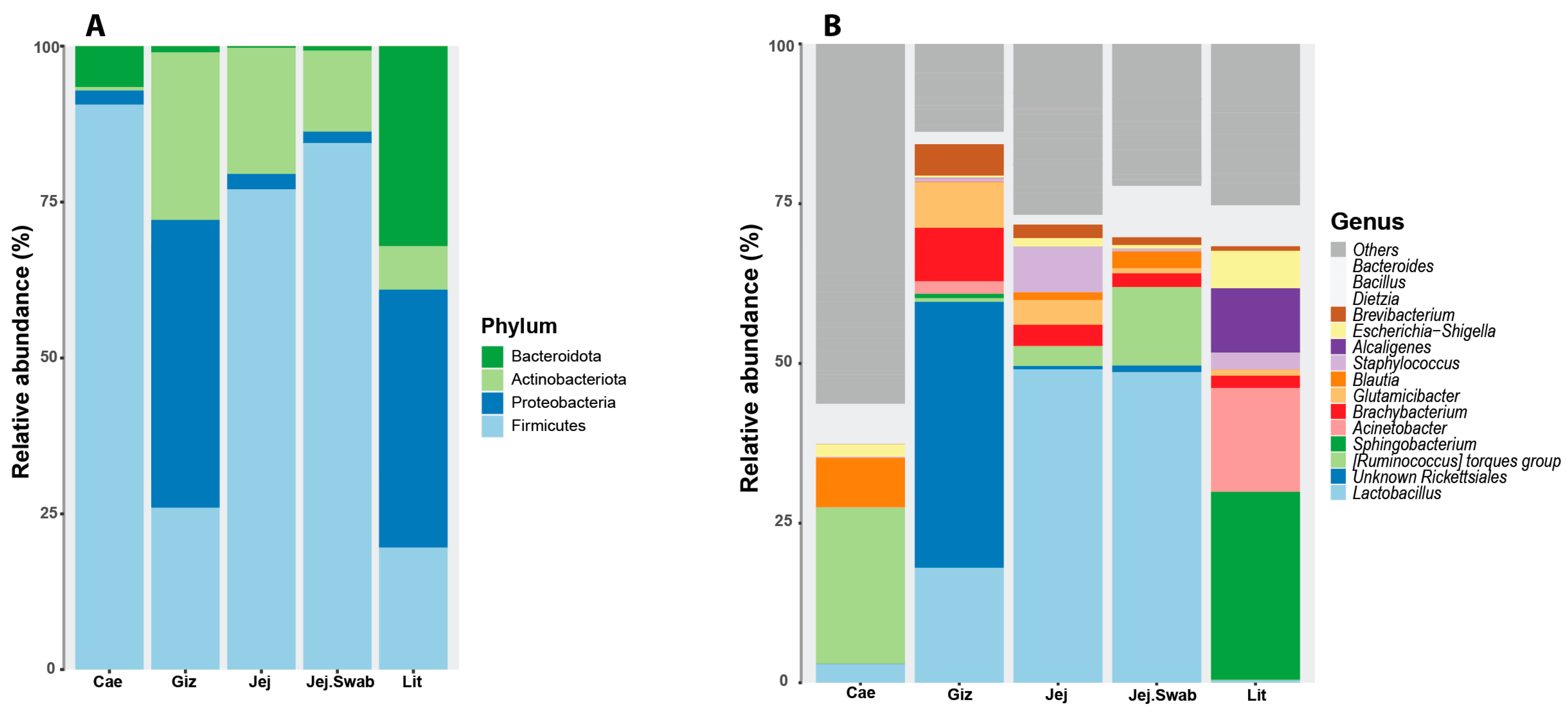
3.3. Litter Microbial Community
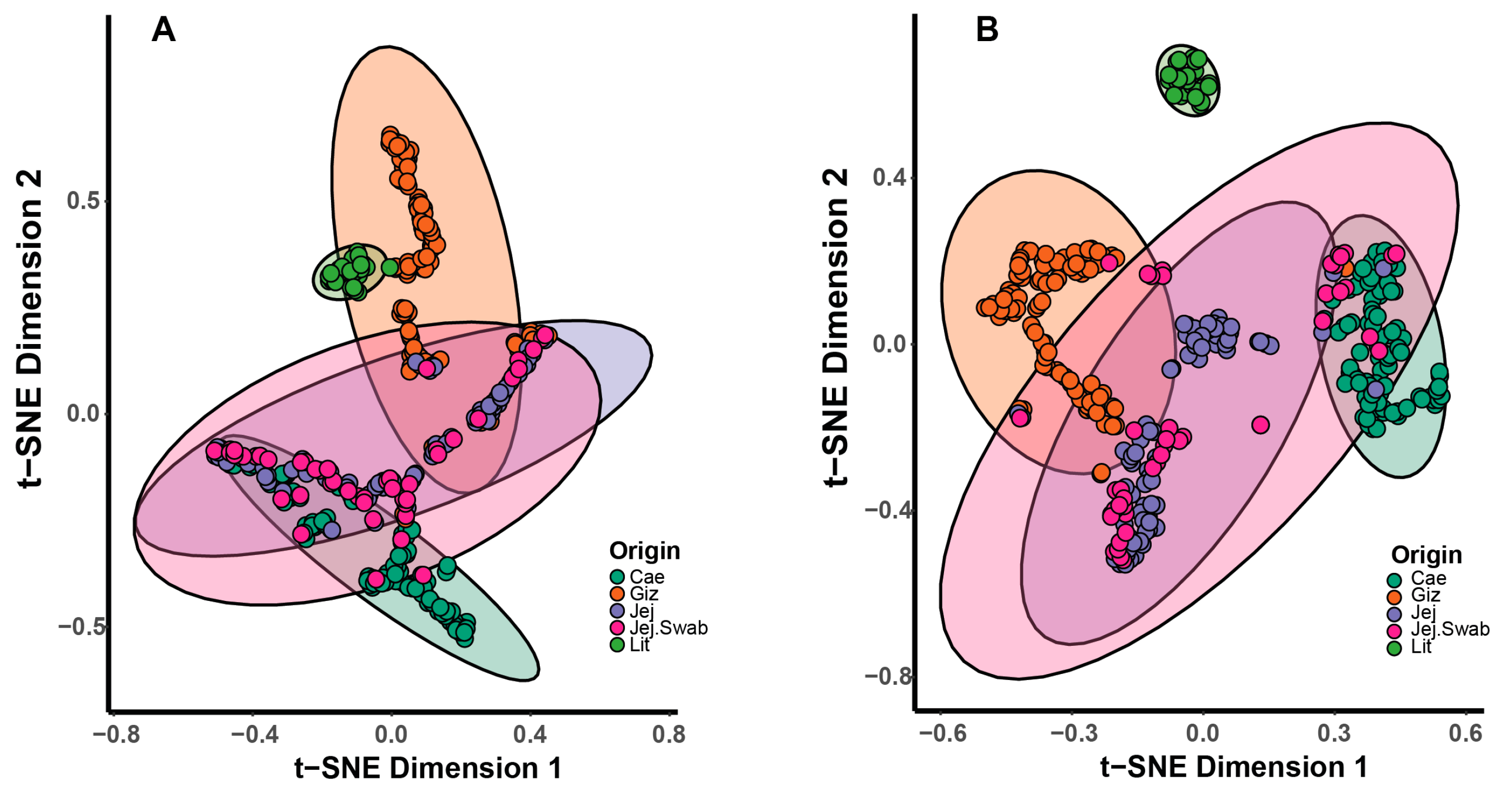
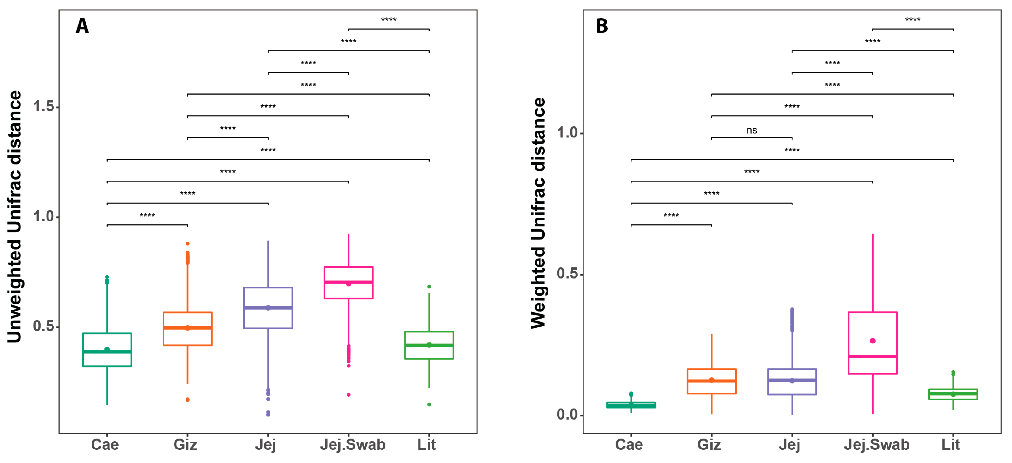
The phylum-level litter microbiota showed similarities with the gizzard regarding a high abundance of Firmicutes and Proteobacteria (Figure 2), but it shared the same phylum-level membership with the other gut sections. On the other hand, strong dissimilarities between the litter and the other gut sections’ microbiota became apparent at the genus level. Some dominant genera present in the litter samples detected using 16S amplicon sequencing included Sphingobacterium, Acinetobacter, Brachybacterium, Glutamicibacter, Staphylococcus, Alcaligenes, Escherichia-Shigella and Brevibacterium. The known genera present in the gut but not in the litter and conversely the known genera present only in the litter but not in any gut sections detected using 16S amplicon sequencing are shown in Supplementary Tables S1 and S2, respectively.

3.4. Beta Diversity

Overall, non-metric multidimensional scaling (NMDS), principal coordinate analysis (PCoA) and stochastic neighbour embedding (t-SNE) plots showed a clear separation by sample origin, as demonstrated using t-SNE in Figure 3. The litter microbiota samples were highly uniform, clustered closely and separated from the other origins at the genus level, but they were close to those from the gizzard at the phylum level, which agrees with the data presented in Figure 2.
PERMANOVA of the litter microbiota showed significant differences introduced by the DEX challenge based on both the Unweighted (p = 0.001) and Weighted UniFrac distances (p < 0.022). The treatments, including C, CD, P and PD, were only significant using Unweighted UniFrac (p = 0.010) (Table 2). Moreover, paired MANOVA presented significant differences between C and CD (p = 0.007), CD and P (p = 0.001) and P and PD (p = 0.021) using the Unweighted UniFrac distances. However, no significant differences were observed using the Weighted UniFrac distance (Table 3).
Paired MANOVA performed between the gut sections and litter microbiota showed highly significant (p < 0.001) differences in all the comparisons between gut sections and between the litter and gut sections using either Weighted or Unweighted UniFrac, as expected.
Further comparisons of the beta diversity within each sampled origin revealed highly significant differences in the sample distribution using Unweighted UniFrac (Figure 4A) and Weighted UniFrac (Figure 4B), indicating high within-group sample-to-sample microbiota differences based on microbiota membership and abundance. The gizzard and jejunum had similar sample-to-sample distances based on weighted UniFrac.
The litter samples were highly uniform (smaller sample-to-sample distance) compared to the gizzard, jejunum and jejunal swabs samples based on both Weighted and Unweighted UniFrac. The within-sample origin similarity (lower sample-to-sample distance) between the litter samples was most comparable to that represented by the smaller distances (higher similarities) between the caecum samples.

3.5. Effects of Treatment on Litter Microbial Taxa

The linear discriminant analysis (LDA) effect size (LEfSe) analysis in Figure 5 presents the distinct microbial taxa across different groups. The litter microbiota were characterised by a higher abundance of Moraxellaceae, Acinetobacter, Enterococcaceae and Enterococcus in the C group and Aerococcaceae, Aerosphaera, Flavobacteriaceae, Flavobacteriales, Myroides, Alphaproteobacteria, Cardiobacteriales, Wohlfahrtiimodaceae, Rhizobiales and Rhizobiaceae in the CD group. The PD group was represented by Burkholderiales, Alcaligeceae, Alcaligenes, Planococcaceae, Clostridia, Peptostreptococcales-Tissierellales, Capnocytophaga spp., Gottschalkia, Flavobacterium and Tissierella (Figure 5). No genera were associated with the P group. The genera associated with the supplementation of DEX and the probiotic according to the LEfSe analysis (LDA > 3.5) are presented in Supplementary Figures S3 and S4.

3.6. Litter Quality

The moisture content is presented in Figure 6. The litter in the DEX groups had a significantly higher moisture content than that in the non-challenged groups, indicating that the diarrhea leaky gut challenge was successful (p < 0.05). Moreover, there were no significant differences in the moisture levels due to probiotic supplementation (p > 0.05). There were no significant differences in the litter pH between the four treatments (p > 0.05) (Supplementary Figure S5: Litter pH in C, CD, P and PD groups).

4. Discussion

As expected, the moisture content was significantly higher in the leaky gut DEX-challenged groups (CD and PD, p < 0.05). The glucocorticoid DEX induces gut inflammation and promotes significant alterations in the intestinal structure, stimulating a condition known as leaky gut, as documented by previous studies [43,44]. This disruption in the gut barrier contributes to decreased digestibility, water loss, and diarrhea, consequently increasing the moisture levels in the litter [45]. Moreover, a leaky gut is associated with the movement of gut bacteria, microbial compounds and/or antigens, prompting a systemic immune response. The systemic inflammation generated by DEX demands energy, decreasing bird production performance [46].
The poultry GIT harbours complex microbiota, and the 16S amplicon methodology exclusively investigates a major portion of the bacterial community, which can profoundly impact broiler performance. The absence of significant variations in the alpha diversity indices (Figure 1) suggests DEX and probiotic supplementation exert minimal impacts on overall microbial diversity. A more prominent distinction is seen in beta diversity. The litter in the t-SNE plots is closely clustered and separated from the other sample origins at the genus level but is proximate to the gizzard at the phylum level. The phylum-level litter microbiota showed a high abundance of Firmicutes and Proteobacteria. This could be a result of ingested feed and litter microbiota passing through the gizzard. The observed variations in microbial diversity between the litter and intestinal samples highlight the importance of considering environmental context [47]. The litter microbial profile is influenced by factors such as the substrate composition and environmental conditions, which exhibit distinct microbial dynamics compared to the gut, which is mainly shaped by environment (moisture content, pH), host physiology and diet [48].
Litter containing 43–67% moisture has a greater bacterial abundance compared to that with 10–25% moisture content [49]. This confirms our observation in Figure 1, where higher moisture leads to higher diversity in the litter samples as measured using the Simpson diversity index. A litter moisture content surpassing 40% has been associated with poor digestion and changes in digesta viscosity and protein levels [50] and is often an indicator of diarrhea. The high moisture content, approaching 60%, in the DEX groups provides ideal conditions for anaerobes, such as Clostridia and Peptostreptococcales-Tissierellales, whereas a ~40% moisture content in the non-DEX groups may have been slightly aerobic, which is ideal for aerobes, such as Moraxellaceae and Acinetobacter. Another study observed something similar, where Clostridia is predominant in fresh litter and ileal mucosa microbiota [18]. Hence, a high moisture content or “wet litter” is a safe haven for pathogens (C. perfringens, Escherichia coli), which can increase the incidence of necrotic enteritis (NE) and secondary health problems, such as footpad dermatitis, ammonia proliferation and poor overall welfare and performance in broilers [49].
No genera were significantly associated with probiotic supplementation, according to the LEfSe analysis, when observing all four treatments. The litter microbiota in the DEX groups were characterised by Acinetobacter, Enterococcaceae, Enterococcus, Myroides, Burkholderiales, Alcaligeceae, Alcaligenes, Clostridia, Peptostreptococcales-Tissierellales, Capnocytophaga spp., Gottschalkia, Flavobacterium and Tissierella. These bacteria can cause chronic diseases in poultry. Enterococcus faecium is deemed a member of the gut microbiota in both humans and poultry. Acinetobacter, part of the Moraxellaceae family, is a strictly aerobic microorganism that mostly occurs in spoiled meats and is considered a mild spoiler of meat and dairy products because of its poor proteolytic activity but good lipolytic activity [51].
Members of the Myroides genus associated with the CD group originate from the family Flavobacteriaceae and are considered opportunistic pathogens in poultry. Moreover, some species of the genus Flavobacterium associated with the PD group are related to poultry diseases and respiratory infections [52]. The order Burkholderiales includes a diverse group of bacteria, but a study by Campos et al. (2022) [53] pointed out that Burkholderiales is more abundant in birds infected by Eimeria tenella.
Some species of Alcaligeceae like Bordetella avium are often present in soil and water environments and are considered zoonotic, contagious and a common cause of a disease called bordetellosis in birds [9,54]. Some clostridia are considered very important agents of enteric disease in poultry. For example, the incidence of C. perfringens-associated NE in poultry has increased significantly in countries discontinuing the use of AGPs [55]. The growth, survival and spread of both clinical and subclinical NE, a major disease in the broiler industry, is promoted by a high moisture content in the litter [49]. These anaerobic microorganisms are also involved in cattle disease pathogenesis and act as secondary opportunistic pathogens [56]. Capnocytophaga spp. are commonly seen in the oral cavities of dogs and cats [57], whereas Gottschalkia are pathogens and were also present in layer manure samples from different-scale farms in a study conducted by Wang and Chai (2022) [58].
Planococcaceae, which are selected as characteristic of PD birds, are common inhabitants of poultry GITs. A study by Abdel-Kafy et al. (2022) identified Planococcaceae in the mucosa of high-weight birds [59]. Moreover, Mon et al. (2020) stated Planococcaceae and Lactobacillaceae are often highly co-abundant [60]. Aerococcaceae were also present in ileal and caecal content samples in a study by Bindari et al. (2021) [7]. Alphaproteobacteria, a class of Proteobacteria, was seen in the CD birds. Proteobacteria is a phylum commonly present in younger birds before the microbial succession moves towards high domination by Firmicutes [4]. Another study stated that the highly pathogenic phylum Proteobacteria is a predominant phylum in cloacae because of its oxygen tolerance [61]. The order Cardiobacteriales is closely correlated with Wohlfahrtiimonas [62]. Wohlfahrtiimonas is isolated from parasitic fly larvae, Wohlfahrtia magnifica [63], and could be present in litter via insect vectors. Finally, Rhizobiales commonly colonise plants and live in symbiosis, promoting N-cycling. There is minimal information on the family of Rhizobiaceae in poultry. One study mentioned that Rhizobiaceae were abundant on eggshell surfaces post-hatching [64], but another study correlated Rhizobiaceae and a decrease in the FCR [65].
While poultry litter contains microbiota originating from excreta, its microbial community cannot be considered similar to the microbiota from poultry gut sections based on their shared intestinal taxa. Considering that the methodology we use in microbiota studies detects taxa based on the DNA extracted from the source, this methodology cannot distinguish between DNA from live and dead bacteria. It is improbable that strict anaerobes from the intestine would survive and thrive in the aerobic environment of the litter. However, dead excreta bacteria could represent a rich source for the growth of different environmental bacteria. A high level of pathogenic Proteobacteria genera in cloacal swabs is likely influenced by litter exposure and Proteobacteria’s ability to tolerate oxygen, leading to the overgrowth of poultry pathogens in the litter. At the same time, some of the most beneficial intestinal microbiota used as next-generation probiotics, like Akkermania and Faecalibacterium, are strict anaerobes. The top layer of the litter (we sampled the top 3 cm core) is exposed to the environment, oxygen, insects and bedding, feed, water and soil microbiota. Thus, controlling the microbial load and moisture content of the litter should be considered in broiler house monitoring programs.
Controlling environmental microbial counts is a complex process. Most commercial broiler houses have management programs in place, such as providing effective ventilation to simultaneously dry litter and reduce air dustiness [66], as well as using antimicrobial and anti-biofilm agents. Proper ventilation is important, as studies on broiler litter have shown a water activity (AW) of <0.84 is suggested as a successful control measure in minimising Salmonella populations [67] and microbial activity [25]. This study provides a passive sampling and analysis protocol proposal to assess microbial contamination in litter samples. The litter microbiota also mature and alter with time, and ongoing monitoring may be useful for providing an early warning of potential disease outbreaks.
Co-infection in commercial poultry flocks is likely to occur due to the close interactions between birds and the litter. This is important, as we know birds continuously practice coprophagy and are exposed to the microbiota present in the bedding particles, where pathogens are commonly present [16]. For example, poultry infected by Histomonas meleagridis and E. coli experienced more severe pathogenesis, a sudden shift in the caecal mucosa-associated microbiota and the colonisation of pathogenic bacteria [68]. Moreover, increased pathogens post-infection or changes in the environmental conditions could decrease the population of beneficial bacteria such as Lactobacillus. A study by Oh et al. (2017) observed stabilised and improved gut microbiota post-supplementation of Bacillus subtilis against Salmonella in hens, which is very positive [69].
As mentioned above, hatchlings can be infected vertically (via hens) or horizontally (via the environment). The ban on AGPs and vaccines whose success depends on specific serovar and host species could alter the microbiota colonisation of mucosal surfaces in young birds [70]. Thus, strategies like proper ventilation and in-feed supplementation of probiotic spores may be successful in minimising water activity and producing competitive exclusion against pathogens [71], modulating the gut microbiota and decreasing the shedding of pathogens, such as Salmonella, into broiler environments and meat [70].

5. Conclusions

In conclusion, the dexamethasone leaky gut challenge introduced a significant increase in diarrhea, evident from a significant increase in the litter moisture, and resulted in significant shifts in the litter microbiota, more prominent in influencing the presence and absence of species (Unweighted) than their abundance and dominance (Weighted UniFrac). Probiotic supplementation did not alter the litter microbiota.
Litter samples have very high microbial diversity, significantly higher than that of any other gut origin, including the caecum. However, the litter does not have the highest microbial richness, in this study being slightly behind the caecum and gizzard. Compared to those from the gut sections, the litter microbial samples (sample-to-sample distance) were highly similar to one another. This uniformity can be utilised for both diagnostic and therapeutic purposes. The use of shotgun metagenomic sequencing of the litter could suggest novel biomarkers and open new directions in using litter to improve bird health.

Supplementary Materials

The following supporting information can be downloaded at https://www.mdpi.com/article/10.3390/ani14121758/s1. Figure S1: Broiler performance metrics, including body weight (BW), average daily gain (ADG), average daily feed intake (ADFI) and feed conversion ratio (FCR), measured before the supplementation of DEX; Figure S2: Broiler performance metrics, including body weight (BW), average daily gain (ADG), average daily feed intake (ADFI) and feed conversion ratio (FCR), between days 28 and 35 after the DEX period; Figure S3: Genera associated with dexamethasone supplementation according to LEfSe analysis (LDA > 3.5); Figure S4: Genera associated with probiotic supplementation according to LEfSe analysis (LDA > 3.5); Figure S5: Litter pH in C, CD, P and PD groups. No statistically significant differences were observed between the groups (p > 0.05); Table S1: Known genera present in the gut but not in the litter detected using 16S amplicon sequencing; Table S2: Known genera present only in the litter but not in any gut sections detected via 16S amplicon sequencing.

Author Contributions

Conceptualisation, D.H., Y.S.B., J.v.H., X.C. and D.S.; methodology, D.H., Y.S.B., J.v.H., X.C., T.T.H.V., M.W.D. and D.S.; formal analysis, D.H., Y.S.B. and D.S.; investigation, D.H., Y.S.B., J.v.H., X.C., T.T.H.V., M.W.D. and D.S.; resources, Y.S.B., J.v.H., X.C. and D.S.; writing—original draft preparation, D.H.; writing—review and editing, D.H., Y.S.B., J.v.H., X.C., T.T.H.V., M.W.D. and D.S.; supervision, Y.S.B., X.C., T.T.H.V. and D.S.; project administration, Y.S.B. All authors have read and agreed to the published version of the manuscript.

Funding

This research was funded by Bioproton Pty Ltd. Bioproton funded the ELEVATE scholarship to Darwin Horyanto.

Institutional Review Board Statement

The study was conducted according to the Australian Code For The Care And Use Of Animals For Scientific Purposes (https://www.nhmrc.gov.au/about-us/publications/australian-code-care-and-use-animals-scientific-purposes, accessed on 1 June 2021). The handling of the commercial poultry samples was approved by the Animal Ethics Committee of Central Queensland University (approval number approval number 0000023123, approved June 2021).

Informed Consent Statement

Not applicable.

Data Availability Statement

The raw sequence data are available from the NCBI SRA database under accession number PRJNA1068165: https://www.ncbi.nlm.nih.gov/bioproject/1068165 (accessed on 26 May 2024).

Acknowledgments

This study was supported by Bioproton Pty Ltd. The data were analysed using the Marie Curie High Performance Computing System at CQU. We acknowledge and appreciate Jason Bell’s help with every aspect of the high-performance computing.

Conflicts of Interest

D.H. received a scholarship from Bioproton Pty Ltd.; D.H., J.v.H. and X.C. are employed by Bioproton Pty Ltd.; D.S. and Y.S.B. received funding from Bioproton Pty Ltd. M.W.D. and T.T.H.V. have no conflicts of interest to declare.

References

  1. Pan, D.; Yu, Z. Intestinal microbiome of poultry and its interaction with host and diet. Gut Microbes 2014, 5, 108–119. [Google Scholar] [CrossRef] [PubMed]
  2. Rychlik, I. Composition and Function of Chicken Gut Microbiota. Animals 2020, 10, 103. [Google Scholar] [CrossRef] [PubMed]
  3. Oakley, B.B.; Lillehoj, H.S.; Kogut, M.H.; Kim, W.K.; Maurer, J.J.; Pedroso, A.; Lee, M.D.; Collett, S.R.; Johnson, T.J.; Cox, N.A. The chicken gastrointestinal microbiome. FEMS Microbiol. Lett. 2014, 360, 100–112. [Google Scholar] [CrossRef] [PubMed]
  4. Aruwa, C.E.; Pillay, C.; Nyaga, M.M.; Sabiu, S. Poultry gut health-microbiome functions, environmental impacts, microbiome engineering and advancements in characterization technologies. J. Anim. Sci. Biotechnol. 2021, 12, 119. [Google Scholar] [CrossRef] [PubMed]
  5. Wise, M.G.; Siragusa, G.R. Quantitative analysis of the intestinal bacterial community in one- to three-week-old commercially reared broiler chickens fed conventional or antibiotic-free vegetable-based diets. J. Appl. Microbiol. 2007, 102, 1138–1149. [Google Scholar] [CrossRef] [PubMed]
  6. Schokker, D.; Hoekman, A.J.W.; Smits, M.A.; Rebel, J.M.J. Gene expression patterns associated with chicken jejunal development. Dev. Comp. Immunol. 2009, 33, 1156–1164. [Google Scholar] [CrossRef] [PubMed]
  7. Bindari, Y.R.; Moore, R.J.; Van, T.T.H.; Hilliar, M.; Wu, S.B.; Walkden-Brown, S.W.; Gerber, P.F. Microbial communities of poultry house dust, excreta and litter are partially representative of microbiota of chicken caecum and ileum. PLoS ONE 2021, 16, e0255633. [Google Scholar] [CrossRef] [PubMed]
  8. Bajagai, Y.S.; Van, T.T.H.; Joat, N.; Chousalkar, K.; Moore, R.J.; Stanley, D. Layer chicken microbiota: A comprehensive analysis of spatial and temporal dynamics across all major gut sections. J. Anim. Sci. Biotechnol. 2024, 15, 20. [Google Scholar] [CrossRef]
  9. Ahaduzzaman, M.; Groves, P.J.; Sharpe, S.M.; Williamson, S.L.; Gao, Y.K.; Nguyen, T.V.; Gerber, P.F.; Walkden-Brown, S.W. A practical method for assessing infectious laryngotracheitis vaccine take in broilers following mass administration in water: Spatial and temporal variation in viral genome content of poultry dust after vaccination. Vet. Microbiol. 2020, 241, 108545. [Google Scholar] [CrossRef]
  10. Assen, A.M.; Stillman, M.; Alfirevich, S.; Gerber, P.F.; Groves, P.J.; Walkden-Brown, S.W. Assessment of A20 infectious laryngotracheitis vaccine take in meat chickens using swab and dust samples following mass vaccination in drinking water. Vet. Microbiol. 2020, 251, 108903. [Google Scholar] [CrossRef]
  11. Ahaduzzaman, M.; Keerqin, C.; Kumar, A.; Musigwa, S.; Morgan, N.; Kheravii, S.K.; Sharpe, S.; Williamson, S.; Wu, S.-B.; Walkden-Brown, S.W. Detection and quantification of Clostridium perfringens and Eimeria spp. in poultry dust using real-time PCR under experimental and field conditions. Avian Dis. 2021, 65, 77–85. [Google Scholar] [CrossRef] [PubMed]
  12. Liebhart, D.; Bilic, I.; Grafl, B.; Hess, C.; Hess, M. Diagnosing Infectious Diseases in Poultry Requires a Holistic Approach: A Review. Poultry 2023, 2, 252–280. [Google Scholar] [CrossRef]
  13. Diaz Carrasco, J.M.; Casanova, N.A.; Fernández Miyakawa, M.E. Microbiota, Gut Health and Chicken Productivity: What Is the Connection? Microorganisms 2019, 7, 374. [Google Scholar] [CrossRef] [PubMed]
  14. Mulchandani, R.; Wang, Y.; Gilbert, M.; Van Boeckel, T.P. Global trends in antimicrobial use in food-producing animals: 2020 to 2030. PLoS Glob. Public Health 2023, 3, e0001305. [Google Scholar] [CrossRef] [PubMed]
  15. Kogut, M.H.; Genovese, K.J.; Swaggerty, C.L.; He, H.; Broom, L. Inflammatory phenotypes in the intestine of poultry: Not all inflammation is created equal. Poult. Sci. 2018, 97, 2339–2346. [Google Scholar] [CrossRef] [PubMed]
  16. Feye, K.M.; Baxter, M.F.A.; Tellez-Isaias, G.; Kogut, M.H.; Ricke, S.C. Influential factors on the composition of the conventionally raised broiler gastrointestinal microbiomes. Poult. Sci. 2020, 99, 653–659. [Google Scholar] [CrossRef] [PubMed]
  17. Cockerill, S.A.; Gerber, P.F.; Walkden-Brown, S.W.; Dunlop, M.W. Suitability of litter amendments for the Australian chicken meat industry. Anim. Prod. Sci. 2020, 60, 1469–1481. [Google Scholar] [CrossRef]
  18. Cressman, M.D.; Yu, Z.; Nelson, M.C.; Moeller, S.J.; Lilburn, M.S.; Zerby, H.N. Interrelations between the microbiotas in the litter and in the intestines of commercial broiler chickens. Appl. Environ. Microbiol. 2010, 76, 6572–6582. [Google Scholar] [CrossRef]
  19. Wei, S.; Morrison, M.; Yu, Z. Bacterial census of poultry intestinal microbiome. Poult. Sci. 2013, 92, 671–683. [Google Scholar] [CrossRef]
  20. Stanley, V.; Gray, C.; Daley, M.; Krueger, W.; Sefton, A. An alternative to antibiotic-based drugs in feed for enhancing performance of broilers grown on Eimeria spp.-infected litter. Poult. Sci. 2004, 83, 39–44. [Google Scholar] [CrossRef]
  21. Cambra-López, M.; Hermosilla, T.; Lai, H.T.; Montero, M.; Aarnink, A.J.; Ogink, N.W. Source identification and quantification of particulate matter emitted from livestock houses. In Proceedings of the International Symposium on Air Quality and Manure Management for Agriculture Conference Proceedings, Dallas, TX, USA, 13–16 September 2010; p. 41. [Google Scholar]
  22. O’Brien, K.M.; Chimenti, M.S.; Farnell, M.; Tabler, T.; Bair, T.; Bray, J.L.; Nonnenmann, M.W. High throughput genomic sequencing of bioaerosols in broiler chicken production facilities. Microb. Biotechnol. 2016, 9, 782–791. [Google Scholar] [CrossRef] [PubMed]
  23. Yan, W.; Sun, C.; Zheng, J.; Wen, C.; Ji, C.; Zhang, D.; Chen, Y.; Hou, Z.; Yang, N. Efficacy of Fecal Sampling as a Gut Proxy in the Study of Chicken Gut Microbiota. Front. Microbiol. 2019, 10, 2126. [Google Scholar] [CrossRef] [PubMed]
  24. Shang, Y.; Kfzumar, S.; Oakley, B.; Kim, W.K. Chicken Gut Microbiota: Importance and Detection Technology. Front. Vet. Sci. 2018, 5, 254. [Google Scholar] [CrossRef] [PubMed]
  25. Dunlop, M.W.; McAuley, J.; Blackall, P.J.; Stuetz, R.M. Water activity of poultry litter: Relationship to moisture content during a grow-out. J. Environ. Manag. 2016, 172, 201–206. [Google Scholar] [CrossRef]
  26. Krysiak, K.; Konkol, D.; Korczyński, M. Overview of the Use of Probiotics in Poultry Production. Animals 2021, 11, 1620. [Google Scholar] [CrossRef] [PubMed]
  27. Ogbuewu, I.P.; Mabelebele, M.; Sebola, N.A.; Mbajiorgu, C. Bacillus Probiotics as Alternatives to In-feed Antibiotics and Its Influence on Growth, Serum Chemistry, Antioxidant Status, Intestinal Histomorphology, and Lesion Scores in Disease-Challenged Broiler Chickens. Front. Vet. Sci. 2022, 9, 876725. [Google Scholar] [CrossRef] [PubMed]
  28. Attia, Y.A.; Basiouni, S.; Abdulsalam, N.M.; Bovera, F.; Aboshok, A.A.; Shehata, A.A.; Hafez, H.M. Alternative to antibiotic growth promoters: Beneficial effects of Saccharomyces cerevisiae and/or Lactobacillus acidophilus supplementation on the growth performance and sustainability of broilers’ production. Front. Vet. Sci. 2023, 10, 1259426. [Google Scholar] [CrossRef]
  29. Costa, F.G.P.; Goulart, C.; Figueiredo, D.; Oliveira, C.; Silva, J. Economic and environmental impact of using exogenous enzymes on poultry feeding. Int. J. Poult. Sci. 2008, 7, 311–314. [Google Scholar]
  30. Vicuña, E.A.; Kuttappan, V.A.; Galarza-Seeber, R.; Latorre, J.D.; Faulkner, O.B.; Hargis, B.M.; Tellez, G.; Bielke, L.R. Effect of dexamethasone in feed on intestinal permeability, differential white blood cell counts, and immune organs in broiler chicks. Poult. Sci. 2015, 94, 2075–2080. [Google Scholar] [CrossRef]
  31. Horyanto, D.; Bajagai, Y.S.; Kayal, A.; von Hellens, J.; Chen, X.; Van, T.T.H.; Radovanović, A.; Stanley, D. Bacillus amyloliquefaciens Probiotics Mix Supplementation in a Broiler Leaky Gut Model. Microorganisms 2024, 12, 419. [Google Scholar] [CrossRef]
  32. Yu, Z.; Morrison, M. Improved extraction of PCR-quality community DNA from digesta and fecal samples. Biotechniques 2004, 36, 808–812. [Google Scholar] [CrossRef] [PubMed]
  33. Fadrosh, D.W.; Ma, B.; Gajer, P.; Sengamalay, N.; Ott, S.; Brotman, R.M.; Ravel, J. An improved dual-indexing approach for multiplexed 16S rRNA gene sequencing on the Illumina MiSeq platform. Microbiome 2014, 2, 6. [Google Scholar] [CrossRef] [PubMed]
  34. Elias, D.M.O.; Robinson, S.; Both, S.; Goodall, T.; Majalap-Lee, N.; Ostle, N.J.; McNamara, N.P. Soil Microbial Community and Litter Quality Controls on Decomposition Across a Tropical Forest Disturbance Gradient. Front. For. Glob. Chang. 2020, 3, 81. [Google Scholar] [CrossRef]
  35. Martin, M. CUTADAPT removes adapter sequences from high-throughput sequencing reads. EMBnet J. 2011, 17, 10–12. [Google Scholar] [CrossRef]
  36. Bolyen, E.; Rideout, J.R.; Dillon, M.R.; Bokulich, N.A.; Abnet, C.C.; Al-Ghalith, G.A.; Alexander, H.; Alm, E.J.; Arumugam, M.; Asnicar, F.; et al. Reproducible, interactive, scalable and extensible microbiome data science using QIIME 2. Nat. Biotechnol. 2019, 37, 852–857. [Google Scholar] [CrossRef] [PubMed]
  37. Callahan, B.J.; McMurdie, P.J.; Rosen, M.J.; Han, A.W.; Johnson, A.J.; Holmes, S.P. DADA2: High-resolution sample inference from Illumina amplicon data. Nat. Methods 2016, 13, 581–583. [Google Scholar] [CrossRef] [PubMed]
  38. Quast, C.; Pruesse, E.; Yilmaz, P.; Gerken, J.; Schweer, T.; Yarza, P.; Peplies, J.; Glöckner, F.O. The SILVA ribosomal RNA gene database project: Improved data processing and web-based tools. Nucleic Acids Res. 2013, 41, D590–D596. [Google Scholar] [CrossRef] [PubMed]
  39. McMurdie, P.J.; Holmes, S. phyloseq: An R Package for Reproducible Interactive Analysis and Graphics of Microbiome Census Data. PLoS ONE 2013, 8, e61217. [Google Scholar] [CrossRef] [PubMed]
  40. Smith, S. phylosmith: An R-package for reproducible and efficient microbiome analysis with phyloseq-objects. J. Open Source Softw. 2019, 4, 1442. [Google Scholar] [CrossRef]
  41. Dixon, P. VEGAN, a package of R functions for community ecology. J. Veg. Sci. 2003, 14, 927–930. [Google Scholar] [CrossRef]
  42. Liu, C.; Cui, Y.; Li, X.; Yao, M. microeco: An R package for data mining in microbial community ecology. FEMS Microbiol. Ecol. 2021, 97, fiaa255. [Google Scholar] [CrossRef] [PubMed]
  43. Chen, J.; Tellez, G.; Richards, J.D.; Escobar, J. Identification of Potential Biomarkers for Gut Barrier Failure in Broiler Chickens. Front. Vet. Sci. 2015, 2, 14. [Google Scholar] [CrossRef] [PubMed]
  44. Awad, W.A.; Hess, C.; Hess, M. Enteric Pathogens and Their Toxin-Induced Disruption of the Intestinal Barrier through Alteration of Tight Junctions in Chickens. Toxins 2017, 9, 60. [Google Scholar] [CrossRef] [PubMed]
  45. Cardoso Dal Pont, G.; Farnell, M.; Farnell, Y.; Kogut, M.H. Dietary Factors as Triggers of Low-Grade Chronic Intestinal Inflammation in Poultry. Microorganisms 2020, 8, 139. [Google Scholar] [CrossRef] [PubMed]
  46. Williams, J.M.; Duckworth, C.A.; Burkitt, M.D.; Watson, A.J.; Campbell, B.J.; Pritchard, D.M. Epithelial cell shedding and barrier function: A matter of life and death at the small intestinal villus tip. Vet. Pathol. 2015, 52, 445–455. [Google Scholar] [CrossRef] [PubMed]
  47. Hanhimäki, E.; Watts, P.C.; Koskela, E.; Koteja, P.; Mappes, T.; Hämäläinen, A.M. Evolved high aerobic capacity has context-specific effects on gut microbiota. Front. Ecol. Evol. 2022, 10, 934164. [Google Scholar] [CrossRef]
  48. Maki, J.J.; Klima, C.L.; Sylte, M.J.; Looft, T. The Microbial Pecking Order: Utilization of Intestinal Microbiota for Poultry Health. Microorganisms 2019, 7, 376. [Google Scholar] [CrossRef] [PubMed]
  49. Fancher, C.A.; Zhang, L.; Kiess, A.S.; Adhikari, P.A.; Dinh, T.T.N.; Sukumaran, A.T. Avian Pathogenic Escherichia coli and Clostridium perfringens: Challenges in No Antibiotics Ever Broiler Production and Potential Solutions. Microorganisms 2020, 8, 1533. [Google Scholar] [CrossRef]
  50. Choct, M.; Hughes, R.J.; Wang, J.; Bedford, M.R.; Morgan, A.J.; Annison, G. Increased small intestinal fermentation is partly responsible for the anti-nutritive activity of non-starch polysaccharides in chickens. Br. Poult. Sci. 1996, 37, 609–621. [Google Scholar] [CrossRef]
  51. Teider, P.I.; Ribeiro, J.C.; Ossugui, E.H.; Tamanini, R.; Ribeiro, J.; Santos, G.A.; Alfieri, A.A.; Beloti, V. Pseudomonas spp. and other psychrotrophic microorganisms in inspected and non-inspected Brazilian Minas Frescal cheese: Proteolytic, lipolytic and AprX production potential. Pesqui. Veterinária Bras. 2019, 39, 807–815. [Google Scholar] [CrossRef]
  52. Yehia, N.; Salem, H.M.; Mahmmod, Y.; Said, D.; Samir, M.; Mawgod, S.A.; Sorour, H.K.; AbdelRahman, M.A.A.; Selim, S.; Saad, A.M.; et al. Common viral and bacterial avian respiratory infections: An updated review. Poult. Sci. 2023, 102, 102553. [Google Scholar] [CrossRef] [PubMed]
  53. Campos, P.M.; Darwish, N.; Shao, J.; Proszkowiec-Weglarz, M. Research Note: Choice of microbiota database affects data analysis and interpretation in chicken cecal microbiota. Poult. Sci. 2022, 101, 101971. [Google Scholar] [CrossRef] [PubMed]
  54. Temple, L.M.; Weiss, A.A.; Walker, K.E.; Barnes, H.J.; Christensen, V.L.; Miyamoto, D.M.; Shelton, C.B.; Orndorff, P.E. Bordetella avium virulence measured in vivo and in vitro. Infect. Immun. 1998, 66, 5244–5251. [Google Scholar] [CrossRef] [PubMed]
  55. Van Immerseel, F.; De Buck, J.; Pasmans, F.; Huyghebaert, G.; Haesebrouck, F.; Ducatelle, R. Clostridium perfringens in poultry: An emerging threat for animal and public health. Avian Pathol. 2004, 33, 537–549. [Google Scholar] [CrossRef] [PubMed]
  56. Khalil, A.; Batool, A.; Arif, S. Healthy Cattle Microbiome and Dysbiosis in Diseased Phenotypes. Ruminants 2022, 2, 134–156. [Google Scholar] [CrossRef]
  57. Mally, M.; Shin, H.; Paroz, C.; Landmann, R.; Cornelis, G.R. Capnocytophaga canimorsus: A human pathogen feeding at the surface of epithelial cells and phagocytes. PLoS Pathog. 2008, 4, e1000164. [Google Scholar] [CrossRef] [PubMed]
  58. Wang, L.; Chai, B. Fate of Antibiotic Resistance Genes and Changes in Bacterial Community with Increasing Breeding Scale of Layer Manure. Front. Microbiol. 2022, 13, 857046. [Google Scholar] [CrossRef]
  59. Abdel-Kafy, E.-S.M.; Youssef, S.F.; Magdy, M.; Ghoneim, S.S.; Abdelatif, H.A.; Deif-Allah, R.A.; Abdel-Ghafar, Y.Z.; Shabaan, H.M.A.; Liu, H.; Elokil, A. Gut Microbiota, Intestinal Morphometric Characteristics, and Gene Expression in Relation to the Growth Performance of Chickens. Animals 2022, 12, 3474. [Google Scholar] [CrossRef]
  60. Mon, K.K.; Zhu, Y.; Chanthavixay, G.; Kern, C.; Zhou, H. Integrative analysis of gut microbiome and metabolites revealed novel mechanisms of intestinal Salmonella carriage in chicken. Sci. Rep. 2020, 10, 4809. [Google Scholar] [CrossRef]
  61. Wilkinson, T.J.; Cowan, A.A.; Vallin, H.E.; Onime, L.A.; Oyama, L.B.; Cameron, S.J.; Gonot, C.; Moorby, J.M.; Waddams, K.; Theobald, V.J.; et al. Characterization of the Microbiome along the Gastrointestinal Tract of Growing Turkeys. Front. Microbiol. 2017, 8, 1089. [Google Scholar] [CrossRef]
  62. Montecillo, J.A.V. Phylogenomics and comparative genomic analyses support the creation of the novel family Ignatzschineriaceae fam. nov. comprising the genera Ignatzschineria and Wohlfahrtiimonas within the order Cardiobacteriales. Res. Microbiol. 2023, 174, 103988. [Google Scholar] [CrossRef] [PubMed]
  63. Kopf, A.; Bunk, B.; Riedel, T.; Schröttner, P. The zoonotic pathogen Wohlfahrtiimonas chitiniclastica—Current findings from a clinical and genomic perspective. BMC Microbiol. 2024, 24, 3. [Google Scholar] [CrossRef] [PubMed]
  64. Maki, J.J.; Bobeck, E.A.; Sylte, M.J.; Looft, T. Eggshell and environmental bacteria contribute to the intestinal microbiota of growing chickens. J. Anim. Sci. Biotechnol. 2020, 11, 60. [Google Scholar] [CrossRef] [PubMed]
  65. Iqbal, Y.; Cottrell, J.J.; Suleria, H.A.R.; Dunshea, F.R. Gut Microbiota-Polyphenol Interactions in Chicken: A Review. Animals 2020, 10, 1391. [Google Scholar] [CrossRef] [PubMed]
  66. Gomes, B.; Pena, P.; Cervantes, R.; Dias, M.; Viegas, C. Microbial Contamination of Bedding Material: One Health in Poultry Production. Int. J. Environ. Res. Public Health 2022, 19, 16508. [Google Scholar] [CrossRef] [PubMed]
  67. Chinivasagam, H.N.; Tran, T.; Blackall, P.J. Impact of the Australian litter re-use practice on Salmonella in the broiler farming environment. Food Res. Int. 2012, 45, 891–896. [Google Scholar] [CrossRef]
  68. Abd El-Hamid, M.I.; Awad, N.F.S.; Hashem, Y.M.; Abdel-Rahman, M.A.; Abdelaziz, A.M.; Mohammed, I.A.A.; Abo-Shama, U.H. In vitro evaluation of various antimicrobials against field mycoplasma gallisepticum and mycoplasma synoviae isolates in Egypt. Poult. Sci. 2019, 98, 6281–6288. [Google Scholar] [CrossRef] [PubMed]
  69. Oh, J.K.; Pajarillo, E.A.B.; Chae, J.P.; Kim, I.H.; Kang, D.K. Protective effects of Bacillus subtilis against Salmonella infection in the microbiome of Hy-Line Brown layers. Asian-Australas J. Anim. Sci. 2017, 30, 1332–1339. [Google Scholar] [CrossRef]
  70. Knap, I.; Kehlet, A.B.; Bennedsen, M.; Mathis, G.F.; Hofacre, C.L.; Lumpkins, B.S.; Jensen, M.M.; Raun, M.; Lay, A. Bacillus subtilis (DSM17299) significantly reduces Salmonella in broilers. Poult. Sci. 2011, 90, 1690–1694. [Google Scholar] [CrossRef]
  71. La Ragione, R.M.; Woodward, M.J. Competitive exclusion by Bacillus subtilis spores of Salmonella enterica serotype Enteritidis and Clostridium perfringens in young chickens. Vet. Microbiol. 2003, 94, 245–256. [Google Scholar] [CrossRef]
Figure 1. Litter sample alpha diversity presented as Observed Species (A) and Simpson metrics (B). Lit = litter, Giz = gizzard, Jej = jejunum lumen, Jej.Swab = jejunum mucosal swabs. (* = p ≤ 0.05; ** = p ≤ 0.01; *** = p ≤ 0.001; **** = p ≤ 0.0001).
Animals 14 01758 g001
Figure 2. Litter sample microbial community membership compared to other gut sections at phylum (A) and genus (B) levels.
Animals 14 01758 g002
Figure 3. The t-SNE plot demonstrates the grouping of phylum-level (A) and genus-level (B) microbiota profiles based on individual origins. Ellipses represent 95% confidence intervals. Lit = litter, Giz = gizzard, Jej = jejunum lumen, Jej.Swab = jejunum mucosal swabs.
Animals 14 01758 g003
Figure 4. Within-group beta diversity measures show that litter samples are more similar to one another than most other gut sections by Unweighted UniFrac (A) and Weighted UniFrac distance (B). (ns = p > 0.05; **** = p ≤ 0.0001).
Animals 14 01758 g004
Figure 5. LEfSe analysis presents taxa at all taxonomic levels in litter microbiota (p < 0.05 and LDA > 3.5). LDA = linear discriminatory analysis; LEfSe = LDA effect size.
Animals 14 01758 g005
Figure 6. The average moisture content values expressed as percentages (%) and standard error (SE) of C, P, CD and PD groups. a,b Statistically significant = p < 0.05.
Animals 14 01758 g006
Table 1. Probiotic strains and specification in CFU/g.
Probiotic StrainsSpecification
Bacillus amyloliquefaciens (BPR-11)2 × 10⁸ CFU/g
Bacillus amyloliquefaciens (BPR-16)2 × 10⁸ CFU/g
Bacillus amyloliquefaciens (BPR-17) 2 × 10⁸ CFU/g
Table 2. PERMANOVA of litter by both Weighted and Unweighted UniFrac.
GroupsR2p-ValueDistance
DEX0.1115640.001 *Unweighted UniFrac
Probiotic0.0322660.710Unweighted UniFrac
Treatments0.1720540.010 *Unweighted UniFrac
DEX0.1197810.022 *Weighted UniFrac
Probiotic0.0105000.933Weighted UniFrac
Treatments0.1408970.287Weighted UniFrac
DEX × probiotic interactions were significant (p < 0.05) according to Unweighted and Weighted UniFrac. Treatment PERMANOVA compared differences between four groups (C, CD, P and PD). * Statistically significant = p < 0.05.
Table 3. Paired MANOVA between experimental groups in the litter. There was a significant difference between groups, according to both Weighted and Unweighted UniFrac, each time when comparing the DEX-challenged and unchallenged groups.
GroupsR2p (>F)Distance
C vs. CD0.1532350.007 *Unweighted UniFrac
C vs. P0.0790200.563Unweighted UniFrac
C vs. PD0.1233300.069Unweighted UniFrac
CD vs. P0.1684610.001 *Unweighted UniFrac
CD vs. PD0.0568010.849Unweighted UniFrac
P vs. PD0.1374870.021 *Unweighted UniFrac
C vs. CD0.1185150.257Weighted UniFrac
C vs. P0.0211700.943Weighted UniFrac
C vs. PD0.1759910.078Weighted UniFrac
CD vs. P0.0916730.289Weighted UniFrac
CD vs. PD0.0268850.878Weighted UniFrac
P vs. PD0.1428750.085Weighted UniFrac
* Statistically significant = p < 0.05.
Disclaimer/Publisher’s Note: The statements, opinions and data contained in all publications are solely those of the individual author(s) and contributor(s) and not of MDPI and/or the editor(s). MDPI and/or the editor(s) disclaim responsibility for any injury to people or property resulting from any ideas, methods, instructions or products referred to in the content.

