Article

Identification of Two Linear Epitopes on MGF_110-13L Protein of African Swine Fever Virus with Monoclonal Antibodies

1
State Key Laboratory of Animal Disease Control and Prevention, Harbin Veterinary Research Institute of Chinese Academy of Agricultural Sciences, Harbin 150069, China
2
Jiangsu Co-Innovation Center for Prevention and Control of Important Animal Infectious Diseases and Zoonoses, Yangzhou University, Yangzhou 225009, China
*
Authors to whom correspondence should be addressed.
Animals 2024, 14(13), 1951; https://doi.org/10.3390/ani14131951
Submission received: 27 May 2024 / Revised: 24 June 2024 / Accepted: 30 June 2024 / Published: 1 July 2024

Abstract

:

Simple Summary

Due to the complexity of the structure of African swine fever virus (ASFV), the structure and function of most viral encoded proteins are uncharacterized. A full understanding and identification of ASFV-coding proteins is conducive to the prevention and control of ASFV. In this study, a previously uncharacterized protein, MGF_110-13L, was identified as an immunogenic protein and expressed as a glycosylated homodimer in eukaryotic cells. Two monoclonal antibodies against MGF_110-13L were generated, and two linear epitopes were further identified using monoclonal antibodies. One epitope peptide was evaluated as a potential antigen for ASFV antibody detection. This study will provide a basis for further understanding the structure and function of the MGF_110-13L protein of ASFV.

Abstract

African swine fever caused by African swine fever virus (ASFV) is an acute, highly contagious swine disease with high mortality. To facilitate effective vaccine development and find more serodiagnostic targets, fully exploring the ASFV antigenic proteins is urgently needed. In this study, the MGF_110-13L was identified as an immunodominant antigen among the seven transmembrane proteins. The main outer-membrane domain of MGF_110-13L was expressed and purified. Two monoclonal antibodies (mAbs; 8C3, and 10E4) against MGF_110-13L were generated. The epitopes of two mAbs were preliminary mapped with the peptide fusion proteins after probing with mAbs by enzyme-linked immunosorbent assay (ELISA) and Western blot. And the two target epitopes were fine-mapped using further truncated peptide fusion protein strategy. Finally, the core sequences of mAbs 8C3 and 10E4 were identified as 48WDCQDGICKNKITESRFIDS67, and 122GDHQQLSIKQ131, respectively. The peptides of epitopes were synthesized and probed with ASFV antibody positive pig sera by a dot blot assay, and the results showed that epitope 10E4 was an antigenic epitope. The epitope 10E4 peptide was further evaluated as a potential antigen for detecting ASFV antibodies. To our knowledge, this is the first report of antigenic epitope information on the antigenic MGF_110-13L protein of ASFV.

1. Introduction

ASF is a highly contagious and severe hemorrhagic swine disease caused by ASFV. Both domestic and wild boars are susceptible to ASFV. The World Organization for Animal Health (WOAH) has listed ASF as a notifiable disease due to the severe pathogenicity of ASFV and its ability to spread rapidly. Since its first description in Kenya in 1921, ASF has subsequently spread throughout sub-Saharan Africa [1]. In addition, there have been several transcontinental spreads of the disease, such as when it was introduced into the Caucasus region and Eastern Europe in 2007 [2], and emerged in China and Southeast Asia in 2018 [3,4,5]. Worldwide, the ASF outbreak and epidemic have resulted in huge economic losses as well as negative social consequences. There is currently no reliable commercial vaccine for ASF. Currently, the only effective control and eradication measures are strict stamping-out policies based on early detection.
ASFV belongs to and is the only member of the family Asfarviridae and the genus Asfivirus [6]. The linear, double-stranded DNA genome is approximately 170–190 kbp in size, with over 150 open reading frames, including more than 50 structural proteins and over 100 nonstructural proteins [6,7,8]. However, the structural and functional properties of the majority of ASFV proteins are unknown, which has hampered research into ASF vaccines and diagnostics. Till now, it has been known that some structural proteins of ASFV, such as P72, P30, and P54, are highly immunogenic. These proteins have been extensively used as immunoserological diagnosis targets and subunit vaccine components [9,10,11,12,13]. In addition to these major structural proteins, there are proteins in the ASFV-encoded proteomic that have good antigenicity and potential value in inducing immune protection and serological diagnosis. For example, the nonstructural protein K205R is a serological immunodeterminant of ASFV [14,15,16]. The immunogenic K205R protein stimulates a robust antibody response in animals that have been immunized, no matter whether it is expressed prokaryotically or eukaryotically [17,18]. To facilitate ASFV subunit vaccine research and explore new serodiagnostic targets, it is necessary to fully explore the antigenic proteins in the ASFV proteome. In particular, because the ASFV possesses a multilayered structure [19,20], the inner and outer membrane proteins are not fully characterized.
In the present study, using ASFV antibody positive pig sera, we identified MGF_113-13L as a novel antigenic protein from seven uncharacterized transmembrane proteins. Using mammalian cell line expressing MGF_110-13L protein as an immunogen, we generated two MGF_110-13L protein-specific monoclonal antibodies (mAbs). The target epitopes of these mAbs were fine-mapped and the potential application of the epitope peptides as serodiagnostic antigens was evaluated.

2. Materials and Methods

2.1. Cells, Sera, and Viruses

SP2/0 cells (ATCC, CRL-1581) were maintained in RPMI (Gibco, Beijing, China); baby hamster kidney BHK-21 cells (ATCC, CCL-10) and human embryo kidney 293 HEK-293 cells (ATCC, CRL-1573) were maintained in Dulbecco’s modified Eagle medium (DMEM; Gibco, Beijing, China). All cell media were supplemented with 10% fetal bovine serum, 100 µg/mL streptomycin and 100 U/mL penicillin. All cells were maintained in a humidified 5% CO2 atmosphere at 37 °C. The ASFV (HLJ/18 strain) was propagated and titrated in primary porcine alveolar macrophages (PAMs) as described [4]. As stated in a prior study [21], pig sera against ASFV were obtained from pigs immunized with the attenuated ASFV strain HLJ/18-7GD, either with or without challenge with the virulent ASFV strain HLJ/18. The Diagnostic Center of Harbin Veterinary Research Institute provided the ASFV antibody-positive serum samples from pigs naturally ASFV-infected. Specific pathogen-free (SPF) pig serum was used as a negative control.

2.2. Plasmids Construction and Transient Transfection

According to the TMHMM transmembrane helix prediction (https://services.healthtech.dtu.dk/services/TMHMM-2.0/, accessed on 1 February 2020), the main outer-membrane domains of seven ASFV transmembrane proteins were selected for transient expression in HEK-293 cells. The main outer-membrane domains were 1 to 160 amino acids of C257L protein, 1 to 237 amino acids of I329L protein, 22 to 177 amino acids of I177L protein, 47 to 158 amino acids of EP153R protein, 29 to 475 amino acids of B475L protein, 35 to 196 amino acids of K196R protein, and 1 to 131 amino acids of MGF_110-13L protein, respectively. All protein sequences were referenced to ASFV strain HLJ/18 in the African Swine Fever Virus Database (http://asfvdb.popgenetics.net/index, accessed on 6 January 2020). The genetic codon-optimized cDNAs encoding the respective outer-membrane domains were synthesized and cloned into the expression vector pCAGneo with a 6× His tag at the carboxyl terminal [18,22]. DNA sequencing was used to verify all of the recombinant plasmids. The plasmids were transfected into HEK-293 cells with ExFect Transfection Reagent (Vazyme, Nanjing, China). After 48 h of transfection, the protein samples were collected from the transfected cells for Western blot analysis.

2.3. Expression and Purification of the MGF_110-13L Protein

Stable expression cell lines were constructed as previously described [18,22]. Briefly, the ectodomain of MGF_110-13L (amino acid positions 1 to 131, reference sequence: QBH90491) expression plasmids were linearized with SspI. Then, BHK-21 cells were transfected with linearized plasmids. Transfected cells were cloned and selected using G418. The engineered cell clones were tested using an indirect immunofluorescence assay (IFA) and Western blot with a His tag-specific mouse monoclonal antibody. One clone, designated BHK-110-13L, demonstrated more efficient expression of the recombinant protein and was thus chosen and maintained in G418-supplemented medium for further characterization and production. The His-tagged recombinant protein was purified from the supernatants of the stable expression cell line via affinity chromatography with an Ni-NTA agarose column. The protein concentration was determined using a BCA protein assay kit (Biosharp, Hefei, China). Purified proteins were stored at −80 °C until use.

2.4. Generation of Monoclonal Antibodies against MGF_110-13L

Monoclonal antibodies against MGF_110-13L protein were generated as previously described [16,23]. Briefly, BALB/c mice were immunized subcutaneously with purified protein emulsified with Freund’s adjuvant (Sigma, St. Louis, MO, USA) three times at an interval of 21 days. A final booster immunization by intraperitoneal injection with soluble 13L-His protein alone was given 3 days before the cell fusion. Hybridomas were obtained by fusing mouse splenocytes with SP2/0 cells. Hybridomas were screened by ELISA, and positive cell lines were subcloned by limiting dilution method. Monoclonal antibodies were purified using affinity chromatography with a Protein G column (Abbkine, Wuhan, China). Each mAb’s heavy and light chains were identified using a mouse antibody isotyping kit (Thermo Scientific, Rockford, IL, USA).

2.5. Immunofluorescence Assay

Indirect immunofluorescence assays were carried out as previously described [16]. Briefly, the MGF_110-13L expressing cells were fixed and permeabilized. After blocking with BSA, the cells were incubated with His tag specific antibodies. After washing, the cells were incubated with fluorescein-conjugated goat anti-mouse IgG. Nuclei were stained by 4′,6-diamidino-2-phenylindole (DAPI). Finally, after three PBS washes, the cells were visualized using fluorescence microscopy. Untransfected BHK-21 cells were used as the negative control.

2.6. Expression of Short Peptide Fusion Proteins

Short peptides were expressed as fusion proteins with maltose-binding protein (MBP) as previously described [16,24]. Briefly, peptide-encoding DNA fragments were synthesized and cloned into plasmid pMAL-c5X between the sites of NdeI and BamHI. After being verified by DNA sequencing, the recombinant plasmids were transformed into expressing host E. coli ER2523. After inducing with isopropyl-beta D-thiogalactopyranoside (IPTG), the cell lysates were analyzed with sodium dodecyl sulfate-polyacrylamide gel electrophoresis (SDS-PAGE).

2.7. SDS-PAGE and Western Blotting

Lysates of bacterial-expressed peptide fusion proteins or ASFV-infected cell lysates were resolved on 4–20% SDS-PAGE (GenScript, M42015C), then transferred onto nitrocellulose membranes and blocked with 5% skimmed milk at 4 °C for an overnight period. After that, the membranes were incubated for 1 h at 37 °C with either the specific monoclonal antibody or pig serum. After three washes with PBS plus 0.5% Tween-20 (PBST), the membranes were then incubated for 1 h at 37 °C with either horseradish peroxidase (HRP)-conjugated goat anti-pig IgG or anti-mouse Alexa Fluor 680-conjugated secondary antibodies (Thermo, A10038). The blots were developed with ECL substrate or visualized using the Li-Cor Odyssey system (Li-Cor Biosciences, Lincoln, NE, USA).

2.8. ELISA

ELISA tests were performed as previously described [16,24]. Briefly, microplates were coated with MBP-peptide fusion proteins and blocked with skimmed milk. After three washes, the plates were incubated with hybridoma supernatants. After washing, the plates were incubated with HRP-conjugated goat anti-mouse IgG (ZSGB-BIO, Beijing, China). Following three washes, the plates were developed with 3,3′,5,5′-tetramethyl benzidine (TMB) and stopped with 2 M H2SO4. The optical density was then measured with an ELISA plate reader.

2.9. Dot Blot Assay

The epitope peptides were chemically synthesized (GenScript, Nanjing, China) and dissolved in dH2O or DMSO according to the manufacturer’s instructions. Dot blot assays were performed as previously described [16]. Briefly, synthesized peptides were spotted onto the nitrocellulose membranes and incubated with pig sera. After washing, the membranes were incubated with HRP-conjugated goat anti-pig IgG. Then, after three washes, the blots were developed with ECL substrate and visualized with a chemilluminescence imaging system.

2.10. Immunization of Pigs

Six landrace pigs were randomly divided into two groups, with three pigs in each group. These pigs were approximately 30 days of age and had been determined to be ASFV antibody-negative prior to the immune experiment. Each pig in group one was immunized with purified MGF_110-13L protein at a dose of 100 μg protein emulsified with oil adjuvant. Twenty eight days after the first immunization, the pigs were given a booster immunization at the same dose. Blood samples were collected prior to and at 14, 28, and 42 days post-immunization. Another group of pigs served as a control group and were injected with the same volume of PBS each time. The antibody titers of pig sera were examined by ELISA as described in Section 2.8, with the exception that the coating antigen was purified MGF_110-13L protein and the secondary antibody was HRP-conjugated goat anti-swine IgG.

3. Results

3.1. MGF_110-13L Was Identified as an Antigenic Protein

To explore ASFV genome-encoded antigenic proteins, seven predicted transmembrane proteins were selected to express in mammalian cells. The main outer-membrane domain expression plasmids were constructed and expressed in mammalian cells by transient transfection. The expression of constructs was verified by Western blot analysis with 6× His tag-specific antibody. The results demonstrated that all seven constructs were expressed in transfected cells (Figure 1A). The transfected cell lysates were then probed with ASFV-infected pig serum by Western blot. As shown in Figure 1B, among the seven transmembrane proteins, only MGF_110-13L exhibited a strong blotting signal. These results suggested that MGF_110-13L was antigenic, which could induce antibody responses and be recognized by ASFV-infected pig serum.

3.2. Expression of MGF_110-13L Protein and Generation of Monoclonal Antibodies

To express MGF_110-13L (1–131aa) in mammalian cells, BHK-21 cells were transfected with the plasmid pCAG-MGF110-13L. After limited dilution cloning and G418 selection, a stable expression cell line (BHK-110-13L) was established (Figure 2A). The recombinant MGF_110-13L protein was purified by nickel affinity chromatography from the supernatants of BHK-110-13L cells (Figure 2B). The purified recombinant protein MGF_110-13L exhibited two main bands near or above 15 kDa in SDS-PAGE analysis (Figure 2B). To survey whether cell line-expressed proteins exhibit two bands due to the presence of partially glycosylated modification, the purified protein was treated with the PNGase F enzyme and subjected to SDS-PAGE analysis. The results showed that the purified MGF_110-13L protein appeared as a single band of nearly 15 kDa after PNGase F treatment (Figure 2C). When analyzed by SDS-PAGE under the non-reducing condition (in the absence of β-mercaptoethanol), the purified MGF_110-13L protein exhibited three bands near or above 35 kDa (Figure 2D). From these results, it can be deduced that the MGF_110-13L protein secreted in the cell line is expressed as a partially glycosylated homodimer. To survey the antigenicity of the MGF_110-13L protein, three pigs were immunized with the purified protein. The sera were tested by ELISA with purified MGF_110-13L protein as the coating antigen. The results showed that MGF_110-13L-specific antibodies were developed in all immunized pigs but not in pigs in the control group (Figure 2E).
To generate MGF_110-13L-specific monoclonal antibody, purified recombinant protein was used to immunize BALB/c mice. After three immunizations, a booster was given intraperitoneally with purified soluble protein without adjuvant. After cell fusion, two hybridomas (8C3 and 10E4) were generated. The light chains of both mAbs were kappa. The isotypes of two mAbs were all tested as IgG1. mAb titers were tested by ELISA. The titers of mAbs 8C3 and 10E4 at a concentration of 1 mg/mL were above 51,200 and 102,400, respectively (Figure 2F). The Western blot results showed that the two mAbs specifically recognized the MGF_110-13L protein in lysates of ASFV-infected cells (Figure 2G). These data suggest that the two mAbs both recognized the linear epitope of the MGF_110-13L protein.

3.3. Epitope Mapping of mAbs

To map the linear epitopes of mAbs, we designed eight partially overlapping short peptides (P1 to P8). P1 to P7 were 30 amino acids long and P8 was 26 amino acids in length. These peptides covered the entire length of the MGF_110-13L outer-membrane domain (Figure 3A). After inducing with IPTG, all eight fusion proteins were expressed in soluble form (Figure 3B). To map the epitopes of the mAbs, the fusion proteins were scanned using mAbs by ELISA. The results indicated that mAb 8C3 recognized peptides P3 and P4 (Figure 3C). However, only peptide P8 was recognized by mAb 10E4 (Figure 3D). We further conducted Western blot assays to verify the ELISA results. As expected, the Western blot results also showed that peptides P3 and P4 were recognized by mAb 8C3 (Figure 3E), and peptide P8 was recognized by mAb 10E4 (Figure 3F). These results demonstrated that two different antigenic epitopes of the MGF_110-13L protein were preliminarily identified.

3.4. Fine-Mapping of the Epitope of 8C3

Though both peptides P3 and P4 were recognized by mAb 8C3, P4 showed a relatively stronger reactive signal than P3 did. So, it was supposed that the peptide 46FCWDCQDGICKNKITESRFIDSNHSIVNCR75 should contain the epitope of mAb 8C3. To determine the core sequence of the epitope of mAb 8C3, the amino acid residues of the P4 peptide 46FCWDCQDGICKNKITESRFIDSNHSIVNCR75 were deleted from the C and N terminals sequentially (Figure 4A). The peptide deletion mutants were also expressed as MBP fusion proteins (Figure 4B). The deletion mutants were scanned with mAb 8C3 by ELISA and Western blot, respectively. The results showed that the OD450 values did not significantly decrease when the C terminus’ eight amino acid residues were deleted. Nevertheless, the binding signals of mutants with mAb 8C3 were sharply reduced when ten or more residues were deleted from the C terminus. The reaction signal did not decrease when 46FC47 was removed from the N terminus. However, the reaction signal drastically decreased when two additional amino acid residues were eliminated. Additionally, the peptide binding reaction with mAb 8C3 almost completely disappeared when four more amino acid residues were deleted (Figure 4C,D). These results indicated that the core sequence of the epitope of mAb 8C3 was 48WDCQDGICKNKITESRFIDS67.

3.5. Fine-Mapping the Epitope of 10E4

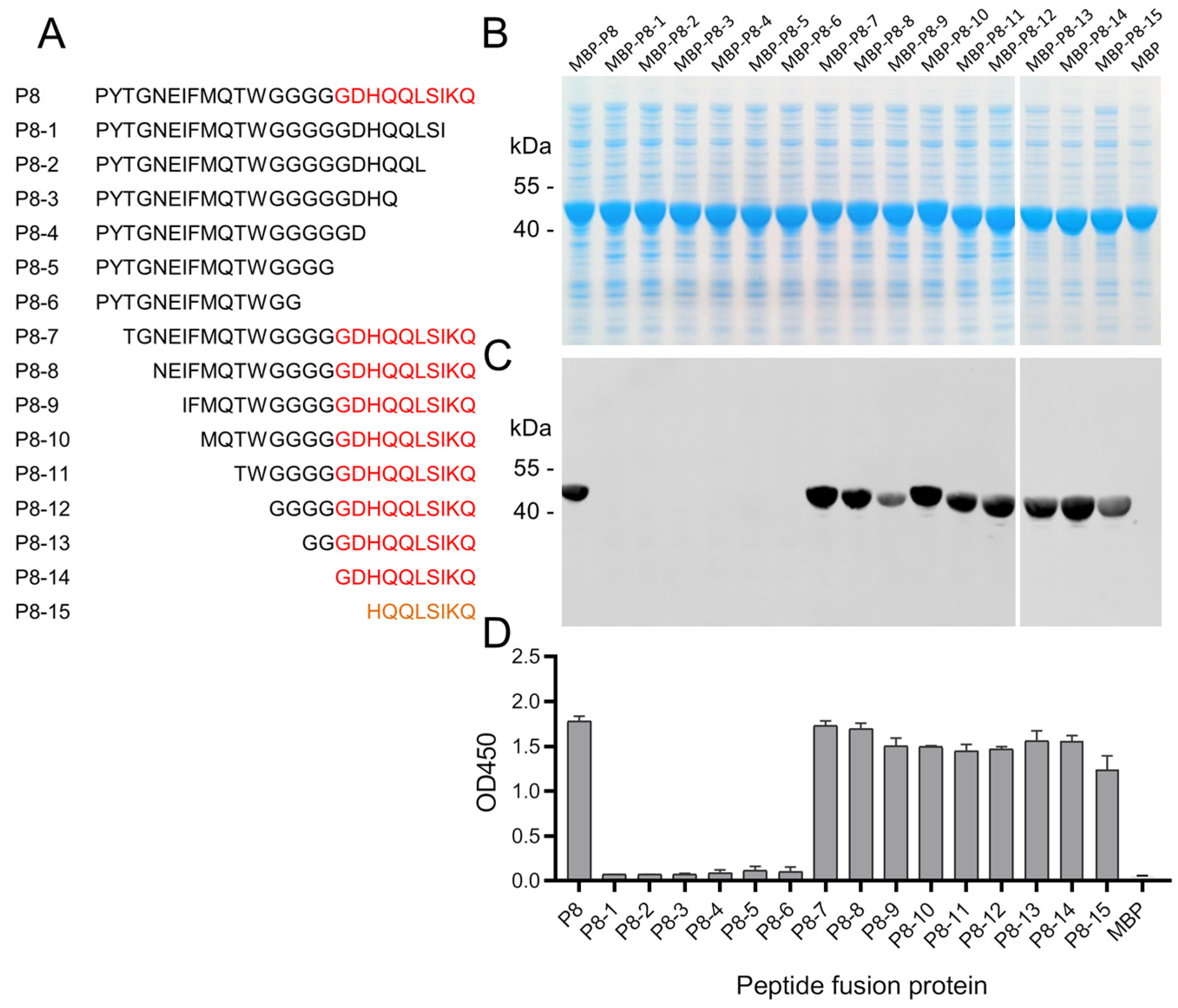
Peptide P8 was truncated sequentially from the C and N termini, respectively, in order to identify the basic unit of the epitope of mAb 10E4 (Figure 5A). The same method was used to express the peptide deletion mutants as MBP fusion proteins; all MBP-peptide fusion proteins were expressed as soluble proteins in cell lysates (Figure 5B). ELISA and Western blot were used to test the binding activities of fusion proteins with mAb 10E4. The results indicated that 16 amino acid residue deletions from the N terminus of P8 did not significantly affect the binding of peptides with mAb 10E4. However, with the deletion of only two amino acid residues (130KQ131) from the C terminus of P8, the peptide almost completely lost its ability to bind to the mAb 10E4 (Figure 5C,D). These findings showed that the linear epitope of mAb 10E4 has a core sequence of 122GDHQQLSIKQ131.

3.6. Immunoreactivity of the Epitope Peptides with Pig Sera

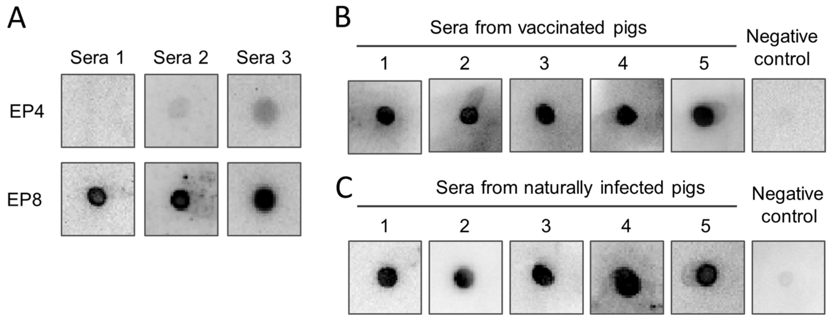
To survey whether the epitopes could bind with pig anti-ASFV sera, the peptides of mAb 8C3 epitope (EP4, 48WDCQDGICKNKITESRFIDS67) and 10E4 epitope (EP8, 122GDHQQLSIKQ131) were synthesized chemically and used to test the reactivity with pig sera from attenuated-ASFV-vaccinated and virulent-ASFV-strain-challenged pigs by dot blot assay. The results demonstrated that the EP8 peptide strongly reacted with all three serum samples, but the peptide EP4 could only react with two out of three serum samples weakly (Figure 6A). These results demonstrated that epitope 10E4 was an antigenic epitope of the MGF_110-13L protein. To survey the potential usage of the epitope 10E4 as an antigen for serological testing of ASFV antibodies, the synthesized peptide EP8 was further evaluated with five more attenuated ASFV-vaccinated pig sera and five naturally ASFV-infected pig sera. As the data showed, EP8 could be recognized by both artificially immunized pig sera (Figure 6B) and naturally ASFV-infected pig sera (Figure 6C).

4. Discussion

Although a gene-deleted modified live vaccine (MLV) has been approved in Vietnam [25,26], ASF still causes significant economic losses in the pig industry worldwide. Currently, for most of the world, well-acknowledged, safe, and effective ASF vaccines are not available. ASFV is a large and complex DNA virus with a double-stranded DNA genome that contains more than 150 ORFs. Over half of ASFV genes lack any known or predictable function. Recently, some virulence-related genes, especially host innate immune suppressor genes, were identified [27,28,29,30,31,32,33]. These findings may provide important bases for the development of gene-deletion-attenuated vaccines. Compared with attenuated live vaccines, it is generally considered that subunit vaccines have better safety. For the development of effective subunit vaccines and the development of novel serological assays, fully exploring the antigenic proteins of ASFV is necessary. Several key immunodeterminants, such as P72, p30, p54, K205R, and CD2v, have been identified. These proteins constitute potential serological assay targets and subunit vaccine components. To explore more potential antigenic proteins, in this study, we focused on predicted transmembrane proteins. Seven transmembrane proteins (C257l, I329L, I177L, EP153R, B475L, K196R, and MGF-110-13L) were transiently expressed in mammalian cells and screened with anti-ASFV pig serum. C257L, EP153R, and B475R showed weak reaction signals. EP153R was reported as a glycosylated C-type lectin-like protein, which may play crucial roles in the virulence of ASFV [34]. Deletion of EP153R attenuated the virus [35]. However, EP153R did not show good reactive activity with ASFV-positive pig serum. B475L also showed a weak reaction signal with ASFV-infected pig serum. Xu et al. recently also reported that prokaryotic-expressed B475L protein showed positive reactions with clinical ASFV-infected pig sera [36]. Here, among the seven selected transmembrane proteins, we found that MGF_110-13L showed the strongest reaction signal. These results demonstrated that MGF_110-13L is an antigenic protein of ASFV. However, the function of MGF_110-13L in inducing immune protection needs further investigation.
To express and characterize the MGF_110-13L protein, a cell line stably expressing the outer-membrane domain of the MGF_110-13L protein was generated. The purified recombinant protein exhibited two main bands by SDS-PAGE analysis. The higher molecular weight band was sensitive to PNGase F. After treatment with PNGase F, the purified protein exhibited only one main band at the same molecular weight as the lower band of the untreated protein. Furthermore, there is a predicted N-glycosylation site (68NHS70) in the outer-membrane domain of MGF_110-13L. The recombinant MGF_110-13L protein is secreted in incomplete glycosylated homodimer form. Under the non-reducing condition of SDS-PAGE, the purified protein exhibited three bands. The three bands were presumed to be glycosylated-glycosylated, glycosylated-unglycosylated, and unglycosylated-unglycosylated dimers, respectively. Whether the MGF_110-13L protein is a structural protein located in the inner membrane or outer membrane requires further investigation.
Antigenic epitopes are important characteristics of antigenic proteins. Identification of antigenic epitopes facilitates the understanding of the structure and function of antigenic proteins. To date, the antigenic epitopes of P72 [37,38], p54 [39,40], p30 [41], CD2v [42,43], and K205R [16] proteins have been reported. In the present study, using recombinant protein as an immunogen, we generated two MGF_110-13L protein-specific mAbs. To identify the epitopes of the two mAbs, a set of truncated overlapping peptides was designed and expressed as MBP fusion proteins. After being screened with mAbs by ELISA and Western blot, two epitopes located at 46 to 75 amino acids and 106 to 131 amino acids of the MGF_110-13L protein were preliminary identified, respectively. With the same strategy of expressing truncated peptides as fusion proteins, the two epitopes’ core sequences were identified as 48WDCQDGICKNKITESRFIDS67 and 122GDHQQLSIKQ131, respectively. To see if the epitopes were also recognized by sera from ASFV-infected pigs, the two epitope peptides were synthesized and tested with sera from ASFV-vaccinated and -challenged pigs. Among the two epitope peptides, peptide EP8 produced strong reaction signals, indicating that it is the antigenic epitope of MGF_110-13L. The potential application of peptide EP8 in serological testing was further investigated using five additional experimentally infected pig sera and five naturally infected pig sera. All ASFV-infected pig sera reacted positively with the peptide EP8. The findings indicate that the mAb 10E4 epitope is a potential candidate marker antigen for the serological diagnosis of ASF.
Due to the occurrence of indels or frame shift mutations, the genetic diversity of the MGF genes varied widely [44]. For MGF_110-13L, there are three variants: full-length (272–275 aa), mutants with deletion of the N-terminal outer membrane domain, and mutants with deletion of the N-terminal outer membrane domain and transmembrane domains. Up to May 11, 2024, there were 191 MGF_110-13L protein sequences in the NCBI database. Among the 191 MGF_110-13L protein sequences, there were 59 full-length MGF_110-13L protein sequences. The sequence alignment results showed that both epitope 8C3 (48WDCQDGICKNKITESRFIDS67) and epitope 10E4 (122GDHQQLSIKQ131) are highly conserved (59/59). These results suggest that the mAbs generated here could provide useful tools for further study on the MGF_110-13L protein. In addition, the epitope 10E4 has the potential to be used as an antigen in the development of serological assays. To our knowledge, this is the first study to characterize the MGF_110-13L protein epitopes. The findings of this study provide insights into future structural and functional research on the transmembrane protein MGF_110-13L.

5. Conclusions

In conclusion, we identified MGF_110-13L as an antigenic protein and fine-mapped the two linear epitopes of the MGF_110-13L protein of ASFV using two MGF_110-13L-specific mAbs. We also evaluated the synthesized peptide EP8 as a potential antigen for detecting ASFV antibodies by dot blot assay.

Author Contributions

S.-J.Z.: investigation, methodology, data curation, visualization. B.N.: investigation, methodology, data curation. S.-M.L.: investigation, methodology. Y.-M.Z.: investigation, methodology. D.-M.Z.: resources. Z.-G.B.: project administration, resources. R.-H.H.: conceptualization, resources, supervision, investigation, data curation, formal analysis, methodology, visualization, funding acquisition, writing—original draft, writing—review and editing. All authors have read and agreed to the published version of the manuscript.

Funding

This work was supported by the National Natural Science Foundation of China [grant number 32172866]; the National Key R&D Program of China [grant number 2021YFD1801301].

Institutional Review Board Statement

All experiments were performed strictly in accordance with the guidelines of care and use of laboratory animals by the Ministry of Science and Technology of the People’s Republic of China. The mouse experiment protocols were approved by the Harbin Veterinary Research Institute (221025-01-GR). The pig experiment protocols were approved by the Harbin Veterinary Research Institute (230414-01-SW). All animals used in this study were carefully fed, and the suffering of animals was minimized.

Informed Consent Statement

Not applicable.

Data Availability Statement

The authors declare that the data supporting the findings of this study are available within the article or upon request to the corresponding authors.

Acknowledgments

We thank the Diagnostic Center of Harbin Veterinary Research Institute for providing the naturally ASFV-infected pig sera.

Conflicts of Interest

The authors declare that they have no conflicts of interest.

References

  1. Penrith, M.-L. History of ‘swine fever’ in Southern Africa. J. S. Afr. Vet. Assoc. 2013, 84, a1106. [Google Scholar] [CrossRef]
  2. Rowlands, R.J.; Michaud, V.; Heath, L.; Hutchings, G.; Oura, C.; Vosloo, W.; Dwarka, R.; Onashvili, T.; Albina, E.; Dixon, L.K. African Swine Fever Virus Isolate, Georgia, 2007. Emerg. Infect. Dis. 2008, 14, 1870–1874. [Google Scholar] [CrossRef] [PubMed]
  3. Zhou, X.; Li, N.; Luo, Y.; Liu, Y.; Miao, F.; Chen, T.; Zhang, S.; Cao, P.; Li, X.; Tian, K.; et al. Emergence of African Swine Fever in China, 2018. Transbound. Emerg. Dis. 2018, 65, 1482–1484. [Google Scholar] [CrossRef] [PubMed]
  4. Zhao, D.; Liu, R.; Zhang, X.; Li, F.; Wang, J.; Zhang, J.; Liu, X.; Wang, L.; Zhang, J.; Wu, X.; et al. Replication and virulence in pigs of the first African swine fever virus isolated in China. Emerg. Microbes Infect. 2019, 8, 438–447. [Google Scholar] [CrossRef] [PubMed]
  5. Dixon, L.K.; Stahl, K.; Jori, F.; Vial, L.; Pfeiffer, D.U. African Swine Fever Epidemiology and Control. Annu. Rev. Anim. Biosci. 2020, 8, 221–246. [Google Scholar] [CrossRef] [PubMed]
  6. Dixon, L.K.; Chapman, D.A.G.; Netherton, C.L.; Upton, C. African swine fever virus replication and genomics. Virus Res. 2013, 173, 3–14. [Google Scholar] [CrossRef] [PubMed]
  7. De Villiers, E.P.; Gallardo, C.; Arias, M.; Da Silva, M.; Upton, C.; Martin, R.; Bishop, R.P. Phylogenomic analysis of 11 complete African swine fever virus genome sequences. Virology 2010, 400, 128–136. [Google Scholar] [CrossRef]
  8. Alejo, A.; Matamoros, T.; Guerra, M.; Andrés, G. A Proteomic Atlas of the African Swine Fever Virus Particle. J. Virol. 2018, 92, e01293-18. [Google Scholar] [CrossRef] [PubMed]
  9. Gómez-Puertas, P.; Rodríguez, F.; Oviedo, J.M.; Ramiro-Ibáñez, F.; Ruiz-Gonzalvo, F.; Alonso, C.; Escribano, J.M. Neutralizing antibodies to different proteins of African swine fever virus inhibit both virus attachment and internalization. J. Virol. 1996, 70, 5689–5694. [Google Scholar] [CrossRef]
  10. Gómez-Puertas, P.; Rodríguez, F.; Oviedo, J.M.; Brun, A.; Alonso, C.; Escribano, J.M. The African swine fever virus proteins p54 and p30 are involved in two distinct steps of virus attachment and both contribute to the antibody-mediated protective immune response. Virology 1998, 243, 461–471. [Google Scholar] [CrossRef]
  11. Giménez-Lirola, L.G.; Mur, L.; Rivera, B.; Mogler, M.; Sun, Y.; Lizano, S.; Goodell, C.; Harris, D.L.H.; Rowland, R.R.R.; Gallardo, C.; et al. Detection of African Swine Fever Virus Antibodies in Serum and Oral Fluid Specimens Using a Recombinant Protein 30 (p30) Dual Matrix Indirect ELISA. PLoS ONE 2016, 11, e0161230. [Google Scholar] [CrossRef]
  12. Gaudreault, N.N.; Richt, J.A. Subunit Vaccine Approaches for African Swine Fever Virus. Vaccines 2019, 7, 56. [Google Scholar] [CrossRef]
  13. Muñoz, A.L.; Tabarés, E. Characteristics of the major structural proteins of African swine fever virus: Role as antigens in the induction of neutralizing antibodies. A review. Virology 2022, 571, 46–51. [Google Scholar] [CrossRef]
  14. Kollnberger, S.D.; Gutierrez-Castañeda, B.; Foster-Cuevas, M.; Corteyn, A.; Parkhouse, R.M.E. Identification of the principal serological immunodeterminants of African swine fever virus by screening a virus cDNA library with antibody. J. Gen. Virol. 2002, 83, 1331–1342. [Google Scholar] [CrossRef] [PubMed]
  15. Gutiérrez-Castañeda, B.; Reis, A.L.; Corteyn, A.; Parkhouse, R.M.E.; Kollnberger, S. Expression, cellular localization and antibody responses of the African swine fever virus genes B602L and K205R. Arch. Virol. 2008, 153, 2303–2306. [Google Scholar] [CrossRef]
  16. Zhang, S.-J.; Liu, J.; Niu, B.; Zhu, Y.-M.; Zhao, D.-M.; Chen, W.-Y.; Liu, R.-Q.; Bu, Z.-G.; Hua, R.-H. Comprehensive mapping of antigenic linear B-cell epitopes on K205R protein of African swine fever virus with monoclonal antibodies. Virus Res. 2023, 328, 199085. [Google Scholar] [CrossRef] [PubMed]
  17. Wu, X.; Xiao, L.; Peng, B.; Wang, Y.; Yang, Z.; Yao, X.; Hu, L.; Lin, X. Prokaryotic expression, purification and antigenicity analysis of African swine fever virus pK205R protein. Pol. J. Vet. Sci. 2016, 19, 41–48. [Google Scholar] [CrossRef]
  18. Hua, R.-H.; Liu, J.; Zhang, S.-J.; Liu, R.-Q.; Zhang, X.-F.; He, X.-J.; Zhao, D.-M.; Bu, Z.-G. Mammalian Cell-Line-Expressed CD2v Protein of African Swine Fever Virus Provides Partial Protection against the HLJ/18 Strain in the Early Infection Stage. Viruses 2023, 15, 1467. [Google Scholar] [CrossRef] [PubMed]
  19. Salas, M.L.; Andrés, G. African swine fever virus morphogenesis. Virus Res. 2013, 173, 29–41. [Google Scholar] [CrossRef]
  20. Wang, N.; Zhao, D.; Wang, J.; Zhang, Y.; Wang, M.; Gao, Y.; Li, F.; Wang, J.; Bu, Z.; Rao, Z.; et al. Architecture of African swine fever virus and implications for viral assembly. Science 2019, 366, 640–644. [Google Scholar] [CrossRef]
  21. Chen, W.; Zhao, D.; He, X.; Liu, R.; Wang, Z.; Zhang, X.; Li, F.; Shan, D.; Chen, H.; Zhang, J.; et al. A seven-gene-deleted African swine fever virus is safe and effective as a live attenuated vaccine in pigs. Sci. China Life Sci. 2020, 63, 623–634. [Google Scholar] [CrossRef] [PubMed]
  22. Hua, R.-H.; Li, Y.-N.; Chen, Z.-S.; Liu, L.-K.; Huo, H.; Wang, X.-L.; Guo, L.-P.; Shen, N.; Wang, J.-F.; Bu, Z.-G. Generation and characterization of a new mammalian cell line continuously expressing virus-like particles of Japanese encephalitis virus for a subunit vaccine candidate. BMC Biotechnol. 2014, 14, 62. [Google Scholar] [CrossRef] [PubMed]
  23. Hua, R.H.; Bu, Z.G. A monoclonal antibody against PrM/M protein of Japanese encephalitis virus. Hybridoma 2011, 30, 451–456. [Google Scholar] [CrossRef] [PubMed]
  24. Hua, R.-H.; Zhang, S.-J.; Niu, B.; Ge, J.-Y.; Lan, T.; Bu, Z.-G. A Novel Conserved Linear Neutralizing Epitope on the Receptor-Binding Domain of the SARS-CoV-2 Spike Protein. Microbiol. Spectr. 2023, 11, e01190-23. [Google Scholar] [CrossRef]
  25. Borca, M.V.; Ramirez-Medina, E.; Silva, E.; Vuono, E.; Rai, A.; Pruitt, S.; Holinka, L.G.; Velazquez-Salinas, L.; Zhu, J.; Gladue, D.P. Development of a Highly Effective African Swine Fever Virus Vaccine by Deletion of the I177L Gene Results in Sterile Immunity against the Current Epidemic Eurasia Strain. J. Virol. 2020, 94, e02017-19. [Google Scholar] [CrossRef]
  26. Brake, D.A. African Swine Fever Modified Live Vaccine Candidates: Transitioning from Discovery to Product Development through Harmonized Standards and Guidelines. Viruses 2022, 14, 2619. [Google Scholar] [CrossRef]
  27. Li, J.; Song, J.; Kang, L.; Huang, L.; Zhou, S.; Hu, L.; Zheng, J.; Li, C.; Zhang, X.; He, X.; et al. pMGF505-7R determines pathogenicity of African swine fever virus infection by inhibiting IL-1β and type I IFN production. PLoS Pathog. 2021, 17, e1009733. [Google Scholar] [CrossRef] [PubMed]
  28. Wang, T.; Luo, R.; Zhang, J.; Lan, J.; Lu, Z.; Zhai, H.; Li, L.-F.; Sun, Y.; Qiu, H.-J. The African swine fever virus MGF300-4L protein is associated with viral pathogenicity by promoting the autophagic degradation of IKK β and increasing the stability of I κ B α. Emerg. Microbes Infect. 2024, 13, 2333381. [Google Scholar] [CrossRef]
  29. Wang, T.; Luo, R.; Zhang, J.; Lu, Z.; Li, L.-F.; Zheng, Y.-H.; Pan, L.; Lan, J.; Zhai, H.; Huang, S.; et al. The MGF300-2R protein of African swine fever virus is associated with viral pathogenicity by promoting the autophagic degradation of IKKα and IKKβ through the recruitment of TOLLIP. PLoS Pathog. 2023, 19, e1011580. [Google Scholar] [CrossRef]
  30. Yang, J.; Li, S.; Feng, T.; Zhang, X.; Yang, F.; Cao, W.; Chen, H.; Liu, H.; Zhang, K.; Zhu, Z.; et al. African Swine Fever Virus F317L Protein Inhibits NF-κB Activation To Evade Host Immune Response and Promote Viral Replication. mSphere 2021, 6, e00658-21. [Google Scholar] [CrossRef]
  31. Zhang, K.; Yang, B.; Shen, C.; Zhang, T.; Hao, Y.; Zhang, D.; Liu, H.; Shi, X.; Li, G.; Yang, J.; et al. MGF360-9L Is a Major Virulence Factor Associated with the African Swine Fever Virus by Antagonizing the JAK/STAT Signaling Pathway. mBio 2022, 13, e02330-21. [Google Scholar] [CrossRef] [PubMed]
  32. Zhou, P.; Dai, J.; Zhang, K.; Wang, T.; Li, L.-F.; Luo, Y.; Sun, Y.; Qiu, H.-J.; Li, S. The H240R Protein of African Swine Fever Virus Inhibits Interleukin 1β Production by Inhibiting NEMO Expression and NLRP3 Oligomerization. J. Virol. 2022, 96, e00954-22. [Google Scholar] [CrossRef] [PubMed]
  33. Huang, L.; Liu, H.; Ye, G.; Liu, X.; Chen, W.; Wang, Z.; Zhao, D.; Zhang, Z.; Feng, C.; Hu, L.; et al. Deletion of African Swine Fever Virus (ASFV) H240R Gene Attenuates the Virulence of ASFV by Enhancing NLRP3-Mediated Inflammatory Responses. J. Virol. 2023, 97, e01227-22. [Google Scholar] [CrossRef] [PubMed]
  34. Galindo, I.; Almazán, F.; Bustos, M.J.; Viñuela, E.; Carrascosa, A.L. African Swine Fever Virus EP153R Open Reading Frame Encodes a Glycoprotein Involved in the Hemadsorption of Infected Cells. Virology 2000, 266, 340–351. [Google Scholar] [CrossRef] [PubMed]
  35. Petrovan, V.; Rathakrishnan, A.; Islam, M.; Goatley, L.C.; Moffat, K.; Sanchez-Cordon, P.J.; Reis, A.L.; Dixon, L.K. Role of African Swine Fever Virus Proteins EP153R and EP402R in Reducing Viral Persistence in Blood and Virulence in Pigs Infected with BeninΔDP148R. J. Virol. 2022, 96, e0134021. [Google Scholar] [CrossRef] [PubMed]
  36. Xu, Z.; Hu, Y.; Li, J.; Wang, A.; Meng, X.; Chen, L.; Wei, J.; Tong, W.; Kong, N.; Yu, L.; et al. Screening and identification of the dominant antigens of the African swine fever virus. Front. Vet. Sci. 2023, 10, 1175701. [Google Scholar] [CrossRef] [PubMed]
  37. Heimerman, M.E.; Murgia, M.V.; Wu, P.; Lowe, A.D.; Jia, W.; Rowland, R.R. Linear epitopes in African swine fever virus p72 recognized by monoclonal antibodies prepared against baculovirus-expressed antigen. J. Vet. Diagn. Investig. 2018, 30, 406–412. [Google Scholar] [CrossRef] [PubMed]
  38. Tesfagaber, W.; Wang, W.; Wang, L.; Zhao, R.; Zhu, Y.; Li, F.; Sun, E.; Liu, R.; Bu, Z.; Meng, G.; et al. A highly efficient blocking ELISA based on p72 monoclonal antibody for the detection of African swine fever virus antibodies and identification of its linear B cell epitope. Int. J. Biol. Macromol. 2024, 268, 131695. [Google Scholar] [CrossRef]
  39. Cao, Y.; Han, D.; Zhang, Y.; Zhang, K.; Du, N.; Tong, W.; Li, G.; Zheng, H.; Liu, C.; Gao, F.; et al. Identification of one novel epitope targeting p54 protein of African swine fever virus using monoclonal antibody and development of a capable ELISA. Res. Vet. Sci. 2021, 141, 19–25. [Google Scholar] [CrossRef]
  40. Petrovan, V.; Murgia, M.V.; Wu, P.; Lowe, A.D.; Jia, W.; Rowland, R.R.R. Epitope mapping of African swine fever virus (ASFV) structural protein, p54. Virus Res. 2020, 279, 197871. [Google Scholar] [CrossRef]
  41. Zhou, G.; Shi, Z.; Luo, J.; Cao, L.; Yang, B.; Wan, Y.; Wang, L.; Song, R.; Ma, Y.; Tian, H.; et al. Preparation and epitope mapping of monoclonal antibodies against African swine fever virus P30 protein. Appl. Microbiol. Biotechnol. 2022, 106, 1199–1210. [Google Scholar] [CrossRef] [PubMed]
  42. Jia, R.; Zhang, G.; Bai, Y.; Liu, H.; Chen, Y.; Ding, P.; Zhou, J.; Feng, H.; Li, M.; Tian, Y.; et al. Identification of Linear B Cell Epitopes on CD2V Protein of African Swine Fever Virus by Monoclonal Antibodies. Microbiol. Spectr. 2022, 10, e01052-21. [Google Scholar] [CrossRef] [PubMed]
  43. Ren, D.; Ding, P.; Liu, S.; Zhang, N.; Chen, Y.; Li, Q.; Fan, L.; Chang, Z.; Zhang, G. Development and characterization of recombinant ASFV CD2v protein nanoparticle-induced monoclonal antibody. Int. J. Biol. Macromol. 2022, 209, 533–541. [Google Scholar] [CrossRef]
  44. Zhu, Z.; Chen, H.; Liu, L.; Cao, Y.; Jiang, T.; Zou, Y.; Peng, Y. Classification and characterization of multigene family proteins of African swine fever viruses. Brief Bioinform. 2021, 22, bbaa380. [Google Scholar] [CrossRef] [PubMed]
Figure 1. Identification of antigenic transmembrane proteins with pig anti-ASFV serum. Seven predicted ASFV transmembrane protein outer-membrane domain-expressing plasmids were transiently transfected into the HEK-293 cells. The protein expression was verified with 6× His tag specific antibody by Western blot (A). After verification, transiently expressed proteins were probed with pig anti-ASFV serum from pigs vaccinated with attenuated ASFV and challenged with a virulent ASFV strain (B).
Animals 14 01951 g001
Figure 2. Expression of MGF_110-13L protein and generation of mAbs against the MGF_110-13L protein of ASFV. After transfection and selection, a stable cell line expressing the MGF_110-13L protein was established. The cell line was identified by IFA with 6× His tag specific antibody (A). The recombinant protein was purified by nickel affinity chromatography from the supernatants of the cell line and analyzed by SDS-PAGE and Western blot (B). The purified recombinant protein was treated with PNGase F and analyzed by SDS-PAGE (C). The purified proteins were analyzed by SDS-PAGE under non-reducing conditions (without β-mercaptoethanol) and reducing conditions (with β-mercaptoethanol) (D). The titers of MGF_110-13L-specific antibody in immunized pig sera were detected by ELISA (E). The titrations of monoclonal antibodies 8C3 and 10E4 were detected by ELISA (F). The MGF_110-13L proteins in ASFV-infected cell lysates were detected by Western blot with mAbs 8C3 and 10E4, respectively (G). **, glycosylated monomer. *, unglycosylated monomer.
Animals 14 01951 g002
Figure 3. Expression of peptide fusion proteins and mapping the epitopes of mAbs. (A) Diagrammatic representation of the truncated overlapping short peptides that span the outer-membrane domain of the MGF_110-13L protein. (B) SDS-PAGE analysis of recombinant peptide fusion proteins. ELISA analysis results show that peptides P3 and P4 were recognized by mAb 8C3 (C), and peptide P8 was recognized by mAb 10E4 (D). Western blot results showed that mAb 8C3 recognized peptides P3 and P4 (E) and mAb 10E4 recognized peptide P8 (F).
Animals 14 01951 g003
Figure 4. Fine-mapping of the epitope of mAb 8C3. Peptide P4 was truncated sequentially at the carboxyl or amino terminus, as shown in the schematic diagram (A). The MBP-peptide fusion proteins were expressed and analyzed by SDS-PAGE (B). The MBP-peptide fusion proteins were probed with mAb 8C3 using Western blot (C) and ELISA (D). The core sequence of the epitope of mAb 8C3 was deduced to be 48WDCQDGICKNKITESRFIDS67. The core sequences of the epitope are highlighted in red, and the truncated epitope sequences with reduced binding capacity to the mAb are shown in orange or yellow (A).
Animals 14 01951 g004
Figure 5. Fine-mapping of the epitope of mAb 10E4. Peptide P8 was truncated sequentially at the carboxyl or amino terminus, as shown in the schematic diagram (A). The MBP-peptide fusion proteins were expressed and analyzed by SDS-PAGE (B). The MBP-peptide fusion proteins were probed with mAb 10E4 using Western blot (C) and ELISA (D). The core sequence of the epitope of mAb 10E4 was deduced to be 122GDHQQLSIKQ131. The core sequences of the epitope are highlighted in red, and the truncated epitope sequences with reduced binding capacity to the mAb are shown in orange (A).
Animals 14 01951 g005
Figure 6. Detection of African swine fever virus antibodies with synthesized peptides. The synthesized peptides EP4 of the mAb 8C3 epitope and EP8 of the mAb 10E4 epitope were subjected to detection of epitope specific antibodies in ASFV-infected pig sera using the dot blot assay (A). The synthesized peptide EP8 of the mAb 10E4 epitope was further evaluated in detecting ASFV antibodies with attenuated ASFV-infected pig sera (B) and naturally ASFV-infected pig sera (C) by dot blot assay.
Animals 14 01951 g006
Disclaimer/Publisher’s Note: The statements, opinions and data contained in all publications are solely those of the individual author(s) and contributor(s) and not of MDPI and/or the editor(s). MDPI and/or the editor(s) disclaim responsibility for any injury to people or property resulting from any ideas, methods, instructions or products referred to in the content.