
New Myzostomids (Annelida) in Symbiosis with Feather Stars in the Shallow Waters of the South China Sea (Hainan Island)
 , and
, and
Abstract
:Simple Summary
Abstract
1. Introduction
2. Materials and Methods
2.1. Sample Collection and Processing
2.2. Morphological Analysis
2.3. Molecular Analysis
3. Results
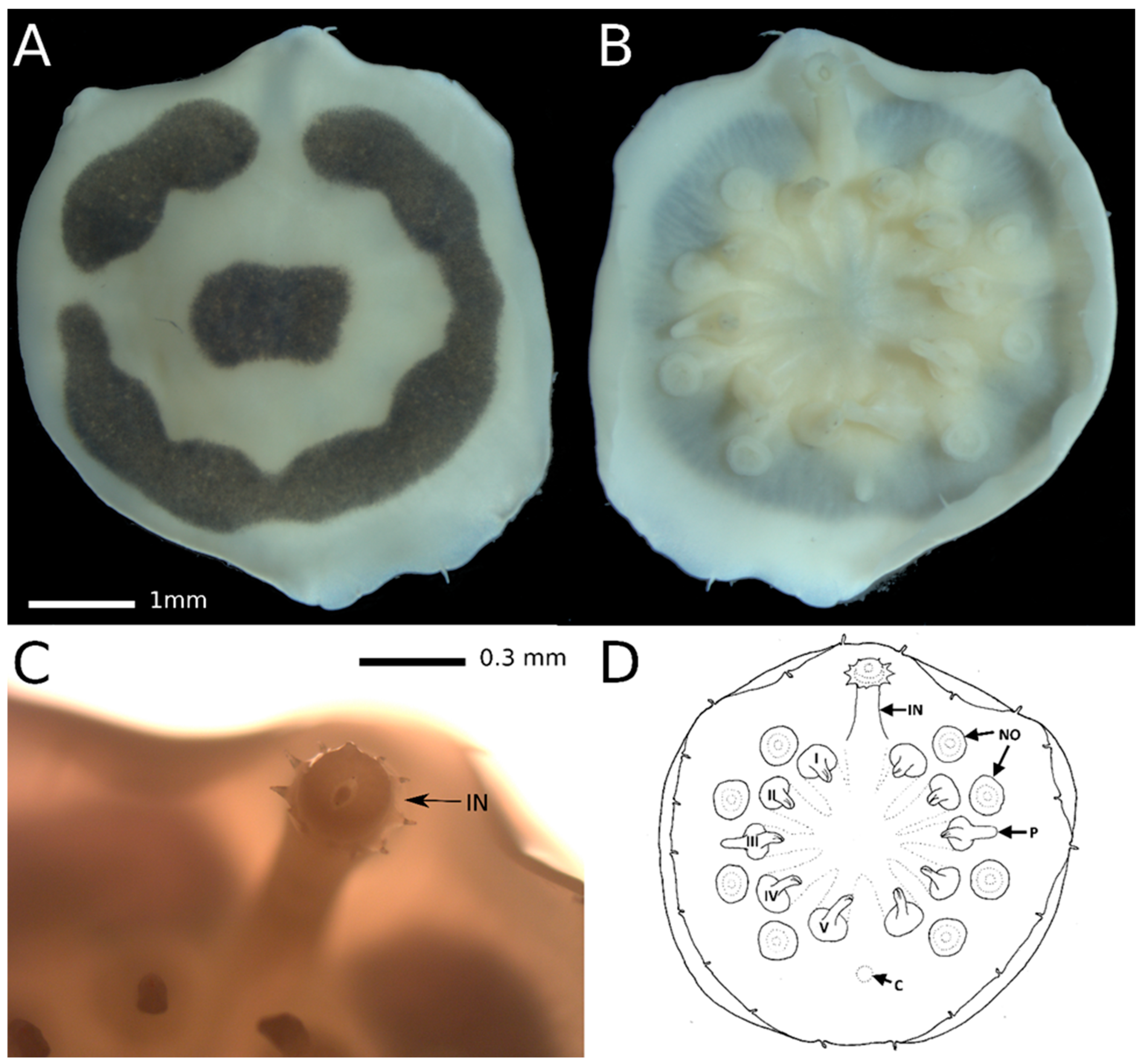
3.1. Taxonomy
3.2. Molecular Analysis
4. Discussion
5. Conclusions
Supplementary Materials
Author Contributions
Funding
Institutional Review Board Statement
Informed Consent Statement
Data Availability Statement
Acknowledgments
Conflicts of Interest
References
- Eeckhaut, I.; Lanterbecq, D. Myzostomida: A review of the phylogeny and ultrastructure. Hydrobiologia 2005, 535/536, 253–275. [Google Scholar] [CrossRef]
- Summers, M.M.; Rouse, G.W. Phylogeny of Myzostomida (Annelida) and their relationships with echinoderm hosts. BMC Evol. Biol. 2014, 14, 170. [Google Scholar] [CrossRef] [PubMed]
- Lanterbecq, D.; Rouse, G.W.; Eeckhaut, I. Bodyplan diversification in crinoid-associated myzostomes (Myzostomida, Protostomia). Invertebr. Biol. 2009, 128, 283–301. [Google Scholar] [CrossRef]
- Summers, M.M.; Al-Hakim, I.I.; Rouse, G.W. Turbo-taxonomy: 21 new species of Myzostomida (Annelida). Zootaxa 2014, 3873, 301–344. [Google Scholar] [CrossRef]
- von Graff, L. Verzeichniss der von den United States Coast Survey steamers “Hassler” und “Blake” von 1867 bis 1879 gesammelten Myzostomiden. Bull. Mus. Comp. Zool. 1883, 11, 125–133. [Google Scholar]
- von Graff, L. Report on the Myzostomida collected during the voyage of H. M. S. Challenger during the years 1873–76. Supplement. report on the scientific results of the Voyage of H. M. S. Challenger. Zoology 1887, 20, 1–16. [Google Scholar]
- World Polychaeta Database. Myzostomida. Read, G.; Fauchald, K. (Eds.) 2024. Available online: https://www.marinespecies.org/aphia.php?p=taxdetails&id=233983 (accessed on 21 July 2024).
- Kolbasova, G.D.; Mekhova, E.S. Myzostoma khanhkhoaensis (Myzostomida), a new myzostomid species from the Nhatrang Bay, Vietnam. Zootaxa 2019, 4691, 235–249. [Google Scholar] [CrossRef] [PubMed]
- Lanterbecq, D.; Rouse, G.W.; Milinkovitch, M.C.; Eeckhaut, I. Molecular phylogenetic analyses indicate multiple independent emergences of parasitism in Myzostomida (Protostomia). Syst. Biol. 2006, 55, 208–227. [Google Scholar] [CrossRef] [PubMed]
- Terrana, L.; Eeckhaut, I. Taxonomic description and 3D modelling of a new species of myzostomid (Annelida, Myzostomida) associated with black corals from Madagascar. Zootaxa 2017, 4244, 277–295. [Google Scholar] [CrossRef] [PubMed]
- Xiao, Y.; Chen, S.B.; Zhang, L.; Yue, P.; Ouyang, Z.Y.; Liu, X.C. Designing nature reserve systems in Hainan Island based on ecosystem services. Acta Ecol. Sin. 2011, 31, 7357–7369. [Google Scholar]
- Liu, J.Y. Status of Marine Biodiversity of the China Seas. PLoS ONE 2013, 8, e50719. [Google Scholar] [CrossRef] [PubMed]
- Venmathi Maran, B.A.; Kim, I.H.; Bratova, O.A.; Ivanenko, V.N. Two new species of poecilostomatoid copepods symbiotic on the venomous echinoid Toxopneustes pileolus (Lamarck) (Echinodermata) from Vietnam. Syst. Parasitol. 2017, 94, 227–241. [Google Scholar] [CrossRef] [PubMed]
- Ivanenko, V.N.; Hoeksema, B.W.; Mudrova, S.V.; Nikitin, M.A.; Martínez, A.; Rimskaya-Korsakova, N.N.; Berumen, M.L.; Fontaneto, D. Lack of host specificity of copepod crustaceans associated with mushroom corals in the Red Sea. Mol. Phylogenet. Evol. 2018, 127, 770–780. [Google Scholar] [CrossRef] [PubMed]
- Shelyakin, P.V.; Garushyants, S.K.; Nikitin, M.A.; Mudrova, S.V.; Berumen, M.; Speksnijder, A.G.; Ivanenko, V.N. Microbiomes of gall-inducing copepod crustaceans from the corals Stylophora pistillata (Scleractinia) and Gorgonia ventalina (Alcyonacea). Sci. Rep. 2018, 8, 11563. [Google Scholar] [CrossRef] [PubMed]
- Korzhavina, O.A.; Hoeksema, B.W.; Ivanenko, V.N. A review of Caribbean Copepoda associated with reef-dwelling cnidarians, echinoderms, and sponges. Contrib. Zool. 2019, 88, 297–349. [Google Scholar] [CrossRef]
- Montano, S.; Reimer, J.D.; Ivanenko, V.N.; García-Hernández, J.E.; van Moorsel, G.W.N.M.; Galli, P.; Hoeksema, B.W. Widespread Occurrence of a Rarely Known Association between the Hydrocorals Stylaster roseus and Millepora alcicornis at Bonaire, Southern Caribbean. Diversity 2020, 12, 218. [Google Scholar] [CrossRef]
- Korzhavina, O.A.; Reimer, J.D.; Ehrlich, H.; Ivanenko, V.N. Global diversity and distribution of Lamippidae copepods symbiotic on Octocorallia. Symbiosis 2021, 83, 265–277. [Google Scholar] [CrossRef]
- Tchesunov, A.V.; Ivanenko, V.N. What is the difference between marine and limnetic-terrestrial associations of nematodes with invertebrates? Integr. Zoöl. 2021, 17, 481–510. [Google Scholar] [CrossRef] [PubMed]
- Korzhavina, O.A.; Grishina, D.Y.; Chen, X.; Fontaneto, D.; Ivanenko, V.N. Diving into diversity: Copepod crustaceans in octocoral associations. Diversity 2023, 15, 1140. [Google Scholar] [CrossRef]
- Korzhavina, O.A.; Gubareva, N.V.; Kitashov, A.V.; Britayev, T.A.; Ivanenko, V.N. From microscale interactions to macroscale patterns in copepod–crinoid symbiosis. Animals 2024, 14, 877. [Google Scholar] [CrossRef] [PubMed]
- Korzhavina, O.A.; Nikitin, M.A.; Hoeksema, B.W.; Armenteros, M.; Reimer, J.D.; Ivanenko, V.N. Tracing geographic and molecular footprints of copepod crustaceans causing Multifocal Purple Spots Syndrome in the Caribbean sea fan Gorgonia ventalina. Diversity 2024, 16, 280. [Google Scholar] [CrossRef]
- Vihtakari, M. ggOceanMaps: Plot Data on Oceanographic Maps Using ‘ggplot2’. R Package Version 2.1.1. 2023. Available online: https://CRAN.R-project.org/package=ggOceanMaps (accessed on 19 November 2023).
- Rennstam, R.O.; Sint, D.; Horngacher, N.; Traugott, M. A broadly applicable COI primer pair and an efficient single-tube amplicon library preparation protocol for metabarcoding. Ecol. Evol. 2018, 8, 12335–12350. [Google Scholar] [CrossRef] [PubMed]
- Geller, J.; Meyer, C.; Parker, M.; Hawk, H. Redesign of PCR primers for mitochondrial cytochrome c oxidase subunit I for marine invertebrates and application in all-taxa biotic surveys. Mol. Ecol. Resour. 2013, 13, 851–861. [Google Scholar] [CrossRef] [PubMed]
- Messing, J. New M13 vectors for cloning. In Methods in Enzymology; Academic Press: Cambridge, MA, USA, 1983; Volume 101, pp. 20–78. [Google Scholar]
- Kearse, M.; Moir, R.; Wilson, A.; Stones-Havas, S.; Cheung, M.; Sturrock, S.; Buxton, S.; Cooper, A.; Markowitz, S.; Duran, C.; et al. Geneious Basic: An integrated and extendable desktop software platform for the organization and analysis of sequence data. Bioinformatics 2012, 28, 1647–1649. [Google Scholar] [CrossRef] [PubMed]
- Altschul, S.F.; Gish, W.; Miller, W.; Myers, E.W.; Lipman, D.J. Basic local alignment search tool. J. Mol. Biol. 1990, 215, 403–410. [Google Scholar] [CrossRef] [PubMed]
- Edgar, R.C. MUSCLE: Multiple sequence alignment with high accuracy and high throughput. Nucleic Acids Res. 2004, 32, 1792–1797. [Google Scholar] [CrossRef] [PubMed]
- Kumar, S.; Stecher, G.; Li, M.; Knyaz, C.; Tamura, K. MEGA X: Molecular evolutionary genetics analysis across computing platforms. Mol. Biol. Evol. 2018, 35, 1547. [Google Scholar] [CrossRef] [PubMed]
- Ronquist, F.; Huelsenbeck, J.P. MrBayes 3: Bayesian phylogenetic inference under mixed models. Bioinformatics 2003, 19, 1572–1574. [Google Scholar] [CrossRef] [PubMed]
- Stamatakis, A. RAxML version 8: A tool for phylogenetic analysis and post-analysis of large phylogenies. Bioinformatics 2014, 30, 1312–1313. [Google Scholar] [CrossRef] [PubMed]
- Pattengale, N.D.; Alipour, M.; Bininda-Emonds, O.R.; Moret, B.M.; Stamatakis, A. How many bootstrap replicates are necessary? In Research in Computational Molecular Biology: 13th Annual International Conference, RECOMB 2009, Tucson, AZ, USA, 18–21 May 2009; Springer: Berlin/Heidelberg, Germany, 2009; pp. 184–200. [Google Scholar]
- Costello, M.J.; Tsai, P.; Wong, P.S.; Kwok Lun Cheung, A.; Basher, Z.; Chaudhary, C. Marine biogeographic realms and species endemicity. Nat. Commun. 2017, 8, 1057. [Google Scholar] [CrossRef] [PubMed]
- Atkins, D. Report on the Myzostomida collected by Mr. F.A. Potts in Torres Strait, with a description of a species obtained by Professor J. Stanley Gardiner from the Maldives. Proc. Zool. Soc. Lond. 1927, 97, 339–357. [Google Scholar] [CrossRef]
- Eeckhaut, I.; Grygier, M.J.; Deheyn, D. Myzostomes from Papua New Guinea, with related Indo-West Pacific distribution records and description of five new species. Bull. Mar. Sci. 1998, 62, 841–886. [Google Scholar]
- Lanterbecq, D.; Eeckhaut, I. Myzostomida from Madagascar, with the description of two new species. Hydrobiologia 2003, 496, 115–123. [Google Scholar] [CrossRef]
- Sun, R.; Lei, Y.; Zhou, J. Phylum Annelida. In Checklist of Marine Biota of China Seas; Liu, R., Ed.; Science Press, Academia Sinica: Beijing, China, 2008; pp. 405–452. [Google Scholar]
- Liao, Y.; Xiao, N. Species composition and faunal characteristics of echinoderms in China Seas. Biodivers. Sci. 2011, 19, 729–736. [Google Scholar]
- Zeppilli, D.; Sarrazin, J.; Leduc, D.; Arbizu, P.M.; Fontaneto, D.; Fontanier, C.; Gooday, A.J.; Kristensen, R.M.; Ivanenko, V.N.; Sørensen, M.V.; et al. Is the meiofauna a good indicator for climate change and anthropogenic impacts? Mar. Biodiv. 2015, 45, 505–535. [Google Scholar] [CrossRef]


| Host | Myzostoma polycyclus | Myzostoma solare sp. nov. | Myzostoma ordinatum sp. nov. | Myzostoma scopus sp. nov. |
|---|---|---|---|---|
| Comanthus parvicirrus | 18 | |||
| Comanthus parvicirrus | 10 | |||
| Comanthus parvicirrus | 5 | |||
| Comaster schlegelii | 1 | 2 | ||
| Comaster schlegelii | 6 | 25 | ||
| Comaster schlegelii | 3 | 34 | 2 | |
| Comaster schlegelii | 9 | 1 |
| Species | Sampling Locality | Date of Sampling | Depth, m | Host | Voucher | GenBank Accession Number |
|---|---|---|---|---|---|---|
| Myzostoma scopus sp. nov. | 18.20465; 109.16995 | 25 November 2009 | 5–8 | Comaster schlegelii | ZMMU Pl-4886 | OR864678 |
| Myzostoma solare sp. nov. | 18.20465; 109.16995 | 25 November 2009 | 5–8 | Comaster schlegelii | ZMMU Pl-4887 | OR864679 |
| Myzostoma ordinatum sp. nov. | 18.20465; 109.16995 | 25 November 2009 | 5–8 | Comaster schlegelii | ZMMU Pl-4888 | |
| Myzostoma ordinatum sp. nov. | 18.20465; 109.16995 | 25 November 2009 | 5–8 | Comaster schlegelii | ZMMU Pl-4889 | OR864677 |
| Myzostoma ordinatum sp. nov. | 18.20465; 109.16995 | 25 November 2009 | 5–8 | Comaster schlegelii | ZMMU Pl-4890 | |
| Myzostoma polycyclus | 18.20465; 109.16995 | 24 November 2009 | 3–5 | Comanthus parvicirrus | ZMMU Pl-4891 | OR864681 |
| Myzostoma polycyclus | 18.20465; 109.16995 | 24 November 2009 | 3–5 | Comanthus parvicirrus | ZMMU Pl-4892 | |
| Myzostoma polycyclus | 18.20465; 109.16995 | 24 November 2009 | 3–5 | Comanthus parvicirrus | ZMMU Pl-4893 | OR864680 |
Disclaimer/Publisher’s Note: The statements, opinions and data contained in all publications are solely those of the individual author(s) and contributor(s) and not of MDPI and/or the editor(s). MDPI and/or the editor(s) disclaim responsibility for any injury to people or property resulting from any ideas, methods, instructions or products referred to in the content. |
© 2024 by the authors. Licensee MDPI, Basel, Switzerland. This article is an open access article distributed under the terms and conditions of the Creative Commons Attribution (CC BY) license (https://creativecommons.org/licenses/by/4.0/).
Share and Cite
Isaychev, A.; Schepetov, D.; Zhou, Y.; Britayev, T.A.; Ivanenko, V.N. New Myzostomids (Annelida) in Symbiosis with Feather Stars in the Shallow Waters of the South China Sea (Hainan Island). Animals 2024, 14, 2265. https://doi.org/10.3390/ani14152265
Isaychev A, Schepetov D, Zhou Y, Britayev TA, Ivanenko VN. New Myzostomids (Annelida) in Symbiosis with Feather Stars in the Shallow Waters of the South China Sea (Hainan Island). Animals. 2024; 14(15):2265. https://doi.org/10.3390/ani14152265
Chicago/Turabian StyleIsaychev, Alexander, Dimitry Schepetov, Yutong Zhou, Temir A. Britayev, and Viatcheslav N. Ivanenko. 2024. "New Myzostomids (Annelida) in Symbiosis with Feather Stars in the Shallow Waters of the South China Sea (Hainan Island)" Animals 14, no. 15: 2265. https://doi.org/10.3390/ani14152265