Article

Histological Characteristics of Follicles, Reproductive Hormones and Transcriptomic Analysis of White King Pigeon Illuminated with Red Light

College of Animal Science and Technology, Yangzhou University, Yangzhou 225009, China
*
Author to whom correspondence should be addressed.
Animals 2024, 14(16), 2320; https://doi.org/10.3390/ani14162320 (registering DOI)
Submission received: 15 June 2024 / Revised: 4 July 2024 / Accepted: 9 August 2024 / Published: 10 August 2024
(This article belongs to the Section Birds)

Abstract

:

Simple Summary

It has been observed that red light enhances egg production in pigeons. However, the underlying histological characteristics and molecular mechanisms remain unclear. In this study, fifty-four White King pigeons were selected to confirm the effect of red light on egg production in pigeons and to assess the histological characteristics of follicles, reproductive hormone levels and ovarian transcriptomics on the third day of the laying interval under both red light and white light. This study revealed a molecular basis associated with red light promoting hierarchical follicle selection, enabling a better understanding of the underlying mechanisms of red light improving egg production in pigeons.

Abstract

Red light (RL) has been observed to enhance egg production in pigeons, yet the underlying histological characteristics and molecular mechanisms remain less understood. This study included fifty-four pigeons to assess follicular histology, reproductive hormones, and ovarian transcriptomics on the third day of the laying interval under RL and white light (WL). The results showed that the granulosa cell layer was significantly thicker under RL (p < 0.05), whereas the theca cell and connective tissue layers showed no significant differences (p > 0.05). Higher plasma estradiol (E2) levels were recorded in the RL group (p < 0.05), as well as follicle stimulating hormone (FSH), although progesterone (P4) levels were higher under WL (p < 0.05). Moreover, P4 concentrations in follicle yolk significantly decreased under RL (p < 0.01), with higher FSH and E2 levels in F1 yolk and similar increases in SF1 yolk (p < 0.01). Transcriptomic analysis revealed 4991 differentially expressed genes in the pigeon ovary. The protein–protein interaction network highlighted genes like HSD11B1, VEGFD, WNT6, SMAD6, and LGR5 as potential contributors to hierarchical follicle selection under RL. This research provides new insights into the molecular basis by which RL may promote hierarchical follicle selection and improve egg production in pigeons.

1. Introduction

Pigeon products have become popular in the human diet for high nutrition, and they are the forth largest poultry product after chicken, duck, and goose [1,2]. However, due to their unique characteristics, with a long clutch interval, and paired pigeons only having two preovulatory follicles in a laying period [3], it is imperative to improve the egg production of pigeons. Poultry exhibits a heightened sensitivity to the red and blue portions of the light spectrum compared to humans, with longer wavelength radiation more effectively penetrating the hypothalamus, at a rate 100–1000 times greater for pigeons, than shorter wavelengths [4]. In poultry, upon photostimulation, reproductive activity is regulated by hypothalamic neuropeptides, gonadotropin-releasing hormones (GnRHs), which prompts the anterior pituitary gland to release gonadotropins, controlling the synthesis of steroid hormones and follicle development [5], which has been well studied in Japanese quail [6], songbird [7] and Dark-eyed Junco [8]. Furthermore, studies have shown that monochromatic light influences the reproductive performance of poultry. For instance, Baxter et al. (2014) [9] concluded that red light (RL) was required to stimulate the reproductive axis while green light was ineffective. Rozenboim et al. (2013) [10] found that the RL enhanced the expressions of both green and red opsin receptor. However, Gongruttananun (2011) [11] observed that RL did not enhance egg production in Thai-native hens. In our previous study, we verified that RL increased egg production in pigeons [12,13]. However, the specific mechanisms through which RL enhances pigeon egg production, particularly the histological characteristics of follicles, remain unclear.
In chickens, the ovary is a crucial organ directly linked to reproductive performance. It houses a variety of follicles of different sizes, ranging from hierarchical follicles (F1 to F6) to prehierarchical follicles, which include small yellow follicles (SYFs), large white follicles and large yellow follicles [14]. The SYF was selected and developed into hierarchical follicles daily until ovulation. Unlike chickens, paired pigeons lay just two eggs per cycle. Additionally, in our prior research, we described the morphological characteristics of pigeon follicles during the laying interval [15]. We observed that only two preovulatory follicles and small follicles were hierarchical and coupled. Furthermore, we also found that RL prompted the selection of hierarchical follicles on the 3rd day of the laying interval (LI3). Transcriptomics is commonly employed to identify genes associated with tissue function. For example, Brady et al. (2023) [16] identified ovary genes related to the ovulation of turkeys, while Yan et al. (2022) [17] focused on genes in ducks that can improve egg production. In this study, the thickness of the granulosa cell (GC) layer, theca cell (TC) layer and connective tissue (CT) layer were measured. We also assessed the reproductive hormones in the plasma and yolk of various follicles and analyzed the ovarian transcriptome of pigeons in LI3 under both RL and white light (WL). The findings offer fresh insights into the reproductive physiology of pigeons under RL and contribute to understanding the molecular mechanisms by which RL enhances egg production.

2. Materials and Methods

2.1. Animals and Sample Collection

Fifty-four paired White King pigeons (12 months old) were purchased from Taizhou Pigeon Breeding Co., Ltd. (Taizhou, China). The birds were maintained under a light regime of 15 h light and 9 h dark and water and the same food were available ad libitum. The allocation of light colors was carried out within blocks of two compartments. The pairs were housed in laying batteries (length × width × height = 40 cm × 33 cm × 30 cm). The birds were divided into two groups: one exposed to RL at 660 nm and the other to WL covering a spectrum of 400–760 nm, with each group further divided into three replicates. The light intensity was 17.50 ± 2.50 lux, which was measured with a TES-1336A light meter (TES Electrical Electronic Corp., Taiwan, China). The duration of the experiment was six months. Twelve female pigeons from each group with similar physiological states (LI3) were selected for blood sampling. From each bird, a 4 to 5 mL blood sample was drawn from the wing vein into tubes containing 10 μL of 0.8 M heparin sodium. The samples were then centrifuged at 3000× g at 4 °C for five min and stored at −20 °C until analysis. Concurrently, the yolks from the F1, F2 and the largest small follicle (SF1) of pigeons were used for plasma collection and were harvested to measure reproductive hormone concentrations. After gently slicing the follicles with a scalpel, the yolk was collected by penetrating the disposable syringe needle into the incision and sucking [18]. The follicles of other six female pigeons were then fixed in 4% (v/v) buffered paraformaldehyde for 24 h post-yolk removal [19]. Additionally, three female pigeons from each group in LI3 were anesthetized with isoflurane and decapitated, and their ovaries were collected, immediately frozen in liquid nitrogen, and stored at −80 °C until analysis.

2.2. Reproductive Hormone Concentrations Analysis

The concentrations of progesterone (P4), estradiol (E2) and follicle-stimulating hormone (FSH) were measured using enzyme-linked immunosorbent assay (ELISA) kits, following the manufacturer’s instructions, The E2 ELISA kit, P4 ELISA kit and FSH ELISA kit were purchased from ElabScience Biotechnology Co., Ltd. (Wuhan, China) [15,20]. The sensitivity of P4 concentration is 1.25–75 ng/mL, the sensitivity of E2 concentration is 12.5–700 ng/L and the sensitivity of FSH concentration is 0.1875–11.25 IU/L. The intra-assay coefficients of variation for all hormones were less than 10% and interassay coefficients of variation for all hormones were less than 15%. The ELISA Calc software v1.0 (Comple-Software, Iowa City, IA, USA) was used to calculate the concentrations of the various reproductive hormones.

2.3. Histological Analysis of Follicles

After fixation, the follicles were dehydrated and embedded in paraffin wax. Thin sections measuring 5 μm in thickness were prepared, stained with hematoxylin and eosin, and examined under a light microscope (Nikon, Tokyo, Japan). The thicknesses of the GC layer, TC layer and CT layer in the follicles were measured using ImageJ 2X software (National Institutes of Health, Bethesda, MD, USA). The arrangement of GC is relatively neat, and the nucleus presents a square shape; the cell nuclei of the TC and CT layer are long spindle, and the arrangement of CT cells is very loose [21].

2.4. Transcriptome of Pigeon Ovary

Six RNA-seq libraries were constructed from pigeon ovaries raised under red (RO1, RO2, RO3) or white (WO1, WO2, WO3) light. Total RNA was isolated from each sample using TRIzol reagent (Invitrogen, Carlsbad, CA, USA) and RNA samples were prepared following the manufacturers’ instructions. RNA integrity, purity and quantity were assessed using the 2100 Bioanalyzer (Agilent Technologies, Wilmington, DE, USA). Sequencing adaptors were attached to RNA fragments to construct the cDNA libraries, which were then sequenced on the Illumina Hiseq 4000 platform (Illumina, Inc., San Diego, CA, USA). Differentially expressed genes (DEGs) between the groups were identified using the DESeq2 R package, with significance determined by a Q-value < 0.05 and an absolute fold change greater than 2. Functional prediction and classification of the unigenes were performed using the KEGG database and GO unigene annotations. Additionally, the protein–protein interaction (PPI) network for the DEGs, encoding proteins in the pigeon ovary under RL, was analyzed using the STRING protein interaction database and visualized with Cytoscape software (version 3.9.0).

2.5. RT-qPCR Validation

The gene primer sequences have been listed in Table 1. Total RNA was extracted using TRIzol reagent (Invitrogen, Carlsbad, CA, USA) and reverse transcribed with the Fast Quant RT Kit (Tiangen Biotech Co., Ltd., Beijing, China). The expressions of selected genes were detected using SuperReal PreMix (Tiangen Biotech Co., Ltd., Beijing, China). Each sample was analyzed in triplicate, with GAPDH serving as an internal reference gene. Relative gene expression was calculated using the 2−ΔΔCt method.

2.6. Statistical Analysis

Data are expressed as means ± standard deviation. The differences between groups were assessed using the independent-samples t test, with a significance threshold set at p < 0.05.

3. Results

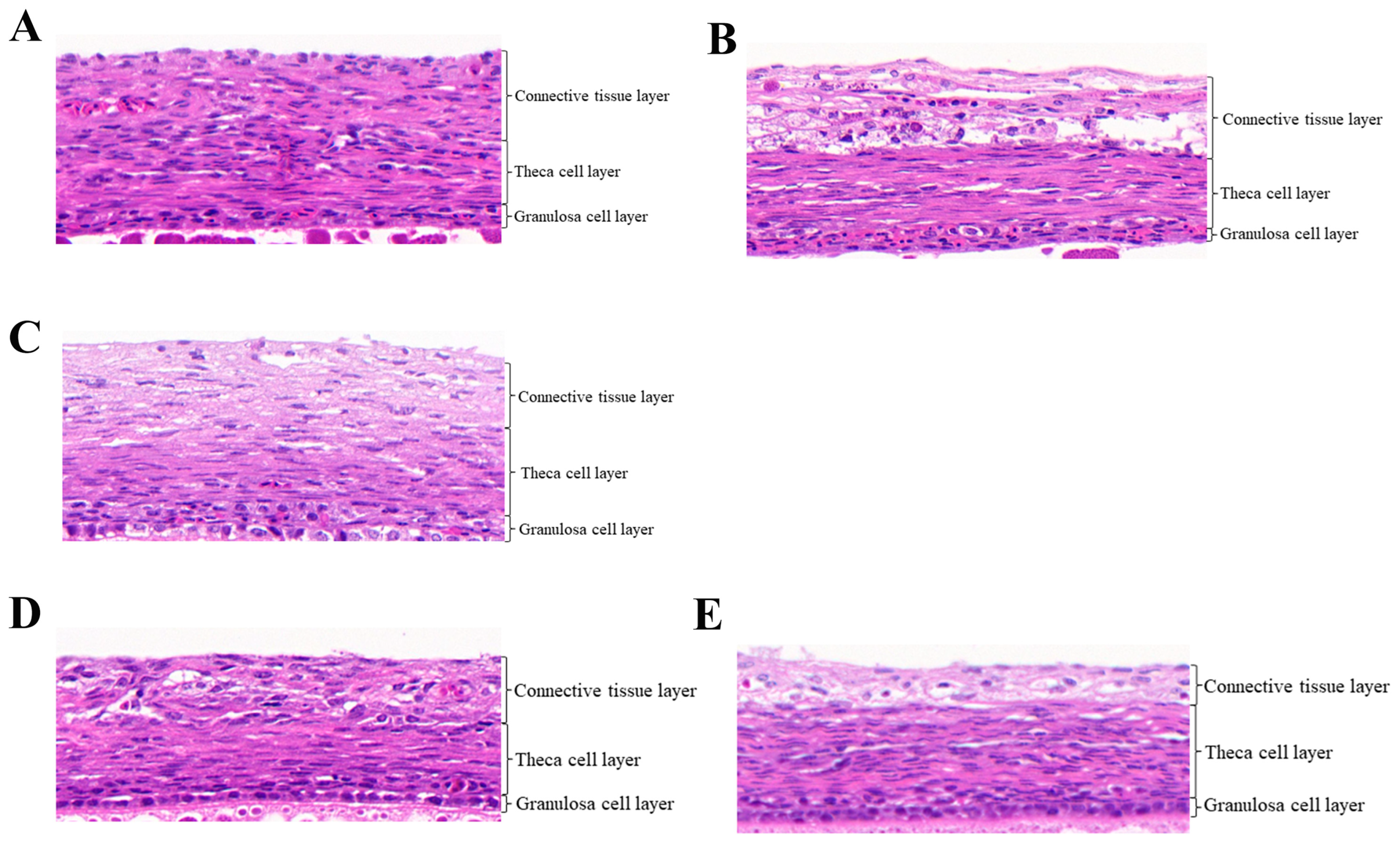
3.1. Histological Observation of Follicles in Pigeon under RL

The thickness of the GC layer, TC layer and CT layer of follicles in pigeons under RL and WL was detected (Figure 1). In the F1 follicles, the thickness of the GC layer in RL was 15.200 μm (Table 2), which was significantly higher than in the WL group (p < 0.05). There were no significant differences in the thickness of the TC and CT layers between the two groups (p > 0.05). In the F2 follicles, the thicknesses were 13.633 μm for the GC layer, 35.667 μm for the TC layer and 49.900 μm for the CT layer. In SF1 follicles, there were no significant differences in the thicknesses of the GC, TC and CT layers between the two light conditions (p > 0.05).

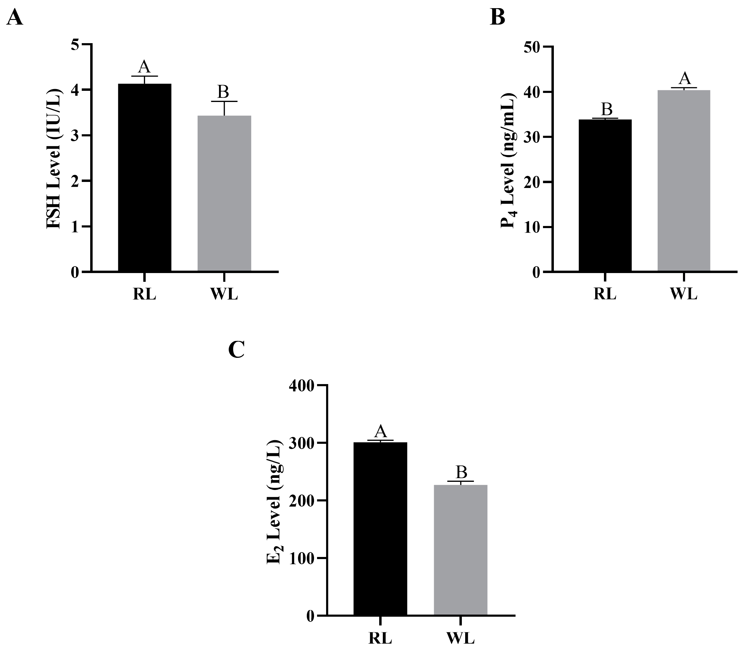
3.2. Concentrations of Reproductive Hormones in Females under RL in LI3

The concentrations of E2, P4 and FSH in the plasma and yolk of females under RL in LI3 were measured. The plasma E2 concentration in the RL group reached 301.12 ng/L, significantly higher than in the WL group (p < 0.05; Figure 2A). Similarly, plasma FSH concentrations followed the same pattern, with RL promoting an increase (p < 0.05; Figure 2C). However, the plasma P4 level was significantly lower in the RL group (p < 0.01; Figure 2B). In the yolk, the concentration of E2 in F1 follicles under RL was 1.83 ng/L higher than under WL, though this difference was not statistically significant (p > 0.05; Figure 3A). The E2 levels in the SF1 yolk showed a similar pattern (p > 0.05), with the F2 yolk E2 levels under RL reaching 36.77 ng/L. The patterns of yolk P4 concentrations mirrored those in the plasma, being significantly higher in the RL group compared to the WL group (p < 0.01; Figure 3B). The FSH concentration in the F1 yolk was significantly higher in the RL group (p < 0.01; Figure 3C), and it was also higher in the SF1 yolk (p > 0.05). Overall, RL appears to positively influence certain reproductive hormone levels, which could impact egg production in pigeons.

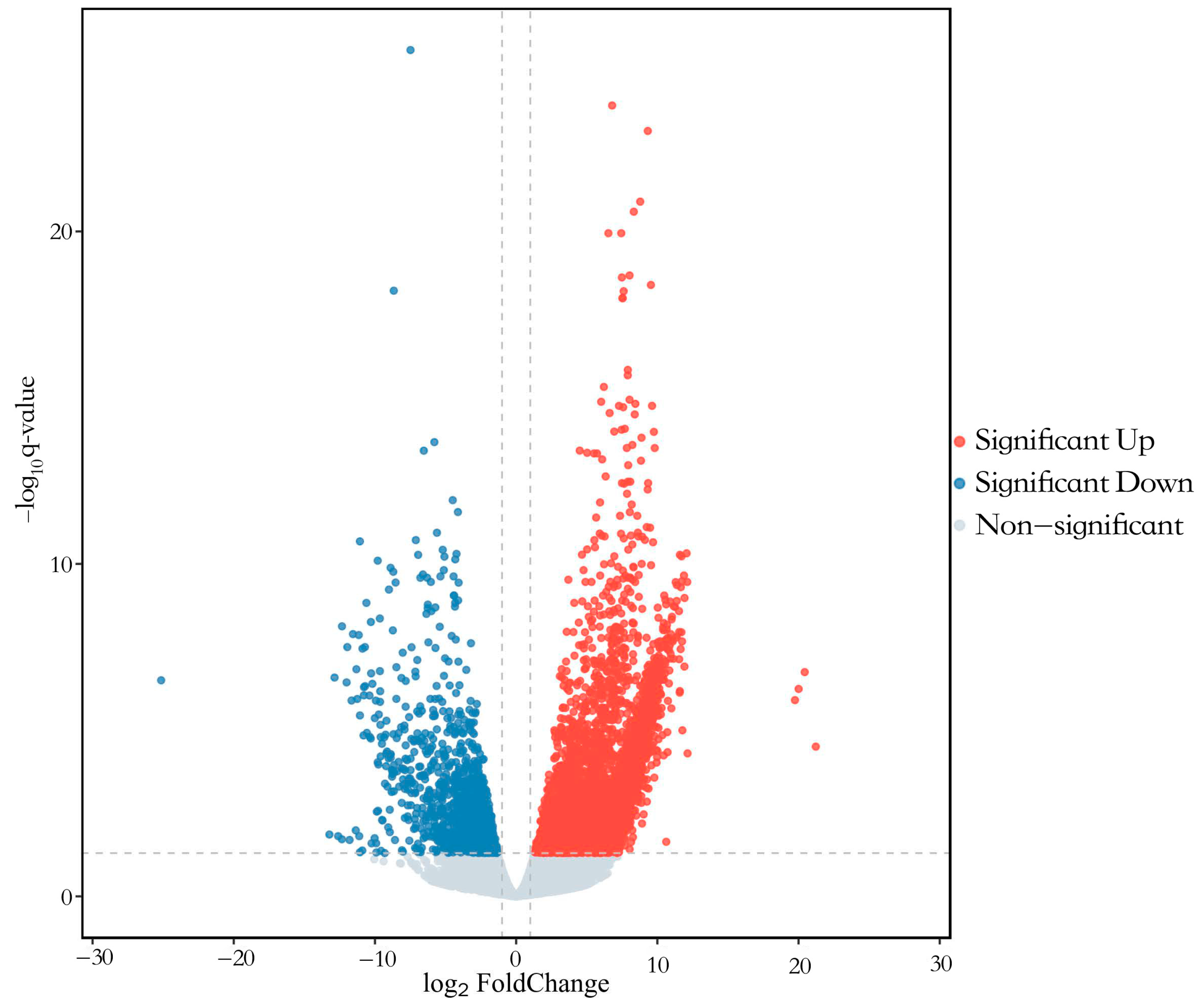
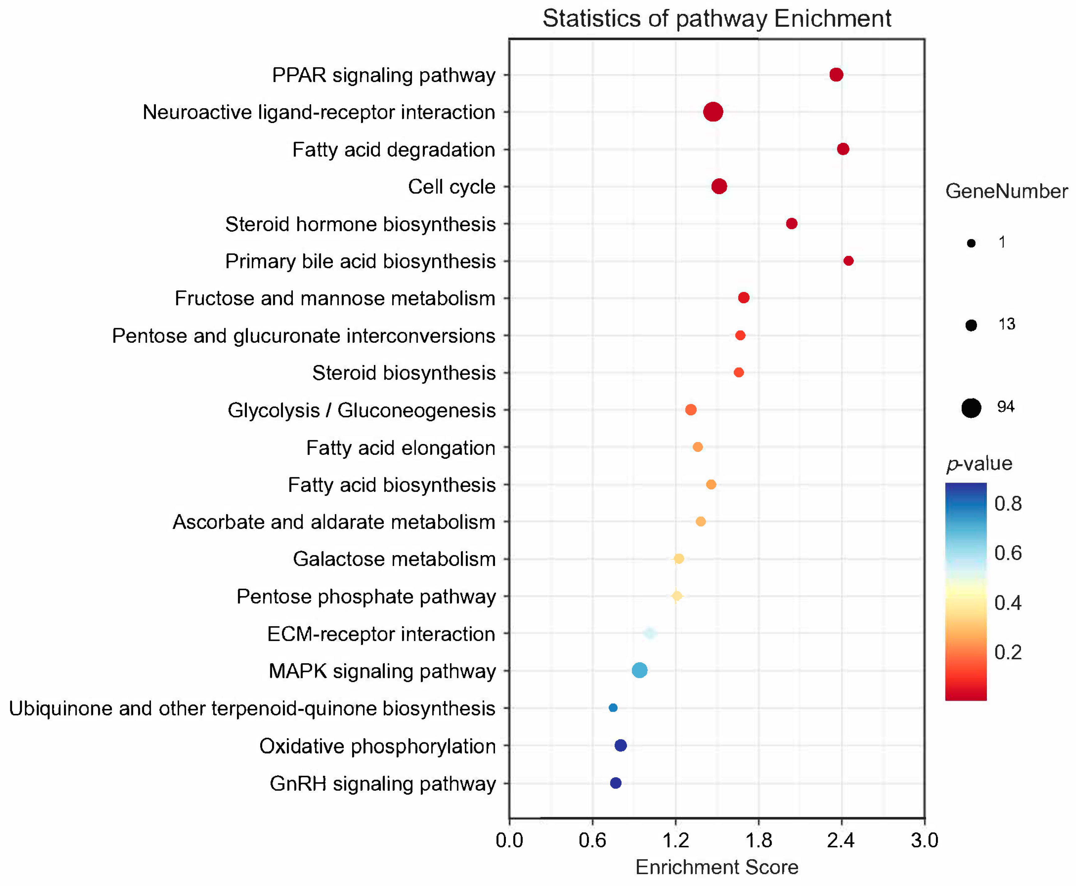
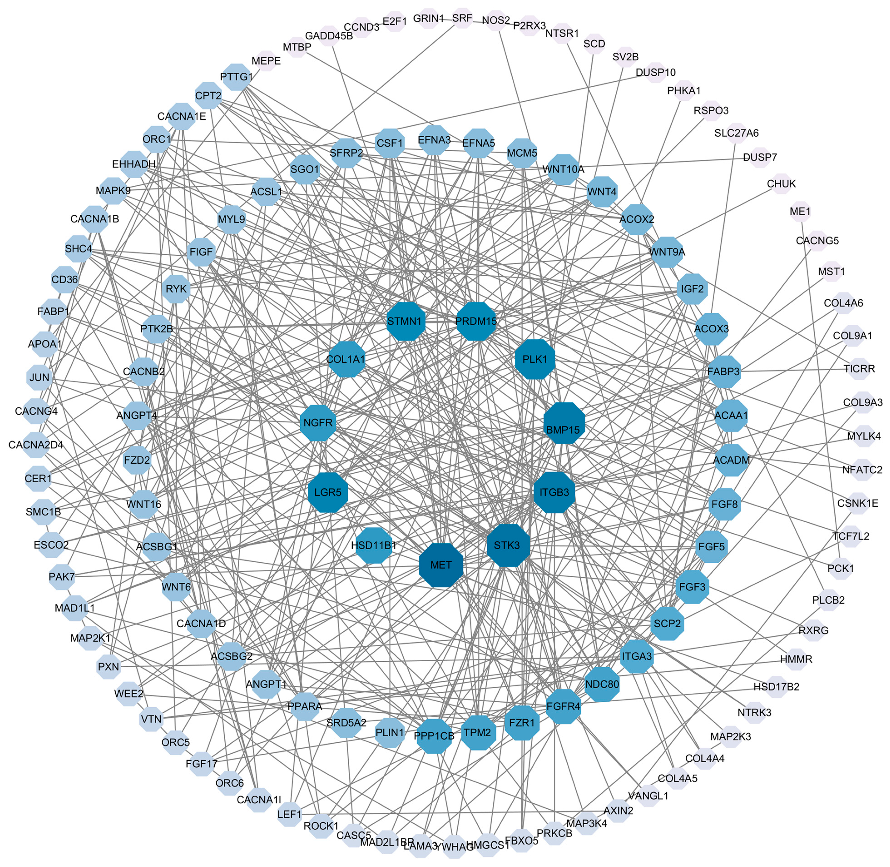
3.3. Transcriptome Analysis of Pigeon Ovary under RL in LI3

Six libraries were constructed, yielding a total of 41.66 Gb of clean bases through high-throughput sequencing (Table 3). The average GC content was 49.20%, with over 93.39% of bases achieving a Q30 quality score. Between 82.07% and 87.11% of reads were successfully mapped to the pigeon reference genome (Columba livia). DEGs were identified in ovaries between the RL and WL groups, with a Fold-Change ≥ 2 and Q-value < 0.05. A total of 4991 genes were differentially expressed, 3769 DEGs were up-regulated and 1222 DEGs were down-regulated (Figure 4). GO analysis classified the DEGs into categories such as biological regulation, cellular processes, metabolic processes, reproduction and reproductive processes for biological processes; cell, cell part, organelle and membrane for cellular components; and binding, catalytic activity, transporter activity and molecular transducer activity for molecular functions (Figure 5). The KEGG analysis of DEGs was categorized into neuroactive ligand–receptor interaction, cell cycle, PPAP signaling pathway, steroid hormone biosynthesis, oxidative phosphorylation and the GnRH signaling pathway (Figure 6). Additionally, a PPI network was constructed, suggesting that genes such as HSD11B1, VEGFD, WNT6, SMAD6 and LGR5 may play roles in follicle selection in pigeons under RL (Figure 7).

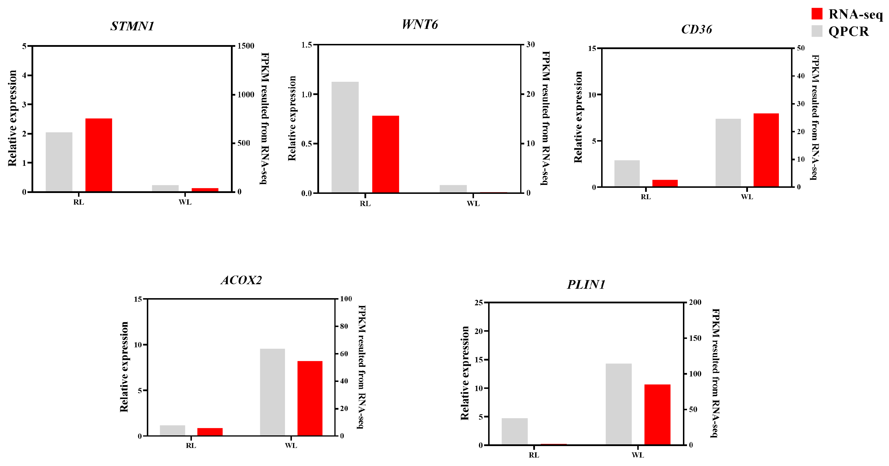
3.4. Validation of Pigeon Ovary Transcriptome

Six DEGs were randomly selected for RT-qPCR to validate the transcriptome data (Figure 8). The expression patterns of these six genes in both the RL and WL groups aligned with the RNA-seq results, further substantiating the accuracy of the transcriptome analysis.

4. Discussion

In our previous research, the second largest follicle (F2) was not selected in LI3 [15]. However, in the current study, F2 was selected in LI3 under RL, suggesting that RL may encourage the selection of hierarchical follicles and thus enhance egg production. The thickness of the GC layer of F1 was significantly higher in RL than in WL, which was in accordance with the higher level of FSH. Yang et al. (2019) [16] concluded that a thicker GC layer aids in the transition of prehierarchical follicles to hierarchical follicles, leading to increased egg production. Similarly, the thicker GC layer of F2 under RL indicates that RL promotes the selection of hierarchical follicles and results in higher egg production.
The initial response of the GC layer to FSH is indicative of its differentiation, which facilitates the synthesis and release of steroids [22,23]. A higher FSH level in the RL group suggests earlier follicular development, and the pattern of E2 changes corresponds with FSH levels. Ma et al. (2020) [24] observed a similar outcome where FSH injections increased plasma E2 levels and expedited follicular development in hens, with higher FSH levels leading to the selection of the second hierarchical follicle in LI3 under RL. Additionally, the FSH concentrations in the follicle yolk were higher in the RL group, indicating accelerated growth of follicles. This aligns with findings by Yang et al. (2019) [19], who reported higher FSH levels in F1 compared to F2 and SYF, consistent with our results. RL stimulated the concentration of E2, which was significantly higher in SF1, as prehierarchical follicles are the primary source of E2 [15,25]. These results indicated that high concentrations of FSH and E2 may facilitate the selection of prehierarchical follicles to hierarchical follicles and enhance egg production under RL. However, the patterns of change in E2 and P4 levels varied under different lighting conditions. Liu et al. (2015) [26] also observed that plasma concentrations of P4 were higher in the WL group than in the RL group. Progesterone produced by GCs might inhibit the production of other steroid hormones [27]. Furthermore, P4 levels were higher in SYF compared to F1 and F2 follicles in Yangzhou geese [19], and the concentration of P4 in SF1 was also higher than in the F1 follicle of pigeons.
The analysis of DEGs was conducted to explore the molecular mechanisms of key genes involved in egg production under RL. The KEGG pathway analysis revealed that processes such as the cell cycle, GnRH signaling pathway and steroid hormone biosynthesis are critical for follicle selection and development. Additionally, a PPI network analysis was used to identify DEGs in core positions, including HSD11B1, VEGFD, WNT6, SMAD6 and LGR5. Notably, 11β-hydroxysteroid dehydrogenase, which includes two enzymes, 11βHSD1 and 11βHSD2, has been identified in bovine ovaries through mRNA studies [28]. Tetsuka et al. (2010) [29] observed that the expression of HSD11B1 mRNA increased as follicles matured in both GC and TC, indicating its importance in follicle development. Latif et al. (2005) [30] found that 11βHSD activity in follicles could be strongly inhibited by progesterone, aligning with the findings of this study where higher concentrations of P4 in serum and follicle yolks were noted in the WL group, correlating with lower expression of HSD11B1. Additionally, vascular changes are a significant aspect of follicle development. In our study, RL inhibited VEGFD mRNA levels; however, Kim et al. (2017) [31] suggested that VEGFD could stimulate early angiogenic events in the primate ovulatory follicle, highlighting that discrepancies in the results may stem from different stages of follicle development. Currently, there is limited research on the expression of VEGFD in relation to follicle development, indicating a need for further investigation. The WNT signaling pathway plays a crucial role in follicle growth, with WNT6 specifically stimulating AMH, which in turn could inhibit the proliferation of GC cells [32,33]. WL significantly stimulated the expression of WNT6 in pigeon ovaries, which may explain the delayed selection of follicles observed under WL conditions. This finding aligns with the known role of the WNT signaling pathway in follicle development. Additionally, LGR5, identified as a potential target of WNT signaling, encodes a G-protein-coupled receptor [34]. This receptor is related to the hormone receptors for FSH and LH, which are critical in regulating reproductive processes. The interaction between WNT signaling and LGR5 suggests a complex network influencing follicle development and hormone interaction [35]. Rastetter et al. (2014) [36] demonstrated that LGR5-positive cells can develop into cortical adult GC. In our study, we observed that RL increased LGR5 mRNA levels, which may accelerate the differentiation of GCs and promote the selection of hierarchical follicles. The SMAD family is divided into receptor-activated, common-partner, or inhibitory SMADs, with SMAD6 functioning as an inhibitor of BMP signaling [37]. Several BMPs are known to act as autocrine/paracrine regulators of ovarian follicle development [38]. Domingues et al. (2023) [39] demonstrated that SMAD6 could significantly inhibit GC proliferation and the rate of follicle growth. Consistently, in our study, SMAD6 was significantly up-regulated in the WL group, which suppressed the proliferation of GCs and delayed the selection of hierarchical follicles. Our results provide a mechanistic insight into how light spectrum can influence follicular dynamics through molecular pathways.

5. Conclusions

In conclusion, our study is the first to present the histological characteristics of pigeon follicles, along with the reproductive hormone levels in plasma and yolk and an ovary transcriptome analysis under RL. RL was found to stimulate the proliferation of GC and TC layers, as well as enhance the secretion of FSH and E2 in both plasma and yolk. This stimulation promotes the selection of hierarchical follicles and increases egg production in pigeons. Additionally, an ovary transcriptome analysis was performed, where 3769 DEGs were up-regulated and 1222 DEGs were down-regulated; furthermore, key genes (HSD11B1, VEGFD, WNT6, SMAD6, and LGR5) involved in follicle selection were identified, providing insights into the molecular mechanisms through which RL improves egg production in pigeons.

Author Contributions

Conceptualization, Y.W., H.Y. and Z.W.; methodology, Y.W., K.Z., C.Z., D.M. and J.C.; validation, C.Z.; writing—original draft preparation, Y.W.; funding acquisition, Y.W. All authors have read and agreed to the published version of the manuscript.

Funding

This work was supported by the National Natural Science Foundation of China (32372875) and the Postgraduate Research & Practice Innovation Program of Jiangsu Province (KYCX24-3811). The funding body did not have any role in the study design, data collection, analysis and interpretation.

Institutional Review Board Statement

The research was approved by the Institutional Animal Care and Use Committee of the Department of Animal Science and Technology, Yangzhou University. It was conducted in line with the Regulations for the Administration of Affairs Concerning Experimental Animals (China, 1988). Additionally, all procedures involving pigeons adhered to the Standards for the Administration of Experimental Practices (Jiangsu, China, 2008).

Informed Consent Statement

Not applicable.

Data Availability Statement

The data were not deposited in an official repository; the accession number can be found below: NCBI; PRJNA1125931. Data are available upon request to the corresponding author.

Acknowledgments

The authors sincerely acknowledge and thank the Taizhou Pigeon Breeding Co., Ltd. for their support.

Conflicts of Interest

The authors declare no conflicts of interest.

References

  1. Kokoszyński, D.; Stęczny, K.; Żochowska-Kujawska, J.; Sobczak, M.; Kotowicz, M.; Saleh, M.; Fik, M.; Arpášová, H.; Hrnčár, C.; Włodarczyk, K. Carcass Characteristics, Physicochemical Properties, and Texture and Microstructure of the Meat and Internal Organs of Carrier and King Pigeons. Animals 2020, 10, 1315. [Google Scholar] [CrossRef]
  2. Jin, C.; He, Y.; Jiang, S.; Wang, X.; Yan, H.; Tan, H.; Gao, C. Chemical composition of pigeon crop milk and factors affecting its production: A review. Poult. Sci. 2023, 102, 102681. [Google Scholar] [CrossRef]
  3. Wang, Y.; Miao, D.Z.; Zhang, C.; Chen, J.; Yang, H.M.; Wang, Z.Y. CircRNAs involved in the red light of effect on follicle selection in pigeons. Poult. Sci. 2024, 103, 104010. [Google Scholar] [CrossRef] [PubMed]
  4. Lewis, P.D.; Morris, T.R. Poultry and coloured light. World Poult. Sci J. 2000, 56, 189–207. [Google Scholar] [CrossRef]
  5. Tsutsui, K.; Bentley, G.E.; Bédécarrats, G.; Osugi, T.; Ubuka, T.; Kriegsfeld, L.J. Gonadotropin-inhibitory hormone (GnIH) and its control of central and peripheral reproductive function. Front. Neuroendocrinol. 2010, 31, 284–295. [Google Scholar] [CrossRef] [PubMed]
  6. Ottinger, M.A.; Abdelnabi, M.; Li, Q.; Chen, K.; Thompson, N.; Harada, N.; Viglietti-Panzica, C.; Panzica, G.C. The Japanese quail: A model for studying reproductive aging of hypothalamic systems. Exp. Gerontol. 2004, 39, 1679–1693. [Google Scholar] [CrossRef]
  7. Stevenson, T.J.; Bernard, D.J.; McCarthy, M.M.; Ball, G.F. Photoperiod-dependent regulation of gonadotropin-releasing hormone 1 messenger ribonucleic acid levels in the songbird brain. Gen. Comp. Endocrinol. 2013, 190, 81–87. [Google Scholar] [CrossRef] [PubMed]
  8. Needham, K.B.; Burns, C.B.; Graham, J.L.; Bauer, C.M.; Kittilson, J.D.; Ketterson, E.D.; Hahn, T.; Greives, T.J. Changes in processes downstream of the hypothalamus are associated with seasonal follicle development in a songbird, the dark-eyed junco (Junco hyemalis). Gen. Comp. Endocrinol. 2019, 270, 103–112. [Google Scholar] [CrossRef]
  9. Baxter, M.; Joseph, N.; Osborne, V.R.; Bédécarrats, G.Y. Red light is necessary to activate the reproductive axis in chickens independently of the retina of the eye. Poult. Sci. 2014, 93, 1289–1297. [Google Scholar] [CrossRef]
  10. Rozenboim, I.; El Halawani, M.E.; Kashash, Y.; Piestun, Y.; Halevy, O. The effect of monochromatic photostimulation on growth and development of broiler birds. Gen. Comp. Endocr. 2013, 190, 214–219. [Google Scholar] [CrossRef]
  11. Gongruttananun, N. Influence of red light on reproductive performance, eggshell ultrastructure, and eye morphology in Thai-native hens. Poult. Sci. 2011, 90, 2855–2863. [Google Scholar] [CrossRef]
  12. Wang, Y.; Li, Y.B.; Yang, H.M.; Wang, Z.Y. Effect of monochromatic lights on egg production, sex hormone levels, and expression of their receptors in pigeons. Livest. Sci. 2018, 216, 233–236. [Google Scholar] [CrossRef]
  13. Wang, Y.; Yang, H.; Zi, C.; Wang, Z. Transcriptomic analysis of the red and green light responses in Columba livia domestica. 3 Biotech 2019, 9, 20. [Google Scholar] [CrossRef]
  14. Chen, Q.; Wang, Y.; Liu, Z.; Guo, X.; Sun, Y.; Kang, L.; Jiang, Y. Transcriptomic and proteomic analyses of ovarian follicles reveal the role of VLDLR in chicken follicle selection. BMC Genom. 2020, 21, 486. [Google Scholar] [CrossRef]
  15. Wang, Y.; Guo, Z.Y.; Zhang, C.; Miao, D.Z.; Mao, X.Y.; Lu, S.M.; Yang, H.M.; Wang, Z.Y. Characterization of ovarian follicles, serum steroid hormone concentration, and steroidogenic gene expression profiles in the developing ovarian follicles in White King pigeons. Poult. Sci. 2023, 102, 102673. [Google Scholar] [CrossRef]
  16. Brady, K.; Liu, H.C.; Hicks, J.; Long, J.A.; Porter, T.E. Global gene expression analysis of the turkey hen hypothalamo-pituitary-gonadal axis during the preovulatory hormonal surge. Poult. Sci. 2023, 102, 102547. [Google Scholar] [CrossRef] [PubMed]
  17. Yan, X.P.; Liu, H.; Hu, J.; Han, X.; Qi, J.; Ouyang, Q.; Hu, B.; He, H.; Li, L.; Wang, J.; et al. Transcriptomic analyses of the HPG axis-related tissues reveals potential candidate genes and regulatory pathways associated with egg production in ducks. BMC Genom. 2022, 23, 281. [Google Scholar] [CrossRef]
  18. Wang, Y.; Wang, J.; Shi, Y.; Ye, H.; Luo, W.; Geng, F. Quantitative proteomic analyses during formation of chicken egg yolk. Food Chem. 2022, 374, 131828. [Google Scholar] [CrossRef] [PubMed]
  19. Yang, Y.Z.; Yao, Y.; Cao, Z.F.; Gu, T.T.; Xu, Q.; Chen, G.H. Histological characteristics of follicles and reproductive hormone secretion during ovarian follicle development in laying geese. Poult. Sci. 2019, 98, 6063–6070. [Google Scholar] [CrossRef]
  20. Miao, D.Z.; Liu, C.; Deng, Z.Y.; Zhang, C.; Guo, Z.Y.; Li, W.Q.; Wang, Y.; Yang, H.M.; Wang, Z.Y. Characterization of reproductive hormones and related gene expression in the hypothalamus and pituitary gland in the egg-laying interval in White King pigeon. Poult. Sci. 2024, 103, 103422. [Google Scholar] [CrossRef]
  21. Gan, X.; Wang, J.W.; Li, Q.; Deng, Y.; Hu, J.W.; Li, L.; Han, C.C. Dynamic development characteristics of mature follicular wall in goose:A new perspective of follicle development and grading. Acta Vet. Zootech. Sin. 2019, 50, 1607–1613. [Google Scholar]
  22. Tilly, J.L.; Kowalski, K.I.; Johnson, A.L. Stage of ovarian follicular development associated with the initiation of steroidogenic competence in avian granulosa cells. Biol. Reprod. 1991, 44, 305–314. [Google Scholar] [CrossRef]
  23. Johnson, A.L. Ovarian follicle selection and granulosa cell differentiation. Poult. Sci. 2015, 94, 781–785. [Google Scholar] [CrossRef] [PubMed]
  24. Ma, Y.; Yao, J.; Zhou, S.; Mi, Y.; Tan, X.; Zhang, C. Enhancing effect of FSH on follicular development through yolk formation and deposition in the low-yield laying chickens. Theriogenology 2020, 157, 418–430. [Google Scholar] [CrossRef] [PubMed]
  25. Senior, B.E.; Furr, B.J.A. A preliminary assessment of the source of oestrogen within the ovary of the domestic fowl, Gallus domesticus. Reproduction 1975, 43, 241–247. [Google Scholar] [CrossRef] [PubMed]
  26. Liu, L.; Li, D.; Gilbert, E.R.; Xiao, Q.; Zhao, X.; Wang, Y.; Yin, H.D.; Zhu, Q. Effect of monochromatic light on expression of estrogen receptor (ER) and progesterone receptor (PR) in ovarian follicles of chicken. PLoS ONE 2015, 10, e0144102. [Google Scholar] [CrossRef] [PubMed]
  27. Woolveridge, I.; Peddie, M.J. The inhibition of androstenedione production in mature thecal cells from the ovary of the domestic hen (Gallus domesticus): Evidence for the involvement of progestins. Steroids 1997, 62, 214–220. [Google Scholar] [CrossRef] [PubMed]
  28. Tetsuka, M.; Yamamoto, S.; Hayashida, N.; Hayashi, K.G.; Hayashi, M.; Acosta, T.J.; Miyamoto, A. Expression of 11beta-hydroxysteroid dehydrogenases in bovine follicle and corpus luteum. J. Endocrinol. 2003, 177, 445–452. [Google Scholar] [CrossRef] [PubMed]
  29. Tetsuka, M.; Nishimoto, H.; Miyamoto, A.; Okuda, K.; Hamano, S. Gene expression of 11b-HSD and glucocorticoid receptor in the bovine (Bos taurus) follicle during follicular maturation and atresia: The role of follicular stimulating hormone. J. Reprod. Dev. 2010, 56, 616–622. [Google Scholar] [CrossRef]
  30. Latif, S.A.; Pardo, H.A.; Hardy, M.P.; Morris, D.J. Endogenous selective inhibitors of 11beta-hydroxysteroid dehydrogenase isoforms 1 and 2 of adrenal origin. Mol. Cell. Endocrinol. 2005, 243, 43–50. [Google Scholar] [CrossRef]
  31. Kim, S.O.; Trau, H.A.; Duffy, D.M. Vascular endothelial growth factors C and D may promote angiogenesis in the primate ovulatory follicle. Biol. Reprod. 2017, 96, 389–400. [Google Scholar] [CrossRef]
  32. Harwood, B.N.; Cross, S.K.; Radford, E.E.; Haac, B.E.; De Vries, W.N. Members of the WNT signaling pathways are widely expressed in mouse ovaries, oocytes, and cleavage stage embryos. Dev. Dyn. 2008, 237, 1099–1111. [Google Scholar] [CrossRef]
  33. Ahmadi, S.; Nemoto, Y.; Ohkubo, T. Impact of In Ovo Leptin Injection and Dietary Protein Levels on Ovarian Growth Markers and Early Folliculogenesis in Post-Hatch Chicks (Gallus domesticus). Biology 2024, 13, 69. [Google Scholar] [CrossRef] [PubMed]
  34. Van de Wetering, M.; Sancho, E.; Verweij, C.; de Lau, W.; Oving, I.; Hurlstone, A.; van der Horn, K.; Batlle, E.; Coudreuse, D.; Haramis, A.P.; et al. The beta-catenin/TCF-4 complex imposes a crypt progenitor phenotype on colorectal cancer cells. Cell 2002, 111, 241–250. [Google Scholar] [CrossRef]
  35. Hermey, G.; Methner, A.; Schaller, H.C.; Hermans-Borgmeyer, I. Identification of a novel seven-transmembrane receptor with homology to glycoprotein receptors and its expression in the adult and developing mouse. Biochem. Biophys. Res. Commun. 1999, 254, 273–279. [Google Scholar] [CrossRef] [PubMed]
  36. Rastetter, R.H.; Bernard, P.; Palmer, J.S.; Chassot, A.A.; Chen, H.; Western, P.S.; Ramsay, R.G.; Chaboissier, M.C.; Wilhelm, D. Marker genes identify three somatic cell types in the fetal mouse ovary. Dev. Biol. 2014, 394, 242–252. [Google Scholar] [CrossRef]
  37. Kaivo-oja, N.; Jeffery, L.A.; Ritvos, O.; Mottershead, D.G. Smad signalling in the ovary. Reprod Biol Endocrin. 2006, 4, 21. [Google Scholar] [CrossRef] [PubMed]
  38. Glister, C.; Kemp, C.F.; Knight, P.G. Bone morphogenetic protein (BMP) ligands and receptors in bovine ovarian follicle cells: Actions of BMP-4,-6 and-7 on granulosa cells and differential modulation of Smad-1 phosphorylation by follistatin. Reproduction 2004, 127, 239–254. [Google Scholar] [CrossRef]
  39. Domingues, R.R.; Andrade, F.S.; Andrade, J.P.N.; Moghbeli, S.M.; Gomez-Leon, V.; Madureira, G.; Mello, M.R.B.; Kirkpatrick, B.W.; Wiltbank, M.C. SMAD6 inhibits granulosa cell proliferation and follicle growth rate in carrier and noncarrier heifers of the Trio allele. Reproduction 2023, 165, 269–279. [Google Scholar] [CrossRef]
Figure 1. Histological characteristics of pigeon follicles under red light. (A) F1 follicle under RL. (B) F1 follicle under WL. (C) F2 follicle under RL. (D) SF1 follicle under RL. (E) SF1 under WL.
Animals 14 02320 g001
Figure 2. The concentration of estradiol (E2, df = 16, A), progesterone (P4, df = 16, B) and follicle-stimulating hormone (FSH, df = 9, C) in pigeon plasma under red light. Values marked with different capital letters on the bars are extremely significantly different (p < 0.01).
Animals 14 02320 g002
Figure 3. The concentration of estradiol (E2, df = 4, A), progesterone (P4, df = 4, B) and follicle-stimulating hormone (FSH, df = 4, C) in pigeon follicle yolk under red light. Values marked with different capital letters on the bars are extremely significantly different (p < 0.01).
Animals 14 02320 g003
Figure 4. The volcano plot of differentially expressed genes.
Animals 14 02320 g004
Figure 5. GO enrichment analysis of differentially expressed genes.
Animals 14 02320 g005
Figure 6. KEGG enrichment analysis of differentially expressed genes.
Animals 14 02320 g006
Figure 7. Protein–protein interaction (PPI) network of differentially expressed genes.
Animals 14 02320 g007
Figure 8. The validation of RNA-Seq using RT-qPCR.
Animals 14 02320 g008
Table 1. Primers for RT-qPCR.
GenePrimer Sequence (5′→3′)Length
STMN1F: ATGCTGAATATCTGTTACACGTC
R: CCATTTTGTTCCGCGTGTC
130 bp
WNT6F: CCAGCAGTTCATGGATGCCAA
R: AAACGTCCGGCTTCATTGTTG
169 bp
CD36F: CCCAAAGAAAATATCACGGAA
R: ATATCAGGTTCAAAACGAGCAA
80 bp
AOCX2F: AAGTGAACGCCACACGTCT
R: CGGTTACCACTCAGCATCGCTTG
169 bp
ANGPT4F: TCTACACCCTGCACATCACC
R: TCCATGTCGCAGTACGCCTT
58 bp
CACNB2F: GACGCTGATACCATTAACCAC
R: TACATCAAACATTTCGGGAGG
171 bp
GAPDHF: CTCTACTCATGGCCACTTCCG
R: ACAACGTATTCAGCACCAGC
138 bp
Table 2. The thickness of the follicular membrane in pigeon follicles in the RL and WL group.
Group 1Granulosa Cell Layer
(μm)
Theca Cell Layer
(μm)
Connective Tissue Layer
(μm)
RLF1 215.200 ± 1.210 a41.866 ± 2.57135.664 ± 4.276
WLF18.767 ± 0.736 b38.433 ± 2.44449.267 ± 6.631
RLF213.633 ± 0.88435.667 ± 2.26749.900 ± 1.513
RLSF17.100 ± 0.45137.267 ± 4.03732.500 ± 3.843
WLSF17.133 ± 0.93930.700 ± 3.00940.100 ± 1.815
1 Values marked with different small letters on the bars are significantly different (p < 0.05). 2 Abbreviations: RLF1 = the largest follicle in the red light group; WLF1 = the largest follicle in the white light group; the df of follicular membrane of RLF1 vs. WLF1 is 4; RLF2 = the second largest follicle in the red light group; RLSF1 = the largest small follicle in the red light group; WLSF1 = the largest small follicle in the white light group; the df of the follicular membrane of RLSF1 vs. WLSF1 is 4.
Table 3. Summary of the sequences’ assembly from pigeon ovaries under red light.
Samples 1Raw Read/MClean Reads/MClean Bases/GMapped Reads/%Q30/%GC/%
RO3-148.9547.016.8783.3394.4549.92
RO3-250.3847.886.9483.9294.3648.95
RO3-348.2047.226.9587.1196.0448.85
WO3-151.2448.396.9984.2693.3949.31
WO3-252.8149.157.0182.0794.0749.16
WO3-348.6647.236.9086.5796.3449.00
1 Abbreviations: RO3 = RNA-seq library constructed from pigeon ovaries raised under red light; WO3 = RNA-seq library constructed from pigeon ovaries raised under white light.
Disclaimer/Publisher’s Note: The statements, opinions and data contained in all publications are solely those of the individual author(s) and contributor(s) and not of MDPI and/or the editor(s). MDPI and/or the editor(s) disclaim responsibility for any injury to people or property resulting from any ideas, methods, instructions or products referred to in the content.

