Article

Evaluation of Reference Gene Stability in Goat Skeletal Muscle Satellite Cells during Proliferation and Differentiation Phases

1
Key Laboratory of Livestock and Poultry Multi-Omics, Ministry of Agriculture and Rural Affairs, College of Animal and Technology, Sichuan Agricultural University, Chengdu 611130, China
2
Farm Animal Genetic Resources Exploration and Innovation Key Laboratory of Sichuan Province, Sichuan Agricultural University, Chengdu 611130, China
*
Author to whom correspondence should be addressed.
These authors contributed equally to this work.
Animals 2024, 14(17), 2479; https://doi.org/10.3390/ani14172479
Submission received: 23 July 2024 / Revised: 16 August 2024 / Accepted: 22 August 2024 / Published: 26 August 2024
(This article belongs to the Section Small Ruminants)

Abstract

:

Simple Summary

Investigating the proliferation and differentiation of skeletal muscle satellite cells (MuSCs) in vitro is crucial for elucidating the underlying mechanisms of skeletal muscle development in goats. Real-time quantitative PCR (RT-qPCR) is a widely utilized technique for quantifying the expression levels of target genes. A significant challenge associated with this method is the identification of optimal reference genes for accurate normalization. In this study, we evaluated ten candidate reference genes for the standardization of gene expression. Our results indicated that RPL14 and RPS15A constituted the most stable reference gene combination during the proliferation and differentiation of goat MuSCs.

Abstract

The process of skeletal muscle development is intricate and involves the regulation of a diverse array of genes. Accurate gene expression profiles are crucial for studying muscle development, making it essential to choose the right reference genes for real-time quantitative PCR (RT-qPCR). In the present study, eight candidate reference genes were identified from our previous transcriptome sequencing analysis of caprine skeletal muscle satellite cells (MuSCs), and two traditional reference genes (ACTB and GAPDH) were assessed. The quantitative levels of the candidate reference genes were determined through the RT-qPCR technique, while the stability of their expression was evaluated utilizing the GeNorm, NormFinder, BestKeeper, and RefFinder programs. Furthermore, the chosen reference genes were utilized for the normalization of the gene expression levels of PCNA and Myf5. It was determined that conventional reference genes, including ACTB and GAPDH, were not appropriate for normalizing target gene expression. Conversely, RPL14 and RPS15A, identified through RNA sequencing analysis, exhibited minimal variability and were identified as the optimal reference genes for normalizing gene expression during the proliferation and differentiation of goat MuSCs. Our research offers a validated panel of optimal reference genes for the detection of differentially expressed genes in goat muscle satellite cells using RT-qPCR.

1. Introduction

Goat is an economically important animal in many communities worldwide, providing meat and milk products with abundant nutritional value [1]. The intricate process of skeletal muscle growth and development encompasses the activation of skeletal muscle satellite cells (MuSCs), the proliferation and differentiation of myoblasts, and the fusion of myoblasts into myofibers, along with the expression of a diverse array of genes [2,3,4]. The examination of gene expression patterns in goat muscle tissues and cells is crucial for identifying pivotal functional genes that impact meat production traits. This knowledge is imperative for marker-assisted selection (MAS) investigations and for improving breeding initiatives.
The RT-qPCR technique is commonly utilized for assessing gene expression due to its robust specificity, heightened sensitivity, and reliable repeatability [5,6,7]. Nonetheless, the accuracy of RT-qPCR results is heavily reliant on the stability of reference genes [8]. An optimal reference gene is anticipated to exhibit consistent expression levels across diverse experimental conditions, including varying developmental stages, tissue types, and cell types. Nevertheless, the universality of reference gene expression is not absolute, as variations can be observed between different tissues, cell types, and stages of cellular growth [9,10,11,12]. The absence of a universally applicable reference gene necessitates careful consideration in selecting an appropriate reference gene for specific cell and tissue types to avoid potential inaccuracies in results and conclusions [13]. Therefore, the selection of suitable reference genes is crucial for an accurate calibration and normalization of target gene expression under specific experimental conditions. Currently, there is a limited number of systematic studies on reference genes in goat muscle cells, despite various scholars utilizing different algorithms to assess reference genes suitable for livestock muscle tissues [14,15,16,17,18]. Furthermore, the internal regulatory mechanism underlying myogenesis in goats remains poorly understood. Therefore, it is crucial to identify appropriate reference genes in order to investigate the internal regulatory mechanism driving this process further.
In this study, ten reference genes (HSPA9, DDOST, RPL5, CAPNS1, YBX1, EEF1G, RPS15A, RPL14, GAPDH, and ACTB) in goat skeletal muscle satellite cells (MuSCs) were examined during the proliferative phase (GM phase) and differentiation phases at day 1 (DM1) and day 5 (DM5). The expression stability of these candidate reference genes was assessed using three established algorithms (GeNorm [19], NormFinder [20], Bestkeeper [21], and RefFinder [22,23]) to gain insights into the proliferation and differentiation processes of goat skeletal muscle satellite cell models in vitro.

2. Materials and Methods

2.1. Ethics Statement

The Animal Care and Use Committee of the College of Animal Science and Technology, Sichuan Agricultural University, Sichuan, Chengdu, China, approved all of the animal care, slaughter, and experimental procedures in accordance with the Regulations for the Administration of Affairs Concerning Experimental Animals (Ministry of Science and Technology, China) [Approval No. SAU2022302131].

2.2. Isolation and Culture of Goat MuSCs

The primary MuSCs were isolated and cultured from longissimus dorsi muscle derived from a fetal goat (Chengdu Ma goat, female, n = 1), as described previously [24,25]. The cells were cultured in DMEM/F12 (Gibco, Shanghai, China) in a 5% CO2 and 95% oxygen incubator at 37 °C, supplemented with 10% FBS (Gibco, Shanghai, China) and 1% penicillin/streptomycin (Solarbio, Beijing, China). When the confluence reached about 80–90%, the MuSCs were seeded into six-well plates. The cells were collected when they achieved about 80–90% confluence in six-well plates in growth medium with three biological replicates, deemed as the proliferation phase (GM). When the cells reached about 80–90% confluence, we replaced the growth medium with differentiation medium containing DMEM/F12, 2% horse serum (Gibco, Shanghai, China), and 1% penicillin/streptomycin. The cells were collected on day 1 (DM1) and day 5 (DM5), with three biological replicates at each time point.

2.3. Selection of Candidate Reference Genes

Candidate reference genes were identified from RNA-seq data (PRJNA779184) of goat skeletal muscle satellite cells cultured under proliferation (GM) and differentiation (DM1/DM5) conditions. The selection process was based on the values of the coefficient of variation (CV, %) and fragments per kb per million reads (FPKM). Genes meeting the criteria of FPKM > 100 and CV < 10% were ranked in ascending order based on CV values, with the top eight genes chosen as candidate reference genes. Additionally, two traditional reference genes, GAPDH and ACTB, were also included in the analysis.

2.4. RNA Extraction and cDNA Synthesis

Total RNA was extracted from goat MuSCs using the Orizol Chloroform-Free RNA Extraction Kit (Oriscience, Chengdu, China) according to instructions. The integrity and concentration of the RNA were determined by agarose gel electrophoresis (Bio-Rad, Richmond, VA, USA) and a NanoDrop 2000 spectrophotometer (Thermo Scientific, Waltham, MA, USA). Then, the cDNA was synthesized using HiScript IV RT SuperMix for qPCR (+gDNA wiper) (Vazyme, Nanjing, China) following the instructions and stored at −20 °C for backup.

2.5. RT-qPCR Analysis

The primers for ten reference genes (HSPA9, DDOST, RPL5, CAPNS1, YBX1, EEF1G, RPS15A, RPL14, GAPDH, and ACTB) were designed using the Primer-Blast program (NCBI tools). The sequences of the primer pairs are outlined in Supplementary Table S1. Agarose gel electrophoresis and melting curve analysis were conducted to assess the specificity of the primer pairs. The RT-qPCR was performed on the CFX96 Real-Time PCR Detection System (Bio-Rad, Hercules, CA, USA) using 2× M5 HiPer SYBR Permix EsTaq (Mei5bio, Beijing, China). Three biological replicates were performed, and each RT-qPCR was performed in technical triplicates. The reaction conditions included an initial denaturation at 95 °C for 3 min, followed by 39 cycles of denaturation at 95 °C for 30 s and annealing at a specific temperature for 30 s. The standard curve of the RT-qPCR was established using the gradient-diluted cDNA. The correlation coefficient and amplification efficiency were calculated using the CFX Manager Software version 3.1 (Bio-Rad, Hercules, CA, USA). The cycle quantification (Cq) values were automatically generated using the default settings of the Real-Time System.

2.6. Stability Analysis of Reference Genes

The expression stability of selected reference genes were evaluated using three programs: GeNorm, NormFinder, and Bestkeeper, following the instructions. The relative expression quantity (Q) of each candidate reference gene was calculated as follows: Q = 2−ΔCq, ∆Cq = Cq (sample) − Cq (minimum), where Cq (sample) was the Cq value of a factor in each sample and Cq (minimum) was the minimum Cq value of this gene in all samples [26].
The GeNorm algorithm employs pairwise comparisons to assess each gene’s expression stability, represented by an M value. A lower M value suggests higher stability. GeNorm was also used to determine the minimum number of reference genes required for accurate normalization. NormFinder was used to calculate stability values for candidate reference genes by analyzing their intragroup and intergroup variation. The reference gene with the lowest stability value was considered to be the most stable gene. BestKeeper was used to analyze the expression stability of candidate reference genes by calculating the coefficient of variance (CV) and the standard deviation (SD) based on the raw Cq values. The reference genes with the highest stability had the lowest values of CV and SD, while harboring the highest value of correlation coefficient (r).

2.7. Validation of Reference Gene Expression

Target genes were chosen to validate the stability of the reference gene by comparing their expression patterns after normalization. PCNA and Myf5 were selected as target genes because they are the marker genes for MuSC proliferation and differentiation. The expression of target genes was normalized using the most stable and the least stable reference genes. All samples were evaluated in triplicate, and their relative expression levels were calculated using the 2−ΔΔCt method [27].

2.8. Statistical Analysis

The results are expressed as means ± standard error of the mean (SEM). All data were evaluated using one-way ANOVA and Duncan’s new multiple range tests using SAS software version 9.2 (SAS, Cary, NC, USA), and p-values lower than 0.05 were considered statistically significant.

3. Results

3.1. Proliferation and Differentiation of Goat MuSCs In Vitro

Quiescent MuSCs were converted to myoblasts and subsequently allowed to proliferate in growth medium (GM) until reaching approximately 80% confluence, marking the proliferation phase as depicted in Figure 1A. Following the replacement of GM with differentiation medium (DM), the cells differentiated into elongated myocytes within one day (Figure 1B). By the fifth day, the myocytes had fused, forming long, multinucleated myotubes (Figure 1C). Cells from the GM, DM1, and DM5 phases were harvested for further experimentation.

3.2. RNA Purity, Primer Verification, and Amplification Efficiency

The RNA samples exhibited OD260/280 ratios ranging from 1.99 to 2.06 and RNA integrity number (RIN) values ranging from 8.2 to 9.4 (Supplementary Table S2), suggesting high quality and suitability for subsequent experimentation. Analysis of Figures S1 and S2 revealed successful amplification of a single band of expected size on agarose gel, as well as detection of a single peak in the melting curve for each primer pair, indicating high specificity in amplifying the target fragment across all 10 primer pairs. Furthermore, the standard curves of the reference genes that were tested exhibited strong linear relationships, with amplification efficiency falling within the range of 90.0% to 103.7% and coefficients of determination (R2) ranging from 0.982 to 0.999 (Supplementary Table S1). These results suggest that the primers performed effectively in the RT-qPCR amplification conditions, producing precise and dependable outcomes.

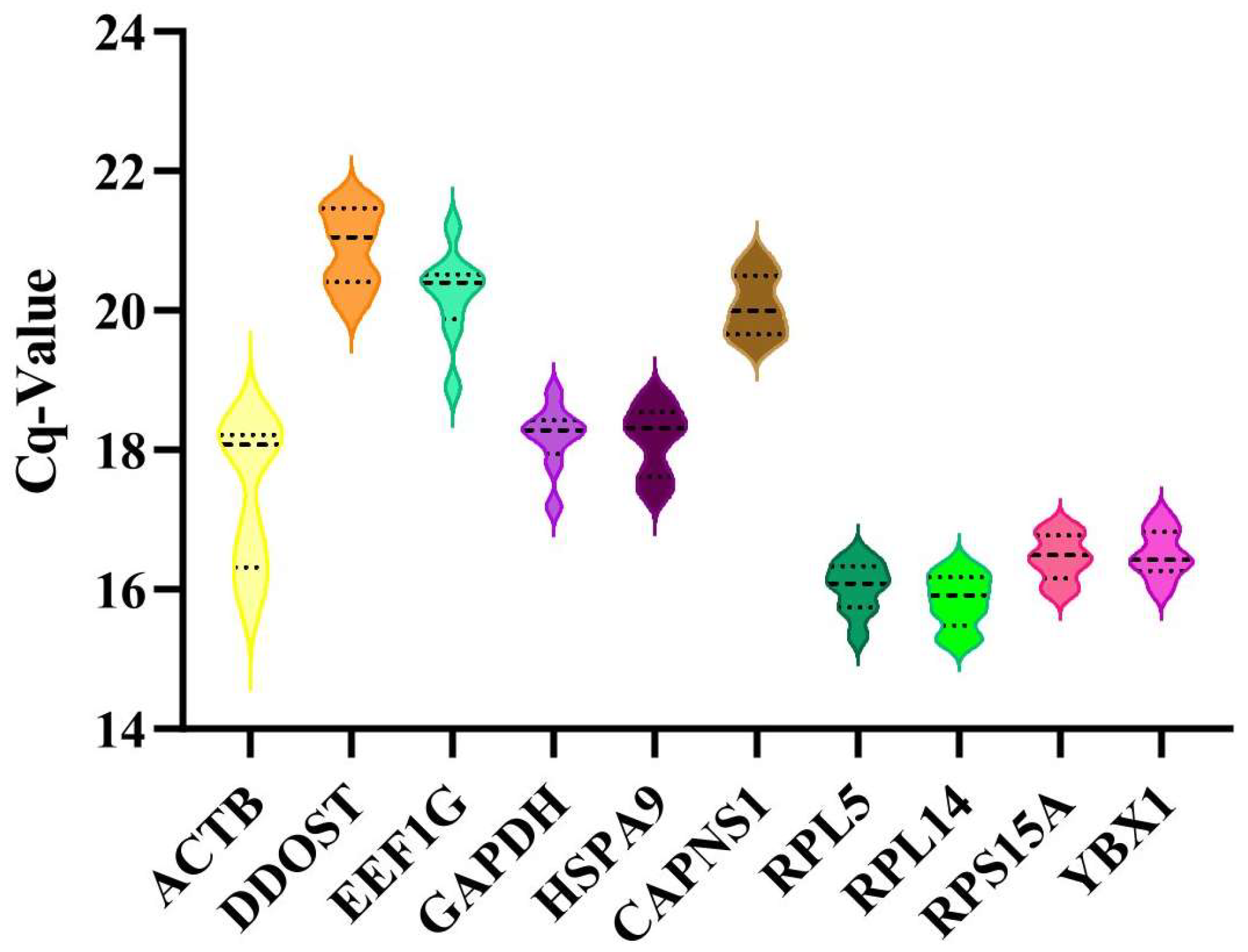
3.3. Analysis of the Expression Levels of the Candidate Reference Genes

The average Cq values of candidate reference genes in all cDNA samples ranged from 15.27 to 20.75 (Figure 2). A lower dispersion of the Cq value indicates higher stability of the gene. Specifically, RPL14 showed the lowest Cq dispersion (Cq values ranged from 15.27 to 16.34), followed by RPL5, RPS15A, and YBX1, while ACTB exhibited the highest variation (Cq values ranged from 15.79 to 18.46). These findings suggest that RPL14 is the most stable gene in terms of mRNA expression levels, whereas ACTB is the least stable.

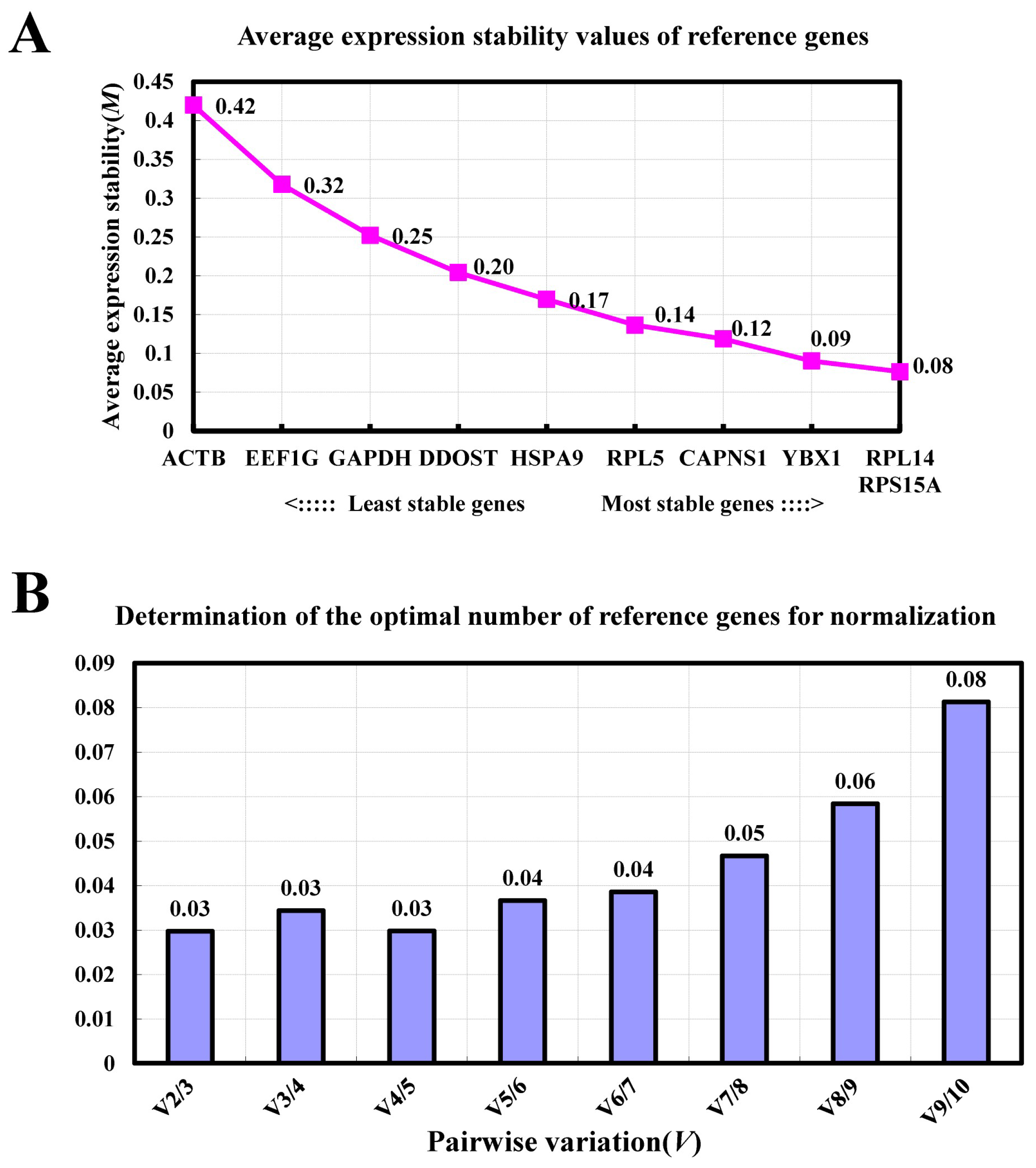
3.4. GeNorm Analysis

The GeNorm program was utilized to calculate the expression stability value (M) of reference genes, with a smaller M value, indicating greater stability. Results revealed that RPL14 and RPS15A exhibited the lowest M values, while ACTB had the highest (Figure 3A). This suggests that RPL14 and RPS15A were the most stable internal reference genes, whereas ACTB was the least stable. Additionally, we also used GeNorm to analyze the optimal number of reference genes required in this study. GeNorm analysis determined that two reference genes were adequate for accurate normalization of target gene expression, as indicated by V2/3 = 0.03 < 0.15 (Figure 3B).

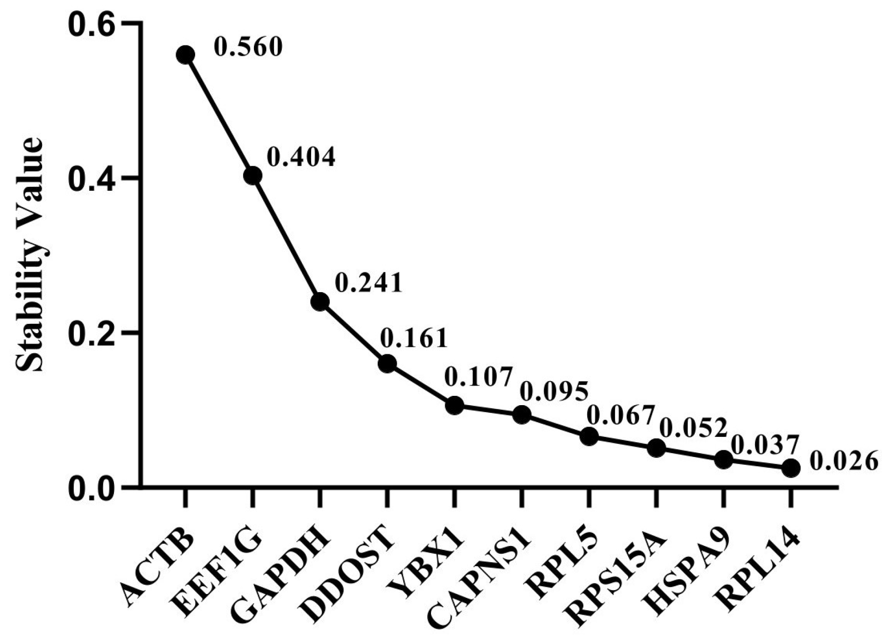
3.5. NormFinder Analysis

The reference genes with the lowest stability values, as determined by the NormFinder method, were deemed to be the most stable. The stability of ten candidate reference genes were then ranked based on the results of the NormFinder analysis (Figure 4). The two most stable reference genes screened by the software were RPL14 (0.026) and HSPA9 (0.037), and ACTB (0.560) was the least stable reference gene.

3.6. Bestkeeper Analysis

Bestkeeper software (version 1) estimates the stability of reference genes based on the coefficient of variation (CV) and standard deviation (SD). A lower CV or SD value indicates a more stable reference gene. In this study, the most stable reference genes (RPS15A and YBX1) and the least stable gene (ACTB) were identified based on stability rankings (Table 1).

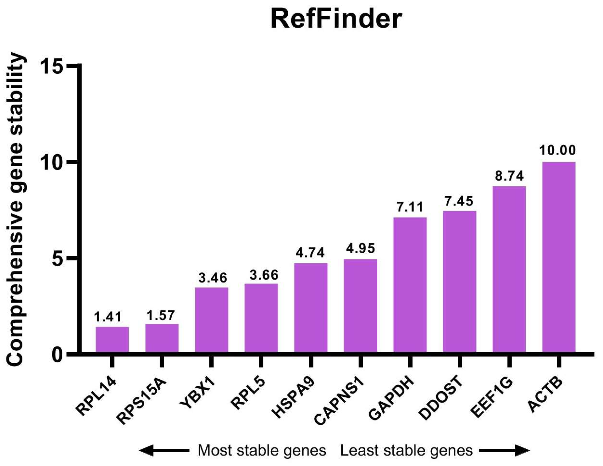
3.7. Comprehensive Analysis of Candidate Reference Genes

To combine the results of analysis by the three programs, the stability ranks of the candidate reference genes were calculated and ranked by geometric mean (Table 2) and RefFinder program (Figure 5). According to the comprehensive stability rankings, RPL14 and RPS15A were the two most stable reference genes, and ACTB was the least stable reference gene. Therefore, RPL14 and RPS15A were the most appropriate reference genes to normalize the expression of target genes during the proliferation and differentiation phases of goat MuSCs in vitro.

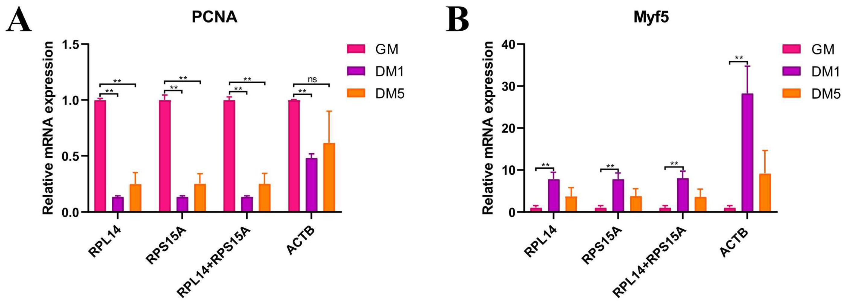
3.8. Expression Validation of Candidate Reference Genes Using Target Genes

In order to further validate the selection of candidate reference genes, the most stable reference genes (RPL14 and RPS15A) as well as the least stable reference gene (ACTB) were utilized to normalize the same target genes. The mRNA expression of PCNA in goat MuSC during the proliferation phase (GM) and differentiation phases (DM1 and DM5) showed a significant difference (p < 0.01) when normalized using RPL14 and RPS15A either individually or in combination. However, there was no significant difference observed between GM and DM5 when normalized using ACTB (Figure 6A). During the transition from the proliferation phase to the differentiation phases of goat muscle satellite cells (MuSCs), the mRNA expression of Myf5 exhibited an initial increase followed by a decrease when normalized using RPL14 and RPS15A as reference genes, either individually or in combination (Figure 6B). Conversely, normalization using ACTB as the reference gene resulted in an anomalous increase in Myf5 mRNA expression between the GM phase and the DM1 phase (Figure 6B). This discrepancy underscores the potential for misinterpretation of target gene expression when inappropriate reference genes are employed. Consequently, the selection of stable and appropriate reference genes is crucial for the accurate normalization of relative gene expression levels.

4. Discussion

The analysis of gene expression is a common measurement in molecular studies, and the current gold standard protocol is RT-qPCR [28,29,30,31,32]. This method requires an accurate and reliable reference gene to standardize the relative expression level of specific target genes [33]. In this regard, the significance of reference genes for the accurate analysis of target gene expression is well established. Ideal reference genes are expected to exhibit stable expression levels across all experimental conditions as well as in various tissues or cell types. However, it has been clearly demonstrated that there is no universal reference gene that maintains stable expression under all conditions [34,35]. In most studies of MuSCs, reference genes are typically selected based on literature reviews and prior experience with other organisms or tissues rather than on empirical evidence supporting their efficacy. Consequently, it is generally recommended that reference genes be validated for each species and specific experimental conditions. This validation is essential for the accurate measurement of gene expression using RT-qPCR, as the selection of inappropriate reference genes may lead to inaccurate values or even contradictory results [36,37,38].
Transcriptome sequencing is a vital research method for analyzing gene expression and is employed to identify differentially expressed and functional genes. Notably, utilizing transcriptome data to screen for reference genes is an effective experimental approach for selecting reference genes in non-model species [39]. In this study, eight candidate reference genes were identified based on our previous RNA-seq data of goat skeletal muscle satellite cells (MuSCs). Additionally, two traditional reference genes were assessed. The Cq values of the candidate reference genes were determined using RT-qPCR, and the stability of their expression was evaluated utilizing the GeNorm, NormFinder, BestKeeper, and RefFinder programs. Our study comprehensively identified RPL14 and RPS15A as the two most stable reference genes during the proliferation and differentiation of goat MuSCs. These genes exhibited greater stability compared to GAPDH and ACTB, indicating that traditional reference genes may not be suitable for certain situations. Previous studies have demonstrated that GAPDH and ACTB are not suitable for the normalization of skeletal muscle development in cattle, with analogous findings reported in pigs, goats, and mice [18,40,41,42], corroborating the results of the present study. Furthermore, our comprehensive ranking analysis indicates that GAPDH exhibits greater stability than ACTB during in vitro differentiation of goat muscle satellite cells. RPL14 and RPS15A are members of the ribosomal protein family. Ribosomal proteins play crucial housekeeping roles in ribosomal biogenesis and protein synthesis and are essential for cell growth, proliferation, differentiation, and development [43,44]. RPL32 and RPS18 have been identified as suitable reference genes for the development of skeletal muscle in pigs [18]. Additionally, a study suggested that RPS4X and RPS6 are stably expressed during rumen development in goats [45]. RPS15A has been determined to be the most appropriate reference gene during the proliferation and differentiation phases of bovine skeletal muscle satellite cells in vitro [46]. Previous studies have demonstrated that RPLP0 is one of the reference genes proposed for data normalization in bovine muscle [47]. RPL15 has been identified as the most stable reference gene in bovine oocytes collected during both winter and summer [48], whereas RPL4 has shown the highest stability as a reference gene during the differentiation of bovine bone marrow mesenchymal stem cells [49]. Additionally, RL13A has been consistently expressed as a stable reference gene for gene expression normalization across various muscle tissues in domestic yaks [50]. Consistent with the results of the present study, these findings suggest that ribosomal protein family genes have the potential to serve as stable internal reference genes. However, it has also been demonstrated that, despite their stability, ribosomal protein family genes may not be universally reliable as reference genes [51]. Therefore, it is essential to evaluate the expression stability of these genes under specific experimental conditions.
To our knowledge, this study represents the first validation of the expression stability of reference genes during the proliferation and differentiation phases of goat muscle satellite cells in vitro. Furthermore, we determined the optimal combination of stably expressed reference genes and identified the least stable ones for use during the in vitro proliferation and differentiation induction of goat MuSCs. Our findings provide a critical reference for selecting appropriate reference genes for gene expression analysis via RT-qPCR in future studies involving goat MuSCs.

5. Conclusions

In summary, RPL14 and RPS15A were identified as the most suitable reference genes for RT-qPCR experiments in goat MuSCs during proliferation and differentiation in vitro. Furthermore, utilizing a combination of RPL14 and RPS15A is recommended as the optimal method for normalizing the expression levels of target genes in goat MuSC experiments.

Supplementary Materials

The following supporting information can be downloaded at: https://www.mdpi.com/article/10.3390/ani14172479/s1, Figure S1: Agarose gel electrophoresis detection of primers; Figure S2: Standard curves and Melting curves of primers; Table S1: The primer sequence information, standard curve amplification efficiency, and R2 values used in this study; Table S2: The RNA quality of all samples.

Author Contributions

Writing—original draft, conceptualization, and funding acquisition, S.Z.; formal analysis, investigation, and validation, L.Z.; methodology and software, T.Z. and L.W.; methodology and visualization, J.G. and J.C.; data curation, L.L.; writing—review and editing, and supervision, H.Z. All authors have read and agreed to the published version of the manuscript.

Funding

This work was supported by grants from the National key Research and Development Program of China (2021YFD1600703) and the Natural Science Foundation of Sichuan Province (2022NSFSC0066).

Institutional Review Board Statement

The animal study was reviewed and approved by the Animal Care and Use Committee of the College of Animal Science and Technology, Sichuan Agricultural University, Sichuan, Chengdu, China [Approval No. SAU2022302131].

Informed Consent Statement

Not applicable.

Data Availability Statement

The original contributions presented in the study are included in the article/Supplementary Material. Further inquiries can be directed to the corresponding author.

Acknowledgments

The authors thank the staff of their laboratory for their assistance.

Conflicts of Interest

The authors declare no conflicts of interest.

References

  1. Dubeuf, J.P.; Morand-Fehr, P.; Rubino, R. Situation, changes and future of goat industry around the world. Small Rumin. Res. 2004, 51, 165–173. [Google Scholar] [CrossRef]
  2. Kopantseva, E.E.; Belyavsky, A.V. Key regulators of skeletal myogenesis. Mol. Biol. 2016, 50, 169–192. [Google Scholar] [CrossRef]
  3. Wu, J.Y.; Yue, B.L. Regulation of myogenic cell proliferation and differentiation during mammalian skeletal myogenesis. Biomed. Pharmacother. 2024, 174, 116563. [Google Scholar] [CrossRef] [PubMed]
  4. Nejad, F.M.; Mohammadabadi, M.; Roudbari, Z.; Gorji, A.E.; Sadkowski, T. Network visualization of genes involved in skeletal muscle myogenesis in livestock animals. Bmc Genom. 2024, 25, 294. [Google Scholar] [CrossRef] [PubMed]
  5. Bustin, S.A. Quantification of mRNA using real-time reverse transcription PCR(RT-PCR): Trends and problems. J. Mol. Endocrinol. 2002, 29, 23–39. [Google Scholar] [CrossRef] [PubMed]
  6. Mackay, I.M. Real-time PCR in the microbiology laboratory. Clin. Microbiol. Infect. 2004, 10, 190–212. [Google Scholar] [CrossRef]
  7. Valasek, M.A.; Repa, J.J. The power of real-time PCR. Adv. Physiol. Educ. 2005, 29, 151–159. [Google Scholar] [CrossRef]
  8. Yuan, J.S.; Reed, A.; Chen, F.; Stewart, C.N., Jr. Statistical analysis of real-time PCR data. BMC Bioinform. 2006, 7, 85. [Google Scholar] [CrossRef]
  9. Bai, W.L.; Yin, R.H.; Zhao, S.J.; Jiang, W.Q.; Yin, R.L.; Ma, Z.J.; Wang, Z.Y.; Zhu, Y.B.; Luo, G.B.; Yang, R.J.; et al. Technical note: Selection of suitable reference genes for studying gene expression in milk somatic cell of yak (Bos grunniens) during the lactation cycle. J. Dairy Sci. 2014, 97, 902–910. [Google Scholar] [CrossRef]
  10. Kishore, A.; Sodhi, M.; Khate, K.; Kapila, N.; Kumari, P.; Mukesh, M. Selection of stable reference genes in heat stressed peripheral blood mononuclear cells of tropically adapted Indian cattle and buffaloes. Mol. Cell. Probes 2013, 27, 140–144. [Google Scholar] [CrossRef]
  11. Radonić, A.; Thulke, S.; Mackay, I.M.; Landt, O.; Siegert, W.; Nitsche, A. Guideline to reference gene selection for quantitative real-time PCR. Biochem. Biophys. Res. Commun. 2004, 313, 856–862. [Google Scholar] [CrossRef] [PubMed]
  12. van Rijn, S.J.; Riemers, F.M.; van den Heuvel, D.; Wolfswinkel, J.; Hofland, L.; Meij, B.P.; Penning, L.C. Expression Stability of Reference Genes for Quantitative RT-PCR of Healthy and Diseased Pituitary Tissue Samples Varies Between Humans, Mice, and Dogs. Mol. Neurobiol. 2013, 49, 893–899. [Google Scholar] [CrossRef] [PubMed]
  13. Bustin, S.A.; Benes, V.; Garson, J.A.; Hellemans, J.; Huggett, J.; Kubista, M.; Mueller, R.; Nolan, T.; Pfaffl, M.W.; Shipley, G.L.; et al. The MIQE guidelines: Minimum information for publication of quantitative real-time PCR experiments. Clin. Chem. 2009, 55, 611–622. [Google Scholar] [CrossRef] [PubMed]
  14. Feng, X.; Xiong, Y.; Qian, H.; Lei, M.; Xu, D.; Ren, Z. Selection of reference genes for gene expression studies in porcine skeletal muscle using SYBR green qPCR. J. Biotechnol. 2010, 150, 288–293. [Google Scholar] [CrossRef]
  15. Zhang, Y.; Zhang, X.-D.; Liu, X.; Li, Y.-S.; Ding, J.-P.; Zhang, X.-R.; Zhang, Y.-H. Reference Gene Screening for Analyzing Gene Expression Across Goat Tissue. Asian-Australas. J. Anim. Sci. 2013, 26, 1665–1671. [Google Scholar] [CrossRef] [PubMed]
  16. Vasu, M.; Ahlawat, S.; Choudhary, V.; Kaur, R.; Arora, R.; Sharma, R.; Sharma, U.; Chhabra, P.; Mir, M.A.; Kumar Singh, M. Identification and validation of stable reference genes for expression profiling of target genes in diverse ovine tissues. Gene 2024, 897, 148067. [Google Scholar] [CrossRef] [PubMed]
  17. Zhao, L.; Yang, H.; Li, X.; Zhou, Y.; Liu, T.; Zhao, Y. Transcriptome-based selection and validation of optimal reference genes in perirenal adipose developing of goat (Capra hircus). Front. Vet. Sci. 2022, 9, 1055866. [Google Scholar] [CrossRef]
  18. Niu, G.L.; Yang, Y.L.; Zhang, Y.Y.; Hua, C.J.; Wang, Z.S.; Tang, Z.L.; Li, K. Identifying suitable reference genes for gene expression analysis in developing skeletal muscle in pigs. Peerj 2016, 4, e2428. [Google Scholar] [CrossRef]
  19. Vandesompele, J.; De Preter, K.; Pattyn, F.; Poppe, B.; Van Roy, N.; De Paepe, A.; Speleman, F. Accurate normalization of real-time quantitative RT-PCR data by geometric averaging of multiple internal control genes. Genome Biol. 2002, 3, Research0034. [Google Scholar] [CrossRef]
  20. Andersen, C.L.; Jensen, J.L.; Ørntoft, T.F. Normalization of Real-Time Quantitative Reverse Transcription-PCR Data: A Model-Based Variance Estimation Approach to Identify Genes Suited for Normalization, Applied to Bladder and Colon Cancer Data Sets. Cancer Res. 2004, 64, 5245–5250. [Google Scholar] [CrossRef]
  21. Pfaffl, M.W.; Tichopad, A.; Prgomet, C.; Neuvians, T.P. Determination of stable housekeeping genes, differentially regulated target genes and sample integrity: BestKeeper–Excel-based tool using pair-wise correlations. Biotechnol. Lett. 2004, 26, 509–515. [Google Scholar] [CrossRef] [PubMed]
  22. Xie, F.L.; Xiao, P.; Chen, D.L.; Xu, L.; Zhang, B.H. miRDeepFinder: A miRNA analysis tool for deep sequencing of plant small RNAs. Plant Mol. Biol. 2012, 80, 75–84. [Google Scholar] [CrossRef]
  23. Xie, F.L.; Wang, J.Y.; Zhang, B.H. RefFinder: A web-based tool for comprehensively analyzing and identifying reference genes. Funct. Integr. Genom. 2023, 23, 125. [Google Scholar] [CrossRef] [PubMed]
  24. Zhao, W.; Chen, L.; Zhong, T.; Wang, L.; Guo, J.; Dong, Y.; Feng, J.; Song, T.; Li, L.; Zhang, H. The differential proliferation and differentiation ability of skeletal muscle satellite cell in Boer and Nanjiang brown goats. Small Rumin. Res. 2018, 169, 99–107. [Google Scholar] [CrossRef]
  25. Li, L.; Chen, Y.; Nie, L.; Ding, X.; Zhang, X.; Zhao, W.; Xu, X.; Kyei, B.; Dai, D.; Zhan, S.; et al. MyoD-induced circular RNA CDR1as promotes myogenic differentiation of skeletal muscle satellite cells. Biochim. Biophys. Acta Gene Regul. Mech. 2019, 1862, 807–821. [Google Scholar] [CrossRef]
  26. Silver, N.; Best, S.; Jiang, J.; Thein, S.L. Selection of housekeeping genes for gene expression studies in human reticulocytes using real-time PCR. BMC Mol. Biol. 2006, 7, 33. [Google Scholar] [CrossRef]
  27. Livak, K.J.; Schmittgen, T.D. Analysis of Relative Gene Expression Data Using Real-Time Quantitative PCR and the 2−ΔΔCT Method. Methods 2001, 25, 402–408. [Google Scholar] [CrossRef] [PubMed]
  28. Higuchi, R.; Dollinger, G.; Walsh, P.S.; Griffith, R. Simultaneous amplification and detection of specific DNA sequences. Biotechnology 1992, 10, 413–417. [Google Scholar] [CrossRef]
  29. Wittwer, C.T.; Herrmann, M.G.; Moss, A.A.; Rasmussen, R.P. Continuous fluorescence monitoring of rapid cycle DNA amplification. 1997. Biotechniques 2013, 54, 314–320. [Google Scholar] [CrossRef]
  30. Bustin, S.A. Absolute quantification of mRNA using real-time reverse transcription polymerase chain reaction assays. J. Mol. Endocrinol. 2000, 25, 169–193. [Google Scholar] [CrossRef]
  31. Kubista, M.; Andrade, J.M.; Bengtsson, M.; Forootan, A.; Jonák, J.; Lind, K.; Sindelka, R.; Sjöback, R.; Sjögreen, B.; Strömbom, L.; et al. The real-time polymerase chain reaction. Mol. Asp. Med. 2006, 27, 95–125. [Google Scholar] [CrossRef] [PubMed]
  32. Higuchi, R.; Fockler, C.; Dollinger, G.; Watson, R. Kinetic PCR analysis: Real-time monitoring of DNA amplification reactions. Biotechnology 1993, 11, 1026–1030. [Google Scholar] [CrossRef] [PubMed]
  33. Almeida-Oliveira, F.; Leandro, J.G.B.; Ausina, P.; Sola-Penna, M.; Majerowicz, D. Reference genes for quantitative PCR in the adipose tissue of mice with metabolic disease. Biomed. Pharmacother. 2017, 88, 948–955. [Google Scholar] [CrossRef]
  34. Cankorur-Cetinkaya, A.; Dereli, E.; Eraslan, S.; Karabekmez, E.; Dikicioglu, D.; Kirdar, B. A novel strategy for selection and validation of reference genes in dynamic multidimensional experimental design in yeast. PLoS ONE 2012, 7, e38351. [Google Scholar] [CrossRef] [PubMed]
  35. Lallemant, B.; Evrard, A.; Combescure, C.; Chapuis, H.; Chambon, G.; Raynal, C.; Reynaud, C.; Sabra, O.; Joubert, D.; Hollande, F.; et al. Reference gene selection for head and neck squamous cell carcinoma gene expression studies. BMC Mol. Biol. 2009, 10, 78. [Google Scholar] [CrossRef] [PubMed]
  36. Jeon, R.H.; Lee, W.J.; Son, Y.B.; Bharti, D.; Shivakumar, S.B.; Lee, S.L.; Rho, G.J. PPIA, HPRT1, and YWHAZ Genes Are Suitable for Normalization of mRNA Expression in Long-Term Expanded Human Mesenchymal Stem Cells. Biomed. Res. Int. 2019, 2019, 3093545. [Google Scholar] [CrossRef]
  37. Palombella, S.; Pirrone, C.; Cherubino, M.; Valdatta, L.; Bernardini, G.; Gornati, R. Identification of reference genes for qPCR analysis during hASC long culture maintenance. PLoS ONE 2017, 12, e0170918. [Google Scholar] [CrossRef]
  38. Dheda, K.; Huggett, J.F.; Bustin, S.A.; Johnson, M.A.; Rook, G.; Zumla, A. Validation of housekeeping genes for normalizing RNA expression in real-time PCR. BioTechniques 2018, 37, 112–119. [Google Scholar] [CrossRef]
  39. Yang, H.; Zhang, L.; Liu, S. Determination of reference genes for ovine pulmonary adenocarcinoma infected lung tissues using RNA-seq transcriptome profiling. J. Virol. Methods 2020, 284, 113923. [Google Scholar] [CrossRef]
  40. Niemann, H.; Najafpanah, M.J.; Sadeghi, M.; Bakhtiarizadeh, M.R. Reference Genes Selection for Quantitative Real-Time PCR Using RankAggreg Method in Different Tissues of Capra hircus. PLoS ONE 2013, 8, e83041. [Google Scholar]
  41. Saremi, B.; Sauerwein, H.; Danicke, S.; Mielenz, M. Technical note: Identification of reference genes for gene expression studies in different bovine tissues focusing on different fat depots. J. Dairy Sci. 2012, 95, 3131–3138. [Google Scholar] [CrossRef]
  42. Thomas, K.C.; Zheng, X.F.; Garces Suarez, F.; Raftery, J.M.; Quinlan, K.G.; Yang, N.; North, K.N.; Houweling, P.J. Evidence based selection of commonly used RT-qPCR reference genes for the analysis of mouse skeletal muscle. PLoS ONE 2014, 9, e88653. [Google Scholar] [CrossRef] [PubMed]
  43. Zhou, X.; Liao, W.J.; Liao, J.M.; Liao, P.; Lu, H. Ribosomal proteins: Functions beyond the ribosome. J. Mol. Cell Biol. 2015, 7, 92–104. [Google Scholar] [CrossRef] [PubMed]
  44. Petibon, C.; Malik Ghulam, M.; Catala, M.; Abou Elela, S. Regulation of ribosomal protein genes: An ordered anarchy. Wiley Interdiscip. Rev. RNA 2021, 12, e1632. [Google Scholar] [CrossRef]
  45. Zhao, J.; Wang, C.; Zhang, L.; Lei, A.A.; Wang, L.J.; Niu, L.L.; Zhan, S.Y.; Guo, J.Z.; Cao, J.X.; Li, L.; et al. Genome-Wide Identification of Reference Genes for Reverse-Transcription Quantitative PCR in Goat Rumen. Animals 2021, 11, 3137. [Google Scholar] [CrossRef]
  46. Wang, G.H.; Liang, C.C.; Li, B.Z.; Du, X.Z.; Zhang, W.Z.; Cheng, G.; Zan, L.S. Screening and validation of reference genes for qRT-PCR of bovine skeletal muscle-derived satellite cells. Sci. Rep. 2022, 12, 5653. [Google Scholar] [CrossRef]
  47. Bonnet, M.; Bernard, L.; Bes, S.; Leroux, C. Selection of reference genes for quantitative real-time PCR normalisation in adipose tissue, muscle, liver and mammary gland from ruminants. Animal 2013, 7, 1344–1353. [Google Scholar] [CrossRef]
  48. Macabelli, C.H.; Ferreira, R.M.; Gimenes, L.U.; de Carvalho, N.A.; Soares, J.G.; Ayres, H.; Ferraz, M.L.; Watanabe, Y.F.; Watanabe, O.Y.; Sangalli, J.R.; et al. Reference gene selection for gene expression analysis of oocytes collected from dairy cattle and buffaloes during winter and summer. PLoS ONE 2014, 9, e93287. [Google Scholar] [CrossRef]
  49. Jang, S.J.; Jeon, R.H.; Kim, H.D.; Hwang, J.C.; Lee, H.J.; Bae, S.G.; Lee, S.L.; Rho, G.J.; Kim, S.J.; Lee, W.J. TATA box binding protein and ribosomal protein 4 are suitable reference genes for normalization during quantitative polymerase chain reaction study in bovine mesenchymal stem cells. Asian-Australas. J. Anim. Sci. 2020, 33, 2021–2030. [Google Scholar] [CrossRef]
  50. Wu, X.; Zhou, X.; Ding, X.; Chu, M.; Liang, C.; Pei, J.; Xiong, L.; Bao, P.; Guo, X.; Yan, P. Reference gene selection and myosin heavy chain (MyHC) isoform expression in muscle tissues of domestic yak (Bos grunniens). PLoS ONE 2020, 15, e0228493. [Google Scholar] [CrossRef]
  51. Thorrez, L.; Van Deun, K.; Tranchevent, L.C.; Van Lommel, L.; Engelen, K.; Marchal, K.; Moreau, Y.; Van Mechelen, I.; Schuit, F. Using ribosomal protein genes as reference: A tale of caution. PLoS ONE 2008, 3, e1854. [Google Scholar] [CrossRef] [PubMed]
Figure 1. Proliferating and differentiating goat skeletal muscle satellite cells in vitro. (A) MuSCs were cultured in the growth medium until they achieved 80% confluence (GM). (B) MuSCs were cultured in the differentiation medium for 1 day (DM1). (C) MuSCs were cultured in the differentiation medium for 5 days (DM5). Scale bars = 100 µm.
Animals 14 02479 g001
Figure 2. Violin plot of Cq values of ten candidate reference genes obtained from all cDNA sample.
Animals 14 02479 g002
Figure 3. Expression stability and the optimal number of candidate reference genes were determined using the GeNorm program. (A) Stability of candidate reference genes during proliferation and differentiation phases of goat MuSCs in vitro. (B) Determination of the optimal number of reference genes required for normalization.
Animals 14 02479 g003
Figure 4. Expression stability of candidate reference genes as analyzed using the NormFinder program.
Animals 14 02479 g004
Figure 5. Comprehensive ranking values of candidate reference genes based on RefFinder program.
Animals 14 02479 g005
Figure 6. Relative expression of PCNA (A) and Myf5 (B) normalized to the most stable reference genes (RPL14, RPS15A, and RPL14+RPS15A combination) and the least stable gene (ACTB) in goat MuSCs. ** p < 0.01. “ns” means no significant difference between the two groups.
Animals 14 02479 g006
Table 1. Expression stability of candidate reference genes estimated by Bestkeeper algorithm.
GeneCoefficient of Variation (CV)Standard Deviation (SD)Rank
RPS15A1.580.261
YBX11.660.272
RPL51.760.283
RPL141.980.314
GAPDH1.790.335
CAPNS11.820.366
EEF1G2.220.457
HSPA92.600.478
DDOST2.300.489
ACTB5.390.9410
Table 2. Stability ranking of candidate reference genes analyzed by the three combined algorithms.
GeneProgramMean RankComprehensive Rank
GeNormNormFinderBestkeeper
RPL141141.591
RPS15A2311.822
YBX13623.303
RPL55433.914
HSPA96284.585
CAPNS14564.936
GAPDH8856.847
DDOST7797.618
EEF1G9978.289
ACTB10101010.0010
Disclaimer/Publisher’s Note: The statements, opinions and data contained in all publications are solely those of the individual author(s) and contributor(s) and not of MDPI and/or the editor(s). MDPI and/or the editor(s) disclaim responsibility for any injury to people or property resulting from any ideas, methods, instructions or products referred to in the content.

