Article

Identification and Functional Analysis of E3 Ubiquitin Ligase g2e3 in Chinese Tongue Sole, Cynoglossus semilaevis

1
College of Fisheries and Life Science, Shanghai Ocean University, Shanghai 201306, China
2
State Key Laboratory of Mariculture Biobreeding and Sustainable Goods, Yellow Sea Fisheries Research Institute, Chinese Academy of Fishery Sciences, Qingdao 266071, China
3
Laboratory for Marine Fisheries Science and Food Production Processes, Qingdao Marine Science and Technology Center, Qingdao 266237, China
*
Author to whom correspondence should be addressed.
These authors contributed equally to this work.
Current address: Pingxiang Aquatic Research Institute, Pingxiang Agricultural Science Research Center, Pingxiang 337099, China.
Animals 2024, 14(17), 2579; https://doi.org/10.3390/ani14172579
Submission received: 23 July 2024 / Revised: 23 August 2024 / Accepted: 30 August 2024 / Published: 5 September 2024
(This article belongs to the Section Animal Genetics and Genomics)

Abstract

:

Simple Summary

This research explores the role of the Cs-g2e3, a type of E3 ubiquitin ligase, in the gametogenesis of the Chinese Tongue Sole (Cynoglossus semilaevis), a marine fish known for its significant sexual dimorphism due to its unique chromosome system. E3 ubiquitin ligases are enzymes that help tag proteins for degradation or other regulatory functions and are involved in various biological processes, including gametogenesis. Our study focused on understanding how Cs-g2e3 influences gametogenesis—the process by which gametes or germ cells are produced. We found that Cs-g2e3 was highly expressed in the gonadal tissues of C. semilaevis, with its expression peaking at 8 months of age. By using techniques such as RNA interference, we discovered that the knockdown of Cs-g2e3 in ovarian and testicular germ cell lines significantly downregulated the expression of spermatogenesis-related and oogenesis-related genes. Additionally, we looked at how different transcription factors, which help turn genes on or off, affect the activity of the Cs-g2e3 promoter. Our findings highlight the crucial role of Cs-g2e3 in gametogenesis, suggesting its potential as a novel genetic tool that could significantly advance artificial reproduction technologies in aquaculture.

Abstract

Gametogenesis, the intricate developmental process responsible for the generation of germ cells (gametes), serves as a fundamental prerequisite for the perpetuation of the reproductive cycle across diverse organisms. The g2e3 enzyme is a putative ubiquitin E3 ligase implicated in the intricate regulatory mechanisms underlying cellular proliferation and division processes. The present study delves into the function of G2/M phase-specific E3 ubiquitin protein ligase (Cs-g2e3) in gametogenesis in Chinese Tongue Sole (Cynoglossus semilaevis). Sequence analysis shows that the Cs-g2e3 mRNA spans 6479 bp, encoding a 733 amino acid protein characterized by three conserved structural domains: PHD, RING, and HECT—typical of HECT E3 ubiquitin ligases. The predominant expression of Cs-g2e3 in the gonad tissues is further verified by qPCR. The expression profile of Cs-g2e3 in the gonads of the Chinese Tongue Sole is analyzed at different ages, and the results show that its expression peaks at 8 months of age and then begins to decline and stabilize. It is noteworthy that the expression level remains significantly elevated compared to that observed during the juvenile period. In situ hybridization shows that the mRNA of Cs-g2e3 is mainly localized in the germ cells of the ovary and the testis. RNA interference experiments show that the knockdown of Cs-g2e3 in ovarian and testicular germ cell lines significantly downregulates the expression of key genes involved in oogenesis (e.g., sox9 and cyp19a) and spermatogenesis (e.g., tesk1 and piwil2), respectively. Furthermore, the analysis of mutations in the transcription factor binding sites reveals that mutations within the Myogenin, YY1, and JunB binding sites significantly impact the transcriptional activity of the Cs-g2e3 gene, with the mutation in the YY1 binding site exhibiting the most pronounced effect (p < 0.001). This study contributes to a deeper understanding of the tissue-specific expression patterns of Cs-g2e3 across various tissues in Cynoglossus semilaevis, as well as the potential regulatory influences of transcription factors on its promoter activity. These findings may facilitate future research endeavors aimed at elucidating the expression and functional roles of the Cs-g2e3 gene.

1. Introduction

In northern China, the Chinese Tongue Sole (Cynoglossus semilaevis) is a widely cultivated marine fish, exhibiting a distinct ZW chromosome sex-determining system and apparent sexual dimorphism, where females are typically two to four times larger than males [1,2,3]. However, the sex ratio problem of Cynoglossus semilaevis has been an important factor restricting the sustainable development of its aquaculture industry [4]. Females are susceptible to sex reversal by external environmental factors during growth, leading to a decrease in the proportion of females, which affects aquaculture efficiency and market supply [5]. Meanwhile, in the artificial breeding process for Chinese Tongue Sole (Cynoglossus semilaevis), the fertilization rate is relatively low, reaching only approximately 25%, necessitating a deeper understanding of its gametogenesis mechanisms [6,7,8]. Therefore, an in-depth exploration of the molecular mechanisms of sex determination and gametogenesis in Cynoglossus semilaevis is of great significance in increasing the proportion of females and optimizing the culture structure.
Ubiquitin, a highly conserved 76-amino-acid polypeptide, serves as a key post-translational modifier, attaching to the lysine side chain of substrates through an isopeptide bond at its C-terminal end [9,10]. The process of ubiquitination is a pervasive regulatory mechanism and profoundly affects the functionality and stability of proteins involved in various cellular activities, including protein degradation pathways, signal transduction cascades, and DNA repair mechanisms [11,12,13]. This complex machinery unfolds through an enzymatic cascade involving three distinct enzymes: the ubiquitin-activating enzyme (E1), the ubiquitin-conjugating enzyme (E2), and the ubiquitin ligase (E3) [14]. This cascade begins with the ATP-dependent activation of the E1 enzyme, which forms a thioester bond with the ubiquitin at the cysteine residue in its active site [15]. Then, the ubiquitin is transferred from the E1 to the active site cysteine of the E2 enzyme through a transthioesterification reaction [16]. Then, the E3 ligase orchestrates the crucial transfer of ubiquitin to either the N-terminal primary amine or a lysine side chain of the target protein [17]. By selectively binding to specific substrates, E3 ligases confer substrate specificity, thereby playing a pivotal role in numerous physiological processes, such as gonadal development and morphogenesis [18,19,20].
G2e3 is a gene encoding a ubiquitin protein ligase, which belongs to a class of enzymes that is categorized into HECT E3, RING E3, and RING-IBR-RING (RBR) E3 enzymes based on their mechanisms for ubiquitin transfer to substrates [21,22]. HECT E3 enzymes, distinguished by a conserved catalytic cysteine residue, function as ubiquitin acceptors from E2 enzymes (E2s), enabling the subsequent transfer of ubiquitin to specific lysine residues on the substrate [23]. Among human ubiquitin ligases, the majority are classified as RING E3 ligases, whereas only 28 types of HECT E3 ligases have been identified, with g2e3 belonging to the HECT category [21,24]. Disruption of the bifunctional ubiquitin ligase g2e3, which is known to inhibit apoptosis during early embryonic stages, results in extensive apoptosis [25]. Wang et al. conducted a phylogenetic analysis and demonstrated that phf7 in Drosophila melanogaster evolved from g2e3, a male-specific gene crucial for spermatogenesis in this species [26,27]. Further studies across various species have highlighted the pivotal role of phf7 in spermatogenesis, with its absence leading to male sterility [28,29,30]. In a transcriptomic comparison between buffalo and cattle, g2e3 was identified as a novel gene associated with antral follicle apoptosis, suggesting its potential involvement in egg formation [31]. Brooks et al. found that the human testis exhibits the highest expression of g2e3 mRNA, a tissue known for its extensive physical interactions with multiple proteins [32]. Moreover, Becker et al. reported that the expression levels of g2e3, along with sox6 and cep41, were reduced in human males with oligospermia compared to controls [33]. Notably, g2e3 is also highly expressed in the testis of humans, rats, chickens, zebrafish, and sea urchins, and significant expression has also been observed in the ovary [26].
This study identifies and characterizes the structure and function of Cs-g2e3 in the Cynoglossus semilaevis through comprehensive expression profiling, sequence analysis, in vitro RNA interference experiments, in situ hybridization experiments, and promoter activity assessments. Our findings highlight the crucial role of Cs-g2e3 in gametogenesis, suggesting its potential as a novel genetic tool that could significantly advance artificial reproduction technologies in aquaculture.

2. Materials and Methods

2.1. Animal Euthanasia and Ethics Statement

Before conducting the experiments, individual fish were anesthetized with MS-222 to minimize any discomfort. This animal-based research underwent thorough scrutiny and obtained the necessary approval from the Animal Care and Use Committee of the Chinese Academy of Fishery Sciences.

2.2. Animals and Samples

Chinese Tongue Soles were cultured at the Weizuo aquaculture base in Tangshan, Hebei, China. These fish were housed in closed indoor concrete ponds equipped with an advanced water circulation system and a real-time environmental monitoring system. The water circulation system purifies the aquaculture water through a series of filters, aeration devices, etc., effectively removes wastes, toxic substances, and excessive nutrients, and thus effectively ensures the quality of aquaculture water. The real-time environmental monitoring system can monitor the temperature, salinity, aquaculture environment temperature, and other indicators of the water in order to regulate the aquaculture environment. During the aquaculture process, the water temperature was maintained between 22 and 26 °C.
DNA was extracted from the fins of each Chinese Tongue Sole using the Marine Tissue Genomic DNA Extraction Kit (TIANGEN Biotech, Beijing, China). Genetic sex determination primers (Cs-sex-F, Cs-sex-R) (Table 1) were then used in PCR amplification to target gender-specific DNA regions. The resulting PCR products were visualized via agarose gel electrophoresis to determine the sex of the fish [34].
Ten different tissues (gonad, liver, spleen, kidney, intestine, brain, skin, muscle, gill, and fin) were collected from four individual 1.5-year-old male fish. Additionally, gonads from four female fish and four male fish at various ages (30 days, 50 days, 100 days, 150 days, 8 months, 1 year, 15 months, and 1.5 years) were harvested. For each age and gender group, at least four fish were sampled to ensure biological replication. All samples were immediately flash-frozen in liquid nitrogen and stored at −80 °C until analysis. Gonads designated for in situ RNA hybridization were fixed overnight in 4% (w/v) paraformaldehyde (PFA) at 4 °C and then transferred to 75% ethanol for preservation. Each analysis of a specific tissue or age/gender cohort included at least four technical replicates of the experimental procedure.

2.3. Cloning and Phylogenetic Analysis of Cs-g2e3

PCR primers (g2e3-F, g2e3-R; Table 1) targeting the g2e3 gene (Gene ID: 103382084, mRNA ID: XM_008314747.3) were designed using GenBank information and Primer Premier 5.0. Primer specificity was confirmed using the NCBI Primer-BLAST tool (https://www.ncbi.nlm.nih.gov/tools/primer-blast/index.cgi, accessed on 21 March 2023). The coding sequence (CDS) region of Cs-g2e3 was cloned and validated by sequencing. Conserved domains of Cs-g2e3 were predicted using SMART (http://smart.embl-heidelberg.de/smart/set_mode.cgi, accessed on 16 April 2023). Homologous protein sequences were identified and compared using NCBI BLAST (https://blast.ncbi.nlm.nih.gov/Blast.cgi, accessed on 25 April 2023). A phylogenetic tree was constructed using the Poisson amino acid evolution model and the neighbor-joining method, employing MEGA7.0 [35]. The resulting phylogenetic tree was visualized and refined using Evolview (http://www.evolgenius.info/evolview/#/treeview, accessed on 26 April 2023).

2.4. Expression Pattern of Cs-g2e3 in Different Tissues and Stages of C. semilaevis

The extraction of total RNA was accomplished from diverse tissues of the Cynoglossus semilaevis, as well as from gonads spanning various stages, utilizing the Trizol reagent sourced from Ambion (Austin, TX, USA). RNA concentration was measured by spectrophotometry, and RNA quality was assessed with the Pultton DNA/Protein Analyzer P100 (Plextech, Los Gatos, CA, USA). Reverse transcription was carried out using the gDNA Eraser (Takara, Otsu, Japan) and the PrimeScriptTM RT reagent kit. The resultant cDNA was then amplified on an ABI 7500 rapid real-time PCR system (Applied Biosystems, Foster City, CA, USA) using quantitative primers (g2e3-RT-F, g2e3-RT-R, Table 1) and a concentration gradient dilution to determine amplification efficiency and to check primer specificity. Subsequently, the cDNA was diluted 10-fold, from which 2 μL of cDNA was mixed with 10 μL of Mix, 0.4 μL of Forward Primer, 0.4 μL of Reverse Primer, and 7.2 μL of ddH2O to form a 20 μL reaction system. After that, qPCR was run according to the corresponding protocol. The following protocol was set for qPCR: 95 °C for 10 s, 95 °C for 5 s, 60 °C for 34 s, 72 °C for 30 s, then 40 cycles are performed. Using β-actin as the internal reference gene, which was previously validated for stability and suitability in similar experiments [36].

2.5. In Situ RNA Hybridization

To localize Cs-g2e3 expression within gonadal cells (testicular and ovarian germ cells) of C. semilaevis, in situ hybridization (ISH) was performed following a previously established protocol [37,38]. Sense and antisense probes specific to the Cs-g2e3 gene were synthesized using primers listed in Table 1 and applied to tissue sections following paraffin dewaxing, proteinase digestion for increased tissue permeability, prehybridization, and hybridization procedures. After hybridization, the slides were washed to remove unbound probes and then observed and analyzed for signal generation using a fluorescence microscope.

2.6. Cell Culture

In this research endeavor, three distinct cell lines were utilized: the ovarian cell line (CO) and testicular cell line (CT) derived from C. semilaevis, as well as human embryonic kidney (HEK) 293T cells. The HEK 293T cells were propagated in Dulbecco’s Modified Eagle Medium (DMEM), enriched with 10% fetal bovine serum sourced from Gibco, Canada, under optimal conditions of 37 °C and 5% CO2, adhering to guidelines akin to ATCC CRL-3216™ standards (LGC Standards S.a.r.l., Molsheim, France). Meanwhile, both C. semilaevis CO cells [39] and C. semilaevis CT cells [40] were maintained in an L-15 medium, fortified with 15% fetal bovine serum (FBS) and a supplement of basic fibroblast growth factor (bFGF) at a concentration of 5 ng/mL, sourced from Invitrogen, Carlsbad, CA, USA. These cells were incubated at 24 °C. Prior to transfection, both cell types were seeded in untreated 24-well or 12-well plates at confluency ranging from 60% to 80% and allowed to proliferate for approximately 24 h.

2.7. The Knockdown Effect of Cs-g2e3 siRNA in C. semilaevis Gonad Cells

Based on the Cs-g2e3 mRNA sequences, the siRNA sites (Table 1) were designed and synthesized by Sangon Co., Ltd. (Shanghai, China). By using the riboFECTTM CP Transfection Kit (Ribobio, Guangzhou, China), the negative control (RNAi-NC), positive control (RNAi-cy3), and the siRNAs for Cs-g2e3 were transfected into C. semilaevis testicular and ovarian germ cells. Prior to dispensing the mixture into each well of a 12-well plate, we prepared a solution by diluting 3 μL of 20 μM siRNA with 60 μL of CP buffer, followed by the incorporation of 5 μL of CP reagent. After 48 h transfection, the silencing efficiency of siRNA was evaluated using the previously mentioned qPCR system and protocols for genes associated with ovarian development, sox9 (NM_001294243.1) [41], cyp19a (NM_001294183.1) [42], and genes associated with testicular development, tesk1 (NM_001319819.1) [43], piwil2 (NM_001294236.1) [44]. For detailed information about siRNA synthesis and transfection, please see the Supplementary Materials.

2.8. In Vivo RNAi-Mediated Cs-g2e3 Knockdown in the Gonads of C. semilaevis

Five individual sexually mature C. semilaevis (all male) were randomly selected from a well-maintained stock and subjected to multiple injections of Cs-g2e3 siRNA directly into the testes at a concentration of 20 μM, with each fish receiving a total of 10 μg siRNA distributed across three injections at 24-h intervals. Seventy-two hours following the final injection, the testes were collected and processed for analysis to assess the effects of the siRNA-mediated knockdown of Cs-g2e3 on the expression of male-related genes tesk1 and piwil2 using qPCR.

2.9. Construction of Promoter Plasmids, the Interaction between Cs-g2e3 and Transcription Factors, and Co-Transfection and Dual Luciferase Assay in C. semilaevis

To elucidate the function of Cs-g2e3 in the Chinese Tongue Sole more comprehensively, studies were initiated by investigating the gene’s promoter and associated transcription factors [45]. Promoter primers (g2e3-P-F, g2e3-P-R, Table 1) were designed utilizing NCBI resources, and a 1498 bp upstream sequence of the Cs-g2e3 promoter was cloned using KOD One polymerase (TOYOBO, Osaka, Japan) following established PCR and cloning protocols. The PCR products were purified and inserted into the pGL3-Basic plasmid using Xhol and HindIII (Promega, Madison, WI, USA) at 37 °C for 3 h, yielding the pGL3-Cs-g2e3 promoter construct. Furthermore, potential transcription factor binding sites for C/EBPα, POU1F1a, myogenin, YY1, and JunB within the Cs-g2e3 promoter were predicted using the Promo tool (http://alggen.lsi.upc.es/cgi-bin/promo_v3/promo/promoinit.cgi?dirDB=TF_8.3, accessed on 23 August 2023). Corresponding plasmids with mutated binding sites for myogenin, YY1, and JunB were constructed using a site-directed mutagenesis kit. These promoter and mutated promoter plasmids were transfected into human embryonic kidney (HEK) 293T cells using Lipofectamine 8000 (Beyotime, Shanghai, China), and the cells were cultured under standard conditions. Luciferase activity was assessed 48 h post-transfection using the Dual-Luciferase Reporter Gene Assay Kit (Beyotime, Shanghai, China), and relative luciferase activities were calculated as Firefly/Renilla ratios. The results, presented as cluster bar graphs using Origin 2017, revealed the influence of specific transcription factors on Cs-g2e3 promoter activity.

2.10. Statistical Analysis

The relative expression levels of Cs-g2e3 mRNA transcripts were quantified using the formula R = 2−ΔΔCt. The expression differences among the experimental groups were analyzed by one-way analysis of variance (ANOVA) and Tukey B using the IBM SPSS Statistics 20. In the siRNA experiments, the data were analyzed with SPSS 25.0 (IBM Corp, Armonk, NY, USA) using a t-test. Statistical significance of the data for each gene of interest was assessed by comparison with a negative control (NC), and p-value < 0.05 was used as the threshold for determining statistical significance.

3. Results

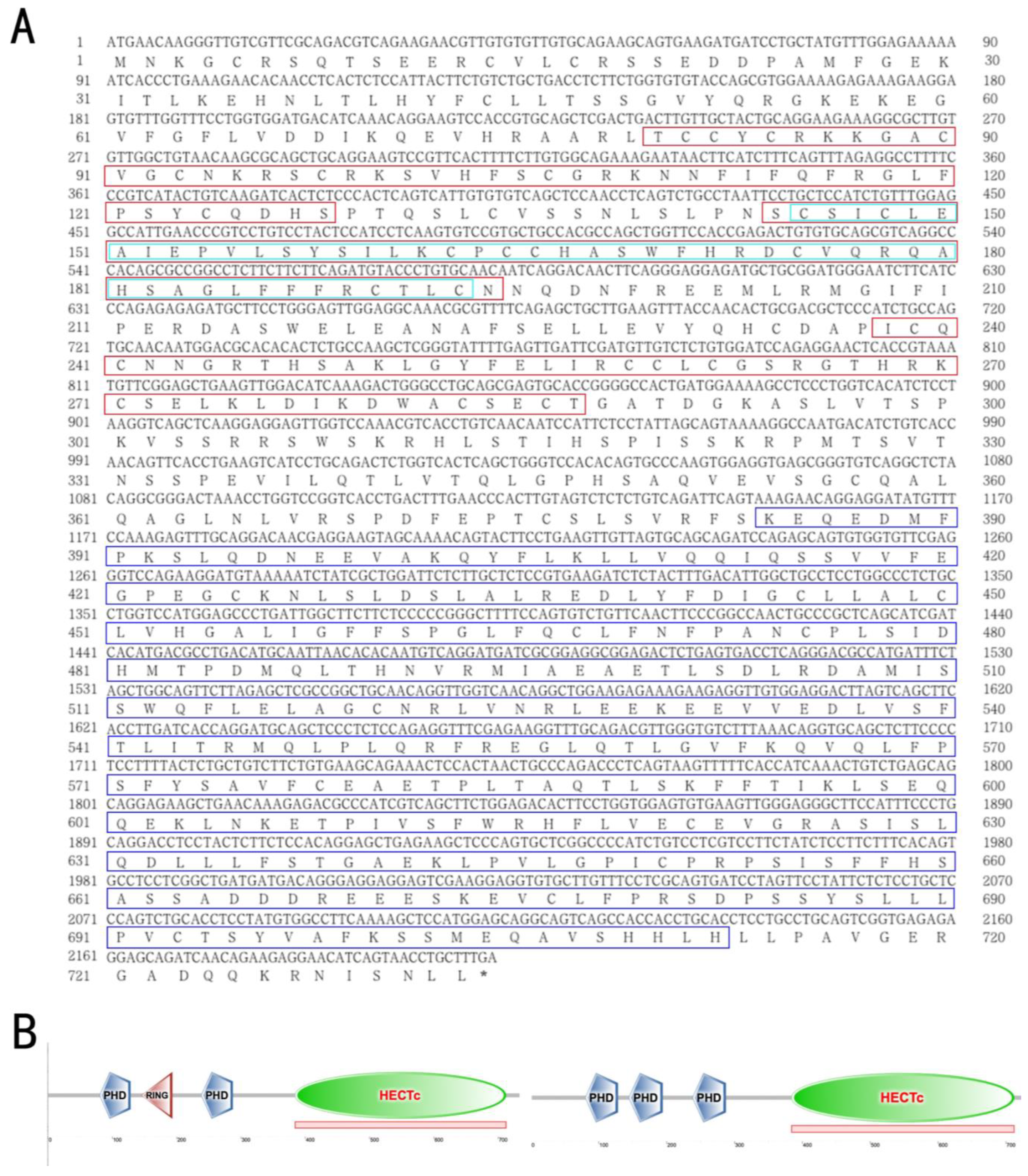
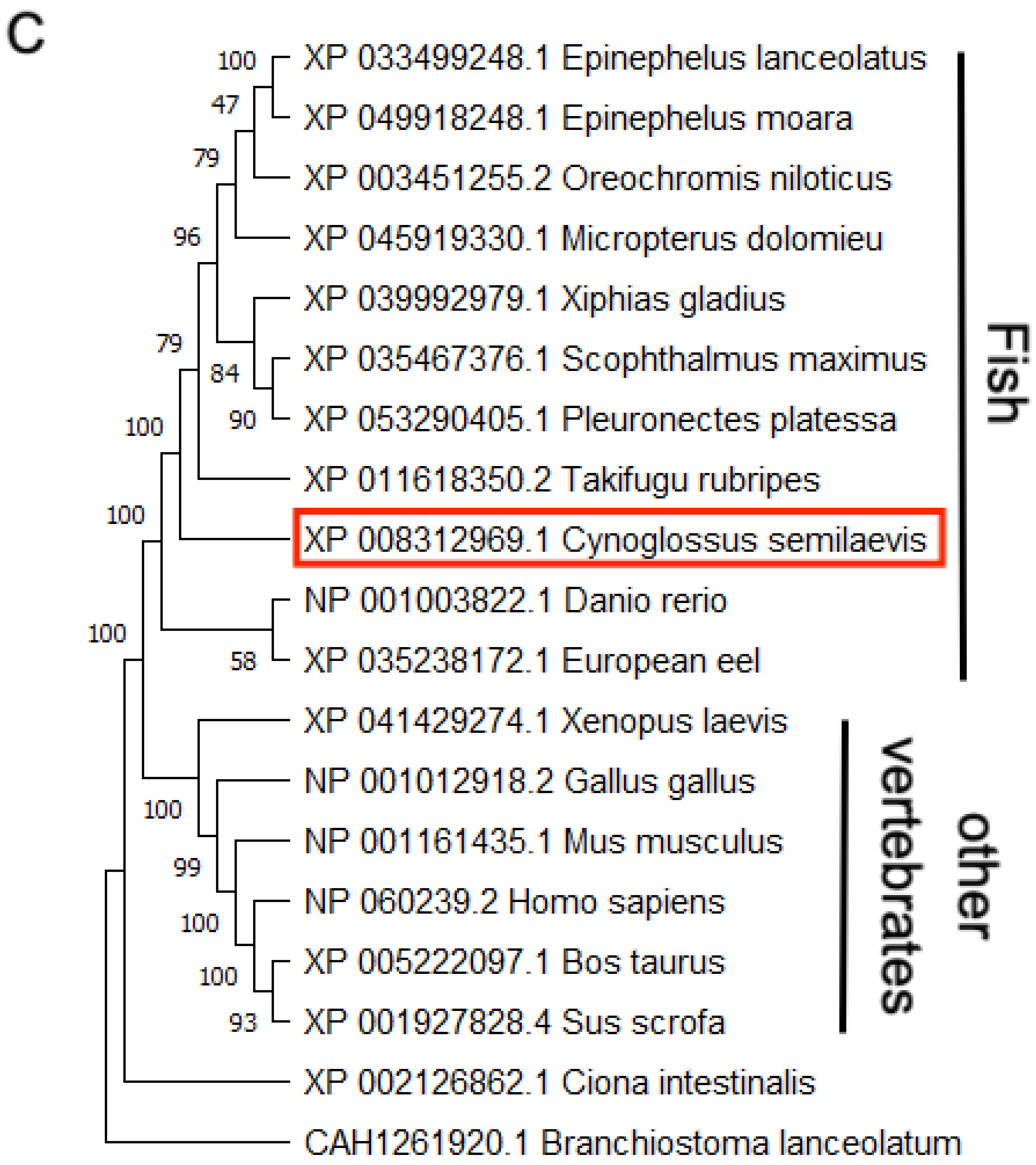
3.1. The Structural and Phylogenetic Analyses of g2e3 in C. semilaevis

The genomic sequence of Cs-g2e3 spans 6479 bp and is mapped to chromosome 1 (NC_024307.1), and includes 17 exons and 16 introns. The total mRNA length of this gene is 3022 bp, encompassing an ORF of 2202 bp that encodes a 733 amino acid protein with a predicted molecular weight of 82.44 kDa and an isoelectric point (PI) of 6.53 (Figure 1A). Predictive analysis of conserved structural domains indicated that the protein contains PHD, Ring, and HECT domains, classifying it as a member of the HECT E3 ubiquitin ligases (Figure 1B). The phylogenetic tree revealed that C. semilaevis clusters with other fish species, while amniotes such as Homo sapiens, Gallus gallus, and Xenopus laevis form a separate group (Figure 1C).

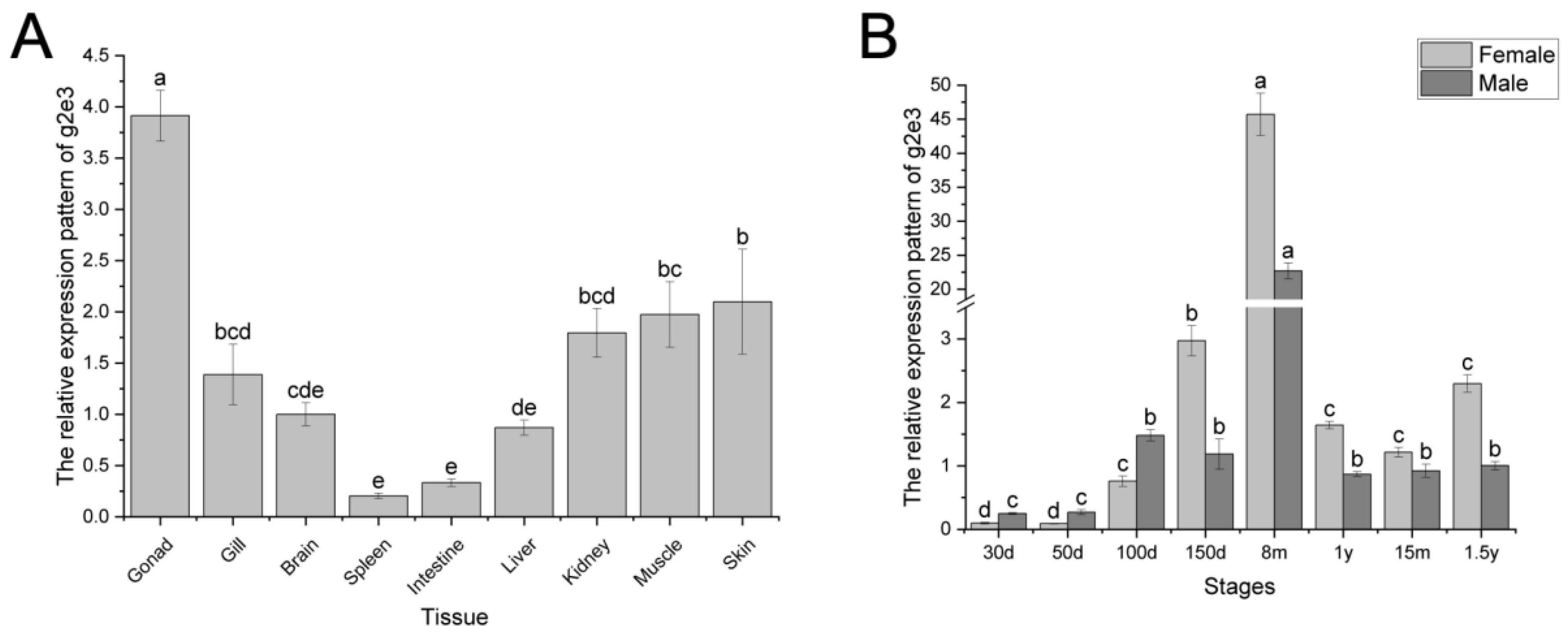
3.2. Expression Pattern of Cs-g2e3 in C. semilaevis

Quantitative PCR analysis revealed that Cs-g2e3 expression is detectable in all sampled tissues of C. semilaevis, with the highest levels observed in the gonads (Figure 2A). Furthermore, monitoring of the gene’s expression across different stages indicated a gradual increase in expression from 30 days to 8 months, reaching a peak at 8 months of age. Subsequently, a decline in expression was observed between 8 months and 1 year of age, followed by stabilization at 1 year (Figure 2B).

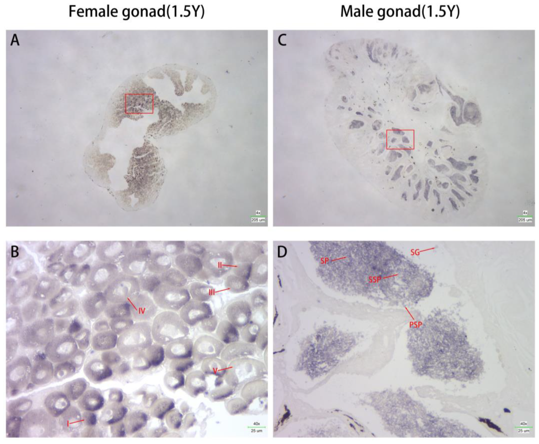
3.3. Localization of Cs-g2e3 mRNA in the Gonads of C. semilaevis

In situ hybridization experiments were conducted to examine the localization of Cs-g2e3 mRNA in male and female gonads. Results indicated that hybridization signals were predominantly localized in sperm and eggs, with no signals detected in connective or other tissues. Strong hybridization signals were observed in both the sperm and egg of C. semilaevis (Figure 3).

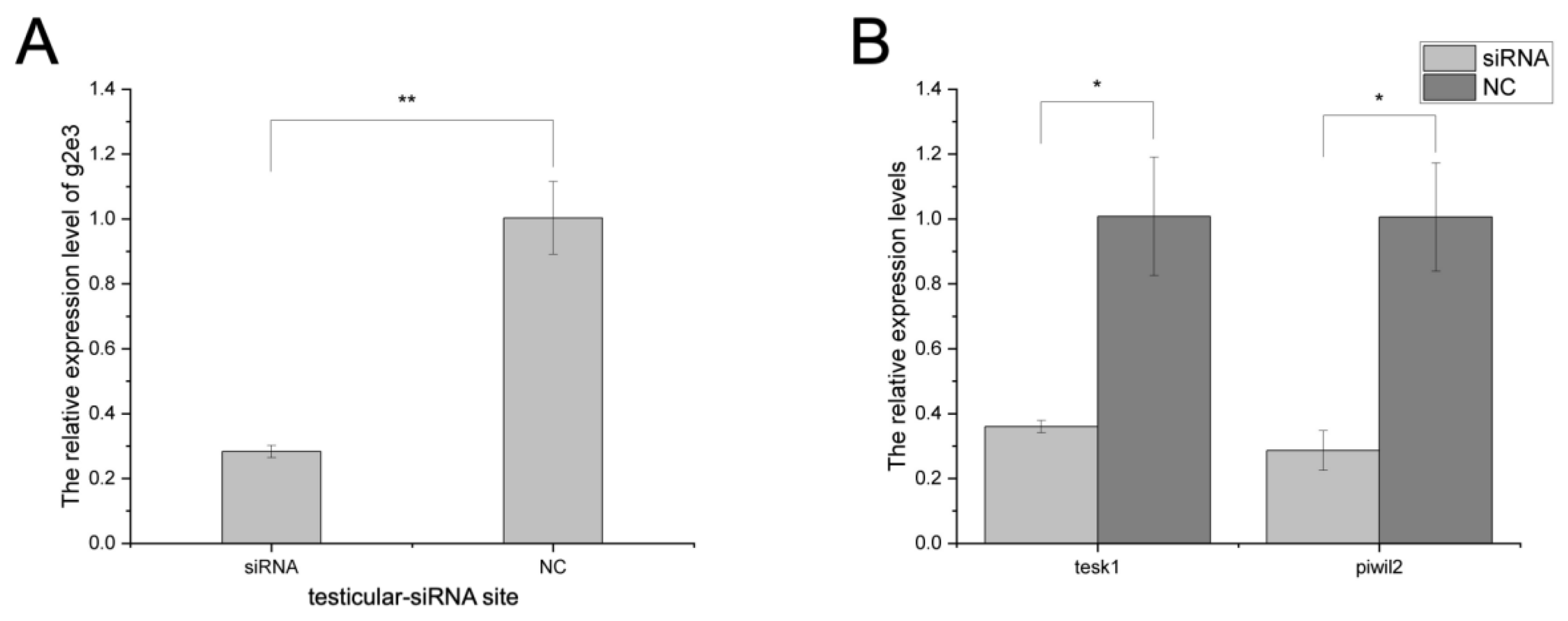
3.4. The Knockdown Effects on Cs-g2e3 and Other Related Genes by siRNA Transfection in Gonadal Germ Cell Lines

In testicular germ cell lines, siRNA knockdown resulted in about a 70% reduction in Cs-g2e3 transcripts (Figure 4A), with consequent significant downregulation of spermatogenesis-related genes piwil2 and tesk1 compared to the negative control (NC) (Figure 4B). In ovarian germ cell lines, siRNA knockdown resulted in about a 70% reduction in Cs-g2e3 transcripts (Figure 4C), with consequent significant downregulation of oogenesis-related genes sox9 and cyp19a compared to the negative control (NC) (Figure 4D).

3.5. In Vivo RNA Interference in C. semilaevis

In vivo RNA interference in the testicular tissue showed that siRNA knockdown was effective, with efficiencies exceeding 70% (Figure 5A). Gene expression of spermatogenesis-related genes piwil2 and tesk1 was significantly downregulated compared to the negative control (NC) (Figure 5B), consistent with the results of RNA interference in testicular germ cell lines.

3.6. Detection and Analysis of the Activity of the Cs-g2e3 Promoter and the Regulation of Transcription Factors

Dual luciferase assays revealed robust activity of the Cs-g2e3 promoter (Table 2). When co-transfected with transcription factors C/EBPα, POU1F1a, myogenin, and JunB, the promoter activity was significantly enhanced, whereas co-transfection with YY1 markedly reduced it to levels comparable to those observed with the pGL3-Basic vector (Figure 6A). Mutations in the transcription factor binding sites for myogenin and JunB resulted in a significant decrease in Cs-g2e3 promoter activity upon co-transfection, highlighting the activating functions of these factors. Conversely, mutation of the YY1 binding site led to a significant increase in promoter activity, indicating a repressive role for YY1 in regulating Cs-g2e3 promoter activity (Figure 6B). These findings suggest that YY1 may serve as a pivotal regulator of Cs-g2e3 transcription.

4. Discussion

Ubiquitination, a ubiquitous post-translational modification, plays a pivotal role in regulating a myriad of cellular processes, notably including the development of mammalian ovarian and testicular germ cells [11,46]. In both amniotes and fish, the levels of ubiquitination undergo a marked increase during gonadal differentiation and gametogenesis [47,48]. Analogously, the ubiquitin ligase gene, designated as g2e3, has been observed to exhibit high levels of expression within the gonads across diverse species, such as humans, rats, chickens, zebrafish, and sea urchins [26]. In the present study, the g2e3 gene was likewise found to exhibit high expression levels in the gonads of Chinese Tongue Sole.
In our study, we successfully cloned Cs-g2e3, determined its sequence, and analyzed its structural features, confirming that Cs-g2e3 belongs to the canonical HECT E3 ligase family, in line with observations made in other species [21,49]. RT-PCR analysis revealed that Cs-g2e3 was predominantly expressed in gonadal tissues, lending support to our hypothesis that Cs-g2e3 is intricately involved in gametogenesis. Further analysis revealed that Cs-g2e3 expression peaked at 8 months of age, subsequently declining from 8 months of age to 1 year of age and stabilizing thereafter. A prior study indicated that sexual differentiation in C. semilaevis occurs around 60 days, which may underlie the initial low expression of Cs-g2e3 [41]. Subsequently, male and female gonads differentiate and develop, producing gametes. By 8 months, the gonads are primarily mature, coinciding with the peak Cs-g2e3 expression. After maturity, the production of gametes is in a dynamic equilibrium, which is consistent with the results of the stabilization of the expression of Cs-g2e3 [50,51]. We also found that Cs-g2e3 expression was higher in male gonads than in female gonads between 30 and 100 days of age and lower in male gonads than in female gonads thereafter, with the most significant difference at 8 months of age, which may be due to sexual dimorphism in C. semilaevis. In situ hybridization experiments conducted on both male and female C. semilaevis specimens yielded strong Cs-g2e3 hybridization signals within the testis and ovary, primarily localized to spermatozoa and eggs, respectively. These findings were reinforced by siRNA knockdown experiments performed in gonadal germ cell lines derived from C. semilaevis. These experiments targeted genes previously implicated in gametogenesis, including tesk1, piwil2, sox9, and cyp19a [41,42,43,44]. Notably, the knockdown of Cs-g2e3 significantly altered the expression patterns of these genes, thereby confirming Cs-g2e3’s functional role in gametogenesis. This conclusion was further validated in vivo through the injection of siRNA directly into the testis of C. semilaevis, yielding results that were consistent with those observed in the germ-cell line experiments.
We also evaluated the activity of the Cs-g2e3 gene promoter and found it to be highly active. By predicting and mutating its binding sites, we observed that the Cs-g2e3 promoter affects these sites to varying degrees, with a particularly strong association with the YY1 transcription factor. This suggests that YY1 may be a key transcription factor that initiates g2e3 gene expression. As a transcription factor, YY1 plays a crucial role in gonadal development and spermatogenesis in many mammals. In studies on chicken testis, YY1 was shown to regulate gonadal development and mediate spermatogenesis and differentiation [52,53,54]. Furthermore, localization of YY1 in the adult testis revealed that it is predominantly present in the nuclei of spermatogonia and spermatozoa, with a particularly strong signal detected in spermatogonia [55]. Similarly, studies in mice have shown the involvement of YY1 in spermatogenesis [56,57]. In addition, YY1 is involved in gonadal development and gametogenesis in fish, such as in yellow catfish, where YY1 can act as a marker of egg quality and play an important role in oogenesis [58], in Pimephales promelas, YY1 is also involved in ovarian development and regulates the synthesis of sex hormones [59]. More importantly, a previously reported study on YY1 in C. semilaevis showed that YY1 is involved in gonadal development in C. semilaevis, which may include gametogenesis [60]. From the experimental results, it was observed that the binding of YY1 to the Cs-g2e3 promoter led to a significant decrease in its activity. Conversely, mutation of the YY1 binding site restored the promoter activity to its original level, further confirming that YY1 exerts an inhibitory effect on the Cs-g2e3 promoter. Previous studies have indeed established that YY1, a crucial transcription factor implicated in spermatogenesis, is localized within spermatogonial stem cells and influences this process by participating in meiosis [55,57]. However, the regulatory mechanisms involved in gametogenesis in C. semilaevis deserve further investigation. Thus, our studies on the Cs-g2e3 promoter and transcription factors further emphasize the possible involvement of Cs-g2e3 in the physiological process of spermatogenesis. This finding serves as a valuable reference for subsequent in-depth studies on transcription factors related to gametogenesis. Meanwhile, given the current low fertilization rate of C. semilaevis, we are considering the possibility of injecting relevant Cs-g2e3 carrier proteins in future production practices of C. semilaevis culture. This would potentially allow the spermatophore to continue spermatogenesis and produce viable sperm for artificial insemination, thereby improving the fertilization rate and promoting the development of the C. semilaevis culture industry. Furthermore, this approach offers valuable ideas for future breeding work and production of C. semilaevis.

5. Conclusions

In this study, we successfully cloned and characterized the cDNA of the ubiquitin ligase gene g2e3 in C. semilaevis (Cs-g2e3). Cs-g2e3 was predominantly expressed in the gonads compared to other tissues. The gene’s expression increased progressively and reached its peak at 8 months of age, then declined and stabilized, maintaining levels significantly higher than those observed in the juvenile period. In situ hybridization demonstrated that Cs-g2e3 was mainly localized in the germ cells. In vitro RNAi studies showed that knockdown of Cs-g2e3 in ovarian and testicular germ cell lines significantly downregulates the expression of key genes involved in oogenesis (e.g., sox9 and cyp19a) and spermatogenesis (e.g., tesk1 and piwil2), respectively. Additionally, in vivo experiments confirmed that the injection of siRNA into the testis of C. semilaevis similarly affected the expression of tesk1 and piwil2. Experiments exploring the regulation of the Cs-g2e3 promoter and its interaction with transcription factors identified a significant relationship between the transcription factor YY1, associated with spermatogenesis, and the Cs-g2e3 promoter. These findings underscore the involvement of Cs-g2e3 in gametogenesis and highlight its critical role in the reproductive biology of C. semilaevis.

Supplementary Materials

The following supporting information can be downloaded at: https://www.mdpi.com/article/10.3390/ani14172579/s1, g2e3 siRNA synthesis and transfection.

Author Contributions

Conceptualization, S.C.; Project administration, S.C. and Z.C.; Funding acquisition, S.C. and Z.C.; Investigation, J.L., F.C., W.X., J.W., M.L. and Y.S.; Writing—original draft, Z.C., J.L. and F.C.; Writing—review and editing, J.W. and W.X. All authors have read and agreed to the published version of the manuscript.

Funding

This research was funded by the National Natural Science Foundation of China (32102779 and 32230107); National Key R&D Program of China (2022YFD2400101 and 2018YFD0901203); Basic Research Funds of Chinese Academy of Fishery Sciences (2023XT01); the Key Research and Development Project of Shandong Province (2022ZLGX01); Central Public-interest Scientific Institution Basal Research Fund, YSFRI, CAFS (No. 20603022023019); the Innovative Team Project of Chinese Academy of Fishery Sciences (2020TD20); Taishan Scholar Climbing Project of Shandong Province, China. The APC was funded by the National Natural Science Foundation of China (32102779).

Institutional Review Board Statement

The study was conducted in accordance with the Declaration of Helsinki and approved by the Ethics Committee of Yellow Sea Fisheries Research Institute (Approval number, YSFRI-2021043).

Informed Consent Statement

Informed consent was obtained from all subjects involved in the study.

Data Availability Statement

The data presented in this study are available in this article.

Acknowledgments

This study was supported by Jiayu Cheng and Jie Zhang. Thanks to Tangshan Weizhuo Aquaculture Company and the Agricultural Technical Extension Station of Caofeidian District for their support during the investigation.

Conflicts of Interest

The authors declare no conflict of interest.

References

  1. Chen, S.L.; Tian, Y.S.; Yang, J.F.; Shao, C.W.; Ji, X.S.; Zhai, J.M.; Liao, X.L.; Zhuang, Z.M.; Su, P.Z.; Xu, J.Y.; et al. Artificial gynogenesis and sex determination in half-smooth tongue sole (Cynoglossus semilaevis). Mar. Biotechnol. 2009, 11, 243–251. [Google Scholar] [CrossRef] [PubMed]
  2. Jiang, L.; Jiang, J.; Liu, J.; Yuan, J.; Chen, Y.; Zhang, Q.; Wang, X. Chromosome mapping of 18S rDNA and 5S rDNA by dual-color fluorescence in situ hybridization in the half-smooth tongue sole (Cynoglossus semilaevis). Genet. Mol. Res. 2014, 13, 10761–10768. [Google Scholar] [CrossRef]
  3. Chen, S.; Zhang, G.; Shao, C.; Huang, Q.; Liu, G.; Zhang, P.; Song, W.; An, N.; Chalopin, D.; Volff, J.N.; et al. Whole-genome sequence of a flatfish provides insights into ZW sex chromosome evolution and adaptation to a benthic lifestyle. Nat. Genet. 2014, 46, 253–260. [Google Scholar] [CrossRef]
  4. Liu, J.; Liu, X.; Jin, C.; Du, X.; He, Y.; Zhang, Q. Transcriptome profiling insights the feature of sex reversal induced by high temperature in Tongue Sole Cynoglossus semilaevis. Front. Genet. 2019, 10, 522. [Google Scholar] [CrossRef]
  5. Wang, Q.; Liu, K.; Feng, B.; Zhang, Z.; Wang, R.; Tang, L.; Li, W.; Li, Q.; Piferrer, F.; Shao, C. Transcriptome of gonads from high temperature induced sex reversal during sex determination and differentiation in Chinese Tongue Sole, Cynoglossus semilaevis. Front. Genet. 2019, 10, 1128. [Google Scholar] [CrossRef] [PubMed]
  6. Jia, Q.; Ni, Y.; Min, S.; Ming, L.; Qian, Y.; Cen, X.; Wang, J.; Tong, X. The ontogenesis of catabolic abilities and energy metabolism during endogenous nutritional periods of tongue sole, Cynoglossus semilaevis. J. Fish Biol. 2021, 99, 1708–1718. [Google Scholar] [CrossRef] [PubMed]
  7. Zhang, Y.; Wang, Y.; Liu, Q.; Wang, H.; Wang, Q.; Shao, C. The role of Z chromosome localization gene psmd9 in spermatogenesis of Cynoglossus semilaevis. Int. J. Mol. Sci. 2024, 25, 6372. [Google Scholar] [CrossRef]
  8. Wang, H.Y.; Liu, X.; Chen, J.Y.; Huang, Y.; Lu, Y.; Tan, F.; Liu, Q.; Yang, M.; Li, S.; Zhang, X.; et al. Single-cell-resolution transcriptome map revealed novel genes involved in testicular germ cell progression and somatic cells specification in Chinese tongue sole with sex reversal. Sci. China Life Sci. 2023, 66, 1151–1169. [Google Scholar] [CrossRef]
  9. Ciechanover, A.; Heller, H.; Elias, S.; Haas, A.L.; Hershko, A. ATP-dependent conjugation of reticulocyte proteins with the polypeptide required for protein degradation. Proc. Natl. Acad. Sci. USA 1980, 77, 1365–1368. [Google Scholar] [CrossRef]
  10. Ramezani, F.; Bahrami-Amiri, A.; Babahajian, A.; Shahsavari Nia, K.; Yousefifard, M. Ubiquitin C-Terminal Hydrolase-L1 (UCH-L1) in prediction of computed tomography findings in traumatic brain injury; a meta-analysis. Emergency 2018, 6, e62. [Google Scholar]
  11. Dikic, I.; Robertson, M. Ubiquitin ligases and beyond. BMC Biol. 2012, 10, 22. [Google Scholar] [CrossRef] [PubMed]
  12. Toma-Fukai, S.; Shimizu, T. Structural diversity of ubiquitin E3 ligase. Molecules 2021, 26, 6682. [Google Scholar] [CrossRef] [PubMed]
  13. Song, L.; Luo, Z.Q. Post-translational regulation of ubiquitin signaling. J. Cell Biol. 2019, 218, 1776–1786. [Google Scholar] [CrossRef] [PubMed]
  14. Pickart, C.M. Back to the future with ubiquitin. Cell 2004, 116, 181–190. [Google Scholar] [CrossRef]
  15. Beaudenon, S.; Huibregtse, J.M. High-level expression and purification of recombinant E1 enzyme. Methods Enzymol. 2005, 398, 3–8. [Google Scholar]
  16. Valimberti, I.; Tiberti, M.; Lambrughi, M.; Sarcevic, B.; Papaleo, E. E2 superfamily of ubiquitin-conjugating enzymes: Constitutively active or activated through phosphorylation in the catalytic cleft. Sci. Rep. 2015, 5, 14849. [Google Scholar] [CrossRef]
  17. Berndsen, C.E.; Wolberger, C. New insights into ubiquitin E3 ligase mechanism. Nat. Struct. Mol. Biol. 2014, 21, 301–307. [Google Scholar] [CrossRef]
  18. Schreiber, A.; Stengel, F.; Zhang, Z.; Enchev, R.I.; Kong, E.H.; Morris, E.P.; Robinson, C.V.; da Fonseca, P.C.; Barford, D. Structural basis for the subunit assembly of the anaphase-promoting complex. Nature 2011, 470, 227–232. [Google Scholar] [CrossRef]
  19. Xu, X.H.; Wang, F.; Chen, H.; Sun, W.; Zhang, X.S. Transcript profile analyses of maize silks reveal effective activation of genes involved in microtubule-based movement, ubiquitin-dependent protein degradation, and transport in the pollination process. PLoS ONE 2013, 8, e53545. [Google Scholar] [CrossRef]
  20. Ohtake, F.; Baba, A.; Takada, I.; Okada, M.; Iwasaki, K.; Miki, H.; Takahashi, S.; Kouzmenko, A.; Nohara, K.; Chiba, T.; et al. Dioxin receptor is a ligand-dependent E3 ubiquitin ligase. Nature 2007, 446, 562–566. [Google Scholar] [CrossRef]
  21. Rotin, D.; Kumar, S. Physiological functions of the HECT family of ubiquitin ligases. Nat. Rev. Mol. Cell Biol. 2009, 10, 398–409. [Google Scholar] [CrossRef] [PubMed]
  22. Wenzel, D.M.; Lissounov, A.; Brzovic, P.S.; Klevit, R.E. UBCH7 reactivity profile reveals parkin and HHARI to be RING/HECT hybrids. Nature 2011, 474, 105–108. [Google Scholar] [CrossRef]
  23. Song, M.S.; Pandolfi, P.P. The HECT family of E3 ubiquitin ligases and PTEN. Semin. Cancer Biol. 2022, 85, 43–51. [Google Scholar] [CrossRef] [PubMed]
  24. Wang, Y.; Argiles-Castillo, D.; Kane, E.I.; Zhou, A.; Spratt, D.E. HECT E3 ubiquitin ligases—Emerging insights into their biological roles and disease relevance. J. Cell Sci. 2020, 133, jcs228072. [Google Scholar] [CrossRef] [PubMed]
  25. Brooks, W.S.; Helton, E.S.; Banerjee, S.; Venable, M.; Johnson, L.; Schoeb, T.R.; Kesterson, R.A.; Crawford, D.F. G2e3 is a dual function ubiquitin ligase required for early embryonic development. J. Biol. Chem. 2008, 283, 22304–22315. [Google Scholar] [CrossRef] [PubMed]
  26. Wang, X.R.; Ling, L.B.; Huang, H.H.; Lin, J.J.; Fugmann, S.D.; Yang, S.Y. Evidence for parallel evolution of a gene involved in the regulation of spermatogenesis. Proc. Biol. Sci. 2017, 284, 20170324. [Google Scholar] [CrossRef]
  27. Yang, S.Y.; Baxter, E.M.; Van Doren, M. Phf7 controls male sex determination in the Drosophila germline. Dev. Cell. 2012, 22, 1041–1051. [Google Scholar] [CrossRef]
  28. Hering, D.M.; Oleński, K.; Ruść, A.; Kaminski, S. Genome-wide association study for semen volume and total number of sperm in Holstein-Friesian bulls. Anim. Reprod. Sci. 2014, 151, 126–130. [Google Scholar]
  29. Paradowska, A.S.; Miller, D.; Spiess, A.N.; Vieweg, M.; Cerna, M.; Dvorakova-Hortova, K.; Bartkuhn, M.; Schuppe, H.C.; Weidner, W.; Steger, K. Genome wide identification of promoter binding sites for H4K12ac in human sperm and its relevance for early embryonic development. Epigenetics 2012, 7, 1057–1070. [Google Scholar] [CrossRef]
  30. Wang, X.; Kang, J.Y.; Wei, L.; Yang, X.; Sun, H.; Yang, S.; Lu, L.; Yan, M.; Bai, M.; Chen, Y.; et al. PHF7 is a novel histone H2A E3 ligase prior to histone-to-protamine exchange during spermiogenesis. Development 2019, 146, dev175547. [Google Scholar] [CrossRef]
  31. Zoheir, K.M.; Darwish, A.M.; Liguo, Y.; Ashour, A.E. Transcriptome comparisons detect new genes associated with apoptosis of cattle and buffaloes preantral follicles. J. Genet. Eng. Biotechnol. 2021, 19, 151. [Google Scholar] [CrossRef]
  32. Brooks, W.S.; Banerjee, S.; Crawford, D.F. RNF138/NARF is a cell cycle regulated E3 ligase that poly-ubiquitinates g2e3. JSM Cell Dev. Biol. 2014, 2, 1005. [Google Scholar] [CrossRef]
  33. Becker, L.S.; Al Smadi, M.A.; Koch, H.; Abdul-Khaliq, H.; Meese, E.; Abu-Halima, M. Towards a more comprehensive picture of the microRNA-23a/b-3p impact on impaired male fertility. Biology 2023, 12, 800. [Google Scholar] [CrossRef]
  34. Yang, L.; Songlin, C.; Fengtao, G.; Liang, M.; Qiaomu, H.; Wentao, S.; Weiqun, L. SCAR-transformation of sex-specific SSR marker and its application in half-smooth tongue sole (Cynoglossus semiliaevis). J. Agric. Biotechnol. 2014, 22, 787–792. [Google Scholar]
  35. Kumar, R.; Kumar, C.; Choudhury, D.R.; Ranjan, A.; Raipuria, R.K.; Dubey, K.K.D.; Mishra, A.; Kumar, C.; Manzoor, M.M.; Kumar, A.; et al. Isolation, characterization, and expression analysis of NAC transcription factor from andrographis paniculata (Burm. f.) nees and their role in andrographolide production. Genes 2024, 15, 422. [Google Scholar] [CrossRef]
  36. Li, Z.; Yang, L.; Wang, J.; Shi, W.; Pawar, R.A.; Liu, Y.; Xu, C.; Cong, W.; Hu, Q.; Lu, T.; et al. beta-Actin is a useful internal control for tissue-specific gene expression studies using quantitative real-time PCR in the half-smooth tongue sole Cynoglossus semilaevis challenged with LPS or Vibrio anguillarum. Fish Shellfish Immunol. 2010, 29, 89–93. [Google Scholar] [CrossRef] [PubMed]
  37. Dong, Y.; Lyu, L.; Zhang, D.; Li, J.; Wen, H.; Shi, B. Integrated lncRNA and mRNA transcriptome analyses in the ovary of Cynoglossus semilaevis reveal genes and pathways potentially involved in reproduction. Front. Genet. 2021, 12, 671729. [Google Scholar] [CrossRef]
  38. Qi, X.; Zhou, W.; Wang, Q.; Guo, L.; Lu, D.; Lin, H. Gonadotropin-Inhibitory hormone, the piscine ortholog of LPXRFa, participates in 17β-Estradiol feedback in female goldfish reproduction. Endocrinology 2017, 158, 860–873. [Google Scholar] [CrossRef]
  39. Sun, A.; Wang, T.Z.; Wang, N.; Liu, X.F.; Sha, Z.X.; Chen, S.L. Establishment and characterization of an ovarian cell line from half-smooth tongue sole, Cynoglossus semilaevis. J. Fish Biol. 2015, 86, 46–59. [Google Scholar] [CrossRef]
  40. Zhang, B.; Wang, X.; Sha, Z.; Yang, C.; Liu, S.; Wang, N.; Chen, S.L. Establishment and characterization of a testicular cell line from the half-smooth tongue sole, Cynoglossus semilaevis. Int. J. Biol. Sci. 2011, 7, 452–459. [Google Scholar] [CrossRef] [PubMed]
  41. Lin, G.; Gao, D.; Lu, J.; Sun, X. Transcriptome profiling reveals the sexual dimorphism of gene expression patterns during gonad differentiation in the half-smooth tongue sole (Cynoglossus semilaevis). Mar. Biotechnol. 2021, 23, 18–30. [Google Scholar] [CrossRef]
  42. Shao, C.; Liu, G.; Liu, S.; Chen, S. Characterization of the cyp19a1a gene from a BAC sequence in half-smooth tongue sole (Cynoglossus semilaevis) and analysis of its conservation among teleosts. Acta Oceanol. 2013, 32, 35–43. [Google Scholar] [CrossRef]
  43. Toshima, J.; Ohashi, K.; Okano, I.; Nunoue, K.; Kishioka, M.; Kuma, K.; Miyata, T.; Hirai, M.; Baba, T.; Mizuno, K. Identification and characterization of a novel protein kinase, TESK1, specifically expressed in testicular germ cells. J. Biol. Chem. 1995, 270, 31331–31337. [Google Scholar] [CrossRef]
  44. Zong, W.; Wang, Y.; Zhang, L.; Lu, W.; Li, W.; Wang, F.; Cheng, J. DNA methylation mediates sperm quality via piwil1 and piwil2 regulation in Japanese Flounder (Paralichthys olivaceus). Int. J. Mol. Sci. 2024, 25, 5935. [Google Scholar] [CrossRef]
  45. Jiang, C.; Ruan, Y.; Li, J.; Huang, J.; Xiao, M.; Xu, H. Tissue expression and promoter activity analysis of the porcine TNFSF11 gene. Theriogenology 2024, 226, 277–285. [Google Scholar] [CrossRef] [PubMed]
  46. Xiong, Y.; Yu, C.; Zhang, Q. Ubiquitin-Proteasome system-regulated protein degradation in spermatogenesis. Cells 2022, 11, 1058. [Google Scholar] [CrossRef]
  47. Sun, J.; Shang, X.; Tian, Y.; Zhao, W.; He, Y.; Chen, K.; Cheng, H.; Zhou, R. Ubiquitin C-terminal hydrolase-L1 (Uch-L1) correlates with gonadal transformation in the rice field eel. FEBS J. 2008, 275, 242–249. [Google Scholar] [CrossRef] [PubMed]
  48. Roman-Trufero, M.; Dillon, N. The UBE2D ubiquitin conjugating enzymes: Potential regulatory hubs in development, disease and evolution. Front. Cell. Dev. Biol. 2022, 10, 1058751. [Google Scholar] [CrossRef] [PubMed]
  49. Kane, E.I.; Beasley, S.A.; Schafer, J.M.; Bohl, J.E.; Lee, Y.S.; Rich, K.J.; Bosia, E.F.; Spratt, D.E. Redefining the catalytic HECT domain boundaries for the HECT E3 ubiquitin ligase family. Biosci. Rep. 2022, 42, BSR20221036. [Google Scholar] [CrossRef]
  50. Kang, X.J.; Liang, C.G.; Guo, M.S.; Ge, S.Q.; Mu, S.M.; Su, W.Q.; Liu, X.H. Ultrastructure of spermatogenesis and spermiogenesis in the half-smooth tongue sole, Cynoglossus semilaevis. Acta Zool. Sin. 2008, 54, 356–365. [Google Scholar]
  51. Ma, X.K.; Liu, X.Z.; Wen, H.S.; Xu, Y.J.; Zhang, L.J. Histological observation on gonadal sex differentiation in Cynoglossus semilaevis Günther. Mar. Fish. Res. 2006, 27, 55–61. [Google Scholar]
  52. Chen, L.; Qiao, L.; Guo, Y.; Huang, Y.; Luo, W.; Feng, Y. Localization and regulatory function of Yin Yang 1 (YY1) in chicken testis. Mol. Genet. Genom. 2022, 297, 113–123. [Google Scholar] [CrossRef]
  53. Cheon, Y.P.; Choi, D.; Lee, S.H.; Kim, C.G. YY1 and CP2c in unidirectional spermatogenesis and stemness. Dev. Reprod. 2020, 24, 249–262. [Google Scholar] [CrossRef] [PubMed]
  54. Kim, J.D.; Kang, K.; Kim, J. YY1’s role in DNA methylation of peg3 and xist. Nucleic Acids Res. 2009, 37, 5656–5664. [Google Scholar] [CrossRef]
  55. Kim, J.S.; Chae, J.H.; Cheon, Y.P.; Kim, C.G. Reciprocal localization of transcription factors YY1 and CP2c in spermatogonial stem cells and their putative roles during spermatogenesis. Acta Histochem. 2016, 118, 685–692. [Google Scholar] [CrossRef] [PubMed]
  56. Kayyar, B.; Kataruka, S.; Suresh Akhade, V.; Rao, M.R.S. Molecular functions of Mrhl lncRNA in mouse spermatogenesis. Reproduction 2023, 166, R39–R50. [Google Scholar] [CrossRef] [PubMed]
  57. Wu, S.; Hu, Y.C.; Liu, H.; Shi, Y. Loss of YY1 impacts the heterochromatic state and meiotic double-strand breaks during mouse spermatogenesis. Mol. Cell Biol. 2009, 29, 6245–6256. [Google Scholar] [CrossRef]
  58. Ren, F.; Zhou, Q.; Meng, Y.; Guo, W.; Tang, Q.; Mei, J. RNA binding proteins are potential novel biomarkers of egg quality in yellow catfish. BMC Genom. 2023, 24, 121. [Google Scholar] [CrossRef]
  59. Garcia-Reyero, N.; Villeneuve, D.L.; Kroll, K.J.; Liu, L.; Orlando, E.F.; Watanabe, K.H.; Sepúlveda, M.S.; Ankley, G.T.; Denslow, N.D. Expression signatures for a model androgen and antiandrogen in the fathead minnow (Pimephales promelas) ovary. Environ. Sci. Technol. 2009, 43, 2614–2619. [Google Scholar] [CrossRef]
  60. Shi, R.; Li, X.; Xu, X.; Chen, Z.; Zhu, Y.; Wang, N. Genome-wide analysis of BMP/GDF family and DAP-seq of YY1 suggest their roles in Cynoglossus semilaevis sexual size dimorphism. Int. J. Biol. Macromol. 2023, 253, 127201. [Google Scholar] [CrossRef]
Figure 1. Sequence analysis, conserved structural domain analysis, and phylogenetic tree analysis of Cs-g2e3 in C. semilaevis. Panel (A) shows the mRNA sequence and protein amino acid sequence of Cs-g2e3. The CDS region of 2202 bp encodes 733 amino acids, with the blue box indicating the conserved HECTc structural domain of the E3 ubiquitin ligase, the red box indicating the conserved PHD structural domain, and the bright green box indicating the conserved RING structural domain. An asterisk (*) represents the stop codon at the end of the ORF. Panel (B) displays a map of the conserved structural domains of Cs-g2e3 predicted by SMART, clearly showing three PHD structures, one HECTc structure, and one RING structure, with the middle PHD and RING structures largely overlapping. Panel (C) presents the phylogenetic tree of g2e3 in fish and other amniotes. C. semilaevis is highlighted in red box to indicate its evolutionary position. Numbers at nodes represent NJ bootstrap values. The phylogenetic tree shows that C. semilaevis clusters with other fish species, forming a separate group from amniotes such as Homo sapiens, Gallus gallus, and Xenopus laevis.
Animals 14 02579 g001aAnimals 14 02579 g001b
Figure 2. Expression pattern of Cs-g2e3 in different tissues and stages of C. semilaevis. Panel (A) represents the expression of Cs-g2e3 in different tissues, and panel (B) represents the expression of Cs-g2e3 in the gonads of C. semilaevis during different stages. The letters above each chart indicate significant differences. The bars represent the standard error.
Animals 14 02579 g002
Figure 3. In situ hybridization of Cs-g2e3 in 1.5-year-old gonads. Panels (A,B) represent the hybridisation signals of the ovary at different magnifications, and panels (C,D) represent the hybridisation signals of the testis at different magnifications, and they all show extremely strong hybridisation signals. Oocytes at different developmental stages are marked by I, II, III, IV, and V (panel (B)). SG: spermatogonia; PSP: primary spermatocyte; SSP: secondary spermatocyte; SP: spermatozoon (panel (D)). Red boxes indicate the same area at different magnifications.
Animals 14 02579 g003
Figure 4. The knockdown effect of Cs-g2e3 siRNAs in C. semilaevis gonadal germ cell lines in mRNA levels. Panel (A) shows the gene silencing effect of Cs-g2e3 siRNA on testicular germ cell lines. Panel (B) illustrates the expression patterns of spermatogenesis-related genes, tesk1, and piwil2, after siRNA transfection. Panel (C) shows the gene silencing effect of Cs-g2e3 siRNA on ovarian germ cell lines. Panel (D) illustrates the expression patterns of oogenesis-related genes sox9 and cyp19a after siRNA transfection. The stars indicate a significant difference (**: p < 0.01, *: 0.01 ≤ p < 0.05). The bars represent the standard error.
Animals 14 02579 g004
Figure 5. The knockdown effect of Cs-g2e3 siRNA in testicular tissue of C. semilaevis in mRNA levels. Panel (A) shows the gene silencing effect of the Cs-g2e3 siRNA site on the testicular tissue of C. semilaevis; panel (B) shows the expression of spermatogenesis-related genes tesk1 and piwil2 after vivo RNA interference. The stars indicate a significant difference (**: p < 0.01, *: 0.01 ≤ p < 0.05). The bars represent the standard error.
Animals 14 02579 g005
Figure 6. Analysis of the activity of the Cs-g2e3 promoter and the regulatory roles of transcription factors. Panel (A) shows the activity of the Cs-g2e3 promoter and the effects of co-transfection with transcription factors C/EBPα, POU1F1a, myogenin, YY1, and JunB on its activity. Panel (B) shows the effects of mutations in the transcription factor binding sites for myogenin, YY1, and JunB on Cs-g2e3 promoter activity. In the one-way ANOVA, different letters are used to indicate statistically significant differences among the groups. The bars represent the standard error.
Animals 14 02579 g006
Table 1. Primers used in experiments.
Primer NameSequence (5′ to 3′)Product Length (bp)Tm Values (°C)Purposes
g2e3-FGCCACGTAACCCCATAT286952sequence validation
g2e3-RAACGCAAGCAGTAGAACAG
β-actin-RT-FTTCCAGCCTTCCTTCCTT12453qRT-PCR
β-actin-RT-RTACCTCCAGACAGCACAG
g2e3-RT-FGTGTTCGAGGGTCCAGAAGG12658qRT-PCR
g2e3-RT-RGAAGCCAATCAGGGCTCCAT
Y-g2e3-FATTTAGGTGACACTATAGAA
CTGCAGGAAGTCCGTTCACT
55665ISH
Y-g2e3-RTAATACGACTCACTATAGGG
GGCCCAGTCTTTGATGTCCA
sox9-RT-FAAGAACCACACAGATCAAGACAGA15057qRT-PCR
sox9-RT-RTAGTCATACTGTGCTCTGGTGATG
cyp19a-RT-FGGTGAGGATGTGACCCAGTGT23056qRT-PCR
cyp19a-RT-RACGGGCTGAAATCGCAAG
tesk1-RT-FGCAGAAACTCTCTCACCCCAACA29059qRT-PCR
tesk1-RT-RCCAGACCAAAGTCCGTCACCA
piwil2-RT-FCGTCACCTTCGCTCCAAAT17156qRT-PCR
piwil2-RT-RTCTTCGTCGTCCGTTCGC
g2e3-P-FAGATCTGCGATCTAAGTAAGCT
GCGTCCTCCAGTTTGGCTA
154068Promoter cloning
g2e3-P-RCAACAGTACCGGAATGCCAAGCT
CGCTTTTCTTCCGTTCCG
Cs-sex-FCCTAAATGATGGATGTAGATTCTGTC169/13456sex determination
Cs-sex-RGATCCAGAGAAAATAAACCCAGG
g2e3-835-FGCAACAAUCAGGACAACUUTT siRNA
g2e3-835-RAAGUUGUCCUGAUUGUUGCTT
g2e3-1562-FCCGUGAAGAUCUCUACUUUTT siRNA
g2e3-1562-RAAAGUAGAGAUCUUCACGGTT
g2e3-1928-FGCAGACGUUGGGUGUCUUUTT siRNA
g2e3-1928-RAAAGACACCCAACGUCUGCTT
Table 2. The luciferase values in human embryonic kidney (HEK) 293T cells.
GroupsFirefly LuciferaseRenilla LuciferaseThe Relative Luciferase Value (±SD)
pGL3-g2e3-pro640,0381,102,3830.56 ± 0.05
571,785954,903
511,2231,016,504
pGL3-g2e3-p + C/EBPα640,123586,9120.99 ± 0.18
706,702644,511
391,717499,763
pGL3-g2e3-p + POU1F1a941,729395,4592.38 ± 0.02
786,581327,948
984,677417,072
pGL3-g2e3-p + myogenin812,5381,023,4840.83 ± 0.07
868,884950,887
883,0881,132,129
pGL3-g2e3-p + YY1365,8761,897,6890.18 ± 0.02
305,7741,580,501
321,8561,940,210
pGL3-g2e3-p + JunB143,58841,269,2351.21 ± 0.10
134,74711,020,311
1,462,6501,236,206
mu-g2e3_myogenin + myogenin841,4081,980,6820.42 ± 0.02
851,1621,927,374
742,4051,831,696
mu-g2e3_YY1 + YY11,565,7323,620,1410.45 ± 0.04
1,682,1823,401,520
1,014,9722,447,655
mu-g2e3_JunB + JunB339,348854,0850.42 ± 0.05
293,453623,706
356,566937,653
pGL3-control7,175,562766,4339.65 ± 0.47
9,262,670909,082
12,162,7821,294,003
pGL3-Basic105,484765,9870.14 ± 0.02
107,500648,147
139,0581,061,613
Disclaimer/Publisher’s Note: The statements, opinions and data contained in all publications are solely those of the individual author(s) and contributor(s) and not of MDPI and/or the editor(s). MDPI and/or the editor(s) disclaim responsibility for any injury to people or property resulting from any ideas, methods, instructions or products referred to in the content.

