Article

Pleiotropic Gene HMGA2 Regulates Myoblast Proliferation and Affects Body Size of Sheep

1
Joint International Research Laboratory of Agriculture and Agri-Product Safety of Ministry of Education of China, Yangzhou University, Yangzhou 225009, China
2
College of Animal Science and Technology, Yangzhou University, Yangzhou 225009, China
3
Jiangsu Key Laboratory of Zoonosis, Yangzhou University, Yangzhou 225009, China
*
Author to whom correspondence should be addressed.
Animals 2024, 14(18), 2721; https://doi.org/10.3390/ani14182721
Submission received: 14 August 2024 / Revised: 8 September 2024 / Accepted: 18 September 2024 / Published: 20 September 2024

Abstract

:

Simple Summary

The HMGA2 gene has been known to regulate body size or muscle development in various animals, including mice. In this study, we explored the role of HMGA2 in sheep. We found that HMGA2 significantly promotes the proliferation of sheep muscle cells. Additionally, a specific genetic variation in the HMGA2 gene was identified, which is associated with important growth traits in sheep. These findings suggest that HMGA2 could be a valuable marker for breeding programs aimed at improving meat production in sheep.

Abstract

Uncovering genes associated with muscle growth and body size will benefit the molecular breeding of meat Hu sheep. HMGA2 has proven to be an important gene in mouse muscle growth and is associated with the body size of various species. However, its roles in sheep are still limited. Using sheep myoblast as a cell model, the overexpression of HMGA2 significantly promoted sheep myoblast proliferation, while interference with HMGA2 expression inhibited proliferation, indicated by qPCR, EdU, and CCK-8 assays. Furthermore, the dual-luciferase reporter system indicated that the region NC_056056.1: 154134300-154134882 (-618 to -1200 bp upstream of the HMGA2 transcription start site) was one of the habitats of the HMGA2 core promoter, given the observation that this fragment led to a ~3-fold increase in luciferase activity. Interestingly, SNP rs428001129 (NC_056056.1:g.154134315 C>A) was detected in this fragment by Sanger sequencing of the PCR product of pooled DNA from 458 crossbred sheep. This SNP was found to affect the promoter activity and was significantly associated with chest width at birth and two months old, as well as chest depth at two and six months old. The data obtained in this study demonstrated the phenotypic regulatory role of the HMGA2 gene in sheep production traits and the potential of rs428001129 in marker-assisted selection for sheep breeding in terms of chest width and chest depth.

1. Introduction

Hu sheep, a unique Chinese breed known for its high prolificacy, is an excellent maternal breed for meat sheep crossbreeding improvement [1]. Recently, to meet the growing demand for mutton, the Ministry of Agriculture and Rural Affairs of the People’s Republic of China issued a National Sheep Genetic Improvement Plan (2021–2035) and claimed that improving the yield and quality of mutton is a top priority in the breeding of native Chinese sheep breeds. To this end, carrying out Hu sheep breeding for the purpose of meat production holds significant importance for the development of the sheep industry in China.
Muscle growth is a crucial factor affecting meat production. Myoblasts proliferate and differentiate to form myotubes, determining the number and volume of muscle fibers [2,3]. This process involves the programmed expression and regulation of a series of related genes, such as the newly identified marker genes of muscle growth, Myomaker and Myomerger [4,5,6]. To date, the molecular regulatory network of muscle growth has not been fully illustrated. Therefore, uncovering more genes associated with muscle growth will benefit the molecular breeding of meat Hu sheep.
In the 1990s, scientists reported a transgenic dwarf mouse with normal growth hormone levels, which was caused by the insertion of exogenous DNA into the HMGA2 gene [7,8,9]. Currently, dwarf phenotypes caused by HMGA2 gene knockout or natural mutations have been reported in pig, chicken, and rabbit [10,11,12,13]. Notably, the functional loss of this gene not only reduces animal size but also causes coordinated changes in the development of tissues and organs. Studies have shown that the HMGA2 gene encodes a structural transcription factor that is exclusively expressed during early animal embryonic development [14]. The HMGA2 protein does not have a transcriptional activation ability itself; instead, it binds to AT-rich motifs in the genome, altering chromatin conformation and promoting the recruitment of transcription factors, thereby regulating the expression of key genes [15,16,17,18,19]. For instance, the HMGA2 protein can bind to the IGF2BP2 gene, recruit nuclear transcription factor NF-κB, and regulate IGF2BP2 gene transcription, thereby participating in muscle development and fat metabolism in mice [20]. Additionally, genome-wide association studies (GWASs) have shown that HMGA2 is significantly associated with body size in horses and dogs and with eye muscle area in cattle and sheep [21,22,23,24,25]. Excitingly, the rs29016809 locus explains ~2% of the body weight and ~4% of the height variation in cattle [26,27]. Therefore, HMGA2 is not only a major gene regulating body size but also a key gene for muscle development.
At present, direct evidence for the regulatory function of the HMGA2 gene in sheep muscle growth remains scarce. This study elucidates the proliferative function of HMGA2 in sheep myoblasts, identifies the core promoter of the HMGA2 gene, and characterizes a SNP within this core promoter associated with sheep’s body size, thereby providing a potential marker for the molecular breeding of sheep.

2. Materials and Methods

2.1. Ethics Statement

The experimental animals used in this study were approved by the Yangzhou University Laboratory Animal Welfare and Ethics Committee (No. 202103279), and all experiments followed the ethical principles of animal welfare.

2.2. Experimental Animals and Isolation of Sheep Myoblasts

After slaughtering a ewe that was two months pregnant, the fetal sheep was collected for the isolation of ovine myoblasts. Sheep myoblasts were isolated from the Longissimus dorsi muscle of the fetal sheep by means of collagenase II as previously described [28]. The cells were cultured in a cell incubator at 37 °C with 5% CO2, using Dulbecco’s Modified Eagle Medium (DMEM) (Gibco, Carlsbad, CA, USA) containing 20% Fetal Bovine Serum (FBS) (Gibco, Carlsbad, CA, USA) and 1% penicillin–streptomycin.
Jugular vein blood samples were collected from 458 six-month-old crossbred sheep (Hu sheep × Dorper sheep, 134 rams, 324 ewes, provided by Xuzhou Suyang Co., Ltd., Xuzhou, China), and growth data including body height, body length, chest circumference, chest depth, chest width, and cannon circumference were recorded at the age of birth, one month, two months, three months, and six months for the association analysis.

2.3. DNA and RNA Extraction and cDNA Synthesis

Genomic DNA was extracted using the phenol–chloroform method and diluted to 20 ng/μL [29]. Total RNA was extracted using the Trizol method and diluted to 100 ng/μL after quality inspection using NanoDrop Microvolume Spectrophotometers (Thermo Scientific, Carlsbad, CA, USA) [30]. A reverse transcription kit (Takara, Kusatsu, Shiga, Japan) was used to remove genomic DNA from the total RNA, with the following program: 42 °C for 2 min, then stored at −80 °C. The genomic DNA-free total RNA was reverse transcribed using the following program: 37 °C for 15 min, then 85 °C for 5 s. The synthesized cDNA was diluted to 10 ng/μL.

2.4. Overexpression and Interference of HMGA2 Gene

Based on the sequence of HMGA2 (accession number: XM_027967479.3), the full-length HMGA2 coding sequence (CDS, 330 bp) was synthesized and cloned into the multiple cloning sites (Hind III and Xho I) of the pcDNA3.1(+) vector (Tsingke Biotechnology Co., Ltd.). HMGA2 siRNAs (Table 1) were designed and synthesized by Genepharma Co., Ltd. (Suzhou, China).

2.5. Detection of Sheep Myoblast Proliferation

When the sheep myoblasts grew to 60% confluence, the recombinant vectors of HMGA2 overexpression (pcDNA3.1-HMGA2, OE), negative control (pcDNA3.1-blank, NC), small interfering RNA (siRNA), or negative control of siRNA (NC) were transfected into cells with a jetPRIME transfection reagent (Polyplus, USA) with at least three replicates for each condition. After 24 h of transfection, qPCR (quantitative polymerase chain reaction), EdU (5-ethynyl-2′-deoxyuridine), and CCK-8 (cell counting kit-8) assays were performed to detect changes in cell proliferation.
qPCR: SYBR Premix Ex Taq II (Takara, Japan) was used for the relative expression of marker genes (CDK2 and CCND1) of cell proliferation [31]. Reaction system: Taq Ⅱ 12.5 μL, 0.4 pmol/μL forward primers (final concentration), 0.4 pmol/μL reverse primers (final concentration), cDNA 2 μL, and ddH2O 8.5 μL. Reaction program: 95 °C for 30 s, 40 cycles of 95 °C for 5 s, 60 °C for 30 s, and 95 °C for 10 s. 2-∆∆Ct was used to determine the mRNA expression levels. The primers used for qPCR are listed in Table 1.
EdU: EdU reagent A (50 μM) was diluted with a complete medium at a ratio of 1000:1. When cells grew to 80% confluence, reagent A was added, and the cells were incubated at 37 °C for 2 h. Cells were then fixed with 4% paraformaldehyde at room temperature for 30 min. Glycine (2 mg/mL), a permeabilization agent (0.5% Triton X-100), a 1× Apollo staining reaction solution, and a Hoechst 33342 reaction solution were added for staining according to the instructions (Ribobio, China). A fluorescent inverted microscope was used to detect the staining.
CCK-8: Cell counting was carried out with a CCK-8 kit (Beyotime, Shanghai, China). Each treatment had six parallel wells. For each well, 10 μL of CCK-8 reagent was added and incubated at 37 °C for 2 h. Finally, cell counting was presented as the OD value at 450 nm with a microplate reader.

2.6. Determination of HMGA2 Promoter Activity

Three regions were predicted to be core promoters of the HMGA2 gene using Neural Network Promoter Prediction (https://www.fruitfly.org/seq_tools/promoter.html, accessed on 17 September 2024). The first of them was predicted at NC_056056.1: 154135457- 154135507, upstream of the HMGA2 transcription start site (TSS) at -1775 to -1825 bp; the second was predicted at NC_056056.1: 154135093- 154135143 (upstream of TSS, -1411 to -1461 bp); the last one was predicted at NC_056056.1: 154134700- 154134750 (upstream of TSS, -1018 to -1068 bp). Based on the locations of the predicted core promoters, primers (Table 1) were designed using Vazyme software (https://crm.vazyme.com/cetool/singlefragment.html, accessed on 17 September 2024). A series of promoter fragments (named P1, P2, P3, and P4) containing different core promoters were amplified by PCR according to the instructions of the Takara PrimeSTAR Max Premix (2×) (Takara, Japan). P1 is 1942 bp and located at NC_056056.1: 154133741-154135682 (upstream of TSS, -59 to -2000 bp); P2 is 1541 bp and located at NC_056056.1: 154133741-154135281 (upstream of TSS, -59 to -1599 bp); P3 is 1142 bp and located at NC_056056.1: 154133741-154134882 (upstream of TSS, -59 to -1200 bp); P4 is 560 bp and located at NC_056056.1: 154133741-154134300 (upstream of TSS, -59 to -618 bp). PCR program: 98 °C for 10 s, 68 °C for 15 s, 72 °C for 1 min, and 39 cycles. The PCR products and pGL3-Basic were digested with Xho I and Nhe I and then purified and recovered according to the instructions of the SanPrep Column PCR Product Purification Kit (Sangon Biotech, Shanghai, China) and SanPrep Column DNA Gel Extraction Kit (Sangon Biotech, Shanghai, China), respectively. The target fragment was ligated with linearized pGL3-Basic using a DNA Ligation Kit (Takara, Kusatsu, Shiga, Japan). The reaction system included 200 U T4 ligase, 50 ng target fragment, and 50 ng linearized pGL3-Basic, and the reaction was incubated overnight at 16 °C. The ligation product (5.0 μL) was added to 30 μL DH5α and incubated in a 37 °C shaker for 1.5 h. Positive clones were selected by Sanger sequencing, and plasmid extraction was performed using the SanPrep Column Plasmid Mini-Preps Kit (Sangon Biotech, Shanghai, China).
The recombinant pGL3-Basic (coding firefly luciferase) was co-transfected with pRL-TK plasmid (internal control, coding Renilla luciferase) into 293T cells. After 24 h of incubation, the luciferase activities were measured with the Dual-Glo Luciferase Assay System (Promega, USA) according to the kit instructions. A microplate reader was used to qualify the luciferase activities of five replicates of each group, and the firefly luciferase activity was normalized against the Renilla luciferase activity.

2.7. Identification and Genotyping of SNP in the HMGA2 Core Promoter

A DNA pool of 50 sheep, each contributing 2.0 μL DNA, was created for SNP identification. The primers SNP-F and SNP-R (Table 1) were used for PCR amplification and SNP scanning. PCR reaction system (25 μL): DNA 20 ng, 0.4 pmol/μL forward primers (final concentration), 0.4 pmol/μL reverse primers (final concentration), PrimeSTAR Max Premix (Takara, Kusatsu, Shiga, Japan) 5.0 μL, and ddH2O 2 μL. PCR program: 30 cycles of 98 °C for 10 s, 60 °C for 15 s, and 72 °C for 1 min. PCR products were sequenced by Sanger sequencing (Sangon Biotech, Shanghai, China) to detect variations. The presence of two peaks at a particular base position in a Sanger sequencing chromatogram is indicative of a single nucleotide polymorphism (SNP) at that locus. Given the lack of appropriate restriction enzymes, Sanger sequencing was chosen as the genotyping method for the rs428001129 locus (NC_056056.1:g.154134315 C>A) in each sheep. The same primers and Sanger sequencing were used for genotyping of the 458 sheep as well.

2.8. Statistical Analysis

One-way ANOVA and t-test were used for the cell proliferation assay data and dual-luciferase reporter assay data. The genotype frequency, allele frequency, and Hardy–Weinberg equilibrium (HWE) were calculated using SHEsis [32]. The population’s genetic indices such as the observed heterozygosity (He), observed homozygosity (Ho), effective allele numbers (Ne), and polymorphism information content (PIC) were calculated with POPGENE. The association analysis was conducted using the general linear model (GLM) of SPSS21.0, with sex and genotype as fixed effects. Results were presented as Mean ± SE. Results were considered significant at * p < 0.05, ** p < 0.01, and *** p < 0.001.

3. Results

3.1. Efficiencies of Overexpression and Interference

The recombinant overexpression vector pcDNA3.1(+)-HMGA2 was digested with Hind Ⅲ and Xho I, resulting in two bands, with the smaller band being the HMGA2 CDS 330 bp (Figure 1A). When the pcDNA3.1(+)-HMGA2 vector was transfected into sheep myoblasts, the HMGA2 expression increased approximately 200-fold (Figure 1B). To screen effective siRNAs, the synthesized HMGA2 siRNAs were transfected into sheep myoblasts, and siRNA-2 was found to have the best interference efficiency, reducing the HMGA2 mRNA expression by approximately 60% compared to siRNA-NC (p < 0.01) (Figure 1C). Therefore, pcDNA3.1(+)-HMGA2 and siRNA-2 could be used for subsequent experiments.

3.2. HMGA2 Promotes Proliferation of Sheep Myoblasts

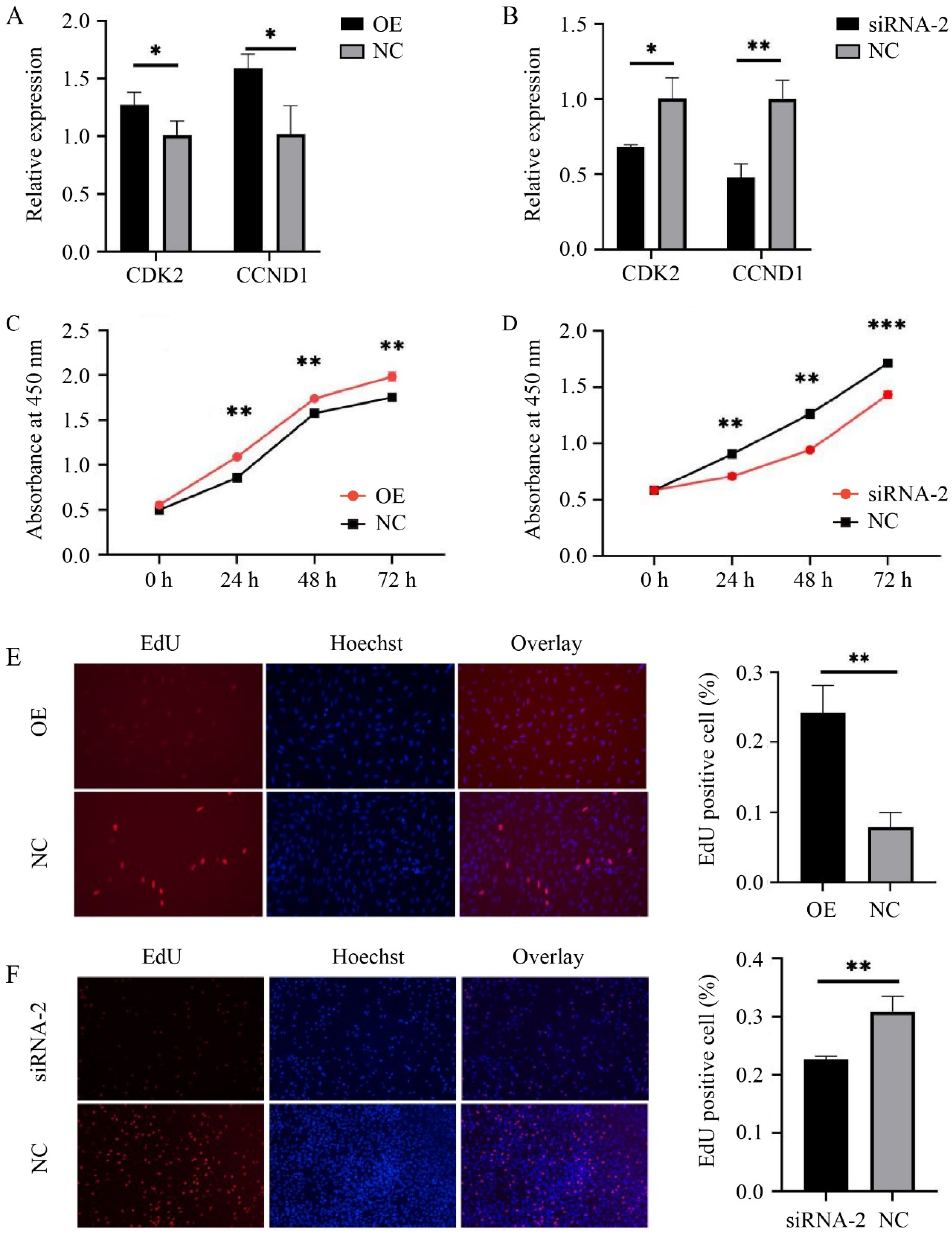
Compared to the control group (NC), the mRNA levels of the cell proliferation marker genes CDK2 (p < 0.05) and CCND1 (p < 0.05) were significantly increased after HMGA2 overexpression (Figure 2A). Conversely, the expression levels of CDK2 (p < 0.05) and CCND1 (p < 0.01) were significantly decreased after interfering with HMGA2 (Figure 2B). The sheep myoblasts were transfected with pcDNA3.1(+)-HMGA2, siRNA-2, and a negative control, respectively, and their cell viability was detected using the CCK-8 assay at 0 h, 24 h, 48 h, and 72 h after transfection (presented as OD value at 450 nm). The results in Figure 2C,D show that the cell viability was significantly higher in the HMGA2 overexpression group compared to the control group (p < 0.01), while the cell viability was significantly lower in the HMGA2 interference group compared to the control group (p < 0.01).
To further determine the effect of the HMGA2 gene on sheep myoblast proliferation, an EdU assay was carried out. As shown in Figure 2E,F, blue fluorescence represents live cells, and red fluorescence represents proliferating cells, with a higher proportion of red indicating more proliferating cells. ImageJ was used for cell counting, and the proportion of EdU-positive cells (red fluorescence) was calculated. Compared to the control group, the proportion of EdU-positive cells was significantly increased by approximately 2.5-fold after HMGA2 overexpression (Figure 2E, p < 0.01), while it was significantly decreased by approximately 1.5-fold after HMGA2 interference (Figure 2F, p < 0.01). These findings indicate that the HMGA2 gene can promote myoblast proliferation in sheep, which is consistent with its role in mice.

3.3. SNP rs428001129 within HMGA2 Promoter Affects Promoter Activity

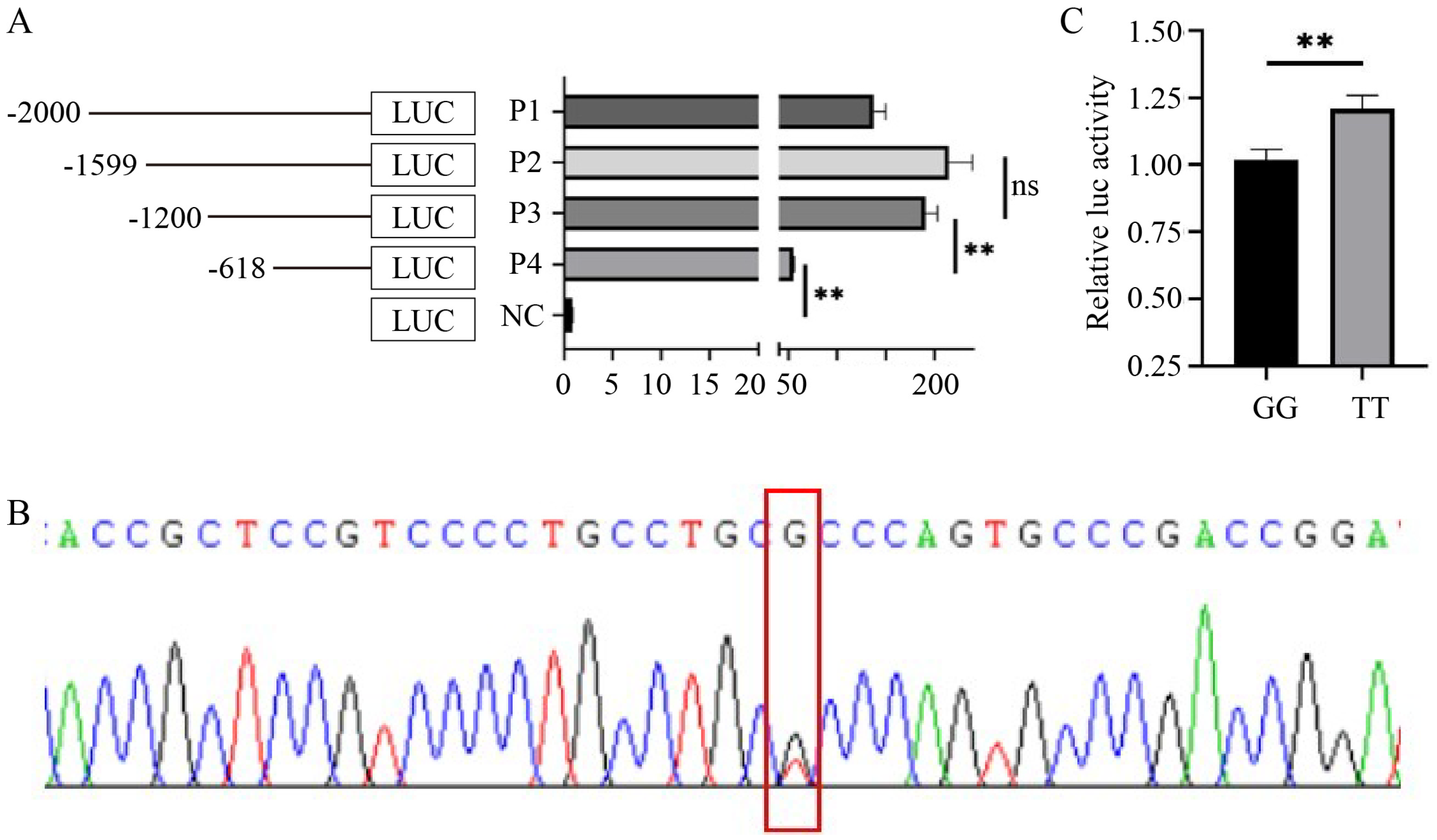
Four different lengths of promoter fragments were amplified, named P1, P2, P3, and P4, and cloned into the pGL3-Basic vector (Figure 3A). The luciferase activities of P1, P2, P3, and P4 were significantly higher than those of the NC (blank pGL3-Basic) group (p < 0.01). However, the luciferase activity of P3 increased ~3-fold compared to P4, while no significant differences between P2 and P3 were observed (p > 0.05), suggesting that the region NC_056056.1: 154134300-154134882 (-618 to -1200 bp upstream of the HMGA2 TSS) contains a core promoter of the HMGA2 gene, but we cannot exclude the possibility that the region P4 has additional core promoter(s).
Using pooled DNA as a template, a fragment containing the HMGA2 core promoter was amplified, followed by Sanger sequencing. An SNP, annotated as rs428001129 (NC_056056.1:g.154134315C>A) in the Ensembl database (ensembl.org), was detected in NC_056056.1: 154134300-154134882 (-618 to -1200 bp upstream of the HMGA2 TSS) (Figure 3B). The amplified products from the DNA of wild-type (GG) and mutant (TT) individuals using SNP-F/R primers were cloned into the pGL3-Basic vector. As shown in Figure 3C, the TT genotype increased the relative luciferase activity remarkably compared to GG (p < 0.01). These results suggest that rs428001129 could change HMGA2 promoter activity and may be associated with sheep growth traits.

3.4. Efficiencies of Overexpression and Interference

Sanger sequencing was used to genotype the SNP rs428001129 in 458 sheep. The genotype frequencies of wild GG, heterozygous GT, and mutant TT were 0.478, 0.454, and 0.068, respectively. The allele frequency of G (0.705) was higher than that of T (0.295). The observed heterozygosity (He) was 0.416, the observed homozygosity (Ho) was 0.584, and the effective allele number (Ne) was 1.712. Notably, rs428001129 was a moderate polymorphism (0.25 < PIC = 0.329 < 0.50) and in Hardy–Weinberg equilibrium, suggesting that rs428001129 does not undergo selection in the crossbred sheep population (Hu sheep × Dorper sheep) used in this study.
The results of the association analysis are shown in Table 2. The SNP rs428001129 was significantly associated with chest width at birth (p < 0.01) and two months old (p < 0.01) and chest depth at two months old (p < 0.05) and six months old (p < 0.01). At birth, the chest width of TT individuals was significantly larger than that of GG and GT individuals, with no significant difference between GG and GT. At two months old, TT individuals had significantly larger chest depth than GG; for chest width, individuals with the TT or GT genotype were larger than GG, with no significant differences between TT and GT. At six months old, TT individuals had significantly larger chest depths than GG and GT individuals, with no significant difference between GT and GG. Notably, at all ages, TT individuals tended to have better performance in body weights than GG and GT individuals, but the differences were not significant. These results suggest that rs428001129 can be used for marker-assisted selection (MAS) in sheep, with the TT being the favorable genotype, conferring significantly larger chest width and depth and slightly higher body weight.

4. Discussion

The HMGA2 gene encodes a non-histone chromatin-binding protein that plays a crucial role in the regulation of gene expression and chromatin remodeling. Using the Expression Atlas in the Ruminant Genome Database (http://animal.omics.pro/code/index.php/RGD, accessed on 17 September 2024), we found that the expression profiling of the HMGA2 gene is consistent across different species. HMGA2 is highly expressed during early embryonic development, playing a vital role in normal embryogenesis. In adult tissues, the expression of the HMGA2 gene is very low or absent, but it may be upregulated in certain tissues and under certain conditions. For example, in testis and ovary, it is involved in maintaining and differentiating stem cells [33]. Furthermore, HMGA2 is often found to be highly expressed in various types of tumors and has garnered significant attention in cancer research, including research on breast cancer, lung cancer, pancreatic cancer, etc. [34]. This overexpression can promote cell proliferation, inhibit apoptosis, and enhance invasive capabilities through different mechanisms, thereby driving tumor development [34].
This study demonstrated that the HMGA2 gene promotes the proliferation of sheep myoblasts and is associated with body size, mirroring its function in various species, including mice, pigs, chickens, and rabbits [10,11,12,13,20]. These results indicated that in addition to its conserved expression pattern, the HMGA2 gene appears to have a conserved phenotypic effect. The conserved phenotypic effect of the HMGA2 gene appears to be related to its sequence. The full length of the sheep HMGA2 gene is approximately 145 kb, with the length of intron 3 alone reaching about 117 kb, while the coding region is only 330 bp, which is almost identical across mammals. To date, the phenotypic effects of the HMGA2 gene and their corresponding molecular mechanisms have been extensively studied, including the LIN28B-let7-HMGA2-IGF2BP2 pathway in muscle development and the HMGA2-PLAG1-IGF2 pathway in body size [35,36,37,38,39].
In contrast, revealing the cis-regulatory elements of the HMGA2 gene is more likely to capture the interest of researchers and breeders. Cis-regulatory elements are specific DNA sequences in or near a gene that are required for the proper spatiotemporal expression of that gene (although the distance of the regulated gene may be many megabases away), often characteristically containing binding sites for transcription factors [40]. Promoters, enhancers, silencers, and insulators are examples of cis-regulatory elements. Genetic variations in cis-regulatory elements could directly impact the gene expression, making them potential markers for disease diagnostics and molecular breeding. The ENCODE cCREs database (https://screen.encodeproject.org/, accessed on 17 September 2024) annotates a large number of cis-regulatory elements within the third intron of the human and mouse HMGA2 genes. Among these, an enhancer within the human HMGA2 gene’s third intron mediates TET1-promoted cell growth in human liver cancer [41]. For livestock, the cattle HMGA2 intronic enhancer (chr5: 47901023–47902297) in a strong sweep region has been confirmed to promote the proliferation of bovine myoblasts [42].
The transcriptional regulation by enhancers depends on their interaction with promoters. Therefore, identifying the core promoter of a gene is beneficial for improving the accuracy of enhancer annotation. To identify the core promoter of the sheep HMGA2 gene, the pGL3-Basic/pRL-TK dual-luciferase reporter system was utilized [43]. Firefly and Renilla luciferases have high sensitivity and efficiency and are popularly employed as reporter genes, which oxidize different substrates to generate quantifiable luminescence [44]. In this study, a series of promoter deletion fragments of HMGA2 were cloned into the promoter region of the firefly luciferase gene of the pGL3-Basic vector (Addgene #212936) with the Renilla luciferase of the pRL-TK vector as an internal control. Therefore, the luminescence ratio can reflect the expression level of firefly luciferase, thereby indicating the promoter activity of different deletion fragments. In other words, a higher luminescence ratio corresponds to higher promoter activity of the deletion fragment. Here, by comparing the relative luciferase activities of four promoter deletion fragments of HMGA2, the region of NC_056056.1: 154134300-154134882 (-618 bp to -1200 bp upstream of TSS) was putatively inferred as one of the habitats of the HMGA2 core promoter. However, this study could not exclude the possibility of other regions also containing the core promoter(s) of the HMGA2 gene, such as the P4 region. This is because further deletion of the P4 fragment was not performed, which is a limitation of this research.
In the fragment of NC_056056.1: 154134300-154134882 (-618 to -1200 bp upstream of the HMGA2 TSS), SNP rs428001129 was detected. It was significantly associated with chest width and depth and had a detectable but not significant effect on the body weight of the crossbred sheep population (Hu sheep × Dorper sheep). A previous study identified SNP and Indel variations in HMGA2, associated with several growth traits in Awassi and Karakul sheep, referred to as ‘intron1:45 A > T’ and ‘intron2:42 A del’ [45]; however, due to the lack of detailed sequence information, we are unable to accurately determine the exact positions of these variations. Together, all these findings provide us with sufficient reason to believe that HMGA2 is the major gene regulating sheep growth and development. Therefore, rs428001129-influenced HMGA2 promoter activity can serve as a molecular marker for the breeding of meat sheep. However, due to the lack of embryonic tissue samples, this study did not investigate the impact of rs428001129 on the HMGA2 gene expression, which is another limitation of this research. In the future, a more in-depth investigation of rs428001129 will be conducted, particularly concerning its potential effects on transcription factor binding.

5. Conclusions

The HMGA2 gene promotes sheep myoblast proliferation, which is consistent with its role in mouse muscle. There is a core promoter of the HMGA2 gene located at NC_056056.1: 154134300-154134882 (-618 to -1200 bp upstream of the HMGA2 TSS). The SNP rs428001129 within the core promoter significantly increases the promoter activity of HMGA2 and is associated with sheep’s chest width and depth, providing candidate sites for marker-assisted breeding in sheep.

Author Contributions

X.C.: conceptualization, data curation, formal analysis, methodology, and writing—original draft; C.L., Y.L. and Y.G.: investigation; J.H.: writing—review and editing; X.C. and W.S.: funding acquisition, project administration, and supervision. All authors have read and agreed to the published version of the manuscript.

Funding

This study was supported by the National Natural Science Foundation of China [32302694], Jiangsu Natural Science Foundation for Colleges and Universities [21KJB230005], Jiangsu Postdoctoral Science Foundation [2021K608C], Major New Varieties of Agricultural Projects in Jiangsu Province [PZCZ201739], and Jiangsu 333 Distinguished Talents Project Foundation [(2022) 2-323].

Institutional Review Board Statement

The experimental animals used in this study were approved by the Yangzhou Univer-sity Laboratory Animal Welfare and Ethics Committee (No. 202103279), and all experiments followed the ethical principles of animal welfare.

Informed Consent Statement

Not applicable.

Data Availability Statement

All data are available within this paper.

Acknowledgments

We are grateful to everyone who helped improve this article.

Conflicts of Interest

The authors declare no conflicts of interest.

References

  1. Yue, G. Reproductive characteristics of Chinese Hu sheep. Anim. Reprod. Sci. 1996, 44, 223–230. [Google Scholar] [CrossRef]
  2. Matarneh, S.K.; Silva, S.L.; Gerrard, D.E. New insights in muscle biology that alter meat quality. Annu. Rev. Anim. Biosci. 2021, 9, 355–377. [Google Scholar] [CrossRef] [PubMed]
  3. Bentzinger, C.F.; Wang, Y.X.; Rudnicki, M.A. Building muscle: Molecular regulation of myogenesis. CSH Perspect. Biol. 2012, 4, a008342. [Google Scholar] [CrossRef] [PubMed]
  4. Millay, D.P.; O’Rourke, J.R.; Sutherland, L.B.; Bezprozvannaya, S.; Shelton, J.M.; Bassel-Duby, R.; Olson, E.N. Myomaker is a membrane activator of myoblast fusion and muscle formation. Nature 2013, 499, 301–305. [Google Scholar] [CrossRef]
  5. Quinn, M.E.; Goh, Q.; Kurosaka, M.; Gamage, D.G.; Petrany, M.J.; Prasad, V.; Millay, D.P. Myomerger induces fusion of non-fusogenic cells and is required for skeletal muscle development. Nat. Commun. 2017, 8, 15665. [Google Scholar] [CrossRef]
  6. Chen, B.; You, W.; Wang, Y.; Shan, T. The regulatory role of Myomaker and Myomixer–Myomerger–Minion in muscle development and regeneration. Cell. Mol. Life Sci. 2020, 77, 1551–1569. [Google Scholar] [CrossRef]
  7. Xiang, X.; Benson, K.F.; Chada, K. Mini-mouse: Disruption of the pygmy locus in a transgenic insertional mutant. Science 1990, 247, 967–969. [Google Scholar] [CrossRef]
  8. Benson, K.F.; Chada, K. Mini-mouse: Phenotypic characterization of a transgenic insertional mutant allelic to pygmy. Genet. Res. 1994, 64, 27–33. [Google Scholar] [CrossRef]
  9. Zhou, X.; Benson, K.F.; Ashar, H.R.; Chada, K. Mutation responsible for the mouse pygmy phenotype in the developmentally regulated factor HMGI-C. Nature 1995, 376, 771–774. [Google Scholar] [CrossRef]
  10. Chung, J.; Zhang, X.; Collins, B.; Sper, R.B.; Gleason, K.; Simpson, S.; Koh, S.; Sommer, J.; Flowers, W.L.; Petters, R.M. High mobility group A2 (HMGA2) deficiency in pigs leads to dwarfism, abnormal fetal resource allocation, and cryptorchidism. Proc. Natl. Acad. Sci. USA 2018, 115, 5420–5425. [Google Scholar] [CrossRef]
  11. Ruyter-Spira, C.P.; de Groof, A.; van der Poel, J.J.; Herbergs, J.; Masabanda, J.; Fries, R.; Groenen, M. The HMGI-C gene is a likely candidate for the autosomal dwarf locus in the chicken. J. Hered. 1998, 89, 295–300. [Google Scholar] [CrossRef] [PubMed]
  12. Ruyter-Spira, C.; Herbergs, J.; Limpens, E.; Marsh, J.; van Der Poel, J.; Ayoubi, T.; Groenen, M. Nucleotide sequence of the chicken HMGI-C cDNA and expression of the HMGI-C and IGF1 genes in autosomal dwarf chicken embryos. BBA-Gene Struct. Expr. 1998, 1399, 83–87. [Google Scholar] [CrossRef] [PubMed]
  13. Carneiro, M.; Hu, D.; Archer, J.; Feng, C.; Afonso, S.; Chen, C.; Blanco-Aguiar, J.A.; Garreau, H.; Boucher, S.; Ferreira, P.G. Dwarfism and altered craniofacial development in rabbits is caused by a 12.1 kb deletion at the HMGA2 locus. Genetics 2017, 205, 955–965. [Google Scholar] [CrossRef]
  14. Hammond, S.M.; Sharpless, N.E. HMGA2, microRNAs, and stem cell aging. Cell 2008, 135, 1013–1016. [Google Scholar] [CrossRef]
  15. Smeti, I.; Watabe, I.; Savary, E.; Fontbonne, A.; Zine, A. HMGA2, the architectural transcription factor high mobility group, is expressed in the developing and mature mouse cochlea. PLoS ONE 2014, 9, e88757. [Google Scholar] [CrossRef]
  16. Reeves, R.; Beckerbauer, L. HMGI/Y proteins: Flexible regulators of transcription and chromatin structure. BBA-Gene Struct. Expr. 2001, 1519, 13–29. [Google Scholar] [CrossRef]
  17. Cui, T.; Leng, F. Specific recognition of AT-rich DNA sequences by the mammalian high mobility group protein AT-hook 2: A SELEX study. Biochemistry 2007, 46, 13059–13066. [Google Scholar] [CrossRef]
  18. Zha, L.; Wang, Z.; Tang, W.; Zhang, N.; Liao, G.; Huang, Z. Genome-wide analysis of HMGA2 transcription factor binding sites by ChIP on chip in gastric carcinoma cells. Mol. Cell. Biochem. 2012, 364, 243–251. [Google Scholar] [CrossRef]
  19. Vignali, R.; Marracci, S. HMGA genes and proteins in development and evolution. Int. J. Mol. Sci. 2020, 21, 654. [Google Scholar] [CrossRef]
  20. Li, Z.; Gilbert, J.A.; Zhang, Y.; Zhang, M.; Qiu, Q.; Ramanujan, K.; Shavlakadze, T.; Eash, J.K.; Scaramozza, A.; Goddeeris, M.M. An HMGA2-IGF2BP2 axis regulates myoblast proliferation and myogenesis. Dev. Cell 2012, 23, 1176–1188. [Google Scholar] [CrossRef]
  21. Makvandi-Nejad, S.; Hoffman, G.E.; Allen, J.J.; Chu, E.; Gu, E.; Chandler, A.M.; Loredo, A.I.; Bellone, R.R.; Mezey, J.G.; Brooks, S.A. Four loci explain 83% of size variation in the horse. PLoS ONE 2012, 7, e39929. [Google Scholar] [CrossRef] [PubMed]
  22. Hayward, J.J.; Castelhano, M.G.; Oliveira, K.C.; Corey, E.; Balkman, C.; Baxter, T.L.; Casal, M.L.; Center, S.A.; Fang, M.; Garrison, S.J. Complex disease and phenotype mapping in the domestic dog. Nat. Commun. 2016, 7, 10460. [Google Scholar] [CrossRef] [PubMed]
  23. Cesar, A.S.; Regitano, L.C.; Reecy, J.M.; Poleti, M.D.; Oliveira, P.S.; de Oliveira, G.B.; Moreira, G.C.; Mudadu, M.A.; Tizioto, P.C.; Koltes, J.E. Identification of putative regulatory regions and transcription factors associated with intramuscular fat content traits. BMC Genom. 2018, 19, 499. [Google Scholar] [CrossRef]
  24. Bolormaa, S.; Pryce, J.E.; Reverter, A.; Zhang, Y.; Barendse, W.; Kemper, K.; Tier, B.; Savin, K.; Hayes, B.J.; Goddard, M.E. A multi-trait, meta-analysis for detecting pleiotropic polymorphisms for stature, fatness and reproduction in beef cattle. PLoS Genet. 2014, 10, e1004198. [Google Scholar] [CrossRef]
  25. Selionova, M.; Aibazov, M.; Sermyagin, A.; Belous, A.; Deniskova, T.; Mamontova, T.; Zharkova, E.; Zinovieva, N. Genome-Wide Association and Pathway Analysis of Carcass and Meat Quality Traits in Karachai Young Goats. Animals 2023, 13, 3237. [Google Scholar] [CrossRef]
  26. Saatchi, M.; Schnabel, R.D.; Taylor, J.F.; Garrick, D.J. Large-effect pleiotropic or closely linked QTL segregate within and across ten US cattle breeds. BMC Genom. 2014, 15, 442. [Google Scholar] [CrossRef]
  27. Bouwman, A.C.; Daetwyler, H.D.; Chamberlain, A.J.; Ponce, C.H.; Sargolzaei, M.; Schenkel, F.S.; Sahana, G.; Govignon-Gion, A.; Boitard, S.; Dolezal, M. Meta-analysis of genome-wide association studies for cattle stature identifies common genes that regulate body size in mammals. Nat. Genet. 2018, 50, 362–367. [Google Scholar] [CrossRef]
  28. Danoviz, M.E.; Yablonka-Reuveni, Z. Skeletal muscle satellite cells: Background and methods for isolation and analysis in a primary culture system. Myogenesis Methods Protoc. 2012, 798, 21–52. [Google Scholar]
  29. Di Pietro, F.; Ortenzi, F.; Tilio, M.; Concetti, F.; Napolioni, V. Genomic DNA extraction from whole blood stored from 15-to 30-years at− 20 C by rapid phenol–chloroform protocol: A useful tool for genetic epidemiology studies. Mol. Cell. Probes 2011, 25, 44–48. [Google Scholar] [CrossRef]
  30. Simms, D.; Cizdziel, P.E.; Chomczynski, P. TRIzol: A new reagent for optimal single-step isolation of RNA. Focus 1993, 15, 532–535. [Google Scholar]
  31. Wang, S.; Cao, X.; Ge, L.; Gu, Y.; Lv, X.; Getachew, T.; Mwacharo, J.M.; Haile, A.; Sun, W. MiR-22-3p inhibits proliferation and promotes differentiation of skeletal muscle cells by targeting IGFBP3 in Hu sheep. Animals 2022, 12, 114. [Google Scholar] [CrossRef] [PubMed]
  32. Yong, Y.; He, L. SHEsis, a powerful software platform for analyses of linkage disequilibrium, haplotype construction, and genetic association at polymorphism loci. Cell Res. 2005, 15, 97. [Google Scholar]
  33. Ashar, H.R.; Chouinard, R.A., Jr.; Dokur, M.; Chada, K. In vivo modulation of HMGA2 expression. BBA-Gene Regul. Mech. 2010, 1799, 55–61. [Google Scholar] [CrossRef] [PubMed]
  34. Mansoori, B.; Mohammadi, A.; Ditzel, H.J.; Duijf, P.H.; Khaze, V.; Gjerstorff, M.F.; Baradaran, B. HMGA2 as a critical regulator in cancer development. Genes 2021, 12, 269. [Google Scholar] [CrossRef]
  35. Zhu, H.; Shah, S.; Shyh-Chang, N.; Shinoda, G.; Einhorn, W.S.; Viswanathan, S.R.; Takeuchi, A.; Grasemann, C.; Rinn, J.L.; Lopez, M.F. Lin28a transgenic mice manifest size and puberty phenotypes identified in human genetic association studies. Nat. Genet. 2010, 42, 626–630. [Google Scholar] [CrossRef]
  36. Copley, M.R.; Babovic, S.; Benz, C.; Knapp, D.J.; Beer, P.A.; Kent, D.G.; Wohrer, S.; Treloar, D.Q.; Day, C.; Rowe, K. The Lin28b–let-7–Hmga2 axis determines the higher self-renewal potential of fetal haematopoietic stem cells. Nat. Cell Biol. 2013, 15, 916–925. [Google Scholar] [CrossRef]
  37. Powers, J.T.; Tsanov, K.M.; Pearson, D.S.; Roels, F.; Spina, C.S.; Ebright, R.; Seligson, M.; De Soysa, Y.; Cahan, P.; Theißen, J. Multiple mechanisms disrupt the let-7 microRNA family in neuroblastoma. Nature 2016, 535, 246–251. [Google Scholar] [CrossRef]
  38. Dai, N.; Zhao, L.; Wrighting, D.; Krämer, D.; Majithia, A.; Wang, Y.; Cracan, V.; Borges-Rivera, D.; Mootha, V.K.; Nahrendorf, M. IGF2BP2/IMP2-deficient mice resist obesity through enhanced translation of Ucp1 mRNA and other mRNAs encoding mitochondrial proteins. Cell Metab. 2015, 21, 609–621. [Google Scholar] [CrossRef]
  39. Abi Habib, W.; Brioude, F.; Edouard, T.; Bennett, J.T.; Lienhardt-Roussie, A.; Tixier, F.; Salem, J.; Yuen, T.; Azzi, S.; Le Bouc, Y. Genetic disruption of the oncogenic HMGA2–PLAG1–IGF2 pathway causes fetal growth restriction. Genet. Med. 2018, 20, 250–258. [Google Scholar] [CrossRef]
  40. Preissl, S.; Gaulton, K.J.; Ren, B. Characterizing cis-regulatory elements using single-cell epigenomics. Nat. Rev. Genet. 2023, 24, 21–43. [Google Scholar] [CrossRef]
  41. Shirai, K.; Nagae, G.; Seki, M.; Kudo, Y.; Kamio, A.; Hayashi, A.; Okabe, A.; Ota, S.; Tsutsumi, S.; Fujita, T. TET1 upregulation drives cancer cell growth through aberrant enhancer hydroxymethylation of HMGA2 in hepatocellular carcinoma. Cancer Sci. 2021, 112, 2855–2869. [Google Scholar] [CrossRef] [PubMed]
  42. Cheng, J.; Cao, X.; Wang, X.; Wang, J.; Yue, B.; Sun, W.; Huang, Y.; Lan, X.; Ren, G.; Lei, C. Dynamic chromatin architectures provide insights into the genetics of cattle myogenesis. J. Anim. Sci. Biotechnol. 2023, 14, 59. [Google Scholar] [CrossRef] [PubMed]
  43. Deng, Z.; Zhang, Y.; Li, L.; Xie, X.; Huang, J.; Zhang, M.; Ni, X.; Li, X. A dual-luciferase reporter system for characterization of small RNA target genes in both mammalian and insect cells. Insect Sci. 2022, 29, 631–644. [Google Scholar] [CrossRef] [PubMed]
  44. Xu, Y.Z.; Kanagaratham, C.; Jancik, S.; Radzioch, D. Promoter deletion analysis using a dual-luciferase reporter system. Gene Regul. Methods Protoc. 2013, 977, 79–93. [Google Scholar]
  45. Aljubouri, T.R.S.; Al-Shuhaib, M.B.S.; Javadmanesh, A. HMGA2 gene polymorphisms and their effects on main growth traits indices in Awassi and Karakul sheep. Agric. Nat. Resour. 2020, 54, 587–594. [Google Scholar]
Figure 1. Efficiency detection of HMGA2 overexpression and interference. (A) Agarose gel electrophoresis of the recombinant overexpression vector pcDNA3.1(+)-HMGA2 digested with Hind Ⅲ and Xho I; (B) overexpression efficiency of pcDNA3.1(+)-HMGA2 (OE); (C) comparison of interference efficiency of three HMGA2 siRNAs. ** p < 0.01.
Animals 14 02721 g001
Figure 2. HMGA2 promotes proliferation of sheep myoblasts. (A,B) The relative expression of the marker genes of cell proliferation after HMGA2 overexpression or interference; (C,D) cell viability, detected by CCK-8 (presented as the OD value at 450 nm); (E,F) the proportion of EdU-positive cells (blue: live cells, red: proliferating cells). * p < 0.05, ** p < 0.01, and *** p < 0.001.
Animals 14 02721 g002
Figure 3. Identification of HMGA2 core promoter and variation. (A) Relative luciferase activities of different lengths of promoter fragments; (B) Sanger sequencing detected SNP rs428001129 in NC_056056.1: 154134300-154134882 (-618 to -1200 bp upstream of the HMGA2 TSS) containing a core promoter; (C) rs428001129 changed HMGA2 promoter activity. P1, NC_056056.1: 154133741-154135682 (upstream of TSS, -59 to -2000 bp); P2, NC_056056.1: 154133741-154135281 (upstream of TSS, -59 to -1599 bp); P3, NC_056056.1: 154133741-154134882 (upstream of TSS, -59 to -1200 bp); P4, NC_056056.1: 154133741-154134300 (upstream of TSS, -59 to -618 bp). ** p < 0.01.
Animals 14 02721 g003
Table 1. Sequences of primers and siRNAs.
IDSequence (5′-3′)Product
siRNA-1CCGGUGAGCCCUCUCCUAATT\
UAGGAGAGGGCUCACCGGTT
siRNA-2GAGACAUCCUCACAAGAGUTT\
ACUCUUGUGAGGAUGUCUCTT
siRNA-3GCCAGCAUUCAAUUUCUACTT\
GUAGAAAUUGAAUGCUGGCTT
siRNA-NCUUCUCCGAACGUGUCACGUTT\
ACGUGACACGUUCGGAGAATT
CDK2-FTGGGCCAGGCAGGATTTTAG166 bp
CDK2-RGTCGAAGGTGAGGTACTGGC
CCND1-FGCTTCCTCTCCTATCACCGC149 bp
CCND1-RGGCTTTGGGGTCCAAGTTCT
GAPDH-FGTCGGAGTGAACGGATTTGG196 bp
GAPDH-RCATTGATGACGAGATTCCCG
HMGA2-FAGACCCAAAGGCAGCAAAAAC100 bp
HMGA2-RGCCATTTCCTAGGTCTGCCTC
P1cgagctcttacgcgtgctagcAAAAGTTTTTATTTTGGAATTG1942 bp
P2cgagctcttacgcgtgctagcTCAGTGGAGGCTGGTGCG1541 bp
P3cgagctcttacgcgtgctagcCAGGTAAAGGCCAAGCCCC1142 bp
P4cgagctcttacgcgtgctagcGGATCCCCGCAGAATCTCC560 bp
RacttagatcgcagatctcgagTCGCCTCTGTCGCCCTGA\
SNP-FGCCTCCCTCCTCCTCATACT492 bp
SNP-RCGGCTTGGAAAGGGAAGAGA
Note: lowercase bases are homology arm sequences matched to pGL3-Basic, and underlined bases are Xho I and Nhe I restriction sites.
Table 2. Association analysis between rs428001129 and sheep growth traits.
AgeTraitsGenotypep Value
GGGTTT
birthbody weight3.67 ± 0.813.73 ± 0.843.95 ± 0.940.250
withers height37.63 ± 3.1337.90 ± 2.8838.85 ± 2.470.141
body length30.46 ± 2.7830.88 ± 3.0131.17 ± 2.990.264
chest girth35.62 ± 2.8935.82 ± 3.0337.17 ± 4.540.055
chest depth15.60 ± 2.1615.58 ± 1.5215.90 ± 1.850.704
chest width10.16 ± 1.48 b10.44 ± 1.50 b11.06 ± 1.65 a0.009
cannon circumference5.83 ± 0.685.93 ± 0.706.06 ± 0.620.183
1 mthbody weight10.22 ± 2.439.82 ± 2.2410.30 ± 2.320.545
withers height45.74 ± 3.0644.93 ± 3.1143.85 ± 3.480.095
body length43.43 ± 3.8342.71 ± 3.4842.70 ± 2.910.455
chest girth50.06 ± 4.3449.66 ± 3.6550.30 ± 3.560.781
chest depth22.23 ± 1.8621.95 ± 1.7121.85 ± 1.620.568
chest width14.43 ± 1.5114.24 ± 1.4714.30 ± 1.670.725
cannon circumference6.50 ± 0.496.38 ± 0.456.45 ± 0.370.292
2 mthbody weight17.85 ± 3.8118.28 ± 3.8918.64 ± 4.490.411
withers height49.75 ± 3.4150.28 ± 3.5850.68 ± 3.950.216
body length51.93 ± 4.5452.53 ± 4.9452.75 ± 4.810.409
chest girth59.85 ± 5.0860.66 ± 4.8260.96 ± 5.660.232
chest depth23.54 ± 2.22 b23.88 ± 2.19 ab24.61 ± 2.31 a0.040
chest width16.34 ± 2.23 b16.94 ± 2.40 a17.79 ± 2.94 a0.002
cannon circumference6.92 ± 0.587.00 ± 0.627.20 ± 0.790.062
3 mthbody weight24.41 ± 3.9524.44 ± 4.0824.70 ± 3.900.981
withers height54.70 ± 3.0254.87 ± 3.3055.19 ± 3.270.912
body length56.64 ± 3.0656.98 ± 3.2458.06 ± 3.120.490
chest girth67.32 ± 4.2767.13 ± 4.7067.38 ± 3.420.973
chest depth26.83 ± 1.6926.90 ± 1.7026.81 ± 1.280.975
chest width17.71 ± 1.6417.95 ± 1.5318.13 ± 1.300.659
cannon circumference7.44 ± 0.447.36 ± 0.547.50 ± 0.380.632
6 mthbody weight37.33 ± 7.3937.50 ± 7.4337.90 ± 8.650.848
withers height61.44 ± 4.2561.83 ± 3.8162.43 ± 4.260.292
body length66.18 ± 4.6865.90 ± 4.6267.33 ± 5.070.313
chest girth77.90 ± 6.0077.70 ± 5.9578.00 ± 7.400.969
chest depth28.66 ± 2.13 b28.82 ± 2.15 b29.72 ± 2.93 a0.047
chest width19.33 ± 2.2119.47 ± 2.3220.02 ± 2.300.269
cannon circumference8.05 ± 0.848.02 ± 0.818.38 ± 1.070.107
a,b Values without letters or with the same letter in the same row indicate no significant differences (p > 0.05); values with different letters in the same row indicate significant differences (p < 0.05). mnt, month(s).
Disclaimer/Publisher’s Note: The statements, opinions and data contained in all publications are solely those of the individual author(s) and contributor(s) and not of MDPI and/or the editor(s). MDPI and/or the editor(s) disclaim responsibility for any injury to people or property resulting from any ideas, methods, instructions or products referred to in the content.