Article

Comparative Transcriptome Analysis of the Hepatopancreas from Macrobrachium rosenbergii Exposed to the Heavy Metal Copper

1
College of Fisheries, Guangdong Ocean University, Zhanjiang 524025, China
2
Guangdong Provincial Key Laboratory of Aquatic Animal Disease Control and Healthy Culture, Zhanjiang 524088, China
*
Author to whom correspondence should be addressed.
Animals 2024, 14(7), 1117; https://doi.org/10.3390/ani14071117
Submission received: 29 February 2024 / Revised: 31 March 2024 / Accepted: 3 April 2024 / Published: 5 April 2024
(This article belongs to the Special Issue Ecotoxicology in Aquatic Animals)

Abstract

:

Simple Summary

The pollution of aquatic ecosystems with the heavy metal copper (Cu) presents considerable risks to the physiological functions of aquatic organisms, including the economically valuable Macrobrachium rosenbergii. In this study, transcriptomic analysis was conducted to explore the molecular response mechanisms in the hepatopancreas of M. rosenbergii exposed to Cu. The results showed that M. rosenbergii attempts to resist the toxicity of Cu by up-regulating the expression of genes related to immunity, metabolism, and detoxification. However, with the excessive accumulation of reactive oxygen species (ROS), the antioxidant enzyme system was destroyed. As a result, DNA damage repair and the cellular stress response were inhibited, thereby exacerbating cell damage. In order to maintain the normal function of the hepatopancreas, M. rosenbergii removes damaged cells by activating the apoptosis mechanism. Differentially expressed genes (DEGs) and KEGG pathways identified in the present study not only facilitate an understanding of the molecular response mechanisms of M. rosenbergii underlying Cu toxicity effects but also help in identifying potential biomarkers associated with the stress response in other crustaceans.

Abstract

The contamination of aquatic ecosystems by the heavy metal copper (Cu) is an important environmental issue and poses significant risks to the physiological functions of aquatic organisms. Macrobrachium rosenbergii is one of the most important freshwater-cultured prawns in the world. The hepatopancreas of crustaceans is a key organ for immune defense, heavy metal accumulation, and detoxification, playing a pivotal role in toxicological research. However, research on the molecular response of the hepatopancreas in M. rosenbergii to Cu exposure is still lacking. In this study, the transcriptomic response in the hepatopancreas of M. rosenbergii was studied after Cu exposure for 3 and 48 h. Compared with the control group, 11,164 (7288 up-regulated and 3876 down-regulated genes) and 10,937 (6630 up-regulated and 4307 down-regulated genes) differentially expressed genes (DEGs) were identified after 3 and 48 h exposure, respectively. Most of these DEGs were up-regulated, implying that gene expressions were largely induced by Cu. Functional enrichment analysis of these DEGs revealed that immunity, copper homeostasis, detoxification, DNA damage repair, and apoptosis were differentially regulated by Cu. Seven genes involved in immunity, detoxification, and metabolism were selected for validation by qRT-PCR, and the results confirmed the reliability of RNA-Seq. All these findings suggest that M. rosenbergii attempts to resist the toxicity of Cu by up-regulating the expression of genes related to immunity, metabolism, and detoxification. However, with the excessive accumulation of reactive oxygen species (ROS), the antioxidant enzyme system was destroyed. As a result, DNA damage repair and the cellular stress response were inhibited, thereby exacerbating cell damage. In order to maintain the normal function of the hepatopancreas, M. rosenbergii removes damaged cells by activating the apoptosis mechanism. Our study not only facilitates an understanding of the molecular response mechanisms of M. rosenbergii underlying Cu toxicity effects but also helps us to identify potential biomarkers associated with the stress response in other crustaceans.

1. Introduction

Copper (Cu), a redox-active transition metal, is a double-edged sword. On the one hand, Cu is an indispensable micronutrient for the development and homeostasis of all living organisms [1]. For example, it is an important cofactor of enzymes, including copper–zinc superoxide dismutase (SOD 1), cytochrome c oxidase (CCO), and lysyl oxidase (LOX) [2]. On the other hand, excessive Cu in the aquatic environment leads to the accumulation of reactive oxygen species (ROS) in aquatic organisms, leading to DNA damage, apoptosis, and tissue structure damage [3,4,5]. In addition, Cu can cause damage to the mitochondrial membrane of crustaceans and disrupt normal metabolism [6]. In recent years, Cu has been widely used in industrial production activities as an important conductive material and raw material for manufacturing alloys, as an effective algaecide for inhibiting toxic algal blooms, and as an essential mineral in feed [7,8,9,10]. These activities undoubtedly cause Cu contamination in aquaculture environments. The latest research showed that Cu pollution in the lower reaches of the Yangtze River is already widespread [11].
Macrobrachium rosenbergii is primarily distributed in inland freshwater areas. Because of its short culture cycle, high nutritional value, and other advantages, it has become an important economic species in China [12]. In addition, M. rosenbergii has always been considered an important model organism with relevance for many research fields such as ecology [13], physiology [14], and toxicology [15]. The hepatopancreas has metabolic, immunological, and antidotal functions, which are important for the healthy growth of crustaceans and resistance to external environmental stimuli [16,17]. Numerous studies have found that the hepatopancreas is a key target organ for heavy metal accumulation [18,19]. High concentrations of heavy metals in aquatic environments can lead to tissue damage, metabolic disorders, and immune suppression in the hepatopancreas [20,21,22]. Up to now, the effects of Cu exposure on the hepatopancreas of M. rosenbergii have mainly focused on tissue enrichment [23], structural damage [24], and enzyme activity [25], while the specific molecular response mechanisms of the hepatopancreas remain poorly understood.
Transcriptome sequencing could identify genes and pathways involved in immunity and metabolism under environmental pollution stress, and it has been widely used in the study of the stress response of aquatic animals [26,27]. In this study, transcriptome sequencing was used to obtain genes and pathways that respond to Cu exposure in the hepatopancreas of M. rosenbergii at different time points, and to explore the molecular mechanisms against Cu toxicity. These findings not only provide new insights into the molecular response mechanisms underlying Cu toxicity effects but also help in identifying potential biomarkers associated with the stress response in other crustaceans.

2. Materials and Methods

2.1. Experimental Animal

The experimental M. rosenbergii (10.89 ± 1.42 g, both genders) were raised at the Donghai Island Research Base of Guangdong Ocean University. M. rosenbergii were acclimatized in three cycling-filtered water tanks containing aerated and filtered freshwater (200 L) for 2 weeks. The water temperature, pH, and dissolved oxygen (DO) were 24–28 °C, 7.5–7.9, and 5.36–6.18 mg/L, respectively. Half of the water was renewed daily to maintain good water quality. During this period, the prawns were fed twice a day with a commercial diet (supplied by Haid Group Co., Ltd., Zhanjiang, China) until 24 h before the beginning of the Cu exposure experiment.

2.2. Cu Exposure Experiment and Sample Collection

In order to investigate the physiological response of M. rosenbergii to Cu exposure, these M. rosenbergii were randomly divided into two groups. The control group was still in the initial conditions, and the exposed group was conducted in the tank containing 200 L of Cu solution (100 μg/L) [28]. Each group was conducted in three replicates, with twenty prawns per tank. The desired Cu concentration was attained by adding CuSO4 to freshwater. The Agilent model 7500cx ICP-MS (Agilent Technologies, Santa Clara, CA, USA) detection system was used to determine the actual Cu concentrations, using operating parameters recommended by the manufacturer. The actual Cu concentrations of the experiment were 11.0 (control) and 98.9 μg/L (mean, n = 3), respectively. At the beginning of the experiment, nine samples were randomly selected from the control group (Hep_0 h) to collect their hepatopancreases. Nine hepatopancreas samples were collected from the exposed group at 3 h (Hep_3 h) and 48 h (Hep_48 h), respectively. All of the hepatopancreas tissues were immediately placed in liquid nitrogen for freezing and then stored at −80 °C until RNA extraction.

2.3. RNA Extraction, Library Construction, and RNA Sequencing

Total RNA was extracted from the hepatopancreas of M. rosenbergii using Trizol reagent (Invitrogen, Carlsbad, CA, USA) according to the manufacturer’s protocol. The quantity and quality of these RNA samples were detected by measuring their absorbance at 260 and 280 nm using a NanoDrop 2000 spectrophotometer (NanoDrop Technologies, Wilmington, DE, USA). The integrity of RNA was assayed by agarose gel electrophoresis. After qualification, equal amounts of RNA from nine individuals in each group were mixed together as a sample to construct the transcriptome library according to the instructions of the Illumina TruSeq RNA Sample Preparation Kit (Illumina Inc., San Diego, CA, USA). In brief, after treating total RNA with DNasel, mRNA was enriched using oligo (dT) magnetic beads. Then, fragmentation buffer was added to break the purified mRNA into short fragments (about 200 bp). These small mRNA fragments were used as templates to synthesize the first complementary DNA (cDNA) strand using random hexamers, Rnase H and SuperScript II Reverse Transcriptase (Invitrogen, Life Technologies). Next, the second strand of cDNA was synthesized, followed by adhesive end repair and the addition of base A at 3′ ends. Proper cDNA fragments were purified by agarose gel electrophoresis and subsequently enriched by PCR amplification. After passing the quality inspection of the Agilent 2100 Bioanalyzer (Agilent Technologies Inc., Santa Clara, CA, USA) and the ABI StepOnePlus Real-Time PCR System (Thermo Fisher Scientific, MA, USA), the constructed libraries were sequenced using the Illumina Hiseq 4000 platform, and 150 bp paired-end raw reads were generated.

2.4. Transcriptome Assembly and Functional Annotation Analysis

Adaptor sequences, sequences with more than 5% N bases, and low-quality sequences (more than 50% bases with quality value ≤ 10) were removed from the raw reads. De novo assembly of the remaining clean reads was performed using Trinity software (version: trinityrnaseq_r20140717) to generate large contigs [29]. TGICL V2.1 was used to eliminate redundant sequences and perform additional assembly, resulting in transcripts called unigenes [30]. All unigenes were annotated by comparison with non-redundant protein (Nr), non-redundant nucleotide (Nt), SwissProt, Gene Ontology (GO), Clusters of Orthologous Groups of Proteins (COG), and Kyoto Encyclopedia of Genes and Genomes (KEGG) databases using BLAST (version: v2.2.26), with a cut-off E-value of 1 × 10⁻⁵ [31]. Based on the Nr BLAST results, Blast2GO version 2.6.6 was used to conduct GO annotation [32]. KEGG pathway analysis was conducted using KOBAS standalone version 2.0 [33].

2.5. Identification of Differentially Expressed Genes (DEGs) and Functional Enrichment Analysis

The expression level of each unigene was calculated and standardized by Fragment Per Kilo bases per Million Reads (FPKM), using the software RNA-Seq by Expectation Maximization (RSEM: v1.2.21) [34]. Differentially expressed genes (DEGs) were identified by the R package DEGseq (version: v1.18.0) [35] with the threshold set to |Log2Fold Change| ≥ 1 and a false discovery rate (FDR) ≤ 0.01 [36]. Then, all DEGs in the three paired comparisons (Hep_0 h vs. Hep_3 h, Hep_0 h vs. Hep_48 h, and Hep_3 h vs. Hep_48 h) were subjected to GO functional and KEGG pathway enrichment analysis using Goseq V1.16.2 [37] and KOBAS [33], respectively. We defined the p-value < 0.05 as significantly enriched.

2.6. Simple Sequence Repeat (SSR) and Single-Nucleotide Polymorphism (SNP) Detection

Simple sequence repeats (SSRs) were identified by using MISA version 2.2.2 [38]. Specific selection criteria were as follows: mono-nucleotide ≥ 12, di-nucleotide ≥ 6, tri-nucleotide and tetra-nucleotide ≥ 5, penta- and hexa-nucleotide ≥ 4. PCR primers for each detected SSR were designed using Primer3 with default parameters. A single-nucleotide polymorphism (SNP) primarily refers to the polymorphism of a DNA sequence caused by the variation of a single nucleotide at the genomic level. We employed the Genome Analysis Toolkit (GATK, version 3.4.0) for the detection of SNP markers [39]. Subsequently, the generated raw Variant Call Format (VCF) files were subjected to filtration using the GATK’s prescribed standard filtering methodology.

2.7. Validation of RNA-Seq Results with Quantitative Real-Time PCR (qRT-PCR)

Seven DEGs involved in immunity, detoxification, and metabolism, such as relish, glucose transporter 2 (Glut 2), JHE-like carboxylesterase 2 (CXE2), hemocyanin (HC), glutathione S-transferase (GST), serine/threonine–protein phosphatase 6 regulatory subunit 3 (SAPS3), and C-type lectin (CTL), were selected to validate the RNA-Seq results, and the elongation factor-1 gene (EF-1) was chosen as an internal reference gene [40]. These genes were analyzed by qRT-PCR. Primers for qRT-PCR were designed by Primer Premier 6.0 (Premier Biosoft, Palo Alto, CA, USA), as shown in Table 1. qRT-PCR was conducted with ChamQ Universal SYBR qPCR Master Mix (Vazyme, Nanjing, China), with the following conditions: 95 °C for 6 min, followed by 40 cycles of 95 °C for 5 s, 60 °C for 15 s, 72 °C for 10 s. The relative gene expression levels of the chosen genes were calculated using the 2−ΔΔCt method [41].

3. Results

3.1. RNA Sequencing and De Novo Assembly

The transcriptome sequencing results are shown in Table 2. A total of 179,549,702 raw reads were generated from the three cDNA libraries of M. rosenbergii. After removing the reads containing adapter sequences and low-quality sequences, a total of 178,793,124 clean reads were obtained. The Trinity software (version: trinityrnaseq_r20140717) was used to assemble the obtained clean reads, and a total of 85,984 unigenes were generated (with a mean length of 1077.23 bp and N50 of 2559 bp) (Table S1). A total of 27,679 unigenes at 200–300 bp accounted for the largest ratio, as high as 32.19%. There were only 8010 unigenes with a length greater than 3000 bp, accounting for 9.32% of the total (Figure S1).

3.2. Functional Annotation and Classification of the Transcriptome

Among all the assembled sequences, a total of 85,984 unigenes were annotated within six functional databases (Table S2), and it was found that 10,741 (12.49%), 13,535 (15.74%), 20,601 (23.96%), 24,394 (28.37%), 13,110 (15.25%), and 20,528 (23.87%) unigenes could find significant hits in the COG, GO, KEGG, Nr, Nt, and SwissProt databases, respectively (Figure S2). A total of 5614 unigenes (6.53%) were mapped to all databases (Figure S3). The species distribution of the most significant hits in the Nr database was analyzed, and the results showed that the maximum number of homologous sequences was found in the arthropods Zootermopsis nevadensis and Limulus polyphemus with high percentages of 10.59% (2583) and 5.55% (1353), respectively. The percentage of unigenes matched to several crustacean species was relatively low, such as in Litopenaeus vannamei (1.29%), Penaeus monodon (0.93%), Marsupenaeus japonicus (0.74%), Macrobrachium nipponense (0.73%), Procambarus clarkii (0.68%), and M. rosenbergii (0.68%) (Figure S4), which may be due to the lack of gene/protein information on crustaceans in the database.
The GO functional analysis showed that a total of 13,535 unigenes were arranged into three main categories: biological process, cellular component, and molecular function. In the biological process category, a large percentage of the unigenes belonged to cellular process (4923), followed by metabolic process (4848) and single-organism process (3746). In the cellular component category, the dominant subcategories were ordered as follows: cell (2100), cell part (2100), membrane (1943). In the molecular function category, most unigenes were related to binding (8403), catalytic activity (4478), and transporter activity (699) (Figure S5). KEGG functional analysis indicated that a total of 20,601 unigenes were mapped to six major types of pathways, namely genetic information processing, metabolism, cellular processes, organismal systems, human diseases, and environmental information processing. The top three pathways with significant unigenes were signal transduction, global and overview maps, and endocrine system. In addition, the immune system pathway was also significantly enriched (Figure S6).

3.3. Identification of Differentially Expressed Genes (DEGs)

Gene expression changes of M. rosenbergii at different treatment time points after Cu exposure were analyzed by pairwise comparison (Table S3). The statistical results showed that, compared with the control group, 11,164 (7288 up-regulated and 3876 down-regulated genes) and 10,937 (6630 up-regulated and 4307 down-regulated genes) DEGs were screened out after 3 h and 48 h exposure, respectively (Figure 1A,B). With regard to the comparison between 3 and 48 h, a total of 6224 down-regulated and 5274 up-regulated genes were identified (Figure S7). Among these DEGs, 4906 genes were both changed at 3 and 48 h, while 6258 genes exhibited differential expression just at 3 h, and 6031 genes exhibited differential expression just at 48 h (Figure 1C). These time-specific genes may be helpful in clarifying stress response indicators at different time points.

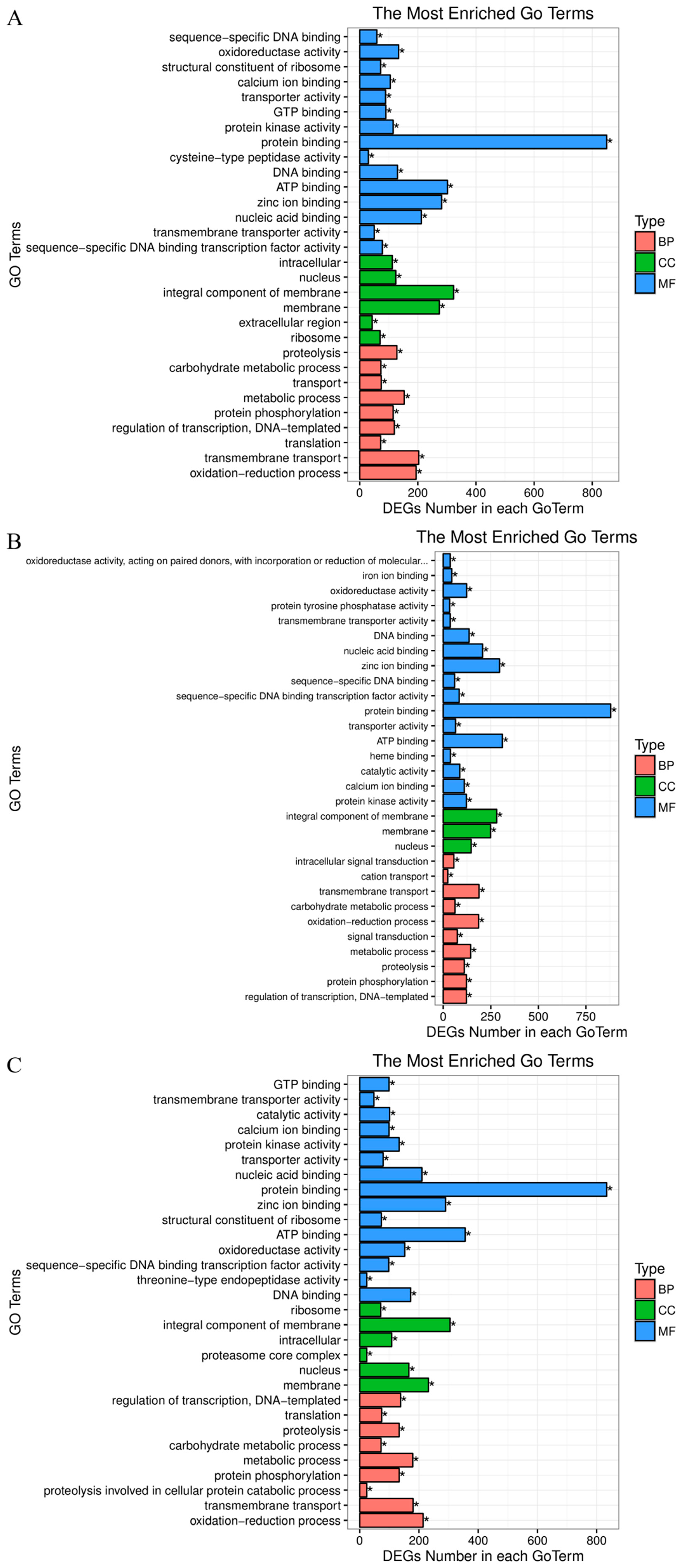
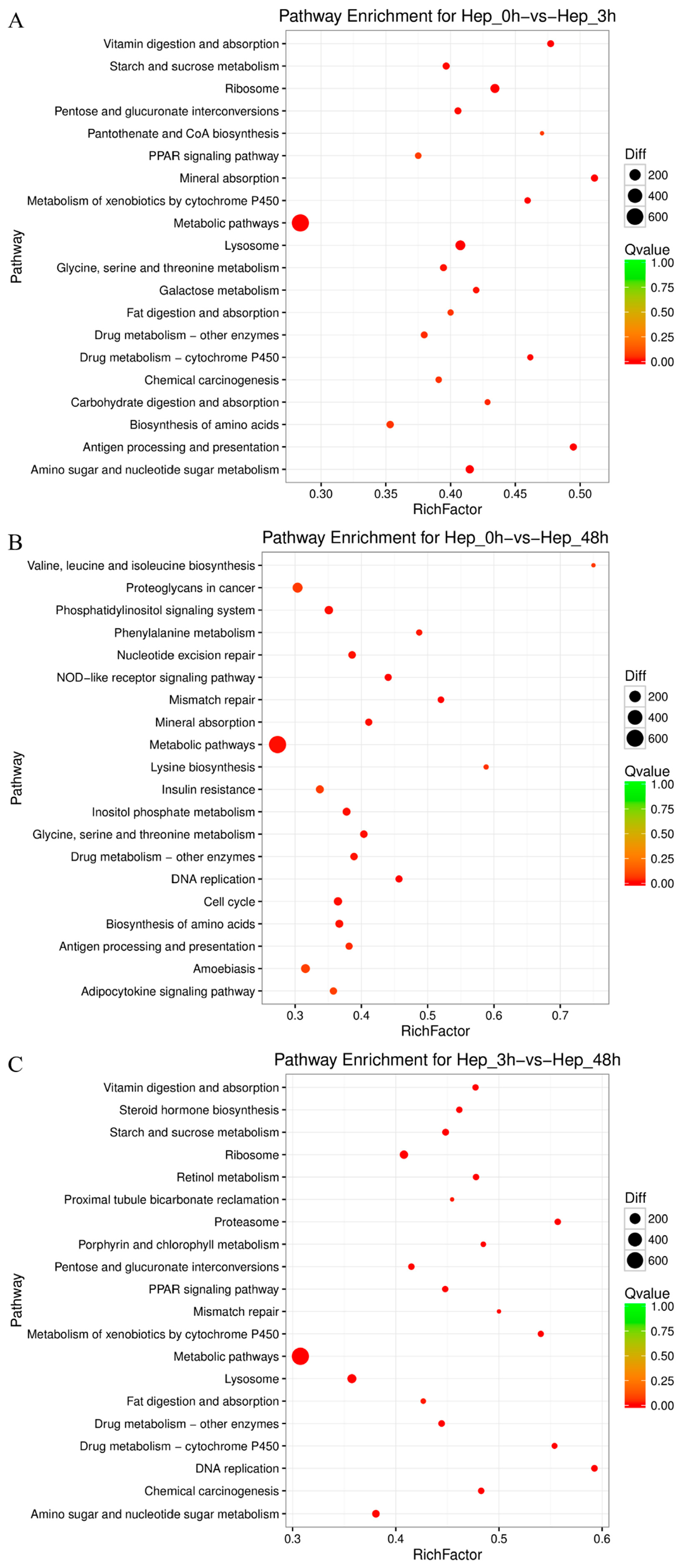
3.4. GO and KEGG Enrichment Analyses of DEGs

In order to identify the biological processes and pathways of DEGs that may be involved in the Cu stress response, GO and KEGG enrichment analyses of all DEGs of the three comparisons (Hep_0 h vs. Hep_3 h, Hep_0 h vs. Hep_48 h, and Hep_3 h vs. Hep_48 h) were conducted. The GO functional enrichment analysis showed that DEGs were classified into three categories: molecular function, biological process, and cell component. The top 30 GO terms enriched in each group are shown in Figure 2. In the molecular function and cellular component categories, the subcategories with the most DEG enrichment among the three comparisons were protein binding (GO:0005515) and integral component of membrane (GO:0016021), respectively. When it comes to biological processes, the most DEGs were enriched in the transmembrane transport subcategory (GO:0055085) in the Hep_0 h vs. Hep_3 h and Hep_0 h vs. Hep_48 h comparisons, while most DEGs were mainly enriched in the oxidation–reduction process subcategory (GO:0055114) in the Hep_3 h vs. Hep_48 h comparison.
To better understand the functional characteristics of these DEGs, the KEGG pathway functional enrichment of DEGs was performed. A total of 299, 298, and 297 pathways were annotated, and 49, 53, and 55 pathways were significantly enriched in Hep_0 h vs. Hep_3 h, Hep_0 h vs. Hep_48 h, and Hep_3 h vs. Hep_48 h, respectively (p-value < 0.05). The top 20 KEGG enrichment analyses of DEGs for each comparison are shown in Figure 3. Of them, multiple pathways are associated with immunity, for example, “Lysosome” (ko04142) and “Metabolism of xenobiotics by cytochrome P450” (ko00980) in Hep_0 h vs. Hep_3 h, “NOD-like receptor signaling pathway” (ko04621) and “Amoebiasis” (ko05146) in Hep_0 h vs. Hep_48 h, and “Proteasome” (ko03050) in Hep_3 h vs. Hep_48 h. It also provided some other molecular pathways related to metabolism and detoxification, such as “Glycine, serine and threonine metabolism” (ko00260), “Metabolic pathways” (ko01100), “Drug metabolism-other enzymes” (ko00983), “Drug metabolism-cytochrome P450” (ko00982), etc. In addition, it is interesting to note that pathways such as “Cell cycle” (ko04110), “Mineral absorption” (ko04978), “Mismatch repair” (ko03430), “Nucleotide excision repair” (ko03420), and “Base excision repair” (ko03410) were also affected after Cu exposure. Taken together, these results indicated that genes related to oxidation–reduction, energy metabolism, apoptosis, copper homeostasis, detoxification, immune response, and DNA damage repair in the hepatopancreas of M. rosenbergii were changed after Cu exposure (Table 3).

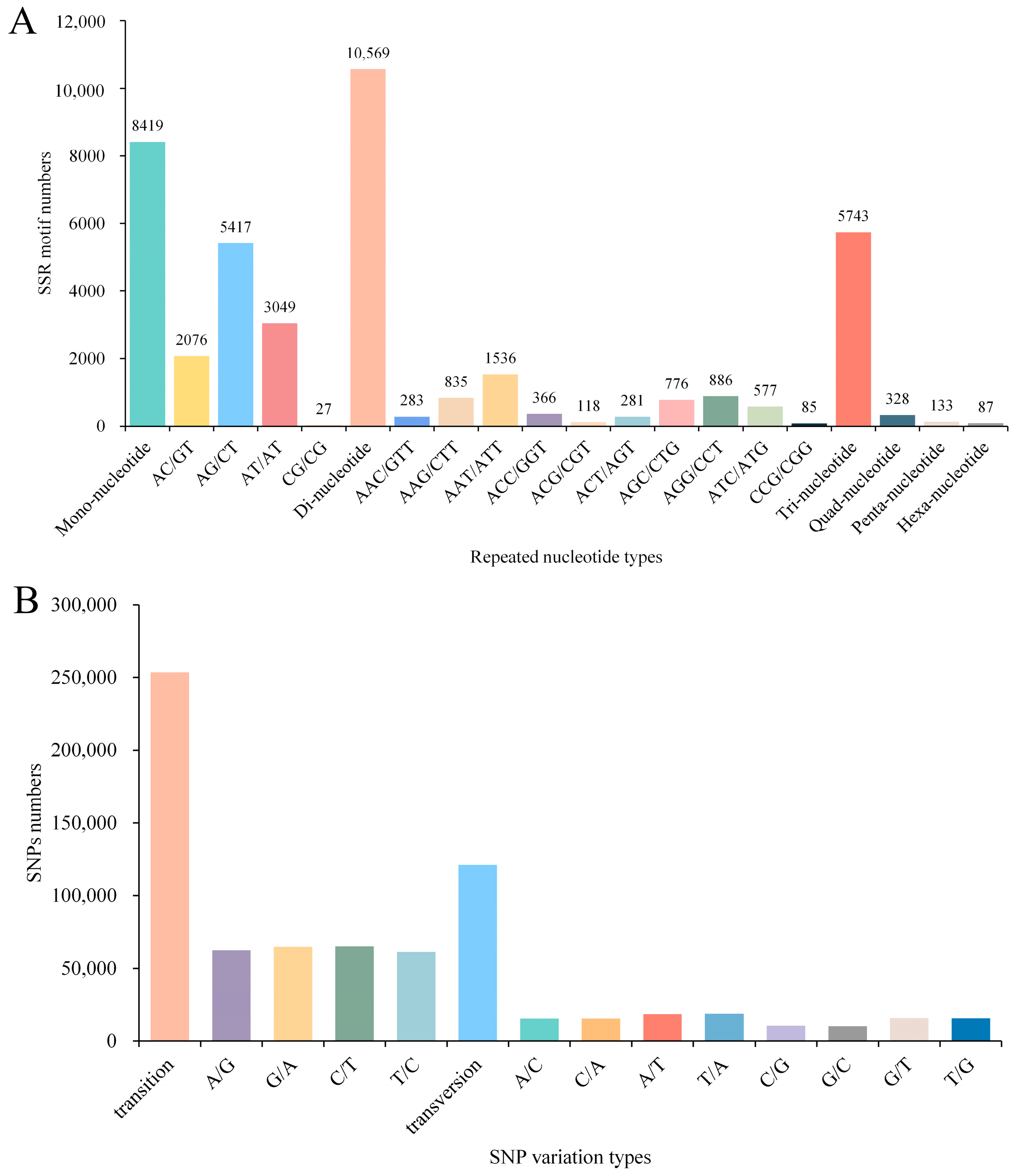
3.5. Detection of SSR and SNP Loci in the Transcriptome

A total of 25,279 SSRs were identified across 85,984 unigenes, with the majority of SSR repeats being di-nucleotide repeats (10,569, 41.81%), followed by mono-nucleotide repeats (8419, 33.30%) and tri-nucleotide repeats (574,379, 22.72%), and the rest being quad-nucleotide repeats (328, 1.30%), penta-nucleotide repeats (133, 0.53%), and hexa-nucleotide repeats (87, 0.3%). Among the di-nucleotide repeats, AG/CT was the main type, followed by AT/AT and AC/GT. The most common repeat sequences in tri-nucleotide repeats were AAT/ATT and AGG/CCT (Figure 4A). Detailed SSR results and corresponding primers are presented in Table S4. In addition, a total of 374,631 SNPs were predicted, including 253,540 transition and 121,091 transversion, with C/T (65,001) and T/A (18,811) SNP types being the most frequent in transition and transversion, respectively (Figure 4B).

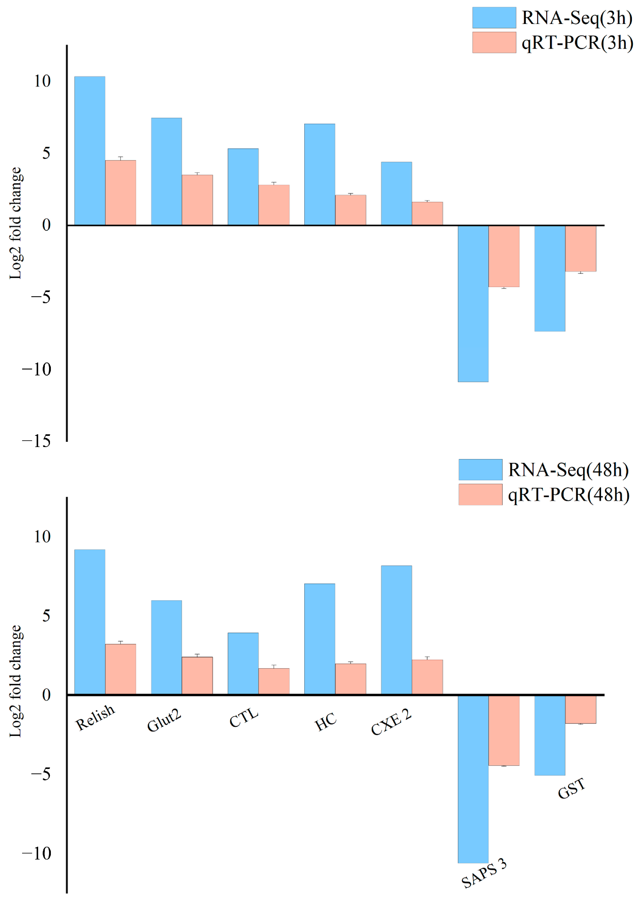
3.6. The qRT-PCR Validation of Differentially Expressed Genes

Seven DEGs were chosen for validation by qRT-PCR. The results showed that, compared to the control group, the expression levels of relish, Glut 2, HC, CXE 2, and CTL were up-regulated, while SAPS 3 and GST were down-regulated. These expression trends were consistent with those observed in the high-throughput sequencing data (Figure 5), confirming the precision and reliability of the sequencing results.

4. Discussion

Cu, a common heavy metal contaminant in aquatic ecosystems, has raised considerable global concern [11]. Currently, the detailed molecular response mechanisms in the hepatopancreas of M. rosenbergii under Cu exposure remain poorly elucidated. In this study, RNA-Seq technology was utilized to reveal the molecular response mechanisms of the hepatopancreas of M. rosenbergii after Cu exposure for 3 and 48 h. Our research was designed to compare the temporal dynamics of stress responses in the hepatopancreas of M. rosenbergii, with the aim of identifying DEGs and disparate pathways involved. A total of 85,984 unigenes were de novo assembled, yet only 28,668 (33.34%) were annotated. The limited number of annotated unigenes may reflect the scarcity of available reference sequences for crustaceans and related species in public databases. Upon Nr database annotation, the largest number of the annotated unigenes were most closely related to Z. nevadensis and L. polyphemus. Notably, a substantial portion remained unannotated; this result was consistent with a previous transcriptomic analysis of M. rosenbergii [42]. The unannotated unigenes might serve as prospective candidates for future explorations in gene discovery.
A total of 11,164 (7288 up-regulated and 3876 down-regulated genes) and 10,937 (6630 up-regulated and 4307 down-regulated genes) DEGs were discovered at 3 and 48 h, respectively. We observed that the number of up-regulated genes was much greater than the down-regulated genes at both time points. Similar results were also reported in previous studies on Neocaridina denticulate sinensis and Anodonta woodiana, indicating that Cu might induce the expression of a greater number of genes in response to Cu toxicity [4,43]. Subsequently, GO and KEGG pathway enrichment analysis of these DEGs revealed that these DEGs play a pivotal role in a suite of fundamental biological processes, such as oxidation–reduction, copper homeostasis, energy metabolism, apoptosis, detoxification, immune response, and DNA damage repair. The temporal specificity of these genes not only highlights the dynamic nature of the stress response but also provides new insights into the time-regulated molecular mechanisms during Cu exposure.
Similar to other invertebrates, M. rosenbergii rely on innate immunity as their primary defense mechanism against external environmental stressors. The Toll-like receptor (TLR) was the first identified pattern recognition receptor (PRR), and its role in innate immunity has been characterized in several crustacean species [44,45,46]. A previous report observed that the expression of TLR in the hepatopancreas was induced by heavy metal stimulation in a time-dependent manner [47]. CTL and ficolin-like protein 2 (FLP 2) are also integral components of the PRRs [48,49]. For crustaceans, CTL and FLP 2 play crucial roles in immune defense, immunosurveillance, and the maintenance of body homeostasis [49,50]. In our transcriptome results, TLR (CL4523.Contig2_All), CTL (CL3394.Contig2_All), and FLP 2 (CL4909.Contig1_All) were up-regulated, implying that M. rosenbergii may mitigate the adverse effects of Cu exposure by enhancing the binding activity of PRRs, thereby bolstering its immune defense mechanisms. Antimicrobial peptides (AMPs) constitute the initial line of defense in the host’s innate immune system. It has been found that domeless, stat, and relish are all involved in the regulation of AMP expression at various levels in P. clarkii, M. rosenbergii, and Eriocheir sinensis [51,52,53]. Hemocyanin (HC), a precursor to AMPs, not only serves as an oxygen carrier but also plays a role in immune defense [54,55,56]. The expression level of HC was up-regulated in crustaceans following heavy metal exposure, thereby facilitating an immunological response [10,42]. In our study, HC (CL2.Contig14_All), domeless (CL5288.Contig1_All), stat (CL607.Contig1_All), and relish (CL2513.Contig3_All) were significantly induced after Cu exposure, indicating that the up-regulation of these genes might be a critical response in the resistance of M. rosenbergii to Cu toxicity. The above findings indicated that M. rosenbergii may activate a diverse array of immune mechanisms to counteract the physiological challenges induced by Cu exposure.
In crustaceans, the hepatopancreas is the target organ for the accumulation of heavy metals as well as a vital detoxification organ. The high-affinity copper uptake protein 1 (Ctr1) is primarily responsible for the absorption of Cu [57]. Excessive Cu decreased the expression of Ctr1, preventing the intracellular accumulation of Cu [58]. In contrast, in our study, Cu induced the expression of Ctr1 (CL2846.Contig2_All). This might lead to an excessive influx of Cu into the cells, resulting in toxicity [59]. The occurrence of this intriguing phenomenon may be due to interspecific differences in the response of organisms to Cu exposure. Metallothioneins (MTs, Cu-MT, and Zn-MT) are the main metal-binding thiol proteins, protecting aquatic organisms against the toxic effects of heavy metals through the sequestration and regulation of essential metal ions [60]. Cytochrome c oxidase Cu chaperone (COX17) is a Cu-chelating protein responsible for the intracellular transport of Cu ions to mitochondria, participating in the enzymatic reactions mediated by cytochrome c oxidase [61]. ATP7A participates in the elimination of excess Cu from cells to regulate the intracellular concentration of Cu [62]. In this study, significant up-regulation of COX17 (Unigene9795_All), Cu-MT (Unigene13182_All), and ATP7A (CL3799.Contig3_All) was observed. We assumed that M. rosenbergii may mitigate the toxicity of intracellular Cu accumulation by sequestering, utilizing, and excreting excess Cu. In addition, JHE-like carboxylesterase 2 (CXE2) and cytochrome P 450 (CYP 450) are involved in detoxification and protection against oxidative damage in crustaceans [63,64]. Cu exposure also induced the expression of CYP450 (CL1886.Contig2_All) and CXE2 (CL426.Contig5_All). The aforementioned findings suggest that the prawn M. rosenbergii attempts to counteract excessive cellular Cu uptake by initiating detoxification mechanisms in the hepatopancreas.
Accumulation of Cu in the organs of crustaceans leads to the overproduction of ROS, resulting in oxidative damage [10]. NADPH oxidase (NOX) is the main source of ROS in living organisms [65,66]. It has been confirmed that NOX was activated and excessive ROS were produced after Cu exposure in P. clarkii [67]. Activation of the antioxidant enzyme system can mitigate the excess ROS [68]. Glutathione peroxidase (GPx) functions in catalyzing the reduction of hydrogen peroxide and lipid peroxide [69]. Glutathione-S-transferase (GST) is involved in cellular antioxidant defense by catalyzing the binding of glutathione to electrophiles [70]. GPx and GST are usually used as indicators of oxidative stress after metal exposure. Moreover, it has been found that up-regulation of GST can enhance an organism’s resistance to Cu exposure [71]. However, the present study revealed that NOX (CL1542.Contig3_All) was significantly induced at both 3 and 48 h time points, whereas GST (CL355.Contig7_All) was suppressed. In addition, the transcription level of glutathione peroxidase (GPx) (CL5985.Contig3_All) was up-regulated at 3 h and down-regulated at 48 h. Similar to our results, in Thamnaconus septentrionalis exposed to cadmium (Cd), the increase in antioxidant enzyme activity in the short term can effectively remove ROS, but long-term Cd exposure weakens the organism’s antioxidant defense ability [72]. Therefore, we speculated that after Cu exposure, GPx has a stronger protective effect on M. rosenbergii compared to GST. Initially, GPx was up-regulated to exert its antioxidant function, but as the duration of exposure prolonged, GPx became insufficient to eliminate the sustained excess ROS production, leading to the inhibition of GPx expression. Consequently, the accumulation of Cu in the hepatopancreas of M. rosenbergii may elevate the risk of oxidative damage.
Oxidative stress caused by ROS can attack biological macromolecules in cells, causing DNA damage and impaired protein function [73]. Previous studies have shown that the heavy metal Cd can inhibit the DNA damage repair mechanism of aquatic organisms [74,75,76]. Similar results were also found in our study. Mismatch repair (MMR) can repair the base mismatch during DNA replication [77]. Nucleotide excision repair (NER) can remove DNA damage caused by various exogenous pressures [78]. After Cu exposure, MutL Homolog 1 (MLH1) (Unigene25307_All), MutS Homolog 2 (MSH2) (Unigene8072_All), and MutS Homolog 6 (MSH6) (Unigene15503_All) in the MMR pathway and DNA damage-binding protein 1 (DDB1) (CL4136.Contig1_All), DNA damage-binding protein 2 (DDB2) (Unigene362_All), replication factor C (RFC) (Unigene27679_All, Unigene768_All, and CL6139.Contig1_All), and DNA polymerase epsilon subunit 2 (POLE2) (Unigene17725_All) in the NER pathway were significantly down-regulated. These results indicated that Cu may have a similar genotoxic mechanism to Cd, aggravating cell damage by inhibiting genes related to oxidative damage repair. Heat shock proteins (HSPs) are a highly conserved protein family, which can maintain cellular protein homeostasis against environmental stress and play an important role in cellular stress response [79]. HSP70 and HSP90 are important members of the heat shock protein family, which are responsible for assisting the correct folding, repair, and degradation of proteins, as well as stabilizing key proteins and promoting signal transduction [80]. They are essential for cell survival and the maintenance of normal functions. In our study, Cu exposure inhibited the expression of HSP70 (CL1286.Contig5_All) and HSP90 (CL1294.Contig2_All). Previous studies have shown that Cu inhibits the expression of HSP40 in Tigriopus japonicus to mediate cell damage [73]. Similarly, decreased expression levels of HSP70 and HSP90 were observed in M. japonicus exposed to Cu or Cd [81]. Therefore, we speculated that when M.rosenbergii is exposed to a Cu-contaminated environment, the DNA damage repair mechanism and cellular stress response system may be inhibited, thereby further exacerbating oxidative damage.
Apoptosis represents a fundamental physiological and biochemical process, serving as a critical mechanism for the elimination of damaged or mutated cells within the host [82]. Cytochrome c (Cyt C) acts as a pivotal initiating factor in the mitochondrial pathway in cell apoptosis [83]. The forkhead box protein O (FOXO) primarily induces and modulates the expression of the pro-apoptotic protein Bim, thereby enabling cytochrome c (Cyt C) to cross the mitochondrial membrane and activate members of the caspase family, such as caspase 2 (Casp 2), to promote the occurrence of apoptosis [84,85,86]. Survivin, a member of the inhibitor of apoptosis protein (IAP) family, has been demonstrated to decrease the inhibitory action against caspases when its expression is decreased, thus promoting the execution of the apoptotic cascade [87]. In the present study, the expression levels of Cyt C (CL4973.Contig1_All), FOXO (CL1823.Contig6_All), and Casp2 (CL1421.Contig1_All) were significantly up-regulated under Cu exposure, while the expression level of survivin (CL7764.Contig1_All) was inhibited. These gene expression changes indicated that apoptosis may occur in the hepatopancreatic cells of M. rosenbergii during Cu exposure. Previous studies have shown that excessive Cu leads to DNA damage, and if such damage remains unrepaired, cells may undergo a series of complex enzymatically catalyzed reactions resulting in apoptosis [88,89]. Based on these findings, we speculated that the up-regulation of apoptosis-related genes observed in this study may be attributed to the inhibition of the DNA repair system and cellular stress response by Cu, resulting in the organism initiating apoptosis to eliminate damaged cells.
Heavy metal stress could increase the energy demand of crustaceans to cope with metal detoxification and damage repair [90,91]. AMP-activated protein kinase (AMPK) is an energy sensor. When environmental stress causes changes in the energy state of organisms, such as the increase of AMP/ATP ratio, AMPK will be phosphorylated to promote glycolysis and fatty acid oxidation, so as to restore the energy homeostasis in the body [92]. Serine/threonine–protein phosphatase 6 regulatory subunit 3 (SAPS3) is a negative regulator of AMPK, and when metabolic disorders occur, SAPS3 is inhibited, leading to AMPK phosphorylation [93]. We observed that the expression of SAPS3 (CL281.Contig5_All) was significantly suppressed after Cu exposure. Moreover, the transcription levels of genes related to fatty acid synthesis, including fatty acid synthase (FAS) (Unigene17066_All), acetyl-CoA carboxylase (ACC) (CL441.Contig3_All), and delta-9 acyl-CoA desaturase (SCD1) (CL800.Contig2_All), were also down-regulated. Glucose transporter 2 (Glut 2) participates in the intracellular uptake and transport of glucose, with an increase in its expression potentially enhancing glucose intake by cells and facilitating glycolysis processes [94,95]. Based on our transcriptome analysis, Cu exposure induced the expression of Glut 2 (CL1337.Contig2_All). The results presented herein suggest that under Cu exposure, M. rosenbergii might potentially enhance its resistance to Cu toxicity by promoting glycolysis and fatty acid oxidation and inhibiting fatty acid synthesis to accommodate the increased energy demand of the organism.
SSRs and SNPs are essential molecular markers in the fields of genetic diversity analysis and molecular-assisted breeding, among others [96,97]. In recent years, they have been applied to the study of genetic variation in numerous crustacean species [98,99,100]. However, there have been no reports of SSRs or SNPs identified in the hepatopancreas of M. rosenbergi under Cu exposure. We identified a significant number of SSRs and SNPs within 85,984 unigenes in the present study. These markers will provide an invaluable genetic resource, facilitating the development of selective breeding programs aimed at enhancing stress resistance in Macrobrachium species.

5. Conclusions

In this study, transcriptomic analysis was conducted to explore the molecular response mechanisms of the hepatopancreas in M. rosenbergii under Cu exposure. The findings indicated that M. rosenbergii attempts to resist the toxicity of Cu by up-regulating the expression of genes related to immunity, metabolism, and detoxification. However, with the excessive accumulation of ROS, the antioxidant enzyme system was destroyed. As a result, DNA damage repair and the cellular stress response were inhibited, thereby exacerbating cell damage. In order to maintain the normal function of the hepatopancreas, M. rosenbergii removed the damaged cells by activating the apoptosis mechanism. Overall, our study not only facilitates an understanding of the molecular response mechanisms of M. rosenbergii underlying Cu toxicity effects but also helps in identifying potential biomarkers associated with the stress response in other crustaceans.

Supplementary Materials

The following supporting information can be downloaded at: https://www.mdpi.com/article/10.3390/ani14071117/s1, Figure S1: Length distribution of all assembled unigenes; Figure S2: Number of unigenes annotated to different databases; Figure S3: Venn diagram of the transcriptomes against the Nr, Nt, COG, SwissProt, and KEGG databases; Figure S4: Distribution of the top 30 species with the most homologous sequences; Figure S5: GO classification of the assembled unigenes; Figure S6: KEGG annotation analysis of the unigenes; Figure S7: Numbers of DEGs in each comparison; Table S1: De novo assembly sequences; Table S2: Annotation results of unigenes in Nr, NT, COG, SwissProt, and KEGG; Table S3: Differentially expressed genes of three comparison groups; Table S4: SSR results and corresponding primers.

Author Contributions

Conceptualization, J.Z., Z.B. and H.G.; Data curation, J.Z. and Z.B.; Formal analysis, X.S.; Funding acquisition, H.G.; Investigation, Z.B. and J.G.; Methodology, J.Z. and J.G.; Project administration, H.G.; Resources, J.Z. and Y.Z.; Software, Y.Z.; Supervision, H.G.; Validation, Z.B. and J.G.; Visualization, X.S.; Writing—original draft, J.Z.; Writing—review and editing, X.S. and H.G. All authors have read and agreed to the published version of the manuscript.

Funding

The Department of Education of Guangdong Province (2023ZDZX4013).

Institutional Review Board Statement

Our research was exempted from ethical evaluation as in China, the Animal Ethics Committee’s scope of approval is limited to studies involving vertebrates (Laboratory Animal Act, Chapter 5 Article 25). Our study is on invertebrate species, and does not need ethical approval in China.

Informed Consent Statement

Not applicable.

Data Availability Statement

The authors declare that all data supporting the conclusions of this study are available within the article.

Conflicts of Interest

The authors declare no conflicts of interest.

References

  1. Dawood, M. Dietary Cu requirements for aquatic animals: A review. Biol. Trace Elem. Res. 2022, 200, 5273–5282. [Google Scholar] [CrossRef] [PubMed]
  2. Bian, C.; Zheng, Z.; Su, J.; Chang, S.; Yu, H.; Bao, J.; Xin, Y.; Jiang, X. Copper homeostasis and cuproptosis in tumor pathogenesis and therapeutic strategies. Front. Pharmacol. 2023, 14, 1271613. [Google Scholar] [CrossRef] [PubMed]
  3. Qian, D.; Xu, C.; Chen, C.; Qin, J.; Chen, L.; Li, E. Toxic effect of chronic waterborne copper exposure on growth, immunity, anti-oxidative capacity and gut microbiota of Pacific white shrimp Litopenaeus vannamei. Fish. Shellfish Immunol. 2020, 100, 445–455. [Google Scholar] [CrossRef] [PubMed]
  4. Chen, X.; Liu, H.; Liber, K.; Jiang, T.; Yang, J. Copper-induced ionoregulatory disturbance, histopathology, and transcriptome responses in freshwater mussel (Anodonta woodiana) gills. Fishes 2023, 8, 368. [Google Scholar] [CrossRef]
  5. Guo, H.; Li, K.; Wang, W.; Wang, C.; Shen, Y. Effects of copper on hemocyte apoptosis, ROS production, and gene expression in white shrimp Litopenaeus vannamei. Biol. Trace Elem. Res. 2017, 179, 318–326. [Google Scholar] [CrossRef] [PubMed]
  6. Wang, L.; Guan, T.; Wang, G.; Gu, J.; Wu, N.; Zhu, C.; Wang, H.; Li, J. Effects of copper on gill function of juvenile oriental river prawn (Macrobrachium nipponense): Stress and toxic mechanism. Aquat. Toxicol. 2023, 261, 106631. [Google Scholar] [CrossRef] [PubMed]
  7. García-Villada, L.; Rico, M.; Altamirano, M.; Sánchez-Martín, L.; López-Rodas, V.; Costas, E. Occurrence of Cu resistant mutants in the toxic cyanobacteria Microcystis aeruginosa: Characterisation and future implications in the use of Cu sulphate as algaecide. Water Res. 2004, 38, 2207–2213. [Google Scholar] [CrossRef]
  8. Zhuang, W.; Zhou, F. Distribution, source and pollution assessment of heavy metals in the surface sediments of the Yangtze River Estuary and its adjacent East China Sea. Mar. Pollut. Bull. 2021, 164, 112002. [Google Scholar] [CrossRef] [PubMed]
  9. Guo, H.; Xian, J.; Zheng, P.; Lu, Y.; Wang, L.; Zhang, X.; Li, J.; Zhang, Z.; Wang, A. Dietary copper affects antioxidant status of shrimp (Penaeus monodon) reared in low salinity water. Aquac. Rep. 2022, 22, 100979. [Google Scholar] [CrossRef]
  10. Xing, K.; Liu, Y.; Yan, C.; Zhou, Y.; Zhang, R.; Sun, Y.; Zhang, J. Transcriptomic analysis of Neocaridina denticulate sinensis hepatopancreas indicates immune changes after Cu exposure. Fish. Shellfish Immunol. 2022, 121, 23–30. [Google Scholar] [CrossRef] [PubMed]
  11. Zhang, Y.; Zhang, M.; Yu, W.; Li, J.; Kong, D. Ecotoxicological risk ranking of 19 metals in the lower Yangtze River of China based on their threats to aquatic wildlife. Sci. Total Environ. 2022, 812, 152370. [Google Scholar] [CrossRef] [PubMed]
  12. Lan, X.; Peng, X.; Du, T.; Xia, Z.; Gao, Q.; Tang, Q.; Yi, S.; Yang, G. Alterations of the gut microbiota and metabolomics associated with the different growth performances of Macrobrachium rosenbergii families. Animals 2023, 13, 1539. [Google Scholar] [CrossRef] [PubMed]
  13. Tu, H.; Peng, X.; Yao, X.; Tang, Q.; Xia, Z.; Li, J.; Yang, G.; Yi, S. Integrated transcriptomic and metabolomic analyses reveal low-temperature tolerance mechanism in giant freshwater prawn Macrobrachium rosenbergii. Animals 2023, 13, 1605. [Google Scholar] [CrossRef] [PubMed]
  14. Zhong, Z.; Chen, G.; Tu, H.; Yao, X.; Peng, X.; Lan, X.; Tang, Q.; Yi, S.; Xia, Z.; Cai, M.; et al. Transcriptomic analysis and functional gene expression in different stages of gonadal development of Macrobrachium rosenbergii. Fishes 2023, 8, 94. [Google Scholar] [CrossRef]
  15. Liang, J.; Abdullah, A.L.B.; Wang, H.; Liu, G.; Han, M. Change in energy-consuming strategy, nucleolar metabolism and physical defense in Macrobrachium rosenbergii after acute and chronic polystyrene nanoparticles exposure. Aquat. Toxicol. 2023, 263, 106711. [Google Scholar] [CrossRef] [PubMed]
  16. Farhadi, A.; Tang, S.; Huang, M.; Yu, Q.; Xu, C.; Li, E. Identification of key immune and stress related genes and pathways by comparative analysis of the gene expression profile under multiple environmental stressors in pacific white shrimp (Litopenaeus vannamei). Fish. Shellfish Immunol. 2023, 135, 108695. [Google Scholar] [CrossRef]
  17. Zhang, R.; Zhao, Z.; Li, M.; Luo, L.; Wang, S.; Guo, K.; Xu, W. Effects of saline-alkali stress on the tissue structure, antioxidation, immunocompetence and metabolomics of Eriocheir sinensis. Sci. Total Environ. 2023, 871, 162109. [Google Scholar] [CrossRef]
  18. Kouba, A.; Buric, M.; Kozák, P. Bioaccumulation and effects of heavy metals in crayfish: A review. Water Air Soil. Poll. 2010, 211, 5–16. [Google Scholar] [CrossRef]
  19. Abbas, M.; Abd El-Aziz, M.; Kaddah, M.; Hassan, A.K.; El-Naggar, H.A.; Radwan, M.; El-Tabakh, M.; Afifi, M.A.; Bashar, M. Bioaccumulation, biosedimentation, and health hazards of elements in crayfish, Procambarus clarkii from El-Rahawi Drain and El-Qanatir in the River Nile, Egypt. Biol. Trace Elem. Res. 2023, 201, 3050–3059. [Google Scholar] [CrossRef] [PubMed]
  20. Sang, W.; Liu, J.; Xing, Y.; Feng, C.; Hu, Q.; Lang, Y.; Li, X.; Bao, J.; Jiang, H. Transcriptome analysis of hepatopancreas of Chinese grass shrimp, Palaemonetes sinensis, infected by Enterocytospora artemiae. Fish. Shellfish Immunol. 2023, 133, 108557. [Google Scholar] [CrossRef]
  21. Zhang, L.; Song, Z.; Zhou, Y.; Zhong, S.; Yu, Y.; Liu, T.; Gao, X.; Li, L.; Kong, C.; Wang, X.; et al. The accumulation of toxic elements (Pb, Hg, Cd, As, and Cu) in red swamp crayfish (Procambarus clarkii) in Qianjiang and the associated risks to human health. Toxics 2023, 11, 635. [Google Scholar] [CrossRef]
  22. Wu, Y.; Tian, W.; Cheng, C.; Yang, L.; Ye, Q.; Li, W.; Jiang, J. Effects of cadmium exposure on metabolism, antioxidant defense, immune function, and the hepatopancreas transcriptome of Cipangopaludina cathayensis. Ecotoxicol. Environ. Saf. 2023, 264, 115416. [Google Scholar] [CrossRef] [PubMed]
  23. Reddy, R.; Pillai, B.R.; Adhikari, S. Bioaccumulation of copper in post-larvae and juveniles of freshwater prawn Macrobrachium rosenbergii (de man) exposed to sub-lethal levels of copper sulfate. Aquaculture 2006, 252, 356–360. [Google Scholar] [CrossRef]
  24. Li, N.; Zhao, Y.; Yang, J. Impact of waterborne copper on the structure of gills and hepatopancreas and its impact on the content of metallothionein in juvenile giant freshwater prawn Macrobrachium rosenbergii (Crustacea: Decapoda). Arch. Environ. Con Tox. 2007, 52, 73–79. [Google Scholar] [CrossRef] [PubMed]
  25. Li, N.; Ma, Q.; Yang, J.; Zhao, Y.; Brochon, J.C. Effects of water-borne copper on the gills and hepatopancreas of Macrobrachium rosenbergii. Integr. Zool. 2008, 3, 299–310. [Google Scholar] [CrossRef]
  26. Pabuwal, V.; Boswell, M.; Pasquali, A.; Wise, S.S.; Kumar, S.; Shen, Y.; Garcia, T.; LaCerte, C., Jr.; Wise, J.P.; Wise, J.P.S.; et al. Transcriptomic analysis of cultured whale skin cells exposed to hexavalent chromium [Cr (VI)]. Aquat. Toxicol. 2013, 134, 74–81. [Google Scholar] [CrossRef] [PubMed]
  27. Jin, J.; Wang, Y.; Wu, Z.; Hergazy, A.; Lan, J.; Zhao, L.; Liu, X.; Chen, N.; Lin, L. Transcriptomic analysis of liver from grass carp (Ctenopharyngodon idellus) exposed to high environmental ammonia reveals the activation of antioxidant and apoptosis pathways. Fish. Shellfish Immunol. 2017, 63, 444–451. [Google Scholar] [CrossRef] [PubMed]
  28. Guo, H.; Miao, Y.; Xian, J.; Qian, K.; Wang, A. Expression profile of antioxidant enzymes in hemocytes from freshwater prawn Macrobrachium rosenbergii exposed to an elevated level of copper. Bull. Environ. Contam. Toxicol. 2015, 95, 447–451. [Google Scholar] [CrossRef] [PubMed]
  29. Grabherr, M.G.; Haas, B.J.; Yassour, M.; Levin, J.Z.; Thompson, D.A.; Amit, I.; Adiconis, X.; Fan, L.; Raychowdhury, R.; Zeng, Q.; et al. Full-length transcriptome assembly from RNA-Seq data without a reference genome. Nat. Biotechnol. 2011, 29, 130–644. [Google Scholar] [CrossRef] [PubMed]
  30. Pertea, G.; Huang, X.; Liang, F.; Antonescu, V.; Sultana, R.; Karamycheva, S.; Lee, Y.; White, J.; Cheung, F.; Parvizi, B. TIGR Gene Indices clustering tools (TGICL): A software system for fast clustering of large EST datasets. Bioinformatics 2003, 19, 651–652. [Google Scholar] [CrossRef] [PubMed]
  31. Altschul, S.F.; Gish, W.; Miller, W.; Myers, E.W.; Lipman, D.J. Basic local alignment search tool. J. Mol. Biol. 1990, 215, 403–410. [Google Scholar] [CrossRef] [PubMed]
  32. Conesa, A.; Gotz, S.; Garcia-Gomez, J.M.; Terol, J.; Talon, M.; Robles, M. Blast2GO: A universal tool for annotation, visualization and analysis in functional genomics research. Bioinformatics 2005, 21, 3674–3676. [Google Scholar] [CrossRef] [PubMed]
  33. Xie, C.; Mao, X.; Huang, J.; Ding, Y.; Wu, J.; Dong, S.; Kong, L.; Gao, G.; Li, C.; Wei, L. KOBAS 2.0: A web server for annotation and identification of enriched pathways and diseases. Nucleic Acids Res. 2011, 39, W316–W322. [Google Scholar] [CrossRef] [PubMed]
  34. Li, B.; Dewey, C.N. RSEM: Accurate transcript quantification from RNA-Seq data with or without a reference genome. BMC Bioinform. 2011, 12, 323. [Google Scholar] [CrossRef] [PubMed]
  35. Wang, L.; Feng, Z.; Wang, X.; Wang, X.; Zhang, X. DEGseq: An R package for identifying differentially expressed genes from RNA-seq data. Bioinformatics 2010, 26, 136–138. [Google Scholar] [CrossRef]
  36. Anders, S.; Huber, W. Differential expression analysis for sequence count data. Genome Biol. 2010, 11, R106. [Google Scholar] [CrossRef] [PubMed]
  37. Young, M.; Wakefield, M.J.; Smyth, G.K.; Oshlack, A. Gene ontology analysis for RNA-seq: Accounting for selection bias. Genome Biol. 2010, 11, R14. [Google Scholar] [CrossRef]
  38. Thiel, T.; Michalek, W.; Varshney, R.K.; Graner, A. Exploiting EST databases for the development and characterization of gene-derived SSR-markers in barley (Hordeum vulgare L.). Theor. Appl. Genet. 2003, 106, 411–422. [Google Scholar] [CrossRef] [PubMed]
  39. McKenna, A.; Hanna, M.; Banks, E.; Sivachenko, A.; Cibulskis, K.; Kernytsky, A.; Garimella, K.; Altshuler, D.; Gabriel, S.; Daly, M.; et al. The genome analysis toolkit: A MapReduce framework for analyzing next-generation DNA sequencing data. Genome Res. 2010, 20, 1297–1303. [Google Scholar] [CrossRef] [PubMed]
  40. Zhou, T.; Liu, J.; Chan, S.; Wang, W. Molecular characterization and expression dynamics of three key genes in the PI3K-AKT pathway reveal its involvement in the immunotoxicological responses of the giant river prawn Macrobrachium rosenbergii to acute ammonia and nitrite stress. Ecotox. Environ. Safe 2021, 208, 111767. [Google Scholar] [CrossRef]
  41. Livak, K.J.; Schmittgen, T.D. Analysis of relative gene expression data using real-time quantitative PCR and the 2−ΔΔCT method. Methods 2001, 25, 402–408. [Google Scholar] [CrossRef]
  42. Guo, H.; Deng, M.; Liang, J.; Lu, W.; Shen, Y. Gill transcriptome alterations in Macrobrachium rosenbergii under copper exposure. Chemosphere 2019, 233, 796–808. [Google Scholar] [CrossRef]
  43. Xing, K.; Liu, Y.; Yan, C.; Zhou, Y.; Sun, Y.; Su, N.; Yang, F.; Xie, S.; Zhang, J. Transcriptome analysis of Neocaridina denticulate sinensis under copper exposure. Gene 2021, 764, 145098. [Google Scholar] [CrossRef]
  44. Habib, Y.J.; Wan, H.; Sun, Y.; Shi, J.; Yao, C.; Lin, J.; Ge, H.; Wang, Y.; Zhang, Z. Genome-wide identification of toll-like receptors in pacific white shrimp (Litopenaeus vannamei) and expression analysis in response to Vibrio parahaemolyticus invasion. Aquaculture 2021, 532, 735996. [Google Scholar] [CrossRef]
  45. Cho, H.; Park, K.H.; Jang, Y.; Cho, Y.; Heo, Y.K.; Kim, M.; Kim, Y.B. Identification and characterization of a toll-like receptor gene from Macrobrachium nipponense. Fish. Shellfish Immunol. 2021, 108, 109–115. [Google Scholar] [CrossRef]
  46. Li, Y.; Yuan, W.; Xu, Q.; Liu, H.; Dai, X. The regulation of immune responses against white spot syndrome virus or Vibrio alginolyticus in toll-like receptors silenced giant freshwater prawn (Macrobrachium rosenbergii). Fish. Shellfish Immunol. 2020, 107, 84–94. [Google Scholar] [CrossRef]
  47. Xu, M.; Wu, J.; Ge, D.; Wu, C.; Chi, C.; Lv, Z.; Liao, Z.; Liu, H. A novel toll-like receptor from Mytilus coruscus is induced in response to stress. Fish. Shellfish Immunol. 2018, 78, 331–337. [Google Scholar] [CrossRef]
  48. Shen, D.; Tong, M.; Guo, J.; Mei, X.; Xia, D.; Qiu, Z.; Zhao, Q. A pattern recognition receptor C-type lectin-S6 (CTL-S6) is involved in the immune response in the Silkworm (Lepidoptera: Bombycidae). J. Insect Sci. 2021, 21, 1–8. [Google Scholar] [CrossRef]
  49. Zhang, X.; Wang, X.; Huang, Y.; Hui, K.; Shi, Y.; Wang, W.; Ren, Q. Cloning and characterization of two different ficolins from the giant freshwater prawn Macrobrachium rosenbergii. Dev. Comp. Immunol. 2014, 44, 359–369. [Google Scholar] [CrossRef]
  50. Luo, J.; Chen, Y.; Huang, Y.; Feng, J.; Yuan, Y.; Jian, J.; Cai, S.; Yang, S. A novel C-type lectin for Litopenaeus vannamei involved in the innate immune response against Vibrio infection. Fish. Shellfish Immunol. 2023, 135, 108621. [Google Scholar] [CrossRef]
  51. Zhang, Z.; Zhang, C.; Dai, X.; Zhang, R.; Cao, X.; Wang, K.; Huang, X.; Ren, Q. Two relish isoforms produced by alternative splicing participate in the regulation of antimicrobial peptides expression in Procambarus clarkii intestine. Fish. Shellfish Immunol. 2020, 99, 107–118. [Google Scholar] [CrossRef]
  52. Ruan, Z.; Wan, Z.; Yang, L.; Li, W.; Wang, Q. JAK/STAT signalling regulates antimicrobial activities in Eriocheir sinensis. Fish. Shellfish Immunol. 2019, 84, 491–501. [Google Scholar] [CrossRef]
  53. Shi, Y.; Jin, M.; Ma, F.; Huang, Y.; Huang, X.; Feng, J.; Zhao, L.; Chen, Y.; Ren, Q. Involvement of relish gene from Macrobrachium rosenbergii in the expression of anti-microbial peptides. Dev. Comp. Immunol. 2015, 52, 236–244. [Google Scholar] [CrossRef]
  54. Coates, C.J.; Decker, H. Immunological properties of oxygen-transport proteins: Hemoglobin, hemocyanin and hemerythrin. Cell Mol. Life Sci. 2017, 74, 293–317. [Google Scholar] [CrossRef]
  55. Zhang, X.; Huang, C.; Qin, Q. Antiviral properties of hemocyanin isolated from shrimp Penaeus monodon. Antivir. Res. 2004, 61, 93–99. [Google Scholar] [CrossRef]
  56. Coates, C.J.; Nairn, J. Diverse immune functions of hemocyanins. Dev. Comp. Immunol. 2014, 45, 43–55. [Google Scholar] [CrossRef]
  57. Minghetti, M.; Leaver, M.J.; Carpenè, E.; George, S.G. Copper transporter 1, metallothionein and glutathione reductase genes are differentially expressed in tissues of sea bream (Sparus aurata) after exposure to dietary or waterborne copper. Comp. Biochem. Phys. C. 2008, 147, 450–459. [Google Scholar] [CrossRef]
  58. Zhao, D.; Zhang, X.; Liu, D.; Ru, S. Cu accumulation, detoxification and tolerance in the red swamp crayfish Procambarus clarkii. Ecotoxicol. Environ. Saf. 2019, 175, 201–207. [Google Scholar] [CrossRef]
  59. Wei, H.; Wang, S.; Xu, E.; Liu, J.; Li, X.; Wang, Z. Synergistic toxicity of microcystin-LR and Cu to Zebrafish (Danio rerio). Sci. Total Environ. 2020, 713, 136393. [Google Scholar] [CrossRef]
  60. Ma’rifah, F.; Saputri, M.R.; Soegianto, A.; Irawan, B.; Putranto, T.W.C. The change of metallothionein and oxidative response in gills of the Oreochromis niloticus after exposure to copper. Animals 2019, 9, 353. [Google Scholar] [CrossRef]
  61. Markossian, K.A.; Kurganov, B.I. Copper chaperones, intracellular copper trafficking proteins. Function, structure, and mechanism of action. Biochemistry 2003, 68, 827–837. [Google Scholar] [CrossRef]
  62. Linz, R.; Lutsenko, S. Copper-transporting ATPases ATP7A and ATP7B: Cousins, not twins. J. Bioenerg. Biomembr. 2007, 39, 403–407. [Google Scholar] [CrossRef]
  63. Guo, H.; Chen, T.; Liang, Z.; Fan, L.; Shen, Y.; Zhou, D. iTRAQ and PRM-based comparative proteomic profiling in gills of white shrimp Litopenaeus vannamei under copper stress. Chemosphere 2021, 263, 128270. [Google Scholar] [CrossRef]
  64. Zang, Y.; Zhang, X.; Huang, P.; Ame, K.H.; Shen, H. Cloning and expression of two carboxylesterases, and their activity modulation in Chinese mitten crab Eriocheir sinensis under pesticide exposer. Turk. J. Fish. Aquat. Sci. 2020, 20, 521–529. [Google Scholar] [CrossRef]
  65. Spencer, N.Y.; Engelhardt, J.F. The basic biology of redoxosomes in cytokine-mediated signal transduction and implications for disease-specific therapies. Biochemistry 2014, 53, 1551–1564. [Google Scholar] [CrossRef]
  66. Ni, Y.; Zhang, J.; Zhu, W.; Duan, Y.; Bai, H.; Luan, C. Echinacoside inhibited cardiomyocyte pyroptosis and improved heart function of HF rats induced by isoproterenol via suppressing NADPH/ROS/ER stress. J. Cell Mol. Med. 2022, 26, 5414–5425. [Google Scholar] [CrossRef]
  67. Zhao, D.; Zhang, X.; Li, X.; Ru, S.; Wang, Y.; Yin, J.; Liu, D. Oxidative damage induced by copper in testis of the red swamp crayfish Procambarus clarkii and its underlying mechanisms. Aquat. Toxicol. 2019, 207, 120–131. [Google Scholar] [CrossRef]
  68. Ou, H.; Liu, J. Role of Nrf2-Keap1 signaling in the antioxidant defense response induced by low salinity in the kuruma shrimp (Marsupenaeus japonicus). Aquac. Int. 2022, 30, 2793–2811. [Google Scholar] [CrossRef]
  69. Cheng, C.; Ma, H.; Deng, Y.; Feng, J.; Chen, X.; Guo, Z. The role of Mu-type glutathione S-transferase in the mud crab (Scylla paramamosain) during ammonia stress. Comp. Biochem. Physiol. C Toxicol. Pharmacol. 2020, 227, 108642. [Google Scholar] [CrossRef]
  70. Liu, K.; Yeh, M.S.; Kou, G.; Cheng, W.; Lo, C.F. Identification and cloning of a selenium-dependent glutathione peroxidase from tiger shrimp, Penaeus monodon, and its transcription following pathogen infection and related to the molt stages. Dev. Comp. Immunol. 2010, 34, 935–944. [Google Scholar] [CrossRef]
  71. Gu, Z.; Ren, Y.; Liang, C.; Zhang, X.; Geng, Y.; Xu, D.; Ye, N. Glutathione s-transferase (GST) identified from giant kelp Macrocystis pyrifera increases the copper tolerance of Synechococcus elongatus pcc 7942. J. Ocean Univ. China 2023, 22, 777–789. [Google Scholar] [CrossRef]
  72. Zhang, X.; Zhang, W.; Zhao, L.; Zheng, L.; Wang, B.; Song, C.; Liu, S. Mechanisms of gills response to cadmium exposure in greenfin horse-faced filefish (Thamnaconus septentrionalis): Oxidative stress, immune response, and energy metabolism. Animals 2024, 7, 561. [Google Scholar] [CrossRef]
  73. Kim, B.; Rhee, J.; Jeong, C.; Seo, J.S.; Park, G.S.; Lee, Y.; Lee, J. Heavy metals induce oxidative stress and trigger oxidative stress-mediated heat shock protein (hsp) modulation in the intertidal copepod Tigriopus Japonicus. Comp. Biochem. Physiol. C Toxicol. Pharmacol. 2014, 166, 65–74. [Google Scholar] [CrossRef] [PubMed]
  74. Jin, Y.; Clark, A.B.; Slebos, R.; Al-Refai, H.; Taylor, J.A.; Kunkel, T.A.; Resnick, M.A.; Gordenin, D.A. Cadmium is a mutagen that acts by inhibiting mismatch repair. Nat. Genet. 2003, 34, 326–329. [Google Scholar] [CrossRef]
  75. Chang, M.; Wang, W.; Wang, A.; Tian, T.; Wang, P.; Zheng, Y.; Liu, Y. Effects of cadmium on respiratory burst, intracellular Ca2+ and DNA damage in the white shrimp Litopenaeus vannamei. Comp. Biochem. Physiol. C Pharmacol. Toxicol. Endocrinol. 2009, 149, 581–586. [Google Scholar] [CrossRef]
  76. Hsu, T.; Tsai, H.; Huang, K.; Luan, M.; Hsieh, C. Sublethal levels of cadmium down-regulate the gene expression of DNA mismatch recognition protein MutS homolog 6 (MSH6) in zebrafish (Danio rerio) embryos. Chemosphere 2010, 81, 748–754. [Google Scholar] [CrossRef]
  77. Olave, M.C.; Graham, R.P. Mismatch repair deficiency: The what, how and why it is important. Gene Chromosome Canc. 2022, 61, 314–321. [Google Scholar] [CrossRef]
  78. Huang, D.; Qiao, X.; Liang, Q.; Wei, W.; Kong, J.; Kang, C.; Liu, Y.; Wang, W. Molecular characterization and function analysis of a nucleotide excision repair gene Rad23 from Litopenaeus Vannamei after Vibrio alginolyticus challenge. Fish. Shellfish Immun. 2018, 83, 190–204. [Google Scholar] [CrossRef]
  79. Kim, B.M.; Rhee, J.S.; Choi, I.Y.; Lee, Y.M. Transcriptional profiling of antioxidant defense system and heat shock protein (Hsp) families in the cadmium- and copper-exposed marine ciliate Euplotes crassu. Genes Genom. 2018, 40, 85–98. [Google Scholar] [CrossRef]
  80. Liang, C.; Liu, J.; Cao, F.; Li, Z.; Chen, T. Transcriptomic analyses of the acute ammonia stress response in the hepatopancreas of the kuruma shrimp (Marsupenaeus japonicus). Aquaculture 2019, 513, 734328. [Google Scholar] [CrossRef]
  81. Ren, X.; Xu, Y.; Zhang, Y.; Wang, X.; Liu, P.; Li, J. Comparative accumulation and transcriptomic analysis of juvenile Marsupenaeus japonicus under cadmium or copper exposure. Chemosphere 2020, 249, 126157. [Google Scholar] [CrossRef] [PubMed]
  82. Tang, D.; Shi, X.; Guo, H.; Bai, Y.; Shen, C.; Zhang, Y.; Wang, Z. Comparative transcriptome analysis of the gills of Procambarus clarkii provides novel insights into the immune-related mechanism of copper stress tolerance. Fish. Shellfish Immunol. 2020, 96, 32–40. [Google Scholar] [CrossRef]
  83. Zaib, S.; Hayyat, A.; Ali, N.; Gul, A.; Naveed, M.; Khan, I. Role of mitochondrial membrane potential and lactate dehydrogenase a in apoptosis. Anticancer Agents Med. Chem. 2022, 22, 2048–2062. [Google Scholar] [CrossRef] [PubMed]
  84. Zhang, X.; Tang, N.; Hadden, T.J.; Rishi, A.K. Akt, FoxO and regulation of apoptosis. Biochim. Biophys. Acta Mol. Cell Res. 2011, 1813, 1978–1986. [Google Scholar] [CrossRef] [PubMed]
  85. Shukla, S.; Rizvi, F.; Raisuddin, S.; Kakkar, P. FoxO proteins’ nuclear retention and BH3-only protein Bim induction evoke mitochondrial dysfunction-mediated apoptosis in berberine-treated HepG2 cells. Free Radic. Biol. Med. 2014, 76, 185–199. [Google Scholar] [CrossRef] [PubMed]
  86. Kopeina, G.S.; Zhivotovsky, B. Caspase-2 as a master regulator of genomic stability. Trends Cell Biol. 2021, 31, 712–720. [Google Scholar] [CrossRef] [PubMed]
  87. Qin, S.; Xu, C.; Li, S.; Wang, X.; Sun, X.; Wang, P.; Zhang, B.; Ren, H. Hyperthermia induces apoptosis by targeting survivin in esophageal cancer. Oncol. Rep. 2015, 34, 2656–2664. [Google Scholar] [CrossRef]
  88. Schwerdtle, T.; Hamann, I.; Jahnke, G.; Walter, I.; Richter, C.; Parsons, J.L.; Dianov, G.L.; Hartwig, A. Impact of copper on the induction and repair of oxidative DNA damage, poly (ADP-ribosyl) ation and PARP-1 activity. Mol. Nutr. Food Res. 2007, 51, 201–210. [Google Scholar] [CrossRef] [PubMed]
  89. Xue, Q.; Kang, R.; Klionsky, D.J.; Tang, D.; Liu, J.; Chen, X. Copper metabolism in cell death and autophagy. Autophagy 2023, 19, 2175–2195. [Google Scholar] [CrossRef] [PubMed]
  90. Sokolova, I.M.; Lannig, G. Interactive effects of metal pollution and temperature on metabolism in aquatic ectotherms: Implications of global climate change. Clim. Res. 2008, 37, 181–201. [Google Scholar] [CrossRef]
  91. Tang, D.; Liu, R.; Shi, X.; Shen, C.; Bai, Y.; Tang, B.; Wang, Z. Toxic effects of metal copper stress on immunity, metabolism and pathologic changes in chinese mitten crab (Eriocheir japonica sinensis). Ecotoxicology 2021, 30, 632–642. [Google Scholar] [CrossRef] [PubMed]
  92. Lee, J.; Jeon, M.J.; Won, E.J.; Yoo, J.W.; Lee, Y.M. Effect of heavy metals on the energy metabolism in the brackish water flea Diaphanosoma celebensis. Ecotoxicol. Environ. Saf. 2023, 262, 115189. [Google Scholar] [CrossRef] [PubMed]
  93. Yang, Y.; Reid, M.A.; Hanse, E.A.; Li, H.; Li, Y.; Ruiz, B.I.; Fan, Q.; Kong, M. SAPS3 subunit of protein phosphatase 6 is an AMPK inhibitor and controls metabolic homeostasis upon dietary challenge in male mice. Nat. Commun. 2023, 14, 1368. [Google Scholar] [CrossRef] [PubMed]
  94. Rencurel, F.; Waeber, G.; Antoine, B.; Rocchiccioli, F.; Maulard, P.; Girard, J.; Leturque, A. Requirement of glucose metabolism for regulation of glucose transporter type 2 (Glut2) gene expression in liver. Biochem. J. 1996, 314 Pt 3, 903–909. [Google Scholar] [CrossRef] [PubMed]
  95. Patil, A.; Dwivedi, P.S.R.; Gaonkar, S.N.; Kumbhar, V.; Shankar Madiwalar, V.; Khanal, P.; Patil, B.M. Glut-2 mediated glucose uptake analysis of Duranta repens: In-silico and In-vitro approach. J. Diabetes Metab. Disord. 2022, 21, 419–427. [Google Scholar] [CrossRef] [PubMed]
  96. Kong, L.; Bai, J.; Li, Q. Comparative assessment of genomic SSR, EST-SSR and EST-SNP markers for evaluation of the genetic diversity of wild and cultured Pacific Oyster, Crassostrea gigas Thunberg. Aquaculture 2014, 420, S85–S91. [Google Scholar] [CrossRef]
  97. Wang, H.; Lv, J.; Zeng, Q.; Liu, Y.; Xing, Q.; Wang, S.; Hu, J.; Bao, L. Genetic differentiation and selection signatures in two bay scallop (Argopecten irradians) breeds revealed by whole-genome resequencing analysis. Aquaculture 2021, 543, 736944. [Google Scholar] [CrossRef]
  98. Lu, Y.; Zheng, P.; Zhang, Z.; Zhang, X.; Li, J.; Wang, D.; Xu, J.; Xian, J.; Wang, A. Hepatopancreas transcriptome alterations in red claw crayfish (Cherax quadricarinatus) under microcystin-LR (MC-LR) stress. Aquac. Rep. 2023, 29, 101478. [Google Scholar] [CrossRef]
  99. Cao, J.; Wu, L.; Jin, M.; Li, T.; Hui, K.; Ren, Q. Transcriptome profiling of the Macrobrachium rosenbergii lymphoid organ under the white spot syndrome virus challenge. Fish. Shellfish Immunol. 2017, 67, 27–39. [Google Scholar] [CrossRef] [PubMed]
  100. Ding, Z.; Jin, M.; Ren, Q. Transcriptome analysis of Macrobrachium rosenbergii intestines under the white spot syndrome virus and poly (I:C) challenges. PLoS ONE 2018, 13, e0204626. [Google Scholar] [CrossRef]
Figure 1. (A) Volcano Plot of DEG distribution trends in the Hep_3 h vs. Hep_0 h comparison. Each dot represents one gene. Red dots represent up-regulated genes and blue dots represent down-regulated genes. Green dots represent no DEGs. (B) Volcano Plot of the distribution trends of DEGs in the Hep_48 h vs. Hep_0 h comparison. (C) Venn diagrams show the DEGs in the Hep_3 h vs. Hep_48 h comparison.
Animals 14 01117 g001
Figure 2. Gene Ontology assignments of DEGs in the (A) Hep_0 h vs. Hep_3 h, (B) Hep_0 h vs. Hep_48 h, and (C) Hep_3 h vs. Hep_48 h comparisons. (* means correct p-value < 0.05).
Animals 14 01117 g002
Figure 3. Comparative KEGG pathways analysis in the (A) Hep_0 h vs. Hep_3 h, (B) Hep_0 h vs. Hep_48 h, and (C) Hep_3 h vs. Hep_48 h comparisons.
Animals 14 01117 g003
Figure 4. Distribution of identified SSRs (A) and putative SNPs (B) in the transcriptome.
Animals 14 01117 g004
Figure 5. Comparison of gene expression data between RNA-Seq and qRT-PCR.
Animals 14 01117 g005
Table 1. Primers for qRT-PCR.
Gene NameGene IDSequence (5′-3′)Product Length
(bp)
Tm (°C)
RelishCL2513.Contig3_AllF: AGGAGGAGGAAGAGGAAGAGGAGAG16461
R: TGGCACTGAAGGCTCATCTGGAA61
Glut 2CL1337.Contig2_AllF: TGGCTATTTCTCTGGCGGCATTG12060
R: CGATGGAGGCGGAAGTGATGAAC60
CXE2CL426.Contig5_AllF: ATTGTCAGAGGCAAATGGCAGTGT14159
R: CGAAGACTTCCAGTTGGTGGTTCT59
HCCL2.Contig14_AllF: GGATGCTCTTGCCCAAGGGTAAG11660
R: AGTGCCGCCGTGCTCTGATT60
GSTCL355.Contig7_AllF: CTTGAAGACCCGAGCCACTATTGAT16658
R: GAAACCATTGAGCCATCCCAAAGC58
SAPS 3CL281.Contig5_AllF: AGAATGGATACATCTTGGGCTCCCT12859
R: GGTATGGCATCACAGGCAGCAAT59
CTLCL3394.Contig2_AllF: AGGGCTACTGGATCTGGGTGGA13860
R: TCCGAGGGTAACTTTCTTCCGAGT60
EF-1 F: GAGGAAGATTGAACGCAAGA15260
R: TTAAGGATGCCAGTCTCCAC60
Table 2. Statistics of the sequencing and assembly data.
Summary0 h3 h48 hTotal
Total raw reads65,384,86258,978,91255,185,928179,549,702
Total clean reads65,117,21858,693,12654,982,780178,793,124
Total clean read ratio (%)99.5999.5299.6399.58
Number of unigenes88,57999,729102,06285,984
Mean length of unigenes (bp)697.92735.61693.591077.23
N50 length of unigenes (bp)1704195417822559
N90 length of unigenes (bp)232237226351
Table 3. DEGs related to oxidation–reduction, energy metabolism, apoptosis, copper homeostasis, detoxification, immune response, and DNA damage repair were significantly regulated at 3 h and 48 h of Cu exposure.
Gene IDGeneDescriptionlog2FC
(3 h)
Regulationlog2FC
(48 h)
Regulation
Oxidation–reduction
CL1542.Contig3_AllNOXNADPH oxidase3.9906up4.2492up
CL5985.Contig3_AllGPxglutathione peroxidase-like2.5799up−8.2860down
CL355.Contig7_AllGSTglutathione S-transferase−7.3801down−5.0590down
Energy metabolism
CL281.Contig5_AllSAPS 3serine/threonine–protein phosphatase 6 regulatory subunit 3−10.9208down−10.6095down
Unigene17066_AllFASfatty acid synthase−1.2389down−2.6630down
CL441.Contig3_AllACCacetyl-CoA carboxylase3.0290up−3.2607down
CL800.Contig2_AllSCD 1delta-9 acyl-CoA desaturase−1.0850down−1.8534down
CL1337.Contig2_AllGlut 2glucose transporter 27.4867up6.0060up
Apoptosis
CL4973.Contig1_AllCyt Ccytochrome c--2.7752up
CL1823.Contig6_AllFOXOforkhead box protein O-like3.5861up2.3504up
CL7764.Contig1_Allsurvivinsurvivin−2.4683down−4.5185down
CL1421.Contig1_AllCasp 2caspase 21.3839up1.2932up
Copper homeostasis
CL2846.Contig2_AllCtr 1High-affinity copper uptake protein 11.3229up1.6525up
Unigene9795_AllCOX 17cytochrome c oxidase copper chaperone1.5825up1.6470up
Unigene13182_AllCu-MTcopper-induced metallothionein−1.2944down4.4623up
CL3799.Contig3_AllATP 7Acopper-transporting ATPase 1-like--4.8592up
Detoxification
CL1886.Contig2_AllCYP450cytochrome P450--5.7927up
CL426.Contig5_AllCXE 2JHE-like carboxylesterase 24.3916up8.2016up
Immune response
CL2.Contig14_AllHChemocyanin7.0572up7.0599up
CL3394.Contig2_AllCTLC-type lectin5.3396up3.9633up
CL4909.Contig1_AllFLP 2ficolin-like protein 21.7688up2.0525up
CL5288.Contig1_AllDomelessdomeless2.1386up2.1734up
CL607.Contig1_AllStatsignal transducer and activator of transcription4.3341up1.5504up
CL4523.Contig2_AllTLRToll-like receptor3.8945up3.0839up
CL2513.Contig3_AllRelishrelish10.3394up9.2218up
DNA damage repair
CL4136.Contig1_AllDDB1DNA damage-binding protein 1−1.1834down−1.3557down
Unigene25307_AllMLH1MutL Homolog 1--−4.1634down
Unigene8072_AllMSH2MutS Homolog 2--−2.0912down
Unigene15503_AllMSH6MutS Homolog 6--−1.8564down
Unigene362_AllDDB2DNA damage-binding protein 2--−1.8626down
Unigene27679_AllRFC2replication factor C subunit 2-like--−2.1294down
Unigene768_AllRFC4replication factor C subunit 4--−2.1227down
CL6139.Contig1_AllRFC5replication factor C subunit 5-like--−2.5631down
Unigene17725_AllPOLE2DNA polymerase epsilon subunit 2--−3.4375down
Disclaimer/Publisher’s Note: The statements, opinions and data contained in all publications are solely those of the individual author(s) and contributor(s) and not of MDPI and/or the editor(s). MDPI and/or the editor(s) disclaim responsibility for any injury to people or property resulting from any ideas, methods, instructions or products referred to in the content.

