
Hepatic Epithelioid Hemangioendothelioma in a Dog
 ,
,  , , and
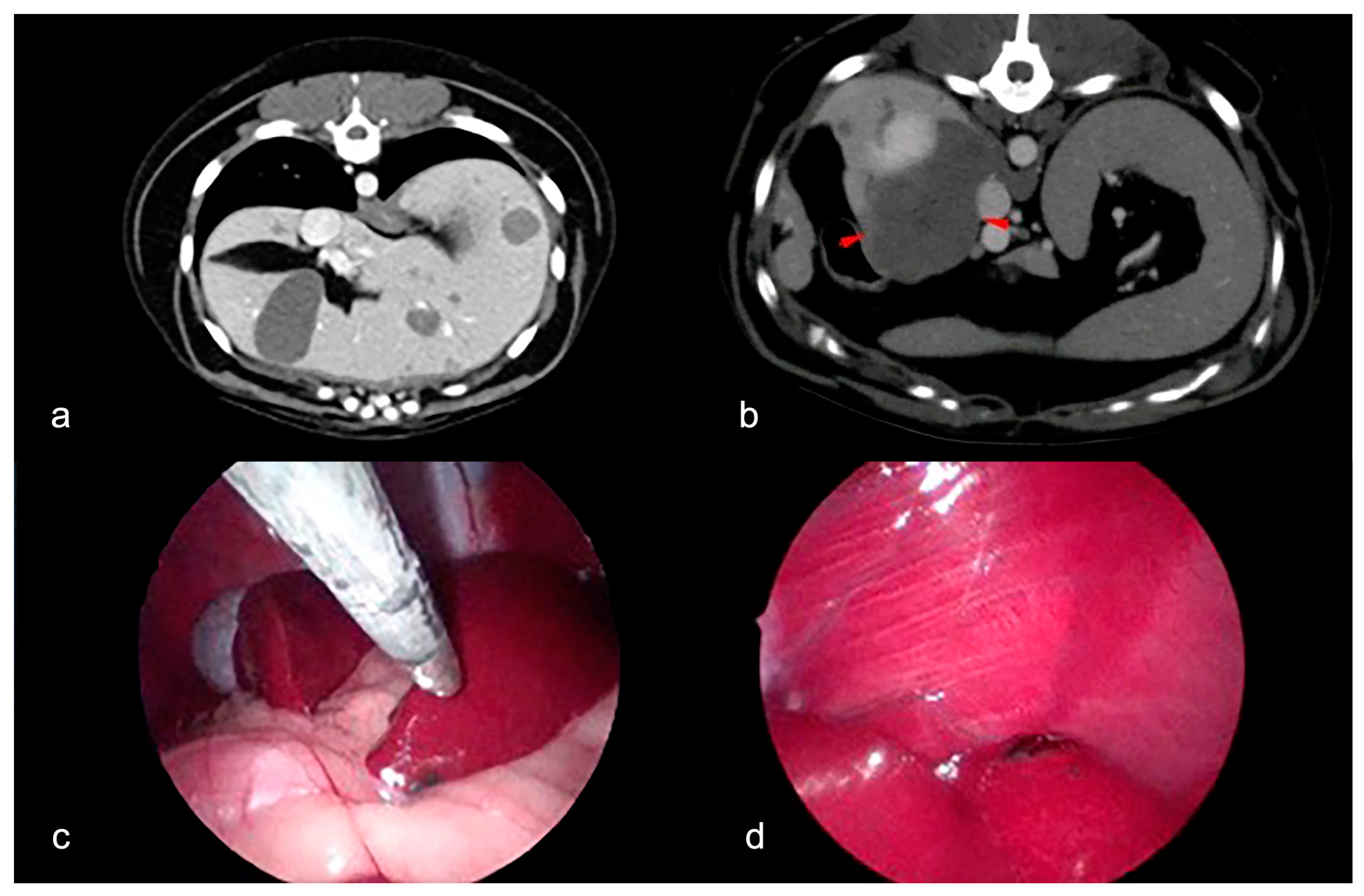
Abstract
:Simple Summary
Abstract
1. Introduction
2. Materials and Methods
3. Results
3.1. Clinical Findings
3.2. Histopathological and Immunohistochemical Features
3.3. Follow Up
4. Discussion
5. Conclusions
Supplementary Materials
Author Contributions
Funding
Institutional Review Board Statement
Informed Consent Statement
Data Availability Statement
Conflicts of Interest
References
- Marr, J.; Miranda, I.C.; Miller, A.D.; Summers, B.A. A Review of Proliferative Vascular Disorders of the Central Nervous System of Animals. Vet. Pathol. 2021, 58, 864–880. [Google Scholar] [CrossRef] [PubMed]
- Fuji, R.N.; Patton, K.M.; Steinbach, T.J.; Schulman, F.Y.; Bradley, G.A.; Brown, T.T.; Wilson, E.A.; Summers, B.A. Feline systemic reactive angioendotheliomatosis: Eight cases and literature review. Vet. Pathol. 2005, 42, 608–617. [Google Scholar] [CrossRef] [PubMed]
- Godizzi, F.; Caniatti, M.; Treggiari, E.; Romanelli, G.; Bonfanti, U.; Ghisleni, G.; Roccabianca, P. Extravascular papillary endothelial hyperplasia mimicking soft tissue sarcoma in 2 cats: A potential diagnostic pitfall. J. Vet. Diagn. Investig. 2022, 34, 552–557. [Google Scholar] [CrossRef] [PubMed]
- Machida, N.; Arimura, T.; Otagiri, Y.; Kiryu, K.; Oka, T. Epithelioid Haemangioendothelioma of the Lung in a Dog. J. Comp. Pathol. 1998, 119, 317–322. [Google Scholar] [CrossRef] [PubMed]
- Pires, I.; Queiroga, F.L.; Silva, F.; Pinto, C.; Lopes, C. Kaposi-like vascular tumor of the urinary bladder in a cow. J. Vet. Med. Sci. 2009, 71, 831–833. [Google Scholar] [CrossRef] [PubMed]
- Pounden, W.D.; Sprunger, E. Malignant hemangioendothelioma of a canine spleen. N. Am. Vet. 1947, 28, 461. [Google Scholar]
- Roccabianca, P.; Schulman, Y.; Avallone, G.; Foster, R.; Scruggs, J.; Dittmer, K. Surgical Pathology of Tumors of Domestic Animals. Volume 3: Tumors of Soft Tissue; Kiupel, M., Ed.; Davis-Thompson DVM Foundation: Gurnee, IL, USA, 2020. [Google Scholar]
- Vincek, V.; Zaulyanov, L.; Mirzabeigi, M. Kaposiform hemangioendothelioma: The first reported case in a nonhuman animal species. Vet. Pathol. 2004, 41, 695–697. [Google Scholar] [CrossRef] [PubMed]
- Yaman, T.; Uyar, A.; Keles, O.F.; Yener, Z. Epithelioid and spindle-cell haemangioendothelioma in the brain of a dog: A case report. Vet. Med. 2018, 63, 193–197. [Google Scholar] [CrossRef]
- Lefkowitch, J.H. Neoplasms and Nodules. In Lefkowitch JHBT-SLBI, 10th ed.; Elsevier: Amsterdam, The Netherlands, 2021; pp. 205–267. [Google Scholar]
- Virarkar, M.; Saleh, M.; Diab, R.; Taggart, M.; Bhargava, P.; Bhosale, P. Hepatic hemangioendothelioma: An update. World J. Gastrointest. Oncol. 2020, 12, 248–256. [Google Scholar] [CrossRef] [PubMed]
- Kou, K.; Chen, Y.G.; Zhou, J.P.; Sun, X.D.; Sun, D.W.; Li, S.X.; Lv, G.Y. Hepatic epithelioid hemangioendothelioma: Update on diagnosis and therapy. World J. Clin. Cases 2020, 8, 3978–3987. [Google Scholar] [CrossRef] [PubMed]
- Errani, C.; Zhang, L.; Sung, Y.S.; Hajdu, M.; Singer, S.; Maki, R.G.; Healey, J.H.; Antonescu, C.R. A novel WWTR1-CAMTA1 gene fusion is a consistent abnormality in epithelioid hemangioendothelioma of different anatomic sites. Genes Chromosomes Cancer 2011, 50, 644–653. [Google Scholar] [CrossRef] [PubMed]
- Mascarenhas, R.C.V.; Sanghvi, A.N.; Friedlander, L.; Geyer, S.J.; Beasley, H.S.; Van Thiel, D.H. Thalidomide inhibits the growth and progression of hepatic epithelioid hemangioendothelioma. Oncology 2004, 67, 471–475. [Google Scholar] [CrossRef] [PubMed]
- Hornick, J. Digestive System Tumours. In The WHO Classification of Tumours; International Agency for Research on Cancer: Lyon, France, 2019. [Google Scholar]
- Mehrabi, A.; Kashfi, A.; Fonouni, H.; Schemmer, P.; Schmied, B.M.; Hallscheidt, P.; Schirmacher, P.; Weitz, J.; Friess, H.; Schmidt, J.; et al. Primary malignant hepatic epithelioid hemangioendothelioma: A comprehensive review of the literature with emphasis on the surgical therapy. Cancer 2006, 107, 2108–2121. [Google Scholar] [CrossRef]
- Griebie, E.R.; David, F.H.; Ober, C.P.; Feeney, D.A.; Anderson, K.L.; Wuenschmann, A.; Jessen, C.R. Evaluation of canine hepatic masses by use of triphasic computed tomography and B-mode, color flow, power, and pulsed-wave Doppler ultrasonography and correlation with histopathologic classification. Am. J. Vet. Res. 2017, 78, 1273–1283. [Google Scholar] [CrossRef] [PubMed]
- Rebhun, R.B.; Thamm, D.H. Multiple distinct malignancies in dogs: 53 cases. J. Am. Anim. Hosp. Assoc. 2010, 46, 20–30. [Google Scholar] [CrossRef] [PubMed]


Disclaimer/Publisher’s Note: The statements, opinions and data contained in all publications are solely those of the individual author(s) and contributor(s) and not of MDPI and/or the editor(s). MDPI and/or the editor(s) disclaim responsibility for any injury to people or property resulting from any ideas, methods, instructions or products referred to in the content. |
© 2024 by the authors. Licensee MDPI, Basel, Switzerland. This article is an open access article distributed under the terms and conditions of the Creative Commons Attribution (CC BY) license (https://creativecommons.org/licenses/by/4.0/).
Share and Cite
Muscatello, L.V.; Massari, F.; Roccabianca, P.; Sarli, G.; Benazzi, C.; Bianchi, M.L. Hepatic Epithelioid Hemangioendothelioma in a Dog. Animals 2024, 14, 1302. https://doi.org/10.3390/ani14091302
Muscatello LV, Massari F, Roccabianca P, Sarli G, Benazzi C, Bianchi ML. Hepatic Epithelioid Hemangioendothelioma in a Dog. Animals. 2024; 14(9):1302. https://doi.org/10.3390/ani14091302
Chicago/Turabian StyleMuscatello, Luisa Vera, Federico Massari, Paola Roccabianca, Giuseppe Sarli, Cinzia Benazzi, and Marco Luigi Bianchi. 2024. "Hepatic Epithelioid Hemangioendothelioma in a Dog" Animals 14, no. 9: 1302. https://doi.org/10.3390/ani14091302