Article

Morphokinetic Analyses of Fishing Cat–Domestic Cat Interspecies Somatic Cell Nuclear Transfer Embryos Through A Time-Lapse System

by
Hai-Jun Liu
1,
Serena Jocelyn Wai Yin Oh
1,
Nicole Liling Tay
1,
Christina Yingyan Lim
1,
Chia-Da Hsu
2,
Delia Hwee Hoon Chua
2,
Winnie Koon Lay Teo
1,
Yuin-Han Loh
1,3,4,5,6 and
Soon Chye Ng
1,7,8,*
1
Endangered Species Conservation via Assisted Reproduction (ESCAR) Lab, Institute of Molecular and Cell Biology (IMCB), Agency for Science, Technology and Research (A*STAR), 61 Biopolis Drive, Proteos, Singapore 138673, Singapore
2
Mandai Wildlife Group, 80 Mandai Lake Road, Singapore 729826, Singapore
3
Cell Fate Engineering and Therapeutics Laboratory, Cell Biology and Therapies Division, Institute of Molecular and Cell Biology (IMCB), Agency for Science, Technology and Research (A*STAR), 61 Biopolis Drive, Proteos, Singapore 138673, Singapore
4
Department of Biological Sciences, National University of Singapore, Singapore 117543, Singapore
5
Department of Physiology, Yong Loo Lin School of Medicine, National University of Singapore, Singapore 117593, Singapore
6
NUS Graduate School for Integrative Sciences and Engineering, National University of Singapore, 28 Medical Drive, Singapore 117456, Singapore
7
Department of Obstetrics and Gynaecology, Yong Loo Lin School of Medicine, National University of Singapore, Singapore 119074, Singapore
8
Sincere Healthcare Group, 8 Sinaran Drive, Singapore 307470, Singapore
*
Author to whom correspondence should be addressed.
Animals 2025, 15(2), 148; https://doi.org/10.3390/ani15020148
Submission received: 6 December 2024 / Revised: 30 December 2024 / Accepted: 3 January 2025 / Published: 9 January 2025
(This article belongs to the Section Animal Reproduction)

Simple Summary

Interspecies somatic cell nuclear transfer (iSCNT) is valuable for preserving endangered animals. The objective of this study was to investigate the morphokinetic parameters of fishing cat–domestic cat (iSCNT) embryos from one cell to blastocyst stages, and in particular, the cleavage patterns of the first division in iSCNT and IVF embryos, as these play a central role in euploidy. A time-lapse imaging system is potentially a powerful tool for selecting early embryos with developmental potential for transfer, and hence, for improving feline iSCNT efficiency.

Abstract

A time-lapse live embryo monitoring system provides a powerful approach to recording dynamic developmental events of cultured embryos in detail. By obtaining continuous short-interval images, blastocyst formation can be predicted and embryos can be selected. The objective of this study was to investigate the morphokinetic parameters of fishing cat–domestic cat interspecies somatic cell nuclear transfer (iSCNT) embryos from one-cell to blastocyst stages, and in particular, the cleavage patterns of the first division in iSCNT and IVF embryos, as these play a central role in euploidy. Domestic cat in vitro fertilization (IVF) embryos were set up as controls. The results show that morula and blastocyst development rates were significantly lower in the iSCNT embryos compared to their IVF counterparts. All earlier time points of embryonic development before the onset of blastulation in the iSCNT embryos were significantly delayed when compared with their IVF counterparts. In iSCNT, normal embryos (defined as those that developed to the blastocyst stage) took a longer time to reach the morula stage, and these morulas were more likely to undergo compaction, compared to their arrested embryo counterparts. Direct cleavage in the first division is a morphological aberration, and was seen with greater prevalence in iSCNT embryos than control IVF embryos; these aberrant embryos displayed a significantly lower blastocyst development rate than embryos that had undergone normal cleavage. In conclusion, the morphokinetic parameters of fishing cat–domestic cat iSCNT embryos at early stages could be used to predict their potential for development to the blastocyst stage. A time-lapse imaging system is potentially a powerful tool for selecting early embryos with developmental potential for transfer, and hence, for improving feline iSCNT efficiency.

1. Introduction

Compared to traditional embryo evaluation approaches, an automated time-lapse monitoring system avoids the need for embryos to be removed from the incubator, thus maintaining the culture in a stable and optimal environment at all times. This approach enables embryos to avoid the stress caused by exposure to adverse conditions such as fluctuations in temperature, gas concentration, pH, and humidity outside the incubator. Another significant advantage is that continuous images of the developing embryos can be acquired automatically at intervals of several minutes. These serial images provide detailed information for a more objective evaluation of the embryos through morphokinetic analysis.
Time-lapse technology is extensively used in human in vitro fertilization (IVF); 93% of blastocyst development stages can be accurately predicted based on three dynamic parameters related to cleavage division 48 h after fertilization [1]. In several retrospective cohort studies, using a time-lapse system for culturing and selecting embryos significantly improved the rates of pregnancy and/or implantation after transfer, compared to using a traditional incubator [2,3,4].
Now, time-lapse technology has extended its applications to bovine, sheep, and domestic cat IVF. The effects of abnormal cleavage patterns on embryonic developmental competence in reaching the blastocyst stage in IVF embryos have been investigated in cats [5,6], cattle [7], and sheep [8]. Morphokinetic parameters of IVF embryos and their relationships to in vitro developmental potential have also been assessed in cats and sheep [6,8].
So far, only three studies have explored somatic cell nuclear transfer (SCNT) embryonic development through a time-lapse system. Two of these studies mainly focused on recording the timing of SCNT embryos reaching different developmental stages in cattle [9] and pigs [10]. In mice, early morphokinetic parameters were identified to predict the blastocyst formation of SCNT embryos [11].
Interspecies SCNT (iSCNT) is valuable for preserving endangered species of animals [12,13]. Assisted reproductive technologies (ARTs), especially iSCNT, have proven its application potential for preserving endangered feline species. Utilizing enucleated domestic cat oocytes as recipients, iSCNT embryos were reconstructed in some endangered felids, such as the African wildcat [14], sand cat [15], leopard cat [16], marbled cat, flat-headed cat [17], and kodkod [18]. However, the development capability of reconstructed embryos in vitro and/or in vivo was low. The taxonomic distance between species from which donor nuclei and recipient oocytes come may negatively affect iSCNT efficiency. To date, live iSCNT offspring in felids were successfully produced only in African wildcats and sand cats, both of which belong to the same genus as the domestic cat [14,15]. However, intergeneric SCNT embryos of the leopard cat–domestic cat or flat-headed cat–domestic cat were unable to develop to term, despite successful implantation [16,17].
The fishing cat (Prionailurus viverrinus) is a unique wetland felid, endemic to South and Southeast Asia. Classified as vulnerable [19], wild populations are most threatened by habitat degradation and fragmentation. For conservation purposes, it is worth attempting to propagate fishing cat individuals through iSCNT technology. However, as with the leopard cat and flat-headed cat, the taxonomic relationship between fishing cats and domestic cats is extrageneric, so intergeneric SCNT between these two species is expected to be challenging.
In several human studies, time-lapse imaging revealed that intracytoplasmic sperm injection (ICSI) embryos with abnormal first cleavage had significantly reduced blastocyst development potential and implantation rates compared to normal cleavage embryos [20,21,22,23]. Such information is valuable for selecting embryos for transfer. In order to reduce the adverse effects caused by long-term in vitro culture, SCNT embryos are often transferred into surrogate oviducts at the early one- to four-cell stages in most species, such as in pigs, goats, sheep, and cats. SCNT outcomes could potentially be improved by the selection of normal cleavage embryos over embryos that underwent an abnormal first cleavage.
To date, morphokenetics parameters of interspecies SCNT embryos as well as feline SCNT embryos have not been investigated.
The three objectives of this current study were (1) to use a time-lapse imaging system to acquire detailed morphokinetic characteristics of fishing cat-domestic cat iSCNT and domestic cat IVF embryos from one-cell to blastocyst stages, (2) to identify differences in morphokinetic parameters between successful fishing cat–domestic cat iSCNT blastocysts and their arrested counterparts as well as domestic cat IVF embryos, and (3) to investigate the relationship between the cleavage patterns of first division in fishing cat–domestic cat iSCNT embryos and their subsequent blastocyst developmental ability.

2. Materials and Methods

2.1. Chemicals

All chemicals were purchased from Sigma-Aldrich (St. Louis, MO, USA) unless specified.

2.2. In Vitro Maturation (IVM) of Oocytes

Domestic cat ovaries were donated by local veterinary clinics, and transported to the laboratory in PBS at 4 °C, where they were stored at 4 °C for 12–24 h. Cumulus–oocyte complexes (COCs) were obtained through the dissection of the ovarian cortex into TCM 199-Hepes medium (Gibco, Grand Island, NY, USA) with 4 mg/mL BSA. Oocytes were classified as previously described [24]. Selected oocytes with homogeneous ooplasm and compact cumulus cells were cultured for in vitro maturation (IVM) in four-well plates (NUNC; Thermo Fisher Scientific, Waltham, MA, USA) in TCM-199 supplemented with 4 mg/mL BSA, 1 mM Sodium Pyruvate, 1.2 mM Cysteine, 2 mM L-Glutamine, 0.1 IU/mL FSH and LH, at 38.5 °C in a humidified atmosphere of 5% CO2, 5%O2, and 90% N2. After culturing for 24–30 h, cumulus cells were removed with 0.5% hyaluronidase. Oocytes with extruded first polar bodies were selected under a stereomicroscope SCNT or IVF.

2.3. Interspecies SCNT

2.3.1. Donor Cells

Donor cells for iSCNT were derived from female fishing cat fibroblasts donated by the Mandai Wildlife Group cell biobank. Dividing cells of passages 6–8 at 80% confluency were dissociated with 0.25% trypsin (Gibco, Grand Island, NY, USA).

2.3.2. Nuclear Transfer

Nuclear transfer was performed as previously described [25,26]. Metaphase II oocytes preincubated with Hoechst 33342 were enucleated by exposure to UV light. Donor cells were introduced into the perivitelline space of enucleated oocytes. Two pulses (2.0 KV/cm, 25 μs) were applied for fusion of the reconstructed oocytes in fusion medium. The fused couplets were activated with 10 μg/mL Calcium Ionophore (A23187) for 5 min, then 10 μg/mL cycloheximide and 5 μg/mL cytochalasin B for 5 h.

2.4. In Vitro Fertilization

Domestic cat testes were donated by local veterinary clinics. Sperm from the epididymis were collected for IVF. Cauda epididymis was cut transversely into small pieces in TCM 199-Hepes (Gibco, Grand Island, NY, USA) with 4 mg/mL BSA, and incubated for 10 min. Collected sperm was stored at 4 °C overnight. The sperm was centrifuged with Sydney IVF Sperm Medium (Cook, Brisbane, Australia). After discarding the supernatant, 1 mL Sydney IVF Sperm Medium was added to the pellet. The sperm samples were incubated for 30 min at 38.5 °C and 5% CO2 for swim-up. The metaphase II oocytes were fertilized with the top motile sperm at a final concentration of 0.5~1 × 10⁶/mL for 15.5 h in Sydney IVF Fertilization Medium (Cook, Brisbane, Australia) at 38.5 °C in a humidified atmosphere of 5% CO2, 5% O2, and 90% N2.

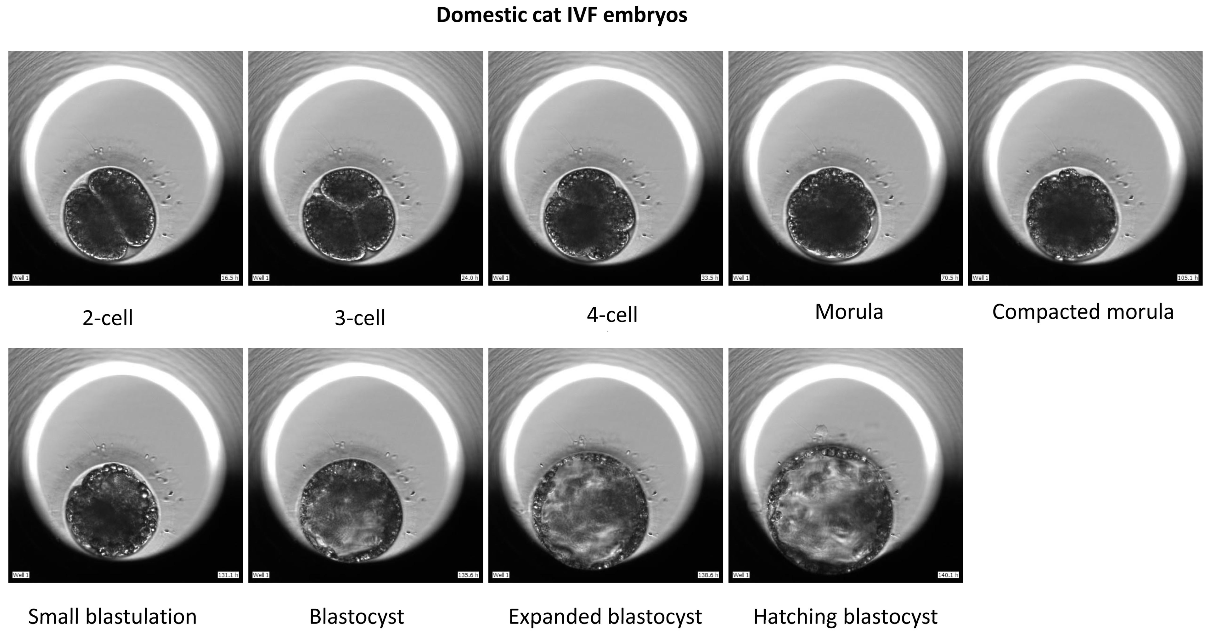
2.5. In Vitro Embryo Culture and Time-Lapse Imaging

The reconstructed iSCNT embryos (after activation) or in vitro fertilized presumptive zygotes were placed individually in droplets of G-TL medium (Vitrolife, Göteburg, Sweden) in an EmbryoscopeTM time-lapse incubator (ES-D2-944; Vitrolife, Göteburg, Sweden) at 38.5 °C with 5% CO2, 5%O2, 90% N2 for 188-215 h (iSCNT embryos) or 207 h (IVF embryos). Images of embryos were acquired every 5 min in five different focal planes.
For IVF embryos, “Time 0” was defined as the time when sperm was added to oocytes; for iSCNT embryos, “Time 0” was defined as the time when fused embryos were placed into the activation medium of cycloheximide and cytochalasin B in M199. Morphokenetic marks included the timing of division to the 2-, 3-, and 4-blastomere stages (t2, t3, t4); morula formation (first signs of morula, tM); compacted morula formation (first signs of compaction, tCM); early blastocyst (tSB); blastocyst (tB); expanded blastocyst (tEB); and hatching blastocyst (tHB) stages. The durations of the second cell cycle (cc2; t3 − t2) and synchronous divisions (s2; t4 − t3) were included.
Cleavage patterns in the first division were classified into normal cleavage and abnormal cleavage. Normal cleavage is defined as the division of a one-cell embryo into two blastomeres of equal sizes. Abnormal cleavage included direct cleavage and uneven cleavage. Direct cleavage is when a one-cell embryo divides directly into three or more blastomeres, obviating the 2-cell stage. Uneven cleavage is defined as the division of a one-cell embryo into two blastomeres with uneven size.
In iSCNT, normal embryos (NE) are defined as those that developed to the blastocyst stage, while arrested embryos (AE) are defined as those whose development was arrested at any stage before reaching the blastocyst stage.

2.6. Blastocyst Cell Counting

Total cell number was compared between iSCNT or IVF blastocysts. Blastocysts derived from iSCNT or IVF were incubated with 10 μg/mL Hoechst 33342 in TCM-199 medium for 15 min and imaged. Blastocyst cell number was counted with the aid of Image J version 1.46r software (NIH, Bethesda, MD, USA) applied to the images.

2.7. Data Analysis

The data were analyzed by Mann–Whitney test with GraphPad InStat 3.06 software (GraphPad, San Diego, CA, USA). Differences were defined as significant at p < 0.05.

3. Results

3.1. Comparison of In Vitro Developmental Capability of Fishing Cat-Domestic Cat iSCNT or Domestic Cat IVF Embryos

Here, 216 oocytes collected from 36 domestic cat ovaries were selected for IVM, and 123 of them extruded first polar bodies. These matured oocytes were used for IVF or iSCNT. We investigated the differences in developmental capabilities between iSCNT embryos of fishing cat–domestic cat and domestic cat IVF embryos. The results show that the morula and blastocyst rates were significantly lower in iSCNT embryos compared with their IVF counterparts (p < 0.05) (Table 1). However, no differences were observed in the rates of cleavage, morula compaction, or hatching blastocyst, or in blastocyst cell numbers (Figure 1, Figure 2 and Figure 3; Videos S1 and S2).

3.2. Comparison of Morphokinetic Parameters of Fishing Cat–Domestic Cat iSCNT Embryos and Domestic Cat IVF Embryos

We compared the morphokinetic parameters of 8 fishing cat–domestic cat iSCNT embryos and 11 domestic cat IVF embryos from the first division to the blastocyst stage (Table 2). All the earlier stages of embryonic development before tSB (i.e., t2, t3, t4, tM, tCM) in iSCNT embryos were significantly delayed compared to their IVF counterparts (p < 0.05). However, all the later stages of embryonic development (i.e., tSB, tB, tEB, tHB) and the durations of cell cycles (cc2, s2) showed no significant difference between iSCNT and IVF embryos.

3.3. Comparison of Morphokinetic Parameters of Normal or Arrested Embryos Derived from Fishing Cat–Domestic Cat iSCNT

We compared the morphokinetic parameters of 8 normal and 24 arrested fishing cat–domestic cat iSCNT embryos from the first division to the compacted morula stage (Table 3). The mean tM and tCM were significantly longer in normal embryos compared to their arrested counterparts (p < 0.05). All earlier cleavage durations of embryonic development (i.e., t2, t3, t4), cc2, and s2 showed no significant differences between these two types of embryos.

3.4. Cleavage Patterns of the First Division in iSCNT and IVF Embryos and Their Relationship to Development to Blastocyst Stage

We investigated the relationship between cleavage patterns of the first division and blastocyst development frequency in iSCNT and IVF embryos. In both iSCNT and IVF embryos, morphologically aberrant direct cleavage at the first division occurred at high proportions (14/32, 43.8%; 5/14, 35.7%; respectively). Compared to normal cleavage embryos, direct cleavage embryos displayed significantly lower blastocyst development rates in iSCNT embryos (43.8% vs. 7.7%; p < 0.05). Also, significantly more IVF embryos from direct cleavage reached the blastocyst stage than their iSCNT counterparts (60% vs. 7.7%; p < 0.05) (Table 4; Figure 4; Videos S3–S7).

4. Discussion

An automated time-lapse monitoring system provides a powerful approach in which embryos can be cultured in a stable environment while recording their dynamic developmental events in detail by continuous short-interval imaging. The resulting information could be used to predict the success of blastocyst formation and to perform quality control to select embryos with greater development potential for transfer.
To our knowledge, this is the first study investigating the morphokenetics parameters of iSCNT embryos and feline SCNT embryos through time-lapse technology.
The present study has shown that fishing cat–domestic cat iSCNT embryos presented compromised abilities to develop to the blastocyst stage compared with their domestic cat IVF counterparts (25% vs. 78.6%). Concordant with our present results, previous studies have demonstrated that iSCNT embryos demonstrated significantly lower blastocyst formation rates than those of IVF embryos in flat-headed cat–domestic cat (8.4% vs. 61.4%) and kodkod–domestic cat (5.9% vs. 44.5%) embryos [17,18]. Recipient domestic cats failed to become pregnant after flat-headed cat iSCNT embryo transfer, but in contrast, all six recipients that received domestic cat IVF embryos established pregnancies, with three of them producing five live kittens [17].
More than two decades have elapsed since the birth of “Dolly” the sheep [27]. However, cloning efficiency remains as low as under 5% (live offsprings/reconstructed embryos) [28]. Incomplete epigenetic reprogramming such as aberrant H3K9me3, H3K27me3, and H3K4me3 could impede the preimplantation or postimplantation development of iSCNT embryos [29]. Such epigenetic modifications also play an important role in in mouse embryonic stem cell pluripotency [30,31]. Mimicking physiological reprogramming in oocytes and sperm in preparation for fertilization may be a more effective strategy to improve nuclear transfer efficiency, compared to the extensively applied epigenetic modification of donor cell reprogramming after nuclear transfer [32]. The overexpression of transcription factor double homeobox (Dux) greatly improves mouse SCNT efficiency [33], and the expression of 2C transcripts was Dux-dependent in 2-cell-like cells of mouse embryonic stem cells (mESCs) [34]. For iSCNT, the incompatibility between nuclear and mitochondrial DNA, mtDNA heteroplasmy, and genome activation (EGA) of reconstructed embryos might be additional barriers impairing embryonic development, compared to SCNT [35].
Our current results indicate that the fishing cat–domestic cat iSCNT developed slower than their domestic cat IVF counterparts at all the earlier embryonic development stages, from two-cell to compacted morula. We presumed that this difference might be caused by the process of the first cell cycle in iSCNT or IVF embryos. Our previous study found that mitotic metaphase and cleavage were first detected at 23 h and 24 h post-activation, respectively, in goat SCNT embryos; this occurred later than in IVF embryos (16 h and 21 h post-insemination, respectively) [25]. Mouse SCNT embryos also took a longer time from the second division to compaction compared to their ICSI counterparts [11]. The current results are consistent with these observations.
The current study found that the arrested fishing cat–domestic cat iSCNT embryos took a shorter time to develop to morula or compacted morula stages. This finding is consistent with that of arrested IVF ovine embryos, which developed to the morula stage faster than normal embryos (72.32 h vs. 104.45 h) [8]. It is possible that prolonged morula formation time might allow normal embryos to undergo the cell divisions necessary to reach the cell numbers required for further development to the blastocyst stage. Mouse arrested SCNT embryos displayed massive levels of dysregulation of genes associated with transcription pathways, compared to their normal counterparts as well as IVF embryos [36]. Our domestic cat IVF embryos developed to the morula stage at 59.05 ± 6.64 h, which is shorter than the 74 h in a previous report [37]. The average cell numbers of domestic cat IVF blastocysts were 121.5 ± 17.5, which are lower than the 156.7 and 204.4 in a previous report [38].
In this study, 50% of the iSCNT embryos showed abnormal cleavage patterns in the first division, indicating the high prevalence of abnormal cleavage in fishing cat–domestic cat iSCNT embryos. Most abnormal cleavages were direct cleavages (87.5%), with uneven cleavage making up the remaining type (12.5%). In contrast, 35.7% of domestic cat IVF embryos underwent direct cleavage. Direct cleavage in iSCNT embryos significantly decreased their developmental capability to the blastocyst stage, compared with normal cleavage embryos, as well as direct cleavage embryos in domestic cat IVF. In contrast with our results, mouse SCNT embryos never underwent direct cleavage, but experienced fragmentation as the most common abnormality [11]. This inconsistency may be due to species differences.
In accordance with our results, previous studies have demonstrated a high frequency of 46–48% of abnormal cleavage in domestic cat IVF embryos [5,6]. Abnormal cleavages include cytoplasmic fragmentation, vacuolation, direct cleavage, additional reverse cleavage, and asymmetry of blastomeres. In these studies, 36.6% of morphologically normal and 48.6% of morphologically abnormal embryos developed to the blastocyst stage [5]. Higher blastocyst and hatching blastocyst rates were obtained when culturing domestic cat IVF embryos in Primo Vision® dishes compared with the classical individual and group culture systems [39].
Through time-lapse imaging, several human studies have showed that the blastocyst development and/or implantation potential of ICSI embryos is severely impaired if direct cleavage occurs at first cleavage after fertilization, compared to embryos that underwent normal cleavage [20,21,22,23]. Bovine IVF embryos exhibiting direct cleavage led to significantly lower blastocyst and hatched blastocyst rates, and decreased chromosomal euploidy rates, than that of their normal counterparts [7]. In sheep IVF, none of the embryos that underwent direct cleavage developed to the blastocyst stage [8].
To date, the cause of direct cleavage remains unclear. A possible explanation for this might be the formation of multipolar spindles and supernumerary centrioles caused by defective sperm [21,23,40]. Recent evidence suggests that PADI6 gene mutations might be related to the pre-implantation embryonic arrest caused by direct cleavage in human IVF/ICSI embryos [41]. Our previous study found multiple polar spindles at the first mitotic metaphase in goat SCNT embryos [25].
Direct cleavage can only be observed by time-lapse imaging, and not by traditional embryo evaluation methods. Due to the impaired blastocyst development and/or implantation potential of direct cleavage embryos, screening out these abnormal embryos for transfer at earlier stages could increase pregnancy and implantation rates for both IVF and SCNT embryos.
Further work that could be done includes investigating the cellular and molecular mechanism underlying direct cleavage patterns in fishing cat–domestic cat iSCNT embryos, and determining the in vivo developmental potential of fishing cat iSCNT embryos by transferring quality-controlled embryos into surrogate cats.

5. Conclusions

In conclusion, the morphokinetics parameters of fishing cat–domestic cat iSCNT embryos at early stages could be used to predict their potential to develop to the blastocyt stage. Time-lapse imaging systems are a powerful tool, whereby early embryos with greater developmental potential could be selected for transfer, thereby improving SCNT efficiency in felid and possibly other species.

Supplementary Materials

The following supporting information can be downloaded at: https://www.mdpi.com/article/10.3390/ani15020148/s1, Video S1: iSCNT embryo development; Video S2: IVF embryo development; Video S3: Normal cleavage of iSCNT embryo; Video S4: Direct cleavage of iSCNT embryo; Video S5: Uneven cleavage of iSCNT embryo; Video S6: Normal cleavage of IVF embryo; Video S7: Direct cleavage of IVF embryo.

Author Contributions

Conceptualization, H.-J.L. and S.C.N.; methodology, H.-J.L.; resources, S.J.W.Y.O., N.L.T., C.Y.L., C.-D.H., D.H.H.C. and W.K.L.T.; investigation, H.-J.L.; data curation, H.-J.L.; writing—original draft, H.-J.L.; writing—review and editing, S.C.N. and Y.-H.L.; funding acquisition, S.C.N.; supervision, Y.-H.L. All authors have read and agreed to the published version of the manuscript.

Funding

This work was supported by a generous grant from Temasek Foundation Ecosperity (TFE-ESF-SG-2017-WRS-01).

Institutional Review Board Statement

Not applicable.

Informed Consent Statement

Not applicable.

Data Availability Statement

The data for this study are included in the article and Supplementary Materials.

Acknowledgments

We thank Mandai Nature for administrative support and stewardship. Great thanks should be given to the late Chai Chou for his significant contributions to this fishing cat iSCNT project.

Conflicts of Interest

Authors Chia-Da Hsu and Delia Hwee Hoon Chua were employed by the company Mandai Wildlife Group, and Author Soon Chye Ng was employed by the company Sincere Healthcare Group. The remaining authors declare that the research was conducted in the absence of any commercial or financial relationships that could be construed as a potential conflict of interest.

References

  1. Wong, C.C.; Loewke, K.E.; Bossert, N.L.; Behr, B.; De Jonge, C.J.; Baer, T.M.; Reijo Pera, R.A. Non-invasive imaging of human embryos before embryonic genome activation predicts development to the blastocyst stage. Nat. Biotechnol. 2010, 28, 1115–1121. [Google Scholar] [CrossRef]
  2. Meseguer, M.; Rubio, I.; Cruz, M.; Basile, N.; Marcos, J.; Requena, A. Embryo incubation and selection in a time-lapse monitoring system improves pregnancy outcome compared with a standard incubator: A retrospective cohort study. Fertil. Steril. 2012, 98, 1481–1489.e10. [Google Scholar] [CrossRef] [PubMed]
  3. Aparicio-Ruiz, B.; Basile, N.; Pérez Albalá, S.; Bronet, F.; Remohí, J.; Meseguer, M. Automatic time-lapse instrument is superior to single-point morphology observation for selecting viable embryos: Retrospective study in oocyte donation. Fertil. Steril. 2016, 106, 1379–1385.e10. [Google Scholar] [CrossRef]
  4. Ma, B.X.; Zhang, H.; Jin, L.; Huang, B. Neonatal Outcomes of Embryos Cultured in a Time-Lapse Incubation System: An Analysis of More Than 15,000 Fresh Transfer Cycles. Reprod. Sci. 2022, 29, 1524–1530. [Google Scholar] [CrossRef] [PubMed]
  5. Kij, B.; Kochan, J.; Nowak, A.; Niżański, W.; Prochowska, S.; Fryc, K.; Bugno-Poniewierska, M. Using Time Lapse Monitoring for Determination of Morphological Defect Frequency in Feline Embryos after In Vitro Fertilization (IVF). Animals 2020, 10, 3. [Google Scholar] [CrossRef] [PubMed]
  6. Kochan, J.; Nowak, A.; Kij, B.; Prochowska, S.; Niżański, W. Analysis of Morphokinetic Parameters of Feline Embryos Using a Time-Lapse System. Animals 2021, 9, 748. [Google Scholar] [CrossRef] [PubMed]
  7. Magata, F.; Ideta, A.; Okubo, H.; Matsuda, F.; Urakawa, M.; Oono, Y. Growth potential of bovine embryos presenting abnormal cleavage observed through time lapse cinematography. Theriogenology 2019, 133, 119–124. [Google Scholar] [CrossRef]
  8. Fryc, K.; Nowak, A.; Kij, B.; Kochan, J.; Bartlewski, P.M.; Murawski, M. Timing of cleavage divisions determined with time-lapse imaging is linked to blastocyst formation rates and quality of in vitro-produced ovine embryos. Theriogenology 2021, 159, 147–152. [Google Scholar] [CrossRef]
  9. Park, M.J.; Kim, E.Y.; Kang, M.J.; Lee, J.B.; Jeong, C.J.; Park, S.P. Investigation of the Developmental Potential and Developmental Kinetics of Bovine Parthenogenetic and Somatic Cell Nuclear Transfer Embryos Using a Time-Lapse Monitoring System. Cell. Reprogram. 2017, 19, 245–254. [Google Scholar] [CrossRef] [PubMed]
  10. Son, Y.J.; Lee, S.E.; Park, Y.G.; Jeong, S.G.; Shin, M.Y.; Kim, E.Y.; Park, S.P. Fibroblast Growth Factor 10 Enhances the Developmental Efficiency of Somatic Cell Nuclear Transfer Embryos by Accelerating the Kinetics of Cleavage During In Vitro Maturation. Cell Reprogram. 2018, 20, 196–204. [Google Scholar] [CrossRef] [PubMed]
  11. Mallol, A.; Piqué, L.; Santaló, J.; Ibáñez, E. Morphokinetics of cloned mouse embryos treated with epigenetic drugs and blastocyst prediction. Reproduction 2016, 151, 203–214. [Google Scholar] [CrossRef] [PubMed]
  12. Swegen, A.; Appeltant, R.; Williams, S.A. Cloning in action: Can embryo splitting, induced pluripotency and somatic cell nuclear transfer contribute to endangered species conservation? Biol. Rev. Camb. Philos. Soc. 2023, 98, 1225–1249. [Google Scholar] [CrossRef] [PubMed]
  13. Cowl, V.B.; Comizzoli, P.; Appeltant, R.; Bolton, R.L.; Browne, R.K.; Holt, W.V.; Penfold, L.M.; Swegen, A.; Walker, S.L.; Williams, S.A. Cloning for the Twenty-First Century and Its Place in Endangered Species Conservation. Annu. Rev. Anim. Biosci. 2024, 12, 91–112. [Google Scholar] [CrossRef] [PubMed]
  14. Gómez, M.C.; Pope, C.E.; Giraldo, A.; Lyons, L.A.; Harris, R.F.; King, A.L.; Cole, A.; Godke, R.A.; Dresser, B.L. Nuclear transfer of sand cat cells into enucleated domestic cat oocytes is affected by cryopreservation of donor cells. Cloning Stem Cells 2004, 6, 247–258. [Google Scholar] [CrossRef] [PubMed]
  15. Gómez, M.C.; Pope, C.E.; Kutner, R.H.; Ricks, D.M.; Lyons, L.A.; Ruhe, M.; Dumas, C.; Lyons, J.; López, M.; Dresser, B.L.; et al. Birth of African Wildcat cloned kittens born from domestic cats. Cloning Stem Cells 2008, 10, 469–483. [Google Scholar] [CrossRef] [PubMed]
  16. Yin, X.J.; Lee, Y.; Lee, H.; Kim, N.; Kim, L.; Shin, H.; Kong, I. In vitro production and initiation of pregnancies in inter-genus nuclear transfer embryos derived from leopard cat (Prionailurus bengalensis) nuclei fused with domestic cat (Felis silverstris catus) enucleated oocytes. Theriogenology 2006, 66, 275–282. [Google Scholar] [CrossRef]
  17. Thongphakdee, A.; Siriaroonrat, B.; Manee-in, S.; Klincumhom, N.; Kamolnorranath, S.; Chatdarong, K.; Techakumphu, M. Intergeneric somatic cell nucleus transfer in marbled cat and flat-headed cat. Theriogenology 2010, 73, 120–128. [Google Scholar] [CrossRef]
  18. Veraguas, D.; Aguilera, C.; Echeverry, D.; Saez-Ruiz, D.; Castro, F.O.; Rodriguez-Alvarez, L. Embryo aggregation allows the production of kodkod (Leopardus guigna) blastocysts after interspecific SCNT. Theriogenology 2020, 158, 148–157. [Google Scholar] [CrossRef]
  19. Mukherjee, S.; Appel, A.; Duckworth, J.W.; Sanderson, J.; Dahal, S.; Willcox, D.H.A.; Herranz Muñoz, V.; Malla, G.; Ratnayaka, A.; Kantimahanti, M.; et al. The IUCN Red List of Threatened Species. 2016. Prionailurus viverrinus, e.T18150A50662615. Available online: https://www.iucnredlist.org/species/18150/221434864 (accessed on 3 June 2022).
  20. Yang, S.T.; Shi, J.X.; Gong, F.; Zhang, S.P.; Lu, C.F.; Tan, K.; Leng, L.Z.; Hao, M.; He, H.; Gu, Y.F.; et al. Cleavage pattern predicts developmental potential of day 3 human embryos produced by IVF. Reprod. Biomed. Online 2015, 30, 625–634. [Google Scholar] [CrossRef]
  21. Zhan, Q.; Ye, Z.; Clarke, R.; Rosenwaks, Z.; Zaninovic, N. Direct Unequal Cleavages: Embryo Developmental Competence, Genetic Constitution and Clinical Outcome. PLoS ONE 2016, 11, e0166398. [Google Scholar] [CrossRef]
  22. Barrie, A.; Homburg, R.; McDowell, G.; Brown, J.; Kingsland, C.; Troup, S. Preliminary investigation of the prevalence and implantation potential of abnormal embryonic phenotypes assessed using time-lapse imaging. Reprod. Biomed. Online 2017, 34, 455–462. [Google Scholar] [CrossRef] [PubMed]
  23. Desai, N.; Goldberg, J.M.; Austin, C.; Falcone, T. Are cleavage anomalies, multinucleation, or specific cell cycle kinetics observed with time-lapse imaging predictive of embryo developmental capacity or ploidy? Fertil. Steril. 2018, 109, 665–674. [Google Scholar] [CrossRef] [PubMed]
  24. Wood, T.C.; Wildt, D.E. Effect of the quality of the cumulus-oocyte complex in the domestic cat on the ability of oocytes to mature, fertilize and develop into blastocysts in vitro. J. Reprod. Fertil. 1997, 110, 355–360. [Google Scholar] [CrossRef] [PubMed]
  25. Liu, H.J.; Liu, R.M. Dynamic changes in chromatin and microtubules at the first cell cycle in SCNT or IVF goat embryos. Cell Biol. Int. 2018, 42, 1401–1409. [Google Scholar] [CrossRef] [PubMed]
  26. Wong, K.W.; Zeng, Y.; Tay, E.; Teo, J.H.J.; Cipta, N.O.; Hamashima, K.; Yi, Y.; Liu, H.; Warrier, T.; Le, M.T.N.; et al. Nuclear receptor-SINE B1 network modulates expanded pluripotency in blastoids and blastocysts. Nat. Commun. 2024, 15, 10011. [Google Scholar] [CrossRef] [PubMed] [PubMed Central]
  27. Wilmut, I.; Schnieke, A.E.; McWhir, J.; Kind, A.J.; Campbell, K.H. Viable offspring derived from fetal and adult mammalian cells. Nature 1997, 385, 810–813. [Google Scholar] [CrossRef]
  28. Klinger, B.; Schnieke, A. 25th ANNIVERSARY OF CLONING BY SOMATIC-CELL NUCLEAR TRANSFER Twenty-five years after Dolly: How far have we come? Reproduction 2021, 162, F1–F10. [Google Scholar] [CrossRef] [PubMed]
  29. Matoba, S.; Zhang, Y. Somatic Cell Nuclear Transfer Reprogramming: Mechanisms and Applications. Cell Stem Cell. 2018, 4, 471–485. [Google Scholar] [CrossRef] [PubMed] [PubMed Central]
  30. Warrier, T.; El Farran, C.; Zeng, Y.; Ho, B.S.Q.; Bao, Q.; Zheng, Z.H.; Bi, X.; Ng, H.H.; Ong, D.S.T.; Chu, J.J.H.; et al. SETDB1 acts as a topological accessory to Cohesin via an H3K9me3-independent, genomic shunt for regulating cell fates. Nucleic Acids Res. 2022, 50, 7326–7349. [Google Scholar] [CrossRef] [PubMed] [PubMed Central]
  31. Hamashima, K.; Wong, K.W.; Sam, T.W.; Teo, J.H.J.; Taneja, R.; Le, M.T.N.; Li, Q.J.; Hanna, J.H.; Li, H.; Loh, Y.H. Single-nucleus multiomic mapping of m6A methylomes and transcriptomes in native populations of cells with sn-m6A-CT. Mol. Cell. 2023, 83, 3205–3216. [Google Scholar] [CrossRef] [PubMed] [PubMed Central]
  32. Loi, P.; Palazzese, L.; Scapolo, P.A.; Fulka, J.; Fulka, H.; Czernik, M. 25th ANNIVERSARY OF CLONING BY SOMATIC-CELL NUCLEAR TRANSFER: Scientific and technological approaches to improve SCNT efficiency in farm animals and pets. Reproduction 2021, 162, F33–F43. [Google Scholar] [CrossRef] [PubMed]
  33. Yang, G.; Zhang, L.; Liu, W.; Qiao, Z.; Shen, S.; Zhu, Q.; Gao, R.; Wang, M.; Wang, M.; Li, C.; et al. Dux-Mediated Corrections of Aberrant H3K9ac during 2-Cell Genome Activation Optimize Efficiency of Somatic Cell Nuclear Transfer. Cell Stem Cell 2021, 28, 150–163.e5. [Google Scholar] [CrossRef] [PubMed]
  34. Yi, Y.; Zeng, Y.; Sam, T.W.; Hamashima, K.; Tan, R.J.R.; Warrier, T.; Phua, J.X.; Taneja, R.; Liou, Y.C.; Li, H.; et al. Ribosomal proteins regulate 2-cell-stage transcriptome in mouse embryonic stem cells. Stem Cell Rep. 2023, 18, 463–474. [Google Scholar] [CrossRef] [PubMed] [PubMed Central]
  35. Mrowiec, P.; Bugno-Poniewierska, M.; Młodawska, W. The perspective of the incompatible of nucleus and mitochondria in interspecies somatic cell nuclear transfer for endangered species. Reprod. Domest. Anim. 2021, 56, 199–207. [Google Scholar] [CrossRef]
  36. Long, C.; Li, H.; Li, X.; Yang, W.; Zuo, Y. Nuclear transfer arrest embryos show massive dysregulation of genes involved in transcription pathways. Int. J. Mol. Sci. 2021, 22, 8187. [Google Scholar] [CrossRef]
  37. Ochota, M.; Niżański, W. Time of early cleavage affects the developmental potential of feline preimplantation embryos in vitro. Theriogenology 2017, 89, 26–31. [Google Scholar] [CrossRef] [PubMed]
  38. Ochota, M.; Wojtasik, B.; Niżański, W. Total Cell Number and its Allocation to Trophectoderm and Inner Cell Mass in In Vitro Obtained Cats’ Blastocysts. Reprod. Domest. Anim. 2016, 51, 339–345. [Google Scholar] [CrossRef] [PubMed]
  39. Kochan, J.; Nowak, A.; Kij, B.; Fryc, K.; Prochowska, S.; Niżański, W. A comparison of in vitro culture systems for cat embryos. Theriogenology 2022, 179, 149–154. [Google Scholar] [CrossRef] [PubMed]
  40. Rubio, I.; Kuhlmann, R.; Agerholm, I.; Kirk, J.; Herrero, J.; Escribá, M.J.; Bellver, J.; Meseguer, M. Limited implantation success of direct-cleaved human zygotes: A time-lapse study. Fertil. Steril. 2012, 98, 1458–1463. [Google Scholar] [CrossRef]
  41. Zheng, W.; Chen, L.; Dai, J.; Dai, C.; Guo, J.; Lu, C.; Gong, F.; Lu, G.; Lin, G. New biallelic mutations in PADI6 cause recurrent preimplantation embryonic arrest characterized by direct cleavage. J. Assist. Reprod. Genet. 2020, 37, 205–212. [Google Scholar] [CrossRef]
Figure 1. Development of fishing cat–domestic cat iSCNT embryos.
Animals 15 00148 g001
Figure 2. Development of domestic cat IVF embryos.
Animals 15 00148 g002
Figure 3. Blastocyst derived from iSCNT or IVF stained with Hoechst 33342.
Animals 15 00148 g003
Figure 4. Cleavage patterns of iSCNT or IVF embryos at the first division.
Animals 15 00148 g004
Table 1. In vitro development of fishing cat–domestic cat iSCNT embryos vs. domestic cat IVF embryos.
GroupNo. of Reconstructed
Couplets
No. of Fused
Couplets (%)
No. of Inseminated
Oocytes
No. of Cultured
Embryos
No. of Cleavage
(%)
iSCNT7249 (68.1)---4432 (72.7)
IVF------161614 (87.5)
No. of Morula
(cleavage %)
No. of Compacted Morula (cleavage %)No. of Blastocyst
(cleavage %)
No. Hatching Blastocyst
(blastocyst %)
Cell Number of Blastocyst
iSCNT13 (40.6) a12 (37.5)8 (25) a4 (50)88.3 + 9.4
IVF14 (100) b12 (85.7)11 (78.6) b10 (90.9)121.5 ± 17.5
Note: Different superscripts within the same column (e.g., a, b) represent significant differences (p < 0.05).
Table 2. Morphokinetic characteristics of fishing cat–domestic cat iSCNT and domestic cat IVF embryos developed to the blastocyst stage.
VariablesiSCNTNo. of Embryos *IVFNo. of Embryos **
t222.5 ± 3.53 a718.75 ± 3.42 b8
t330.61 ± 3.24 a723.3 ± 3.84 b8
t439.4 ± 7.6 a728.84 ± 3.98 b8
cc28.11 ± 3.674.55 ± 2.53 8
s28.79 ± 5.5975.54 ± 2.938
tM81.45 ± 6.03 a859.05 ± 6.64 b11
tCM99 ± 6.42 a884.26 ± 7.94 b11
tSB126.93 ± 9.328125.4 ± 12.7111
tB130.3 ± 5.547135.16 ± 10.1711
tEB140.87 ± 10.336141.5 ± 12.4611
tHB148.2 ± 15.114143.26 ± 6.6110
Notes: Different superscripts within the same row (e.g., a, b) represent significant differences (p < 0.05). * one iSCNT embryo with direct cleavage was excluded from t2 to s2. ** three IVF embryos with direct cleavage were excluded from t2 to s2.
Table 3. Morphokinetic characteristics of normal or arrested embryos derived from fishing cat–domestic cat iSCNT.
VariablesNormal EmbNo. of Embryos *Arrested EmbNo. of Embryos **
t222.5 ± 3.53722.36 ± 6.157 ***
t330.61 ± 3.24733.53 ± 7.817
t439.4 ± 7.6739.5 ± 11.415
cc28.11 ± 3.6711.17 ± 3.47
s29.5 ± 5.7477.58 ± 7.235
tM81.45 ± 6.03 a867.32 ± 2.61 b5
tCM99 ± 6.42 a875.9 ± 1.86 b4
Notes: The final developmental stages of the 24 arrested iSCNT embryos were as follows: 2-cell, 3; 3-cell, 6; 4-cell, 3; 8-cell, 5; 16-cell, 2; morula, 1; compacted morula, 4. Different superscripts within the same row (e.g., a, b) represent significant differences (p < 0.05). * one normal iSCNT embryo with direct cleavage was excluded from t2 to s2. ** thirteen arrested iSCNT embryos with direct cleavage were excluded from t2 to s2. *** four 2-cell stage iSCNT were excluded, including three embryos arrested at the 2-cell stage and one 2-cell embryo cleaved directly into a 4-cell one.
Table 4. Cleavage patterns of the first division in iSCNT and IVF embryos and their relationship to development to the blastocyst stage.
GroupiSCNTIVF
Cleavage
Pattern
CleavedBlastocyst
(Cleavage %)
CleavedBlastocyst
(Cleavage %)
Normal cleavage167 (43.8) a98 (88.9)
Direct cleavage141 (7.7) b,c53 (60) d
Uneven cleavage20
Total328 (25.8)1411 (78.6)
Note: Different superscripts within the same column (e.g., a, b) or same row (e.g., c, d) represent significant differences (p < 0.05).
Disclaimer/Publisher’s Note: The statements, opinions and data contained in all publications are solely those of the individual author(s) and contributor(s) and not of MDPI and/or the editor(s). MDPI and/or the editor(s) disclaim responsibility for any injury to people or property resulting from any ideas, methods, instructions or products referred to in the content.

