Article

Effects of Sampling Time and Depth on Phytoplankton Metrics in Agricultural Irrigation Ponds

by
Jaclyn E. Smith
1,2,*,
Jennifer L. Wolny
3,
Matthew D. Stocker
1 and
Yakov Pachepsky
1
1
United States Department of Agriculture—Agricultural Research Service, Environmental Microbial Food Safety Laboratory, Beltsville, MD 20705, USA
2
Oak Ridge Institute for Science and Education, Oak Ridge, TN 37830, USA
3
Office of Regulatory Science, Center for Food Safety and Applied Nutrition, US Food and Drug Administration, College Park, MD 20740, USA
*
Author to whom correspondence should be addressed.
Environments 2024, 11(4), 74; https://doi.org/10.3390/environments11040074
Submission received: 7 February 2024 / Revised: 28 March 2024 / Accepted: 30 March 2024 / Published: 4 April 2024

Abstract

:
Spatiotemporal variations of phytoplankton populations in agricultural irrigation ponds need to be accounted for in order to properly assess water quality. Phytoplankton cell and photosynthetic pigment concentrations are two common metrics used to characterize phytoplankton communities. This work evaluated depth and time of the day as factors affecting discrete sampling of phytoplankton. The abundance of chlorophytes, diatoms, cyanobacteria, flagellates, and dinoflagellates, as well as chlorophyll-a and phycocyanin pigments, were determined in samples taken at the surface and depth, in 0.5 m increments, in three to five spatial replications at 9 a.m., 12 p.m., and 3 p.m. in two ponds in Maryland, USA. Depth was a significant factor for photosynthetic pigment concentration variations in both ponds on most sampling dates and time of day was a significant factor for photosynthetic pigment concentrations in half of the sampling dates. Depth was not a significant factor in cell concentration variations for any of the phytoplankton groups observed, but time of day was a significant factor in 40% of the sampling dates. Two distinct patterns in pigment concentration daily variation were observed. The first featured a continuous increase with depth throughout the day. The second showed maximum concentrations at the surface in the morning changing to maximum concentrations at 0.5 m depth at 12 p.m. and 3 p.m.; these patterns corresponded to different morning solar irradiance levels. This indicates that sampling depth and time can be a significant factor when evaluating photosynthetic pigments and should be accounted for in monitoring programs that rely on pigments for decision-making.

1. Introduction

The quality of agricultural irrigation water is not only vital to crop development and success, but also has implications for environmental, human, and animal health. Influx of nutrients into a waterbody can result in eutrophication which can stimulate the overgrowth of phytoplankton and subaqueous vegetation. Hypoxic conditions can arise when those organisms eventually die, and the organic material begins to decompose. Hypoxic conditions can negatively affect aquatic organisms and the biodiversity of a waterbody, and can have a negative impact on crop and soil quality, with low oxygenated water leading to poor photosynthetic rates and reduced crop yield [1,2]. Eutrophic waters can also lead to the overgrowth of cyanobacteria, some of which produce toxins that can be transported in irrigation water to crops and soils where they can persist for extended periods of time [3,4]. Certain crops can take cyanotoxins up through their root systems, thus creating a potential for human or animal ingestion [3].
The United States Department of Agriculture (USDA) estimates that over half of all agricultural irrigation water used in the United States is obtained from surface water sources, e.g., streams, lakes, ponds, canal systems, and reservoirs [5]. The total area of agricultural ponds in the United States is about 22,000 km2 [6]. Naturally occurring surface waters and manmade ponds and impoundments provide an easily accessible source of water for irrigation and other agricultural activities. Eutrophication and its implications have been documented in various types of agricultural pond waters including those utilized for aquaculture [7,8,9], irrigation [10,11,12], and livestock watering [13,14].
Since eutrophication increases the phytoplankton biomass that can be supported during an algal bloom [15] various metrics related to the amount of phytoplankton growth have been established as indicators of the overall water quality and trophic status in both fresh and marine aquatic environments [16,17,18,19]. Approaches to quantifying the presence of phytoplankton can be divided into two groups: cell abundance and photosynthetic pigment concentrations. Both cell identification and enumeration, and phytoplankton pigment (mainly chlorophyll-a and phycocyanin) concentrations have been extensively used for surface water quality assessments in lakes [20,21], rivers [22], ponds [23,24], and reservoirs [25,26].
In freshwater, phytoplankton depth dependencies have been described as either horizontal shifts or vertical fluctuations. Vertical diurnal changes have been attributed to the colony size of phytoplankton [27], buoyancy regulation [28], community composition [29], winds [30], water flow patterns [31], thermal stratification [17], and light attenuation [32]. Horizontal shifts in distribution were mostly recognized as being wind-driven [30,33,34]. Qi et al. [28] recognized three vertical diurnal patterns during a cyanobacteria bloom in Lake Taihu (China): these three patterns were described as (1) near continuous increase in cyanobacteria abundance throughout the day, (2) an increase for the first part of the day followed by a decrease in cyanobacteria density later in the day, and (3) near continuous decrease in cyanobacteria throughout the day. Similarly, temporal and spatial trends of phytoplankton metrics have been extensively studied and provided important information about the waterbody biodynamics of freshwater lakes [35,36], reservoirs [37,38], ponds [23,24,39], and rivers [40,41]. In particular, the vertical distribution of phytoplankton pigments and populations have been examined in various large freshwater sources. In these larger systems, the observed gradients were attributed to patterns of water flow [42], winds [43], seasonality [44], light intensity [45], water temperature [46], nutrient availability [47], and a combination of abiotic and biotic processes [48]. Most of these observations have been made in large freshwater systems. Except for a few studies [14,49,50,51], there is little-to-no information available on the diurnal or spatial dynamics of phytoplankton populations and pigments in agricultural irrigation ponds < 8000 m2, which are increasingly being used in farming practices [52].
The presence of spatial and temporal trends in phytoplankton metrics indicates a need for explicitly defining sampling depth and time as elements of the phytoplankton monitoring design. Reviews of phytoplankton water quality assessments indicated that the depth in which a sample is collected is often not reported or is buried within metadata [53,54,55]. Furthermore, sampling of only surface water is common due to the ease of collection [56,57]. Similarly, the time of sampling is often omitted. With two approaches to phytoplankton assessments—pigment concentration quantification and cell enumeration—little is known about the similarity and dissimilarity of spatial and temporal trends of each of these metrics in shallow waterbodies including agricultural irrigation ponds. Consequently, the objective of this work was to evaluate the potential significance of sampling time and depth on phytoplankton community structure and chlorophyll-a and phycocyanin concentrations for characterizing water quality in two agricultural irrigation ponds in Maryland, USA.

2. Materials and Methods

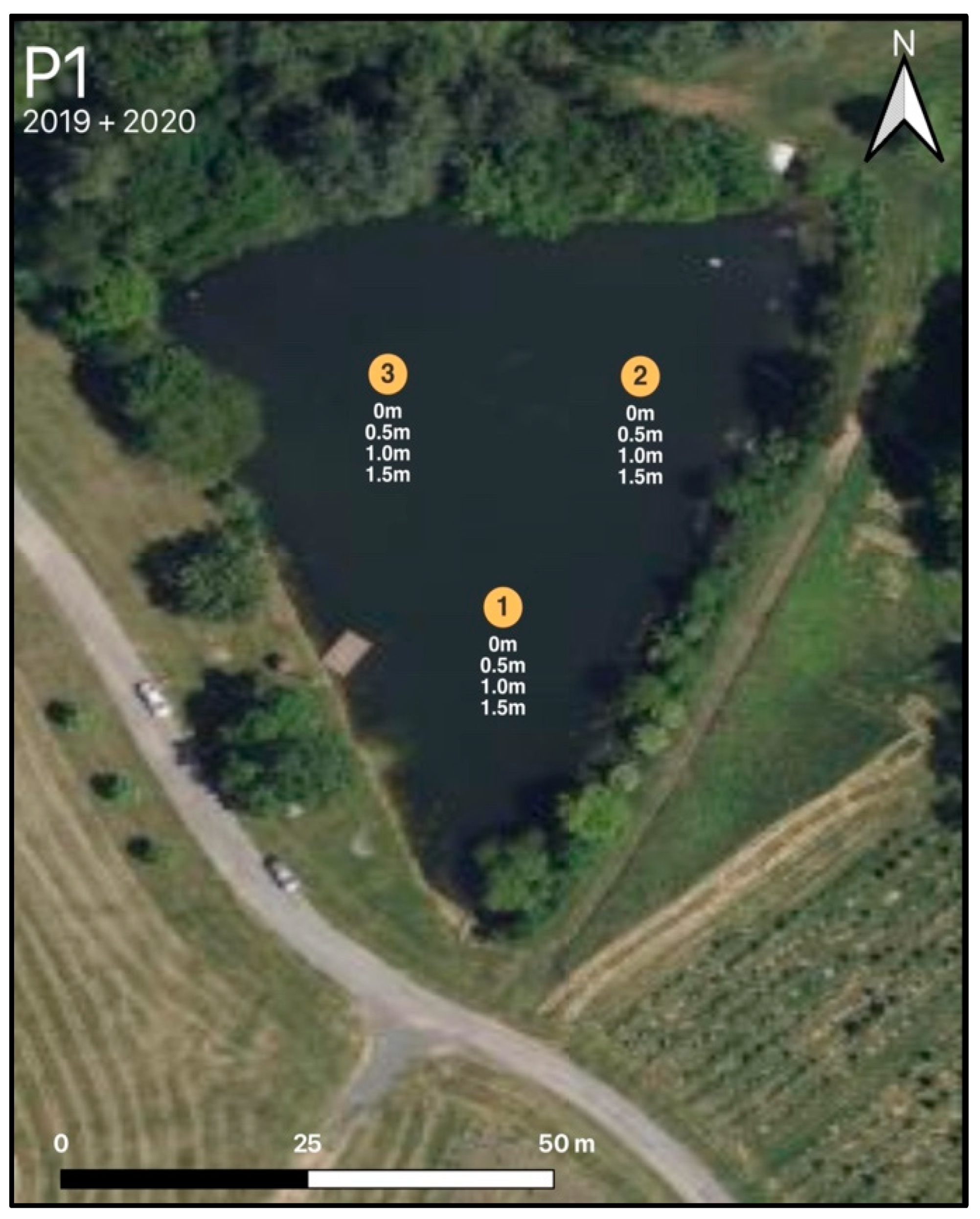
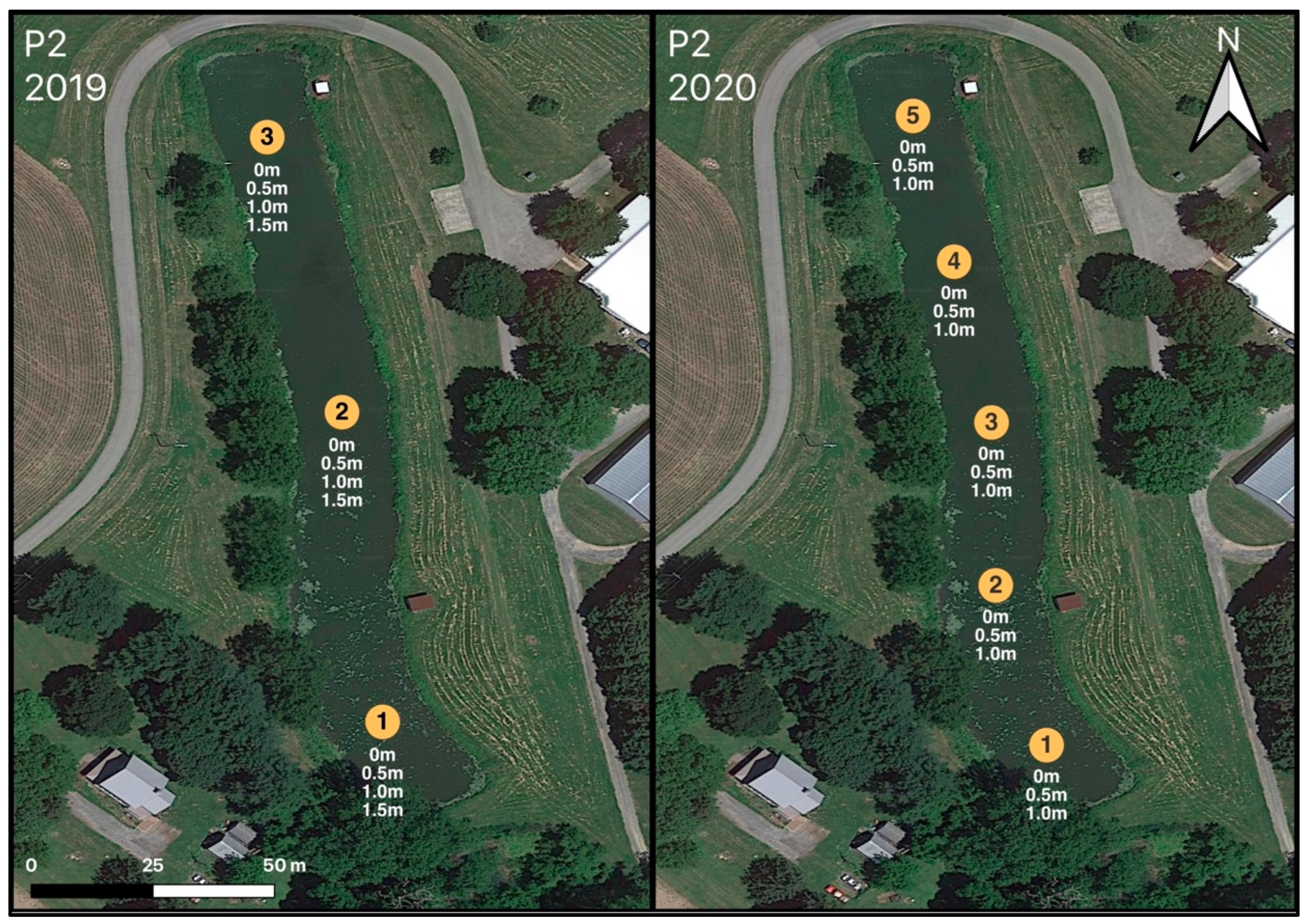
2.1. Sites, Field, and Laboratory

Water sampling was conducted at two agricultural irrigation ponds in Maryland, USA during the growing seasons of 2019 and 2020. Detailed descriptions of both ponds can be found in Smith et al. [24]. Briefly, Pond 1 (P1; Figure 1), located in Central Maryland, is a manmade embankment pond adjacent to crop fields. Pond 2 (P2; Figure 2), located on the Eastern Shore of Maryland, is a manmade excavation pond. The total surface area of the ponds are 4087 and 4249 m2 for P1 and P2, respectively. P1 was sampled once in 2019 (6 September 2019) and once in 2020 (23 July 2020). P2 was sampled twice in 2019 (15 September 2019, 21 September 2019) and three times in 2020 (15 July 2020, 10 August 2020, 26 August 2020). Both ponds are approximately a one-hour drive from the USDA-Agricultural Research Station laboratories, allowing samples to be collected and processed on the same day.
Sample collection consisted of samples being taken from interior locations of the ponds at multiple depths throughout the day. For all sampling dates, sample collection started at 9 a.m., 12 p.m., and 3 p.m. and was completed within 30 min of the start time. Sampling design remained the same at P1 for 2019 and 2020 with three interior sampling locations and four depths (0 m, 0.5 cm, 1.0 m, and 1.5 m). For P2 in 2019 there were three interior sampling locations with four depths as described for P1. In 2020 the sampling design was changed to obtain better spatial coverage at P2 which included five instead of three interior sampling locations and three depths instead of four (0 m, 0.5 m, and 1.0 m). The total number of samples analyzed were 279, with 72 samples from P1 and 207 from P2. Replications of samples and microscopy analysis were not performed due to time and resource limitations. The average photic zones during the growing season (May–October), as determined by Secchi depth, were 0.8 m and 0.5 m for P1 and P2, respectively [24].
Water samples were collected from a boat using a Sigma 900 MAX autosampler (Hach, Loveland, CO, USA). Further details on sampling procedure can be found in Stocker et al. [58]. In brief, the autosampler tubing was marked with tape so samples could be taken at the same depth every time. The intake of the autosampler tubing was attached to a YSI EXO2 multiparameter sonde (Yellow Springs Instruments, Yellow Springs, OH, USA) so that water quality measurements were representative of the water being collected for laboratory analysis. Field measurements collected with the YSI sonde included: temperature (°C), specific conductance (SPC; μS cm−1), dissolved oxygen (DO; mg L−1), pH, fluorescent dissolved organic matter (FDOM; relative fluorescent units [RFU]), and turbidity (NTU). The autosampler tubing was flushed for 30 s at each depth prior to collecting the water sample into a pre-labeled 500 mL amber plastic bottle. Samples were kept in a cooler with ice packs to maintain ambient water temperature.
Sample processing in the laboratory typically occurred within two hours after the 3 p.m. sampling. For phytoplankton identification and enumeration, 50 mL subsamples were portioned and treated with a 5% Lugol’s iodine solution (to a 1% final concentration) immediately after collection and stored in coolers in the field. Upon arrival to the laboratory, phytoplankton samples were then stored in the dark at 4 °C until processing. Water samples were analyzed using an Aquafluor fluorometer (Turner Designs, San Jose, CA, USA) for whole cell chlorophyll-a (RFU), phycocyanin (µg L−1), and colored dissolved organic matter (CDOM; µg L−1). A VarioTOC cube (Elementar, Ronkonkoma, NY, USA) was utilized to analyze water samples for total carbon (TC; mg L−1), total organic carbon (TOC; mg L−1), total inorganic carbon (TIC; mg L−1), and total bound nitrogen (TNB; mg L−1). All analyses were completed according to manufacturer guidelines.

2.2. Microscopy

All phytoplankton samples were examined using a Nikon Ts2R inverted microscope (Nikon Instruments Inc., Melville, NY, USA) using a modified Ütermohl method as described in Marshall & Alden [59] and Garrett et al. [60]. A detailed description of the enumeration method can be found in Smith et al. [24]. Briefly, a Lugol’s iodine preserved sample (2 or 3 mL) was placed into a chambered cover glass slide (Nalgene Nunc #155380, Thermo Scientific, Rochester, NY, USA) and was allowed to settle for 30 to 60 min. Optical frames were counted until either a minimum of 200 cells or 20 frames were evaluated. Identification of phytoplankton was performed to the lowest taxon possible for eukaryotic algae using John et al. [61] and for cyanobacteria using Komárek [62] and Komárek and Anagnostidis [63] and recorded as cells L−1. For data analyses, enumerative phytoplankton data were binned into five major groups: diatoms, chlorophytes, dinoflagellates, flagellates, and cyanobacteria. The chlorophyte group consisted of non-motile single-cell, colonial, and filamentous genera. The flagellate group consisted of euglenophyte, raphidophyte, cryptophyte, and motile chlorophyte taxa which finer scale resolution or morphological overlap was possible due to preservation with Lugol’s iodine and the limitation of analysis only with light microscopy. This binning method was suggested by Davies et al. [64], as a way to use datasets with limited fine-resolution taxonomy in the creation of ecological indicators.

2.3. Statistics and Graphics

To analyze the differences in mean abundance of major phytoplankton groups by time of day and sampling depth, two-way permutational multivariate analysis of variance (PERMANOVA) was performed. Spearman rank correlations were performed between phytoplankton abundance and measured water quality parameters. Due to the infrequent occurrence and minor contribution to the total phytoplankton population, dinoflagellates were not included in statistical analyses. All statistical analyses were computed with PAST4 [65]. The normalized concentration Rij in chlorophyll-a and phycocyanin contour maps was calculated as follows:
R i j = X i j X ¯
where Xij is the average over replications at specific time of day (i) and depth (j) and X ¯ is the daily average over the pond. Figures were created with Sigma Plot v13 (Systat Software, San Jose, CA, USA) and all maps were prepared using QGIS v3.22 (OSGeo, Basel, Switzerland).

3. Results

3.1. Data Summary

Descriptive statistics (minimum, maximum, mean, and median) for measured water quality parameters in both ponds and years can be found in Supplemental Table S1. Normalized temperature measurements as a function of the time of day and sampling depth are shown in Supplemental Figure S1. Average cell abundance of phytoplankton (diatoms, chlorophytes, flagellates, cyanobacteria, and total phytoplankton) at P1 were greater in 2019 than in 2020. Whereas at P2, the average abundance of phytoplankton groups were greater in 2020 than in 2019. Average water temperatures, FDOM, TOC, and TN in P1 and P2 were higher in 2020 when compared to 2019. Chlorophyll-a, phycocyanin, CDOM, turbidity, pH average values were higher in 2019 for both ponds.
Precipitation data over 1-week and 2-week accumulation periods is presented in Supplemental Table S2. Overall, the largest amounts of precipitation accumulations were seen at P2 in 2020. In both ponds, the lowest amounts of precipitation were seen for the 2019 sampling dates, with all dates having < 1 mm of accumulation the week prior to sampling. At P1, the 2020 sampling date had higher precipitation amounts than the 2019 sampling date both one and two weeks prior to sampling. A very large precipitation event took place one week prior to the 10 August 2020 (P2) sampling date with over 200 mm of rainfall documented. Two weeks prior to sampling P2 on 26 August 2020 there was an accumulation of ~200 mm of precipitation. Solar irradiance data are shown in Supplemental Table S3. Variation among 9 a.m. irradiance values was much higher than that among 12 p.m. and 3 p.m. values. A larger spread of irradiance values at 12 p.m. and 3 p.m. samplings was observed at P1 compared with P2.
The relative abundances of phytoplankton groups based on cell abundance over the seven sampling dates in this study are displayed in Figure 3 and identified taxa for both ponds are reported in Table S4. For all sampling dates at both ponds, the phytoplankton community was dominated by either flagellates or chlorophytes. Dinoflagellates were present in two of the seven sampling dates (15 September 2019 and 21 September 2019) as a very small percentage of the total population (<5%). Similarly, diatom abundance was repeatedly a small proportion of the total population, with most dates being less than 10%, except for 23 July 2020 when diatoms (predominately Stephanodiscus and Aulacoseira species) made up ~33% of the total population. While not the most abundant phytoplankton group, cyanobacteria were present in the ponds on all sampling dates, except for 26 August 2020. Based on microscopy analyses, cyanobacteria averaged a 5–10% contribution to the phytoplankton community. During these late summer and early fall sampling dates the cyanobacteria population in P1 was dominated by Microcystis wesenbergii and in P2 by Microcystis wesenbergii and Microcystis aeruginosa. The highest abundance of chlorophytes was observed on 6 September 2019 (predominately Westella spp. and Monoraphidium spp.) and 21 September 2019 (predominately Scenedesmus spp. and Tetraedron spp.), comprising about 63% and 56% of the phytoplankton community, respectively. The flagellate community, which represented a diverse grouping of motile organisms, routinely contained cryptophyte and euglenophyte taxa in both ponds. On 26 August 2020, the phytoplankton community at P2 was dominated by the harmful raphidophyte Gonyostomum semen, which was categorized within the flagellate group for this study.
Correlations between measured water quality parameters, photosynthetic pigments, and cell abundances for each sampling date can be found in Supplemental Tables S5–S11. For these analyses, mild correlations have correlation coefficients of 0.300–0.499, moderate correlations have correlation coefficients of 0.500–0.699, and strong correlations have correlation coefficients of 0.700–0.999. No correlations were found for phycocyanin on 6 September 2019, but chlorophyll-a was strongly correlated with FDOM. On 23 July 2020 phycocyanin and chlorophyll-a were moderately correlated with each other and mildly correlated with NTU. On both dates at P1 the cell abundance and pigments did not show any correlations. At P2 on 15 September 2019 there were moderate correlations for both phycocyanin and chlorophyll-a with chlorophyte abundance. On 21 September 2019 there were no correlations between phycocyanin and cell abundance, and only mild correlations were established between chlorophyte abundance and chlorophyll-a. At P2 on 15 July 2020 and 10 August 2020 there were no correlations established between pigments and cell abundances. However, on 26 August 2020, phycocyanin and chlorophyll-a were moderately correlated with chlorophyte abundance. Additionally, on all three dates in 2020 at P2 both pigments were either moderately or strongly correlated with CDOM and NTU.

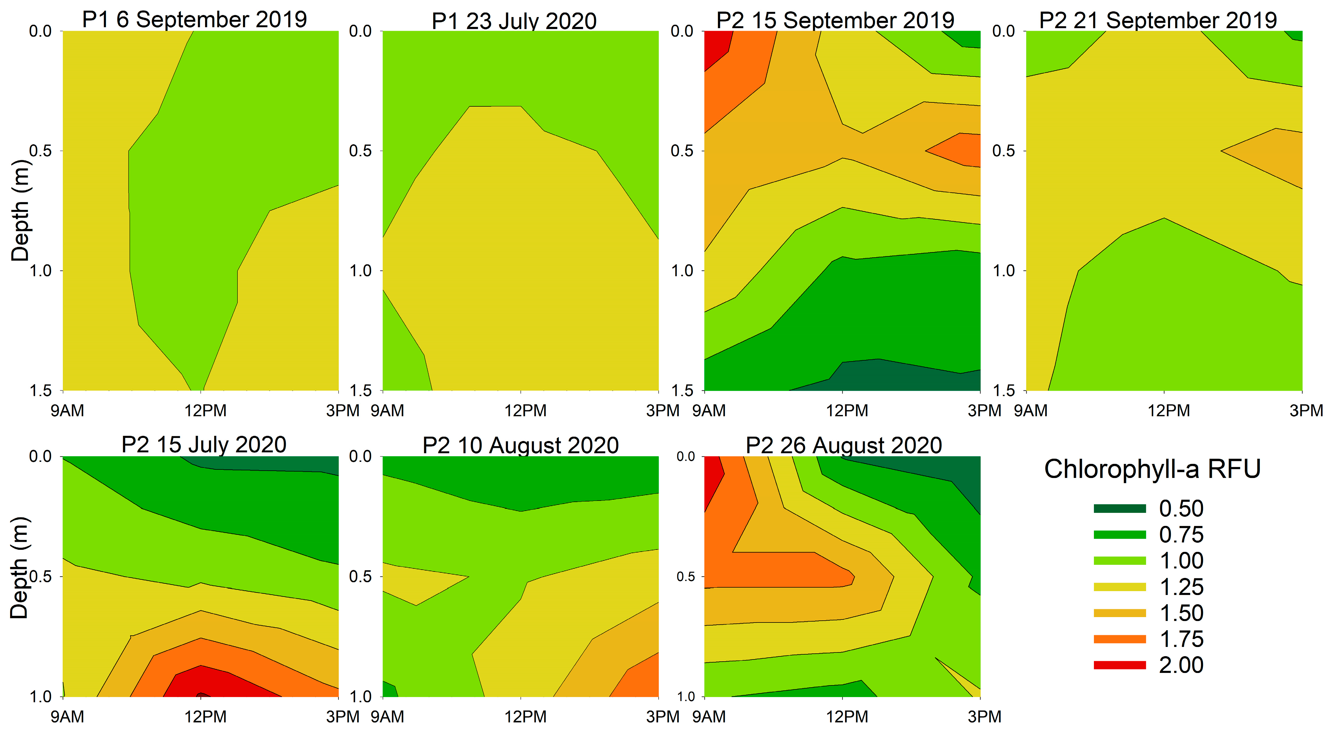
3.2. Diurnal Vertical Variations in Phytoplankton Pigments

The two-way PERMANOVA was chosen to test whether sampling time of day and sampling depth impacted concentrations of phytoplankton pigments. The results of the two-way PERMANOVA for chlorophyll-a and phycocyanin are shown in Table 1. Sampling time of day at P1 was a significant factor for chlorophyll-a for one (6 September 2019) of the two sampling dates. At P1, time of day and sampling depth were not found to be significant factors for phycocyanin on either sampling dates. For P2, there were five instances when time of day was found to be a significant factor for chlorophyll-a and phycocyanin. Depth was a significant factor, in the majority of cases, in both ponds (11 of 14 sampling points) for both phytoplankton pigments. There were three instances when both time of day and depth were significant factors for chlorophyll-a (6 September 2019, 15 July 2020, 10 August 2020) and one instance for phycocyanin (10 August 2020).
Contour graphs of normalized photosynthetic pigment concentrations are shown in Figure 4. Relatively small variations with depth and time were found in P1. On 6 September 2019 there were slightly elevated (~25% larger than daily average) concentrations of chlorophyll-a throughout the water column at 9 a.m. By 3 p.m., the surface water had average chlorophyll-a concentrations and the 1 m to 1.5 m depths had slightly elevated chlorophyll-a concentrations. On 23 July 2020, however, the chlorophyll-a concentrations at the surface were average while concentrations at the 0.5 m to 1.5 m depths remained slightly elevated throughout the day. At P2 a trend was seen on 15 September 2019 and 26 August 2020 where above average chlorophyll-a concentrations were found at the surface at 9 a.m. and by 12 p.m. or 3 p.m. had moved to the 0.5 m depth. For both dates, the 1 m and 1.5 m depths had chlorophyll-a concentrations below or at average of pond chlorophyll-a observations. A different pattern of the chlorophyll-a distributions could be observed on 15 July 2020 and 10 August 2020 in P2 where the surface waters remained below the average chlorophyll-a concentrations for the entire day and higher chlorophyll-a concentrations were found at 1 m depth from 12 p.m. to 3 p.m. On 21 September 2019 in P2, most of the water column throughout the day remained close to the average chlorophyll-a concentration with only a slight elevation at 0.5 m at 3 p.m.
Normalized phycocyanin concentrations as functions of time of day and depth are shown in Figure 5. The normalized phycocyanin concentrations at P1 followed similar patterns on both sampling dates. High or slightly elevated concentrations were found near the surface early in the day followed by slightly elevated concentrations at the 0.5 m or 1.5 m depths later in the day. At P2 on 15 September 2019 and 26 August 2020, phycocyanin concentrations were above average at the surface at 9 a.m. Later in the day the highest phycocyanin concentrations were between the 0.5 m and 1 m depths. On 15 July 2020 and 10 August 2020 at P2, phycocyanin concentrations were below or at average concentrations in surface waters throughout the day. Additionally, throughout the day, slightly above average phycocyanin concentrations were noted below the 0.5 m depth. The 21 September 2019 phycocyanin concentration pattern was similar to the 15 September 2019 pattern, but the spread of relative concentrations in space and time is smaller.

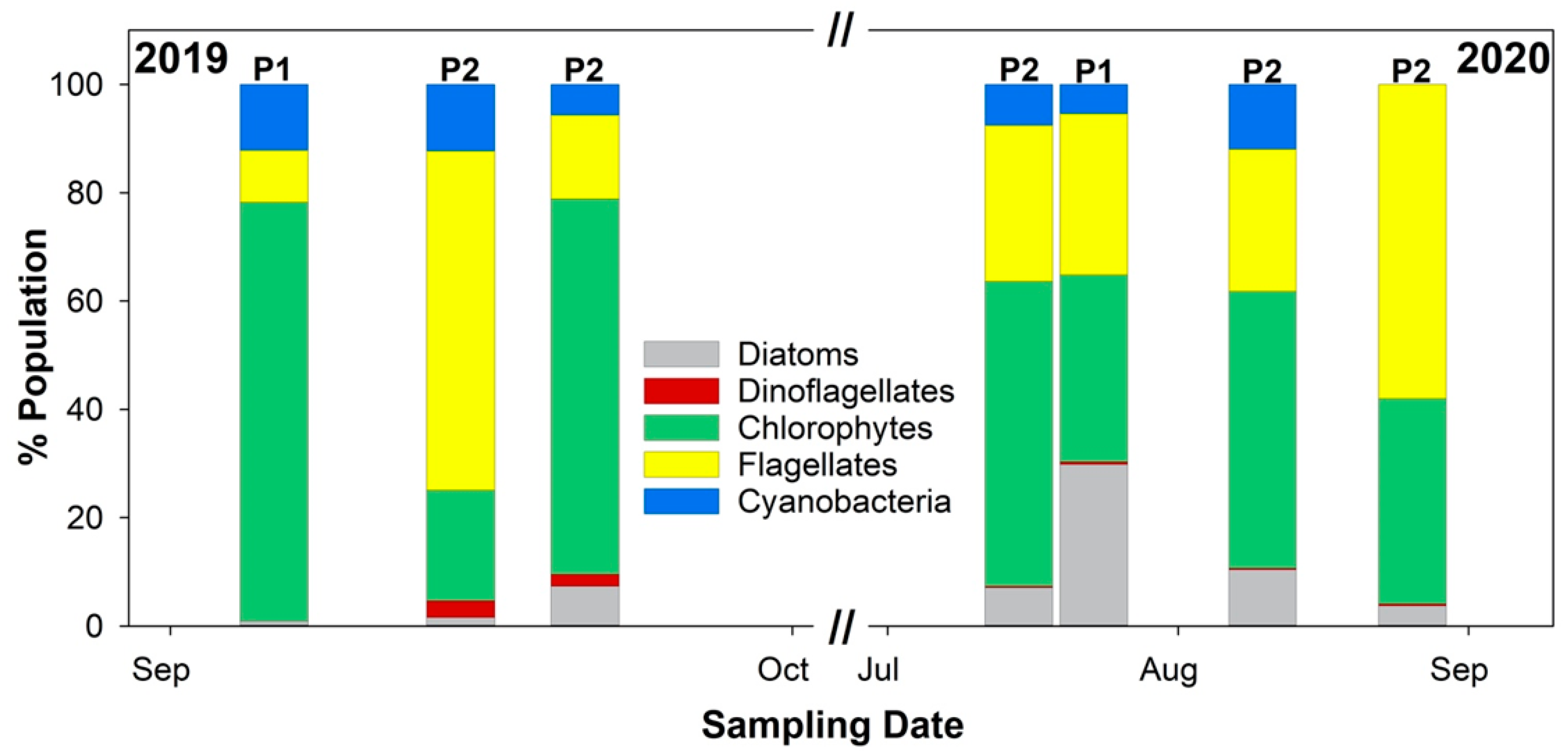
3.3. Diurnal Vertical Variations in Phytoplankton Cell Counts

The results of the two-way PERMANOVA analysis on phytoplankton group cell counts can be found in Table 2. Unlike for chlorophyll-a and phycocyanin concentrations, there were only a few instances when time of day and sampling depth were significant factors for phytoplankton cell abundances. Cyanobacteria saw no significant effect of time of day across all dates. In all but two instances time of day was a significant factor for flagellates. Additionally, on four of the seven sampling dates, sampling time of day was found to be significant for chlorophytes. Depth was not found to be a significant factor for diatom, flagellate, chlorophyte, and cyanobacteria cell abundance across all sampling dates. However, when the total phytoplankton population was considered, sampling depth was significant on one occasion (15 September 2019). Sampling time of day was found to be a significant factor when considering the whole phytoplankton community on 23 July 2020 and 10 August 2020, dates in which the highest percentages of diatoms were found in the phytoplankton population. Only on 15 September 2019 was both sampling time of day and depth a significant factor on the whole phytoplankton community; in this instance the phytoplankton community was dominated by the motile taxa grouped in the flagellate category (Euglena spp. and Trachelomonas spp.).

3.4. Diurnal and Vertical Variations in Water Quality Variables

The results of the two-way PERMANOVA analysis on measured water quality variables is shown in Table 3. Time of day and depth were found to be significant (p < 0.05) factors for temperature, DO, and pH on all dates; in all but two instances temperature, DO, and pH were highly significant (p < 0.001). For carbon concentrations at both ponds, time of day was found to be a significant factor with carbon concentrations being highest at 9 a.m., whereas depth was rarely significant for TC, TOC, TIC, and TNB. For specific conductance, time of day was rarely a significant factor, whereas for depth, for all but one date, was a significant factor. Similarly, for FDOM, NTU, and CDOM, time of day was often not found to be a significant factor, but depth was found to be significant in most instances.

4. Discussion

Significant trends throughout time and sampling depth were established for the phytoplankton pigments, but not for phytoplankton cell abundance on most sampling dates. One reason for this discrepancy can be the absence of persistent correlations between pigment and cell abundance in this study. In many studies, chlorophyll-a concentrations were well-correlated with cell abundances for various phytoplankton groups. Correlations have also been established between phycocyanin and cyanobacteria cell abundance [66,67]. However, other studies indicated that cell abundance and photosynthetic pigments concentrations don’t often correlate to each other and attributed these discrepancies to data collection uncertainties and phytoplankton community structure. Gregor et al. [68] and Rozina et al. [69] found that in scenarios where cell abundances were low, microscopy-based counts were not as accurate and associations to pigments were harder to establish. Harrison et al. [70] stated that microscopy enumeration includes an error rate of ±10–20% from misidentification, counting error, and subsampling, and can be particularly biased by the counting chambers used for microscopy-based analyses [71,72]. While we employed a settling and enumeration technique that has been successfully used for exploring phytoplankton community composition in the marine environment where diatoms and dinoflagellates comprise the bulk of the community [73,74,75], the diversity and abundance of small-sized taxa in other phytoplankton groups (e.g., Cyanophyta, Chlorophyta) in these particular freshwater environments may be better accounted for with a different settling technique or a combination of techniques. Cell abundance correlations to photosynthetic pigments can also be affected by the fact that not all phytoplankton are equal in terms of producing photosynthetic pigments. This is the case for both cyanobacteria and eukaryotic algae where the concentration of phycocyanin or chlorophyll-a in a single cyanobacteria cell [26,76,77] and the concentration of chlorophyll-a in a single cell of eukaryotic algae [70,78,79] varies among species. Additionally, the nutritional state and status [80], growth phase [66,76], and cell size [81,82] may influence pigment concentrations and reliable enumerative capacity.
Another possible explanation for the discrepancies between PERMANOVA results of phytoplankton and photosynthetic pigment concentrations is the difficulty in enumerating phytoplankton size classes smaller than microplankton. According to Hampton et al. [83], picoplankton (cells 0.2–2.0 µm) can represent anywhere from 10–50% of the primary productivity in a waterbody. Pico-sized phytoplankton are a substantial part of the plankton community, particularly in the summer months, and can be the dominate phytoplankton group shortly after rain events [84]. Bowling et al. [85] determined that picocyanobacteria contributed to more than 50% of the cyanobacteria community in a shallow urban pond and similar percentages have been reported for larger fresh waterbodies, such as Lake Huron [86] and Kühwörter Wasser [87]. However, enumeration and identification using the Utermöhl method on an inverted microscope often cannot detect small cells like picoplankton and picocyanobacteria and these taxa are therefore overlooked in phytoplankton enumeration processes [70,78,88]. While picoplankton may not be fully represented in cell concentration data, they are likely influencing the phytoplankton pigment concentrations measured in a waterbody [89,90,91]. This indicates that pigment measurements may provide a broader picture of the phytoplankton biomass in waterbodies, whereas microscopy-based analyses are likely excluding entire groups of chlorophyll-a and phycocyanin containing organisms.
In this study, discrepancies between phycocyanin concentrations and cyanobacteria cell abundance in P2 may also derive from the epipsammic community, which was only qualitatively examined during this study. The bed of P2 is comprised mostly of sandy-muddy sediments, a habitat which favors motile filamentous cyanobacteria [92] such as the genera found within P2 surface sediments (Komvophoron, Lyngbya, and Phormidium). Additionally, these surface sediments also contained a population of Microcystis aeruginosa colonies, which have been shown to undergo a pelagic-benthic oscillation (see [93]). Benthic mats dominated by cyanobacteria can contain high concentrations of phycocyanin [94,95] and the late summer-early fall decay processes of these mats and pelagic cyanobacteria species sequestered within the surface sediments of P2 may explain the phycocyanin detection in the absence of pelagic cyanobacteria cells. The benthic algal community of P1 was not examined during this study but should be investigated during future monitoring efforts.
Two distinct patterns in the phytoplankton pigment concentrations were established when sampling time of day and depth were taken into consideration. In the first pattern, surface water pigment concentrations remained below or about average throughout the day while higher than average concentrations were detected at depth (0.5–1.5 m). In the second pattern, higher-than-average concentrations of pigments were seen on the surface at 9 a.m., followed by a migration of higher-than-average pigments to deeper depths by 12 p.m. or 3 p.m. In P1, chlorophyll-a concentrations tended to be well-mixed or about the average at all depths and across all sampling times for both sampling dates. This well-mixed pattern may be due, in part, to the frequent use of copper sulfate as a treatment to reduce algal biomass, which has been applied to the pond starting in early July every year since 2016. Wang et al. [42] documented similar findings; that on dates when the chlorophyll-a concentrations were low phytoplankton tended to be uniformly distributed throughout the water column.
During this study, ponds were sampled at discrete depths between the hours of 9 a.m. and 3 p.m. This allowed for the examination of phytoplankton community vertical movement during maximum solar irradiance periods via the measurement of the phytoplankton pigments, chlorophyll-a and phycocyanin. Evidence of vertical migration was seen on 15 September 2019, when Microcystis aeruginosa and Microcystis wesenbergii were the dominant cyanobacteria. Here, elevated phycocyanin concentrations were seen at the surface around 9 a.m. and by 3 p.m. these elevated concentrations were detected at the 0.5 m depth. The vertical migration of phytoplankton throughout the day has been attributed to various factors such as solar radiance [28,96,97], wind [43,46], colony density [98,99], water turbulence/mixing [27], and thermal stratification [100]. Specific to Microcystis in shallow waterbodies, the colonies are reportedly responding to thermal stratification (which was not evident in P1 or P2, see Supplemental Figure S1), wind mixing, and solar radiance [101]. Our results indicate that the migration of portions of the phytoplankton community from the surface waters to lower depths later in the day likely has to do with solar irradiance. On dates when higher abundances of phytoplankton were present at the surface in the morning, solar irradiance tended to be lower (Table S3). On 15 September 2019 and 26 August 2020 when the morning solar irradiance was the lowest (Table S3), high chlorophyll-a concentrations were observed in surface waters. These surface chlorophyll-a concentrations decreased during the day and the maximum appeared at 0.5 m later in the day. This pattern was particularly strong on 26 August 2020 when the phytoplankton community in P2 was dominated by the harmful freshwater raphidophyte Gonyostomum semen, a species known to undergo vertical migration, often descending from warm surface waters to exploit nutrients found at depth [102], and to avoid high light intensities [103]. On 15 July 2020 and 10 August 2020, the morning and midday solar irradiance values were the highest. Conversely, the lowest pigment concentrations were seen at the surface and the highest concentrations were observed at 1 m depth. Numerous other studies have reported this trend of phytoplankton abundances being the highest at the surface in the morning followed by a migration to 0.5 m or deeper depths later in the day to avoid UV irradiance and other photoinhibitors [96,97,101,104].
For the dates when higher pigment concentrations were seen at depth, there are two possible explanations. High solar irradiance during the morning sampling time (9 a.m.) may have caused the phytoplankton to migrate to lower depths earlier in the day. Sampling prior to 9 a.m. could have distinguished if the phytoplankton had already migrated to lower depths or could have captured the migration process. The other possibility is that due to solar irradiance being high early in the morning the phytoplankton population did not fully migrate to the surface from deeper depths in the morning and rather remained at the 0.5 m–1 m depths, where optimal light intensity conditions were located. This coincides with the findings of Cui et al. [101] who observed that at open water stations of the Three Gorges Reservoir in China, maximum chlorophyll-a concentrations rarely reached surface waters but aggregated near the 0.5–1.0 m layer. Similar observations were reported by Joniak et al. [105] for oligohumic and mesothermic lakes. Inspection of precipitation data (Table S2) and patterns of chlorophyll-a and phycocyanin did not show any association, but sampling closer to precipitation events may reveal different associations between time, depth, phytoplankton community and pigment concentrations, as suggested by Bergkemper and Weisse [106], Ivey et al. [84], and Lefort and Gasol [107].
The increase in phytoplankton biomass often results in an increase in DO, which in turn causes an increase in pH [108,109]. Results in this work varied with some dates having positive correlations, negative correlations, or no correlations between phycocyanin and chlorophyll-a concentrations and DO measurements. On both 2019 sampling dates at P2 there were positive correlations between DO and the photosynethic pigments, which matches what is typically reported in the literature. Whereas in 2020 at P2, the first sampling date showed strong negative correlations and the remaining two dates had negative or no correlations between the photosynthetic pigments and DO measurements. Similarly, at P1 for both years, there were negligible to negative correlations between the photosynthetic pigments and DO. One possible reason for these negative correlations could be subaqueous vegetation or benthic or macrophytic algae in the ponds. Since 2016, macrophytic algae has been commonly found in the shallower areas of P1 throughout the summer, leading farm managers to apply copper sulfate starting in July. Macrophytes were present in Pond 1 but not quantified during this study. However. their leached pigments would likely be detected by fluorometry measurements and may account for the discrepancies observed in this dataset. Macrophytes and subaqueous vegetation are both known to cause an influx or supersaturation of oxygen into surface waters in freshwaters [110,111,112]. Thus, the macrophyte population could be impacting the DO levels in the water, while not being accounted for in pigment measurements, potentially causing discrepancies in the correlations. If a substantial macrophyte population is present and the phytoplankton population is average or low these higher DO and pigment values might inaccurately represent the phytoplankton community.
The datasets collected during this work pose several questions for further investigation since few strong trends were observed. First, there needs to be further investigations into the discrepancies between photosynthetic pigment concentrations, phytoplankton abundance, and the influences of benthic and/or macroalgae communities to better design a water quality monitoring program that can rely on measurements of photosynthetic pigments coupled with or without corresponding phytoplankton abundances. This could include improved laboratory methodologies that would detect all phytoplankton size classes and better characterize photosynthetic pigments, as well as field work for the characterization of the pond beds. Concurrently, cell abundances could be converted to cell biomass, and this may improve correlations with pigment concentrations. Second, now that blooms of harmful algae species with resting stages, Microcystis spp. (sediment-sequestered cells; [93]) and Gonyostomum semen (cysts; [113]), have been identified the appropriate spatial and temporal scales for water quality monitoring need to be determined. Specifically, studies that address if harmful species are better monitored for by using photosynthetic pigments, microscopy, other laboratory methods not examined here, or a combination approach to offer the best risk protection for the agricultural community should be developed. Thirdly, carbon and nitrogen concentrations did not strongly correlate with phytoplankton abundance or pigment concentrations. Correlations between nutrients and phytoplankton metrics may become more apparent if future studies examine individual N ions, P ions, and the N:P ratios. Finally, this study focused on the growing season for Maryland when farmers would be actively irrigating their fields and risks from cyanotoxins would be greatest. Extending the study outside of the growing season would allow for a larger dataset and would potentially show more or stronger correlations between water quality and pigments or phytoplankton abundance data. Finally, as climate change alters the Mid-Atlantic region’s weather patterns, including the frequency and severity of precipitation events [114], determining how the timing of water quality sampling influences sampling analyses under the constraints of both small- and large-scale precipitation events should be addressed. Overall, agricultural irrigation ponds, albeit being relatively small, are complex systems and water quality monitoring of these systems must account for persistent patterns and features of water constituents.

5. Conclusions

Within two agricultural irrigation ponds, spatiotemporal patterns were observed in phytoplankton photosynthetic pigment concentrations, but not phytoplankton community structure, except in the case of distinct algal bloom events (Microcystis spp. and Gonyostomum semen). Correlations between photosynthetic pigments and phytoplankton abundance were rarely established suggesting that water quality monitoring programs should take into consideration methods that account for the contributions of both the benthic and pico-sized algal communities, which were underrepresented in this study. Water quality monitoring programs focused on harmful algal bloom risk assessment should also recognize the limited utility of pigments concentration data alone. Concentrations of chlorophyll-a and phycocyanin can apprise managers of the presence of an algal bloom but cannot differentiate between harmful and non-harmful taxa at this time making a suite of in-situ observations necessary. This work indicates that sampling depth and sample collection time are important factors to consider and record when assessing a waterbody. Solar irradiance and the vertical migration habits can influence the position of phytoplankton communities within the water column, even in shallow water bodies, throughout the day. If resource managers have a priori knowledge of the phytoplankton communities in waterways they are charged with monitoring, adopting a sampling strategy that integrates samples over a known depth may be preferable to avoid over- or under- estimating phytoplankton abundance. Alternatively, recording water quality parameters, including the chlorophyll maxima, as well as time of day and solar irradiance values when collecting phytoplankton samples would provide context when reviewing phytoplankton community composition and abundance data.

Supplementary Materials

The following supporting information can be downloaded at: https://www.mdpi.com/article/10.3390/environments11040074/s1, Table S1: Descriptive statistics of measured phytoplankton and water quality data at ponds P1 and P2. Figure S1: Normalized temperature measurements as functions to time of day and depth at Pond 1 and Pond 2 for each sampling date. Table S2: Precipitation from one and two weeks prior to sample collection at ponds P1 and P2. Table S3: Average solar irradiance (W/m2) during sampling at ponds P1 and P2. Table S4: List of identified taxa for P1 and P2. Tables S5–S11: Correlations between measured water quality parameters, photosynthetic pigments, and cell abundance for each sampling date.

Author Contributions

Conceptualization, J.E.S. and Y.P.; methodology, J.E.S., M.D.S. and Y.P.; formal analysis, J.E.S. and J.L.W.; investigation, J.E.S. and M.D.S.; writing—original draft preparation, J.E.S., J.L.W. and Y.P.; writing—review and editing, J.E.S., J.L.W., M.D.S. and Y.P.; project administration, Y.P. All authors have read and agreed to the published version of the manuscript.

Funding

This research was co-funded through the USDA’s Agricultural Research Service project number 8042-42610-001-000-D.

Data Availability Statement

The raw data supporting the conclusions of this article will be made available by the authors on request.

Conflicts of Interest

The authors declare that they have no known competing financial interests or personal relationships that could have appeared to influence the work reported in this paper.

References

  1. Bhattarai, S.P.; Midmore, D.J.; Pendergast, L. Yield, Water-Use Efficiencies and Root Distribution of Soybean, Chickpea and Pumpkin under Different Subsurface Drip Irrigation Depths and Oxygation Treatments in Vertisols. Irrig. Sci. 2008, 26, 439. [Google Scholar] [CrossRef]
  2. Maestre-Valero, J.F.; Martínez-Alvarez, V. Effects of Drip Irrigation Systems on the Recovery of Dissolved Oxygen from Hypoxic Water. Agric. Water Manag. 2010, 97, 1806–1812. [Google Scholar] [CrossRef]
  3. Corbel, S.; Mougin, C.; Nélieu, S.; Delarue, G.; Bouaïcha, N. Evaluation of the Transfer and the Accumulation of Microcystins in Tomato (Solanum Lycopersicum Cultivar MicroTom) Tissues Using a Cyanobacterial Extract Containing Microcystins and the Radiolabeled Microcystin-LR ((14)C-MC-LR). Sci. Total Environ. 2016, 541, 1052–1058. [Google Scholar] [CrossRef] [PubMed]
  4. Saqrane, S.; Oudra, B. CyanoHAB Occurrence and Water Irrigation Cyanotoxin Contamination: Ecological Impacts and Potential Health Risks. Toxins 2009, 1, 113–122. [Google Scholar] [CrossRef] [PubMed]
  5. USDA. 2018 Irrigation and Water Management Survey; USDA: Washington, DC, USA, 2019.
  6. Downing, J.A.; Prairie, Y.T.; Cole, J.J.; Duarte, C.M.; Tranvik, L.J.; Striegl, R.G.; McDowell, W.H.; Kortelainen, P.; Caraco, N.F.; Melack, J.M.; et al. The Global Abundance and Size Distribution of Lakes, Ponds, and Impoundments. Limnol. Oceanogr. 2006, 51, 2388–2397. [Google Scholar] [CrossRef]
  7. Hu, X.; Zhang, R.; Ye, J.; Wu, X.; Zhang, Y.; Wu, C. Monitoring and Research of Microcystins and Environmental Factors in a Typical Artificial Freshwater Aquaculture Pond. Environ. Sci. Pollut. Res. 2018, 25, 5921–5933. [Google Scholar] [CrossRef] [PubMed]
  8. Roy, K.; Vrba, J.; Kaushik, S.J.; Mraz, J. Feed-Based Common Carp Farming and Eutrophication: Is There a Reason for Concern? Rev. Aquac. 2020, 12, 1736–1758. [Google Scholar] [CrossRef]
  9. Yang, P.; Lai, D.Y.F.; Jin, B.; Bastviken, D.; Tan, L.; Tong, C. Dynamics of Dissolved Nutrients in the Aquaculture Shrimp Ponds of the Min River Estuary, China: Concentrations, Fluxes and Environmental Loads. Sci. Total Environ. 2017, 603–604, 256–267. [Google Scholar] [CrossRef] [PubMed]
  10. Nguyen, D.T.; Harada, M.; Hiramatsu, K. Evaluation of the Water-Quality Dynamics in a Eutrophic Agricultural Pond by Using a One-Box Ecosystem Model Considering Several Algal Groups. Paddy Water Environ. 2010, 8, 301–318. [Google Scholar] [CrossRef]
  11. Saito, M.; Onodera, S.I.; Shiimizu, Y. Effects of Residence Time and Nutrient Load on Eutrophic Conditions and Phytoplankton Variations in Agricultural Reservoirs. In Proceedings of the 2013 Symposium H04: Understanding Freshwater Quality Problems in a Changing World, Gothenburg, Sweden, 22–26 July 2013; pp. 197–203. [Google Scholar]
  12. Usio, N.; Imada, M.; Nakagawa, M.; Akasaka, M.; Takamura, N. Effects of Pond Draining on Biodiversity and Water Quality of Farm Ponds. Conserv. Biol. 2013, 27, 1429–1438. [Google Scholar] [CrossRef]
  13. Malerba, M.E.; Lindenmayer, D.B.; Scheele, B.C.; Waryszak, P.; Yilmaz, I.N.; Schuster, L.; Macreadie, P.I. Fencing Farm Dams to Exclude Livestock Halves Methane Emissions and Improves Water Quality. Glob. Change Biol. 2022, 28, 4701–4712. [Google Scholar] [CrossRef] [PubMed]
  14. Wilson, A.E.; Chislock, M.F.; Yang, Z.; Barros, M.U.G.; Roberts, J.F. Pond Bank Access as an Approach for Managing Toxic Cyanobacteria in Beef Cattle Pasture Drinking Water Ponds. Environ. Monit. Assess. 2018, 190, 247. [Google Scholar] [CrossRef]
  15. Flynn, K.J.; Clark, D.R.; Mitra, A.; Fabian, H.; Hansen, P.J.; Glibert, P.M.; Wheeler, G.L.; Stoecker, D.K.; Blackford, J.C.; Brownlee, C. Ocean Acidification with (de)Eutrophication Will Alter Future Phytoplankton Growth and Succession. Proc. R. Soc. B Biol. Sci. 2015, 282, 20142604. [Google Scholar] [CrossRef]
  16. Huang, Y.; Shen, Y.; Zhang, S.; Li, Y.; Sun, Z.; Feng, M.; Li, R.; Zhang, J.; Tian, X.; Zhang, W. Characteristics of Phytoplankton Community Structure and Indication to Water Quality in the Lake in Agricultural Areas. Front. Environ. Sci. 2022, 10, 833409. [Google Scholar] [CrossRef]
  17. Rangel, L.M.; Soares, M.C.S.; Paiva, R.; Silva, L.H.S. Morphology-Based Functional Groups as Effective Indicators of Phytoplankton Dynamics in a Tropical Cyanobacteria-Dominated Transitional River–Reservoir System. Ecol. Indic. 2016, 64, 217–227. [Google Scholar] [CrossRef]
  18. Rimet, F.; Druart, J.-C. A Trait Database for Phytoplankton of Temperate Lakes. Ann. Limnol. Int. J. Lim. 2018, 54, 18. [Google Scholar] [CrossRef]
  19. Varol, M. Phytoplankton Functional Groups in a Monomictic Reservoir: Seasonal Succession, Ecological Preferences, and Relationships with Environmental Variables. Environ. Sci. Pollut. Res. 2019, 26, 20439–20453. [Google Scholar] [CrossRef]
  20. Nankabirwa, A.; De Crop, W.; Van der Meeren, T.; Cocquyt, C.; Plisnier, P.-D.; Balirwa, J.; Verschuren, D. Phytoplankton Communities in the Crater Lakes of Western Uganda, and Their Indicator Species in Relation to Lake Trophic Status. Ecol. Indic. 2019, 107, 105563. [Google Scholar] [CrossRef]
  21. Xu, W.; Duan, L.; Wen, X.; Li, H.; Li, D.; Zhang, Y.; Zhang, H. Effects of Seasonal Variation on Water Quality Parameters and Eutrophication in Lake Yangzong. Water 2022, 14, 2732. [Google Scholar] [CrossRef]
  22. Twiss, M.R.; Brahmstedt, E.S.; Cabana, G.; Guillemette, F. Proliferation of Phytoplankton along a 500 Km Transect of the St. Lawrence River from Its Outflow at Lake Ontario. J. Great Lakes Res. 2022, 48, 336–342. [Google Scholar] [CrossRef]
  23. Smith, J.E.; Stocker, M.D.; Wolny, J.L.; Hill, R.L.; Pachepsky, Y.A. Intraseasonal Variation of Phycocyanin Concentrations and Environmental Covariates in Two Agricultural Irrigation Ponds in Maryland, USA. Environ. Monit. Assess. 2020, 192, 706. [Google Scholar] [CrossRef] [PubMed]
  24. Smith, J.E.; Wolny, J.L.; Stocker, M.D.; Hill, R.L.; Pachepsky, Y.A. Temporal Stability of Phytoplankton Functional Groups within Two Agricultural Irrigation Ponds in Maryland, USA. Front. Water 2021, 3, 724025. [Google Scholar] [CrossRef]
  25. Gámez, T.E.; Groeger, A.W.; Manning, S.R. Dynamic Phytoplankton Community Structure in a Subtropical Reservoir during an Extended Drought, Central Texas, USA. Aquat. Sci. 2022, 85, 7. [Google Scholar] [CrossRef]
  26. Kong, Y.; Lou, I.; Zhang, Y.; Lou, C.U.; Mok, K.M. Using an online phycocyanin fluorescence probe for rapid monitoring of cyanobacteria in Macau freshwater reservoir. Hydrobiologia 2014, 741, 33–49. [Google Scholar] [CrossRef]
  27. Aparicio Medrano, E.; Uittenbogaard, R.E.; Dionisio Pires, L.M.; van de Wiel, B.J.H.; Clercx, H.J.H. Coupling Hydrodynamics and Buoyancy Regulation in Microcystis aeruginosa for Its Vertical Distribution in Lakes. Ecol. Model. 2013, 248, 41–56. [Google Scholar] [CrossRef]
  28. Qi, L.; Hu, C.; Visser, P.M.; Ma, R. Diurnal Changes of Cyanobacteria Blooms in Taihu Lake as Derived from GOCI Observations. Limnol. Oceanogr. 2018, 63, 1711–1726. [Google Scholar] [CrossRef]
  29. Zhang, M.; Kong, F.; Wu, X.; Xing, P. Different Photochemical Responses of Phytoplankters from the Large Shallow Taihu Lake of Subtropical China in Relation to Light and Mixing. Hydrobiologia 2008, 603, 267–278. [Google Scholar] [CrossRef]
  30. Vidal, J.; Rigosi, A.; Hoyer, A.; Escot, C.; Rueda, F.J. Spatial Distribution of Phytoplankton Cells in Small Elongated Lakes Subject to Weak Diurnal Wind Forcing. Aquat. Sci. 2014, 76, 83–99. [Google Scholar] [CrossRef]
  31. Santos, R.M.; Saggio, A.A.; Silva, T.L.R.; Negreiros, N.F.; Rocha, O. Short-Term Thermal Stratification and Partial Overturning Events in a Warm Polymictic Reservoir: Effects on Distribution of Phytoplankton Community. Braz. J. Biol. 2015, 75, 19–29. [Google Scholar] [CrossRef] [PubMed]
  32. Becker, V.; de Souza Cardoso, L.; Huszar, V.L.M. Diel Variation of Phytoplankton Functional Groups in a Subtropical Reservoir in Southern Brazil during an Autumnal Stratification Period. Aquat. Ecol. 2009, 43, 285–293. [Google Scholar] [CrossRef]
  33. Patterson, G.; Wilson, K.K. The Influence of the Diel Climatic Cycle on the Depth-Time Distribution of Phytoplankton and Photosynthesis in a Shallow Equatorial Lake (Lake Baringo, Kenya). Hydrobiologia 1995, 304, 1–8. [Google Scholar] [CrossRef]
  34. Vidal, J.; Moreno-Ostos, E.; Escot, C.; Quesada, R.; Rueda, F. The Effects of Diel Changes in Circulation and Mixing on the Longitudinal Distribution of Phytoplankton in a Canyon-Shaped Mediterranean Reservoir. Freshw. Biol. 2010, 55, 1945–1957. [Google Scholar] [CrossRef]
  35. Crossetti, L.O.; Becker, V.; de Souza Cardoso, L.; Rodrigues, L.R.; da Costa, L.S.; da Motta-Marques, D. Is Phytoplankton Functional Classification a Suitable Tool to Investigate Spatial Heterogeneity in a Subtropical Shallow Lake? Limnologica 2013, 43, 157–163. [Google Scholar] [CrossRef]
  36. Jiang, Y.-J.; He, W.; Liu, W.-X.; Qin, N.; Ouyang, H.-L.; Wang, Q.-M.; Kong, X.-Z.; He, Q.-S.; Yang, C.; Yang, B.; et al. The Seasonal and Spatial Variations of Phytoplankton Community and Their Correlation with Environmental Factors in a Large Eutrophic Chinese Lake (Lake Chaohu). Ecol. Indic. 2014, 40, 58–67. [Google Scholar] [CrossRef]
  37. Alexander, R.; Imberger, J. Spatial Distribution of Motile Phytoplankton in a Stratified Reservoir: The Physical Controls on Patch Formation. J. Plankton Res. 2009, 31, 101–118. [Google Scholar] [CrossRef]
  38. Zhao, C.S.; Shao, N.F.; Yang, S.T.; Ren, H.; Ge, Y.R.; Feng, P.; Dong, B.E.; Zhao, Y. Predicting Cyanobacteria Bloom Occurrence in Lakes and Reservoirs before Blooms Occur. Sci. Total Environ. 2019, 670, 837–848. [Google Scholar] [CrossRef]
  39. Touchart, L.; Bouny, J. Phytoplankton Geographic Spatialization in Two Ponds in Limousin (France). Appl. Geogr. 2008, 28, 295–310. [Google Scholar] [CrossRef]
  40. Haque, M.A.; Jewel, M.A.S.; Akhi, M.M.; Atique, U.; Paul, A.K.; Iqbal, S.; Islam, M.S.; Das, S.K.; Alam, M.M. Seasonal Dynamics of Phytoplankton Community and Functional Groups in a Tropical River. Environ. Monit. Assess. 2021, 193, 704. [Google Scholar] [CrossRef]
  41. Yang, M.; Xia, J.; Cai, W.; Zhou, Z.; Yang, L.; Zhu, X.; Li, C. Seasonal and Spatial Distributions of Morpho-Functional Phytoplankton Groups and the Role of Environmental Factors in a Subtropical River-Type Reservoir. Water Sci. Technol. 2020, 82, 2316–2330. [Google Scholar] [CrossRef]
  42. Wang, L.; Cai, Q.; Zhang, M.; Xu, Y.; Kong, L.; Tan, L. Vertical distribution patterns of phytoplankton in summer microcystis bloom period of Xiangxi Bay, Three Gorges Reservoir, China. Fresenius Environ. Bull. 2011, 20, 553–560. [Google Scholar]
  43. Wallace, B.B.; Bailey, M.C.; Hamilton, D.P. Simulation of Vertical Position of Buoyancy Regulating Microcystis Aeruginosa in a Shallow Eutrophic Lake. Aquat. Sci. 2000, 62, 320–333. [Google Scholar] [CrossRef]
  44. Cardoso, L.D.S.; Marques, D.D.M. The Influence of Hydrodynamics on the Spatial and Temporal Variation of Phytoplankton Pigments in a Large, Sub-Tropical Coastal Lake (Brazil). Braz. Arch. Biol. Technol. 2004, 47, 587–600. [Google Scholar] [CrossRef]
  45. Moore, T.S.; Churnside, J.H.; Sullivan, J.M.; Twardowski, M.S.; Nayak, A.R.; McFarland, M.N.; Stockley, N.D.; Gould, R.W.; Johengen, T.H.; Ruberg, S.A. Vertical Distributions of Blooming Cyanobacteria Populations in a Freshwater Lake from LIDAR Observations. Remote Sens. Environ. 2019, 225, 347–367. [Google Scholar] [CrossRef]
  46. Yang, J.; Holbach, A.; Wilhelms, A.; Qin, Y.; Zheng, B.; Zou, H.; Qin, B.; Zhu, G.; Norra, S. Highly Time-Resolved Analysis of Seasonal Water Dynamics and Algal Kinetics Based on in-Situ Multi-Sensor-System Monitoring Data in Lake Taihu, China. Sci. Total Environ. 2019, 660, 329–339. [Google Scholar] [CrossRef]
  47. Sevindik, T.O.; Çelik, K.; Naselli-Flores, L. Spatial Heterogeneity and Seasonal Succession of Phytoplankton Functional Groups along the Vertical Gradient in a Mesotrophic Reservoir. Ann. Limnol. Int. J. Lim. 2017, 53, 129–141. [Google Scholar] [CrossRef]
  48. Perkins, K.R.; Rollwagen-Bollens, G.; Bollens, S.M.; Harrison, J.A. Variability in the Vertical Distribution of Chlorophyll in a Spill-Managed Temperate Reservoir. Lake Reserv. Manag. 2019, 35, 119–126. [Google Scholar] [CrossRef]
  49. Henderson, K.A.; Murdock, J.N.; Lizotte, R.E., Jr. Water Depth Influences Algal Distribution and Productivity in Shallow Agricultural Lakes. Ecohydrology 2021, 14, e2319. [Google Scholar] [CrossRef]
  50. Declerck, S.; De Bie, T.; Ercken, D.; Hampel, H.; Schrijvers, S.; Van Wichelen, J.; Gillard, V.; Mandiki, R.; Losson, B.; Bauwens, D.; et al. Ecological Characteristics of Small Farmland Ponds: Associations with Land Use Practices at Multiple Spatial Scales. Biol. Conserv. 2006, 131, 523–532. [Google Scholar] [CrossRef]
  51. Castro Berman, M.; O’ Farrell, I.; Huber, P.; Marino, D.; Zagarese, H. A Large-Scale Geographical Coverage Survey Reveals a Pervasive Impact of Agricultural Practices on Plankton Primary Producers. Agric. Ecosyst. Environ. 2022, 325, 107740. [Google Scholar] [CrossRef]
  52. López-Felices, B.; Aznar-Sánchez, J.A.; Velasco-Muñoz, J.F.; Piquer-Rodríguez, M. Contribution of Irrigation Ponds to the Sustainability of Agriculture. A Review of Worldwide Research. Sustainability 2020, 12, 5425. [Google Scholar] [CrossRef]
  53. Chen, Y.; Qin, B.; Teubner, K.; Dokulil, M.T. Long-Term Dynamics of Phytoplankton Assemblages: Microcystis-Domination in Lake Taihu, a Large Shallow Lake in China. J. Plankton Res. 2003, 25, 445–453. [Google Scholar] [CrossRef]
  54. Ding, Y.; Pan, B.; Zhao, G.; Sun, C.; Han, X.; Li, M. Geo-Climatic Factors Weaken the Effectiveness of Phytoplankton Diversity as a Water Quality Indicator in a Large Sediment-Laden River. Sci. Total Environ. 2021, 792, 148346. [Google Scholar] [CrossRef] [PubMed]
  55. Ren, X.; Yang, K.; Che, Y.; Wang, M.; Zhou, L.; Chen, L. Spatial and Temporal Assessment of the Initial Pattern of Phytoplankton Population in a Newly Built Coastal Reservoir. Front. Earth Sci. 2016, 10, 546–559. [Google Scholar] [CrossRef]
  56. Song, J.; Hou, C.; Liu, Q.; Wu, X.; Wang, Y.; Yi, Y. Spatial and Temporal Variations in the Plankton Community Because of Water and Sediment Regulation in the Lower Reaches of Yellow River. J. Clean. Prod. 2020, 261, 120972. [Google Scholar] [CrossRef]
  57. Srichandan, S.; Kim, J.Y.; Kumar, A.; Mishra, D.R.; Bhadury, P.; Muduli, P.R.; Pattnaik, A.K.; Rastogi, G. Interannual and Cyclone-Driven Variability in Phytoplankton Communities of a Tropical Coastal Lagoon. Mar. Pollut. Bull. 2015, 101, 39–52. [Google Scholar] [CrossRef] [PubMed]
  58. Stocker, M.D.; Smith, J.E.; Pachepsky, Y.A. Depth-Dependent Concentrations of E. coli in Agricultural Irrigation Ponds. Water 2022, 14, 2276. [Google Scholar] [CrossRef]
  59. Marshall, H.G.; Alden, R.W. A Comparison of Phytoplankton Assemblages and Environmental Relationships in Three Estuarine Rivers of the Lower Chesapeake Bay. Estuaries 1990, 13, 287–300. [Google Scholar] [CrossRef]
  60. Garrett, M.; Wolny, J.; Truby, E.; Heil, C.; Kovach, C. Harmful Algal Bloom Species and Phosphate-Processing Effluent: Field and Laboratory Studies. Mar. Pollut. Bull. 2011, 62, 596–601. [Google Scholar] [CrossRef] [PubMed]
  61. John, D.M.; Whitton, B.A.; Brook, A.J. The Freshwater Algal Flora of the British Isles: An Identification Guide to Freshwater and Terrestrial Algae; Cambridge University Press: London, UK, 2011; 724p, ISBN 978-0-521-19375-7. [Google Scholar]
  62. Komárek, J. Süßwasserflora von Mitteleuropa, Bd. 19/3: Cyanoprokaryota. Tiel/3rd Part: Heterocystous Genera; Süßwasserflora von Mitteleuropa; Spektrum Academischer Verlag: Heidelberg, Germany, 2013. [Google Scholar]
  63. Komárek, J.; Anagnostidis, K. Cyanoprokaryota: Chroococcales; Süßwasserflora von Mitteleuropa; Spektrum Akademischer Verlag: Heidelberg, Germany, 2008; ISBN 978-3-7482-2111-1. [Google Scholar]
  64. Davies, C.H.; Coughlan, A.; Hallegraeff, G.; Ajani, P.; Armbrecht, L.; Atkins, N.; Bonham, P.; Brett, S.; Brinkman, R.; Burford, M.; et al. A Database of Marine Phytoplankton Abundance, Biomass and Species Composition in Australian Waters. Sci. Data 2016, 3, 160043. [Google Scholar] [CrossRef]
  65. Hammer, Ø.; Harper, D.A.T.; Ryan, P.D. PAST: Paleontological Statistics Software Package for Education and Data Analysis. Palaeontol. Electron. 2001, 4, 1. [Google Scholar]
  66. Gregor, J.; Maršálek, B.; Šípková, H. Detection and Estimation of Potentially Toxic Cyanobacteria in Raw Water at the Drinking Water Treatment Plant by in Vivo Fluorescence Method. Water Res. 2007, 41, 228–234. [Google Scholar] [CrossRef]
  67. Kasinak, J.M.E.; Holt, B.M.; Chislock, M.F.; Wilson, A.E. Benchtop Fluorometry of Phycocyanin as a Rapid Approach for Estimating Cyanobacterial Biovolume. J. Plankton Res. 2015, 37, 248–257. [Google Scholar] [CrossRef]
  68. Gregor, J.; Geriš, R.; Maršálek, B.; Heteša, J.; Marvan, P. In Situ Quantification of Phytoplankton in Reservoirs Using a Submersible Spectrofluorometer. Hydrobiologia 2005, 548, 141–151. [Google Scholar] [CrossRef]
  69. Rozina, T.; Eleršek, T.; Justin, M.Z.; Meglič, A. Combined Use of Chlorophyll a and Phycocyanin Fluorescence Sensors for Quantification and Differentiation of Phytoplankton: A Useful Approach for Small Surface Water Bodies. Acta Biol. Slov. 2018, 61, 29–37. [Google Scholar] [CrossRef]
  70. Harrison, J.W.; Howell, E.T.; Watson, S.B.; Smith, R.E.H. Improved Estimates of Phytoplankton Community Composition Based on in Situ Spectral Fluorescence: Use of Ordination and Field-Derived Norm Spectra for the Bbe FluoroProbe. Can. J. Fish. Aquat. Sci. 2016, 73, 1472–1482. [Google Scholar] [CrossRef]
  71. Godhe, A.; Cusack, C.; Pedersen, J.; Andersen, P.; Anderson, D.M.; Bresnan, E.; Cembella, A.; Dahl, E.; Diercks, S.; Elbrächter, M.; et al. Intercalibration of Classical and Molecular Techniques for Identification of Alexandrium fundyense (Dinophyceae) and Estimation of Cell Densities. Harmful Algae 2007, 6, 56–72. [Google Scholar] [CrossRef]
  72. Venrick, E.L. How Many Cells to Count? Phytopalnkton Man. 1978, 167–180. [Google Scholar]
  73. Liefer, J.D.; Robertson, A.; MacIntyre, H.L.; Smith, W.L.; Dorsey, C.P. Characterization of a Toxic Pseudo-Nitzschia spp. Bloom in the Northern Gulf of Mexico Associated with Domoic Acid Accumulation in Fish. Harmful Algae 2013, 26, 20–32. [Google Scholar] [CrossRef]
  74. Corcoran, A.A.; Wolny, J.; Leone, E.; Ivey, J.; Murasko, S. Drivers of Phytoplankton Dynamics in Old Tampa Bay, FL (USA), a Subestuary Lagging in Ecosystem Recovery. Estuarine, Coastal and Shelf Science 2017, 185, 130–140. [Google Scholar] [CrossRef]
  75. Novoveská, L. Study of the Seasonality and Hydrology as Drivers of Phytoplankton Abundance and Composition in a Shallow Estuary, Weeks Bay, Alabama (USA). JAMB 2019, 8. [Google Scholar] [CrossRef]
  76. Bertone, E.; Burford, M.A.; Hamilton, D.P. Fluorescence Probes for Real-Time Remote Cyanobacteria Monitoring: A Review of Challenges and Opportunities. Water Res. 2018, 141, 152–162. [Google Scholar] [CrossRef]
  77. Chang, D.-W.; Hobson, P.; Burch, M.; Lin, T.-F. Measurement of Cyanobacteria Using In-Vivo Fluoroscopy—Effect of Cyanobacterial Species, Pigments, and Colonies. Water Res. 2012, 46, 5037–5048. [Google Scholar] [CrossRef]
  78. Rolland, A.; Rimet, F.; Jacquet, S. A 2-Year Survey of Phytoplankton in the Marne Reservoir (France): A Case Study to Validate the Use of an in Situ Spectrofluorometer by Comparison with Algal Taxonomy and Chlorophyll a Measurements. Knowl. Managt. Aquat. Ecosyst. 2010, 398, 2. [Google Scholar] [CrossRef]
  79. Simonazzi, M.; Pezzolesi, L.; Guerrini, F.; Vanucci, S.; Graziani, G.; Vasumini, I.; Pandolfi, A.; Servadei, I.; Pistocchi, R. Improvement of In Vivo Fluorescence Tools for Fast Monitoring of Freshwater Phytoplankton and Potentially Harmful Cyanobacteria. Int. J. Environ. Res. Public Health 2022, 19, 14075. [Google Scholar] [CrossRef]
  80. Latasa, M.; Scharek, R.; Morán, X.A.G.; Gutiérrez-Rodríguez, A.; Emelianov, M.; Salat, J.; Vidal, M.; Estrada, M. Dynamics of Phytoplankton Groups in Three Contrasting Situations of the Open NW Mediterranean Sea Revealed by Pigment, Microscopy, and Flow Cytometry Analyses. Progress Oceanogr. 2022, 201, 102737. [Google Scholar] [CrossRef]
  81. Schlüter, L.; Møhlenberg, F.; Havskum, H.; Larsen, S. The Use of Phytoplankton Pigments for Identifying and Quantifying Phytoplankton Groups in Coastal Areas: Testing the Influence of Light and Nutrients on Pigment/Chlorophyll a Ratios. Mar. Ecol. Progress Ser. 2000, 192, 49–63. [Google Scholar] [CrossRef]
  82. Uitz, J.; Claustre, H.; Morel, A.; Hooker, S.B. Vertical Distribution of Phytoplankton Communities in Open Ocean: An Assessment Based on Surface Chlorophyll. J. Geophys. Res. Ocean. 2006, 111, 1–23. [Google Scholar] [CrossRef]
  83. Hampton, S.E.; Gray, D.K.; Izmest’eva, L.R.; Moore, M.V.; Ozersky, T. The Rise and Fall of Plankton: Long-Term Changes in the Vertical Distribution of Algae and Grazers in Lake Baikal, Siberia. PLoS ONE 2014, 9, e88920. [Google Scholar] [CrossRef]
  84. Ivey, J.E.; Wolny, J.L.; Heil, C.A.; Murasko, S.M.; Brame, J.A.; Parks, A.A. Urea Inputs Drive Picoplankton Blooms in Sarasota Bay, Florida, USA. Water 2020, 12, 2755. [Google Scholar] [CrossRef]
  85. Bowling, L.C.; Zamyadi, A.; Henderson, R.K. Assessment of in Situ Fluorometry to Measure Cyanobacterial Presence in Water Bodies with Diverse Cyanobacterial Populations. Water Res. 2016, 105, 22–33. [Google Scholar] [CrossRef]
  86. Fahnenstiel, G.L.; Carrick, H.J. Phototrophic Picoplankton in Lakes Huron and Michigan: Abundance, Distribution, Composition, and Contribution to Biomass and Production. Can. J. Fish. Aquat. Sci. 1992, 49, 379–388. [Google Scholar] [CrossRef]
  87. Steitz, A.; Velimirov, B. Contribution of Picocyanobacteria to Total Primary Production and Community Respiratory Losses in a Backwater System. J. Plankton Res. 1999, 21, 2341–2360. [Google Scholar] [CrossRef]
  88. Seoane, S.; Garmendia, M.; Revilla, M.; Borja, Á.; Franco, J.; Orive, E.; Valencia, V. Phytoplankton Pigments and Epifluorescence Microscopy as Tools for Ecological Status Assessment in Coastal and Estuarine Waters, within the Water Framework Directive. Mar. Pollut. Bull. 2011, 62, 1484–1497. [Google Scholar] [CrossRef]
  89. Barnett, M.L.; Kemp, A.E.S.; Nimmo-Smith, W.A.M.; Purdie, D.A. Total Water Column Analysis Shows the Importance of a Single Species in Subsurface Chlorophyll Maximum Thin Layers in Stratified Waters. Front. Mar. Sci. 2022, 8, 733799. [Google Scholar] [CrossRef]
  90. Brient, L.; Lengronne, M.; Bertrand, E.; Rolland, D.; Sipel, A.; Steinmann, D.; Baudin, I.; Legeas, M.; Rouzic, B.L.; Bormans, M. A Phycocyanin Probe as a Tool for Monitoring Cyanobacteria in Freshwater Bodies. J. Environ. Monit. 2008, 10, 248–255. [Google Scholar] [CrossRef]
  91. Stumpf, R.P.; Johnson, L.T.; Wynne, T.T.; Baker, D.B. Forecasting Annual Cyanobacterial Bloom Biomass to Inform Management Decisions in Lake Erie. J. Great Lakes Res. 2016, 42, 1174–1183. [Google Scholar] [CrossRef]
  92. Hašler, P.; Štěpánková, J.; Špačková, J.; Neustupa, J.; Kitner, M.; Hekera, P.; Veselá, J.; Burian, J.; Poulíčková, A. Epipelic Cyanobacteria and Algae: A Case Study from Czech Ponds. Fottea 2008, 8, 133–146. [Google Scholar] [CrossRef]
  93. Rinta-Kanto, J.M.; Saxton, M.A.; DeBruyn, J.M.; Smith, J.L.; Marvin, C.H.; Krieger, K.A.; Sayler, G.S.; Boyer, G.L.; Wilhelm, S.W. The Diversity and Distribution of Toxigenic Microcystis spp. in Present Day and Archived Pelagic and Sediment Samples from Lake Erie. Harmful Algae 2009, 8, 385–394. [Google Scholar] [CrossRef]
  94. Bonilla, S.; Villeneuve, V.; Vincent, W.F. Benthic and Planktonic Algal Communities in a High Arctic Lake: Pigment Structure and Contrasting Responses to Nutrient Enrichment. J. Phycol. 2005, 41, 1120–1130. [Google Scholar] [CrossRef]
  95. Henesy, J.; Wolny, J.; Mullican, J.; Rosales, D.; Pitula, J.; Love, J. Identification of Planktothrix (Cyanobacteria) Blooms and Effects on the Aquatic Macroinvertebrate Community in the Non-Tidal Potomac River, USA. Va. J. Sci. 2020, 72, 1–29. [Google Scholar] [CrossRef]
  96. Köhler, J.; Wang, L.; Guislain, A.; Shatwell, T. Influence of Vertical Mixing on Light-Dependency of Phytoplankton Growth. Limnol. Oceanogr. 2018, 63, 1156–1167. [Google Scholar] [CrossRef]
  97. Öterler, B.; Elipek, B.; Arat, S.M. Influence of Environmental Conditions on the Phytoplankton Community Assemblages in Süloğlu Reservoir (Edirne, Turkey). Turk. J. Fish. Aquat. Sci. 2018, 18, 969–982. [Google Scholar] [CrossRef]
  98. Den Uyl, P.A.; Harrison, S.B.; Godwin, C.M.; Rowe, M.D.; Strickler, J.R.; Vanderploeg, H.A. Comparative Analysis of Microcystis Buoyancy in Western Lake Erie and Saginaw Bay of Lake Huron. Harmful Algae 2021, 108, 102102. [Google Scholar] [CrossRef]
  99. Zhu, W.; Feng, G.; Chen, H.; Wang, R.; Tan, Y.; Zhao, H. Modelling the Vertical Migration of Different-Sized Microcystis Colonies: Coupling Turbulent Mixing and Buoyancy Regulation. Environ. Sci. Pollut. Res. 2018, 25, 30339–30347. [Google Scholar] [CrossRef]
  100. Rangel, L.M.; Silva, L.H.S.; Arcifa, M.S.; Perticarrari, A. Driving Forces of the Diel Distribution of Phytoplankton Functional Groups in a Shallow Tropical Lake (Lake Monte Alegre, Southeast Brazil). Braz. J. Biol. 2009, 69, 75–85. [Google Scholar] [CrossRef]
  101. Cui, Y.-J.; Liu, D.-F.; Zhang, J.; Yang, Z.-J.; Khu, S.-T.; Ji, D.-B.; Song, L.-X.; Long, L.-H. Diel Migration of Microcystis during an Algal Bloom Event in the Three Gorges Reservoir, China. Environ. Earth Sci. 2016, 75, 616. [Google Scholar] [CrossRef]
  102. Rohrlack, T. The Diel Vertical Migration of the Nuisance Alga Gonyostomum semen Is Controlled by Temperature and by a Circadian Clock. Limnologica 2020, 80, 125746. [Google Scholar] [CrossRef]
  103. Hagman, C.H.C.; Skjelbred, B.; Thrane, J.-E.; Andersen, T.; Wit, H.A. de Growth Responses of the Nuisance Algae Gonyostomum semen (Raphidophyceae) to DOC and Associated Alterations of Light Quality and Quantity. Aquat. Microb. Ecol. 2019, 82, 241–251. [Google Scholar] [CrossRef]
  104. Takamura, N.; Yasuno, M. Diurnal Changes in the Vertical Distribution of Phytoplankton in Hypertrophic Lake Kasumigaura, Japan. Hydrobiologia 1984, 112, 53–60. [Google Scholar] [CrossRef]
  105. Joniak, T.; Klimaszyk, P.; Kraska, M. Diel Dynamics of Vertical Changes of Chlorophyll and Bacteriochlorophyll in Small Humic Lakes. Oceanol. Hydrobiol. Stud. 2010, 39, 103–111. [Google Scholar] [CrossRef]
  106. Bergkemper, V.; Weisse, T. Do Current European Lake Monitoring Programmes Reliably Estimate Phytoplankton Community Changes? Hydrobiologia 2018, 824, 143–162. [Google Scholar] [CrossRef]
  107. Lefort, T.; Gasol, J.M. Short-Time Scale Coupling of Picoplankton Community Structure and Single-Cell Heterotrophic Activity in Winter in Coastal NW Mediterranean Sea Waters. J. Plankton Res. 2014, 36, 243–258. [Google Scholar] [CrossRef]
  108. Zang, C.; Huang, S.; Wu, M.; Du, S.; Scholz, M.; Gao, F.; Lin, C.; Guo, Y.; Dong, Y. Comparison of Relationships Between pH, Dissolved Oxygen and Chlorophyll a for Aquaculture and Non-Aquaculture Waters. Water Air Soil Pollut. 2011, 219, 157–174. [Google Scholar] [CrossRef]
  109. Bortolini, J.C.; Moresco, G.A.; de Paula, A.C.M.; Jati, S.; Rodrigues, L.C. Functional Approach Based on Morphology as a Model of Phytoplankton Variability in a Subtropical Floodplain Lake: A Long-Term Study. Hydrobiologia 2016, 767, 151–163. [Google Scholar] [CrossRef]
  110. Caraco, N.F.; Cole, J.J. Contrasting Impacts of a Native and Alien Macrophyte on Dissolved Oxygen in a Large River. Ecol. Appl. 2002, 12, 1496–1509. [Google Scholar] [CrossRef]
  111. Sand-Jensen, K.; Prahl, C.; Stokholm, H. Oxygen Release from Roots of Submerged Aquatic Macrophytes. Oikos 1982, 38, 349–354. [Google Scholar] [CrossRef]
  112. Vilas, M.P.; Marti, C.L.; Adams, M.P.; Oldham, C.E.; Hipsey, M.R. Invasive Macrophytes Control the Spatial and Temporal Patterns of Temperature and Dissolved Oxygen in a Shallow Lake: A Proposed Feedback Mechanism of Macrophyte Loss. Front. Plant Sci. 2017, 8, 270779. [Google Scholar] [CrossRef]
  113. Figueroa, R.I.; Rengefors, K. Life Cyle and Sexuality of the Freshwater Raphidophyte Gonyostomum semen (Raphidophyceae). J. Phycol. 2006, 42, 859–871. [Google Scholar] [CrossRef]
  114. Reidmiller, D.R.; Avery, C.W.; Easterling, D.R.; Kunkel, K.E.; Lewis, K.L.M.; Maycock, T.K.; Stewart, B.C. Impacts, Risks, and Adaptation in the United States: The Fourth National Climate Assessment, Volume II; U.S. Global Change Research Program: Washington, DC, USA, 2017. [CrossRef]
Figure 1. Sampling locations and depths for 2019 and 2020 sampling years at Pond 1.
Environments 11 00074 g001
Figure 2. Sampling locations and depths for 2019 and 2020 sampling years at Pond 2.
Environments 11 00074 g002
Figure 3. Relative abundances of diatoms, dinoflagellates, chlorophytes, flagellates, and cyanobacteria calculated from averages within ponds P1 and P2 on each sampling date in 2019 and 2020.
Environments 11 00074 g003
Figure 4. Normalized chlorophyll-a concentrations as functions of depth and time of day for ponds P1 and P2.
Environments 11 00074 g004
Figure 5. Normalized phycocyanin concentrations as functions of depth and time of day for ponds P1 and P2.
Environments 11 00074 g005
Table 1. Probabilities of time of day and depth being significant factors of variation of chlorophyll-a and phycocyanin concentrations.
Location and DateChlorophyll-aPhycocyanin
TimeDepthTimeDepth
Pond 1
6 September 20190.0210.0060.7180.098
23 July 20200.4780.0230.2680.361
Pond 2
15 September 20190.0970.0060.324<0.001
21 September 20190.7670.0430.8650.012
15 July 20200.046<0.0010.104<0.001
10 August 20200.003<0.0010.0260.001
26 August 20200.0500.2690.0490.679
Bold italic font indicates statistical significance (p ≤ 0.05).
Table 2. Probabilities of time of day and depth being significant factors of variation in phytoplankton group cell abundance at ponds P1 and P2. Assessments could not be made on 26 August 2020 between time, depth, and cyanobacteria abundances because cyanobacteria cell abundances were below the limit of detection with the microscopy method used.
DatesDiatomsFlagellatesChlorophytesCyanobacteriaTotal Cell Count
TimeDepthTimeDepthTime DepthTimeDepthTimeDepth
Pond 1
6 September 20190.7060.3190.8390.6040.8450.5920.6160.7080.5420.963
23 July 2020<0.0010.251<0.0010.305<0.0010.3110.1160.358<0.0010.340
Pond 2
15 September 20190.4790.1790.0470.2360.0430.2280.6330.1400.0540.052
21 September 20190.1790.9810.6430.1860.6650.1820.5560.6080.69650.760
15 July 20200.7870.1310.0150.8410.0130.8360.8090.8970.2370.726
10 August 20200.0020.3390.0020.226<0.0010.2280.3190.589<0.0010.240
26 August 20200.4910.8800.0250.5470.7570.408NDND0.5330.394
Bold italic font indicates statistical significance (p ≤ 0.05). ND—Not detected.
Table 3. Probabilities of time of day and depth being significant factors of variation of water quality parameters at ponds P1 and P2.
Two-Way PERMANOVA
TEMPDOSPCpHNTUFDOMCDOMTCTOCTICTNB
TimeDepthTimeDepthTimeDepthTimeDepthTimeDepthTimeDepthTimeDepthTimeDepthTimeDepthTimeDepthTimeDepth
Pond 1
6 September 2019<0.001<0.001<0.001<0.0010.0410.002<0.001<0.0010.9950.099<0.001<0.0010.0910.2390.0250.8840.0080.5910.0210.9570.0420.629
23 July 2020<0.001<0.001<0.001<0.0010.3960.021<0.001<0.0010.302<0.0010.4020.0030.5270.0020.0230.2600.0730.9180.0220.0250.0140.136
Pond 2
15 September 2019<0.001<0.0010.004<0.0010.457<0.0010.018<0.0010.5890.2200.634<0.0010.4320.0030.0850.4480.9410.0780.0230.9320.0630.903
21 September 2019<0.001<0.001<0.001<0.0010.3350.019<0.001<0.0010.6410.0020.472<0.0010.9760.017<0.0010.5670.0080.1810.0010.8870.0020.813
15 July 2020<0.001<0.001<0.001<0.001<0.001<0.001<0.001<0.0010.107<0.001<0.001<0.0010.256<0.001<0.0010.693<0.0010.519<0.0010.911<0.0010.422
10 August 2020<0.001<0.0010.001<0.0010.1730.095<0.001<0.0010.0430.1290.1520.0580.013<0.0010.0730.8340.1130.9270.0150.0120.0980.668
26 August 2020<0.001<0.001<0.001<0.0010.9830.033<0.001<0.0010.9590.0230.522<0.0010.0170.1720.0010.9030.0040.6800.0170.0200.0030.896
Bold italic font indicates statistical significance (p ≤ 0.05).
Disclaimer/Publisher’s Note: The statements, opinions and data contained in all publications are solely those of the individual author(s) and contributor(s) and not of MDPI and/or the editor(s). MDPI and/or the editor(s) disclaim responsibility for any injury to people or property resulting from any ideas, methods, instructions or products referred to in the content.

