Review

Decellularized Scaffolds for Skin Repair and Regeneration

by
Mélissa Dussoyer
1,
Anna Michopoulou
2 and
Patricia Rousselle
1,*
1
Laboratoire de Biologie Tissulaire et Ingénierie Thérapeutique, UMR 5305, CNRS—Université Lyon 1, Institut de Biologie et Chimie des Protéines, SFR BioSciences Gerland-Lyon Sud, 7 Passage du Vercors, 69367 Lyon, France
2
BIOHELLENIKA SA, Company of Biotechnology, 65 Georgikis Scholis Av., 57001 Thessaloniki, Greece
*
Author to whom correspondence should be addressed.
Appl. Sci. 2020, 10(10), 3435; https://doi.org/10.3390/app10103435
Submission received: 23 April 2020 / Revised: 11 May 2020 / Accepted: 12 May 2020 / Published: 15 May 2020
(This article belongs to the Special Issue Application of Extracellular Matrix in Regenerative Medicine)

Abstract

:
The skin is the largest organ in the body, fulfilling a variety of functions and acting as a barrier for internal organs against external insults. As for extensive or irreversible damage, skin autografts are often considered the gold standard, however inherent limitations highlight the need for alternative strategies. Engineering of human-compatible tissues is an interdisciplinary and active field of research, leading to the production of scaffolds and skin substitutes to guide repair and regeneration. However, faithful reproduction of extracellular matrix (ECM) architecture and bioactive content capable of cell-instructive and cell-responsive properties remains challenging. ECM is a heterogeneous, connective network composed of collagens, glycoproteins, proteoglycans, and small molecules. It is highly coordinated to provide the physical scaffolding, mechanical stability, and biochemical cues necessary for tissue morphogenesis and homeostasis. Decellularization processes have made it possible to isolate the ECM in its native and three-dimensional form from a cell-populated tissue for use in skin regeneration. In this review, we present recent knowledge about these decellularized biomaterials with the potential to be used as dermal or skin substitutes in clinical applications. We detail tissue sources and clinical indications with success rates and report the most effective decellularization methods compatible with clinical use.

Graphical Abstract

1. Introduction

Skin is the largest organ in vertebrates and fulfills a variety of functions. Acting primarily as a barrier protecting the internal organs from external insults, it also fulfills other functions such as controlling fluid homeostasis, sensory detection, vitamin D synthesis, immune surveillance, and self-healing. Vertebrate skin consists of the epidermis anchored firmly to an underlying dermis, conferring elasticity and mechanical resistance [1]. The epidermis, which consists primarily of keratinocytes, is continuously renewed by the proliferation of stem cells, which progressively undergo terminal differentiation as they move upward from the basal layer toward the surface, where they die and slough off [2,3]. The dermis consists of the predominating extracellular matrix (ECM), composed of collagens, elastin, and glycosaminoglycans (GAGs) with embedded fibroblasts, the major cellular constituents. The dermis mainly provides the physical strength and flexibility of skin and supports its extensive vasculature, lymphatic system, and nerve bundles. The upper sublayer of the dermis, called the papillary dermis, is a loose ECM consisting of a fine network of collagen fibers. The papillary dermis includes a large amount of nerve fibers, capillaries, water, and fibroblasts [4,5]. The reticular layer, constituting the lower part of the dermis, has a denser and thicker ECM network with fewer nerve fibers and capillaries. In this sublayer, collagen fibers are aggregated into thick bundles that are largely aligned parallel to the skin surface [6,7]. Beneath the reticular dermis is the adipose tissue, which provides insulation from heat and cold and performs endocrine functions involved in food intake, glucose homeostasis, lipid metabolism, inflammation, and angiogenesis. Recent findings henceforth discriminate the dermal white adipose tissue, which consists of intradermal pre-adipocyte and adipocyte populations, from the subcutaneous white adipose tissue [8].
Damage or loss of integrity of the skin caused by wounds may impair the functions of the skin to varying extents. Wounds can be caused by mechanical trauma, surgical procedures, reduced blood circulation, burns, or aging. Most skin wounds can heal naturally through dynamic and interactive processes, which involve soluble factors, blood elements, ECM components, and cells [9]. However, in cases of extensive or irreversible damage, surgical strategies are often needed to provide immediate skin coverage using skin substitutes or scaffolds to guide repair and regeneration. This scenario is typical for chronic wounds that do not proceed through the normal phases of wound healing for multifactorial reasons [10]. A chronic wound may enter a persistent inflammatory state and perpetual non-healing state characterized by chronicity and frequent relapses. A number of pathophysiological factors can cause a failure of normal wound healing, including inflammation, infection, malnutrition, age, diabetes, tissue maceration, pressure necrosis, and renal impairment [11,12,13]. The restoration of dermis requires three-dimensional (3D) scaffolds to provide elasticity and strength to the epidermal graft and to feed the keratinocytes in the epidermal layer. Thus, one crucial factor in skin tissue engineering is the construction of a tissue scaffold that serves as a template to guide restructuring of cells and subsequent host infiltration of the graft.
In the case of important tissue injury or disease, tissue autografts are often considered the gold standard, but inherent limitations including donor site morbidity, low availability, and troubling failure rates highlight the need for alternative strategies [14,15]. To date, engineering human tissues is an interdisciplinary and active field of research [16,17,18], but fully reproducing the properties of the ECM is a great challenge [19,20,21]. The involvement of ECM proteins in tissue repair is well documented [22,23,24], and the development of ECM-biomimetic and -bioinspired materials is undergoing considerable growth. Moreover, despite technological progress and advances in polymer science, the vast majority of artificial scaffolds do not perfectly mimic ECM scaffolding and bioactive cell-instructive and cell-responsive properties. These aspects are particularly challenging for skin repair because regeneration of the different skin layers requires different cell types and matrix elements.
Among the essential noncellular components, the ECM is a heterogeneous, connective network composed of fibrous glycoproteins, proteoglycans, and small molecules that coordinate in vivo to provide the physical scaffolding, mechanical stability, and biochemical cues necessary for tissue morphogenesis and homeostasis. Decellularization allows acquisition of cell-free, natural ECMs. The pronounced effect of decellularized ECM scaffolds in supporting tissue regeneration is based on two major characteristics: first, the maintenance of the 3D structure, providing support, tensile strength, and attachment sites for cell surface receptors; and second, the availability of bioactive components that modulate angiogenesis, cell migration, and cell proliferation and orientation in wound healing [25].

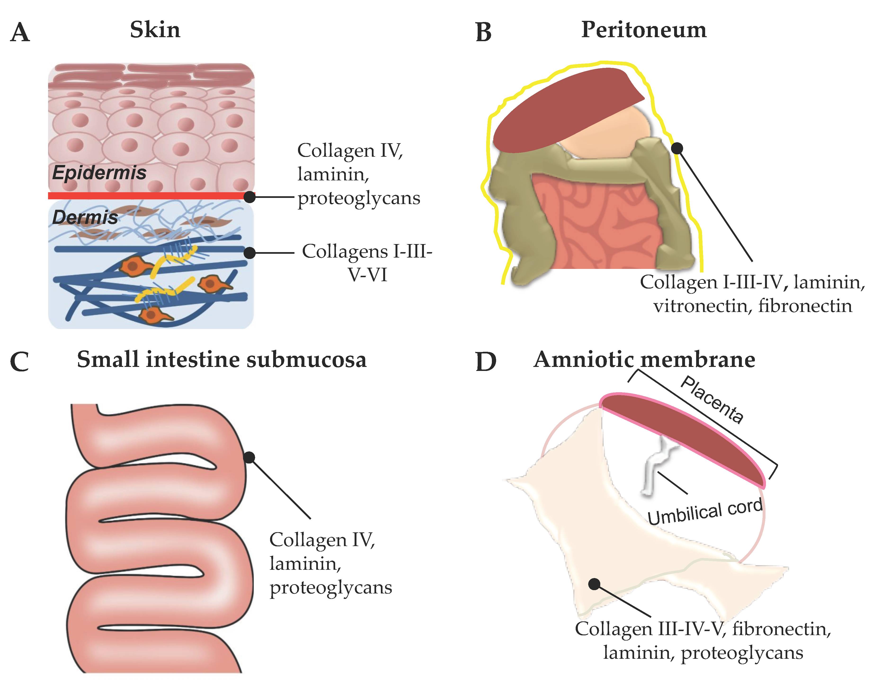
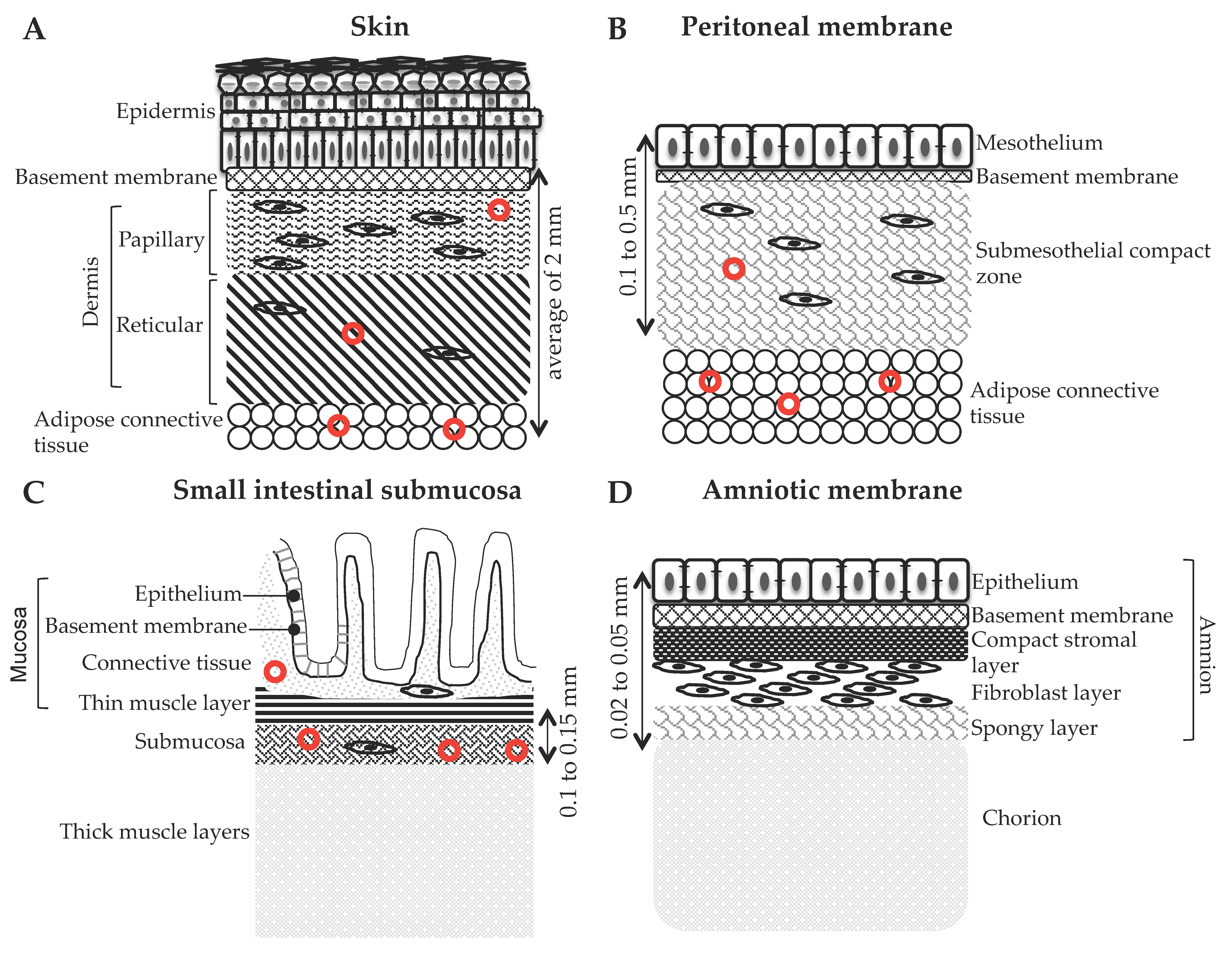
2. Tissue Sources for Preparation of ECM-Based Biomaterials for Skin Repair

In the last decades, decellularized scaffolds for use as skin substitutes have been intensively developed from various tissue sources such as dermis, skin flaps, peritoneum, intestinal tissues, and amnion/chorion tissues (Figure 1).
Allogeneic sources, such as tissues from cadaveric donors, and xenogeneic materials, such as porcine, bovine, and goat tissues, have been used, with different long-term results [26]. The decellularization protocols typically combine physical and chemical treatments to remove cellular antigens, involving freeze-thawing or detergents and/or enzymes that can alter the 3D structure of the ECM [27]. Additionally, the US Food and Drug Administration (FDA) requirements impose that acellular mammalian-derived products undergo “viral inactivation,” which implies the use of detergents resulting in the removal of many important biologic components from the tissue, such as lipids, glycans, and elastins [28]. As an alternative, skin grafts from fish such as Atlantic cod have been proposed because viral and prion transmission risk is nonexistent. These tissues can be subjected to gentle processing that does not disrupt the structure or bioactive composition of the original ECM [29].
Thus, decellularized tissues can be obtained from a variety of sources and implanted either orthotopically, i.e., the reconstructed tissue exhibits similar properties to those of the source, or ectopically, i.e., to a site that is completely different from origin site of the decellularized tissue [30]. In this review, we focus on decellularized biomaterials with the potential to be used as dermal or skin substitutes for clinical applications.

2.1. Acellular Dermal Matrix (ADM)

A decellularized dermal matrix is an acellular tissue made by taking a full-thickness section from a donor source, which in most cases is a human cadaver or of porcine or bovine origin. In the case of human donors, the tissue is screened for infectious agents, such as HIV, hepatitis, and syphilis [31]. In the last two decades, ADM has become increasingly popular for the coverage of soft-tissue open wounds. Artificial dermal substitutes are routinely used in the treatment of chronic and acute injuries, particularly when autologous intact cutaneous tissue is not available [32]. Although skin grafting is significantly less expensive, the use of ADM can be a successful alternative to a painful and aesthetically undesirable donor site [31]. In addition, as has long been recognized, the presence of dermis enhances the success of subsequent skin transplantation, inhibits granulation tissue formation, and prevents scar formation, thereby reducing vascular contraction [26]. Thus, ADM is often used in conjunction with split-thickness grafts for the treatment of full-thickness wounds [31]. Aside from burn wounds and diabetic foot ulcers (DFUs), more recently, ADMs have been suggested as a successful alternative to vascularized flaps for extremity wounds with exposed tendons, bones, and joints [32]. ADMs are currently produced and developed as various commercial products, and here we focus on commercially available skin substitutes with substantial relevant evidence of efficacy.
The gold standard for temporary coverage is a skin allograft, i.e., the skin is transplanted from one person to another. As with autografts involving graft transfer from the same individual, allografts undergo vascularization within 2–3 days and can provide a variable barrier after a severe burn. Unlike autografts, however, the skin is highly immunogenic, and when a viable skin allograft is transplanted onto a healthy recipient, rejection occurs within 6–14 days [33]. Allograft ADM products derived from donated human skin are supplied by tissue banks compliant with standards of the American Association of Tissue Banks and FDA guidelines. A long list of products derived from minimally processed human donor tissue is available, with different durations of covering depending on their design and composition [34] including the following: AlloDermTM regenerative tissue matrix (RTM; LifeCell Corp., Branchburg, New Jersey, USA), AlloPatch HD® Pliable (Musculoskeletal Transplant Foundation), Cymetra™ Micronized AlloDermTM (injectable form of AlloDermTM, LifeCellKCl), Dermacell® (LifeNet Health), Flex HD® (Ethicon and Musculoskeletal Transplant Foundation), GammaGraft® (Promethean Lifesciences, Inc.), GraftJacket® RTM (LifeCell, licensed to Wright Medical Technology and KCl), Glyaderm® (Euro Skin Bank, Beverwijk, The Netherlands), Matrix HD™ (RTI Biologics), Memoderm™ (Memometal, Inc.), Puros Dermis® (Zimmer Dental), and Repliform® (LifeCell and Boston Scientific) [35]. At least five different manufacturing processes and products (AlloDermTM, DermaMatrix, Glyaderm®, GraftJacket®, and SureDerm®) are currently registered for wound care (Table 1) [33]. We discuss here in detail the most cited versions considering their efficacy in treating burns and non-healing/difficult-to-heal wounds.

2.1.1. AlloDermTM RTM (LifeCell Corp., Branchburg, NJ, USA)

AlloDermTM (LifeCell Corp., Branchburg, New Jersey, USA) is one of the first FDA-approved acellular matrix materials and among the most extensively investigated [25]. It has been used since 1992 to treat burns with successful engraftment of both the AlloDermTM and the subsequent overlying skin graft in many individuals [36]. This product was FDA approved for the replacement of inadequate integument tissue, but the most common cited application has been to cover the skin flaps of donor sites as a sheet graft without an overlying split-thickness graft (Table 1) [37]. AlloDermTM is processed directly from fresh cadaver skin treated with high salt to remove the cellular components. The treated skin is then freeze-dried, leaving an ADM with an intact basement membrane complex (Figure 2A) [25]. AlloDermTM is hydrated before use, typically in an antibiotic saline solution according to the manufacturer’s recommendations. The meshing of the ADM at a 1:1 ratio optimizes revascularization and nutrition of the overlying skin graft [25]. Finally, in vitro recellularization of AlloDermTM with allogeneic keratinocytes, adipose-derived stem cells, fibroblasts, and umbilical vein endothelial cells allows for production of biological skin substitutes [25].

2.1.2. AlloPatch® Pliable (Musculoskeletal Transplant Foundation, Edison, NJ, USA)

AlloPatch® Pliable is an aseptically processed human reticular (HR)-ADM, intended for use in chronic or acute wound coverage [34]. It requires no rehydration or refrigeration prior to use and can be stored at ambient temperature according to the manufacturer’s recommendations. This dermis differs from many other ADMs in that it does not contain a papillary portion, resulting in a homogeneous matrix that retains its original architecture and its key ECM components (Figure 2). The absence of asymmetry or orientation is considered beneficial in the clinical setting, having a positive impact on cell infiltration and native tissue remodeling [38]. In a prospective, randomized, controlled, multicenter study, this HR-ADM was shown to be superior in promoting DFU healing as compared to standard of care involving daily dressing changes with a collagen alginate matrix [39].

2.1.3. DermACELL™ (LifeNet Health)

DermACELL™ is a decellularized regenerative human tissue matrix allograft that contains both the reticular and papillary compartments with a basement membrane (Figure 2). Upon application, the reticular site is placed against the surgical wound. The patented preparation process for DermACELL™ includes the use of anionic detergents and endonucleases to eliminate more than 97% of the nucleic acids. It is preserved and stored at room temperature and thus can be delivered hydrated [40]. DermACELL™ is indicated for chronic non-healing wounds [34] (Table 1). In a prospective, multicenter, single-arm clinical trial for the treatment of large complex DFUs with exposed tendon or bone, DermACELL™ rapidly reduced the size of the wound. However, that study favored cases that required a single application of the ADM [41]. Additionally, in a case report [40], DermACELL™ was successfully used to treat a patient with scarring from a severe burn. More recently, a case report publication provided evidence that DermACELL™ can eventually be used to treat fingertip injuries, e.g., after amputation [42]. An ongoing open-label trial will assess the effectiveness of this ADM in patients with chronic venous ulcers.

2.1.4. GraftJacket® RTM (Wright Medical Technology, Inc., Licensed to KCl)

GraftJacket® RTM is an intact human ADM containing a basement membrane (Figure 2), processed with a patented method to minimize damage to the scaffold and freeze-dried to remove moisture. GraftJacket® RTM is intended for replacement of damaged or inadequate integumental tissue, such as DFUs, venous leg ulcers, pressure ulcers, or other homologous uses on human integument [43]. A quantitative analysis of comparative literature including randomized, prospective controlled clinical studies with full-thickness DFU evaluated the efficacy of this ADM compared to standard moist wound care. The results demonstrated improved healing of DFUs in a significantly reduced time with this ADM. The authors highlighted the fact that a single application achieved this outcome, whereas multiple applications are necessary with other commercialized ADMs [44].
Despite their effectiveness, particularly toward wound bed vascularization, skin allografts present some drawbacks. These include limited availability, potential for pathogen transmission, and cost [33].

2.1.5. Animal-Derived ADMs

Grafts from xenogeneic dermal substitutes are often applied to extensive skin defects caused by severe burns. Although human ADMs and engineered human skin matrices have shown satisfactory effects in the treatment of extended second- and/or third-degree burns when xenogeneic ADMs lack histocompatibility, human ADMs are so expensive that their use is often impossible in these indications. The necessity of novel cost-effective dermal substitutes with equivalent efficiency has led to the search for alternative solutions [45]. The critical barrier to xenogeneic scaffolds in translational application is the recipient immune response to the antigenic components of the xenogeneic tissue [46]. Despite initial speculation, the decellularization process is insufficient to eliminate antigens from xenogeneic tissues because both cells and the ECM components themselves can elicit the host immune response. In addition, the remaining cell debris can be immunogenic [47]. Some strategies to achieve removal of specific antigens from xenogeneic scaffolds are being developed and are presented later in this review. These include enzymatic removal of known xenoantigens, such as α-gal epitopes, stepwise solubilization-based antigen removal, or a combination of these approaches [47].
Although the animal skin can be extremely adherent and occasionally incorporated into the wound, xenografts are not yet considered true grafts or transplants. Xenografts do not vascularize, but they can still be beneficial to wound management by reducing the pain in partial-thickness injuries and improving healing rates. Such ADMs are being used for exfoliative skin disorders [48].
An extensive list of animal-derived ADM products is currently available, most of which achieve success in clinical use. Since the 1960s, pigs have served as the primary donors for xenografts in the United States, by virtue of their relative affordability and histological structural similarities to the human skin [48]. Several reports have attested to the benefits of using porcine skin in the treatment of extensive wounds. These advantages include reduced healing rates for partial-thickness wounds and granulating wounds and reduced pain with placement over burns [48]. Currently available acellular porcine dermal matrices include Permacol™ (Tissue Sciences Laboratories), Strattice™ (LifeCell Corp.), CollamendTM (Bard), Xenoderm (mbp), and XenMatrixTM (Davol, Inc.) [31]. According to the UnitedHealthCare® Medicare Advantage Policy Guideline for 2020, porcine skin dressings have been suggested as possibly necessary as an occlusive dressing for burns, donor sites of a homograft, and decubiti and other ulcers [49]. Permacol™ is composed of porcine dermal collagen that is subjected to crosslinking, which is suggested to improve tensile strength. Strattice™ is a non-crosslinked porcine-derived ADM in pliable and firm versions, which can be stored at room temperature and is available fully hydrated.
Materials from different sources and processing exhibit different biochemical and mechanical properties and host responses upon implantation [50]. Therefore, suitable dressing material must be used based on the wound type.
PriMatrix™ (TEI Biosciences) is an ADM processed from fetal bovine skin. It was cleared for marketing by the FDA for partial- and full-thickness wounds, for diabetic, pressure, and venous stasis ulcers, and for surgical wounds [51]. This fetal bovine collagen scaffold is processed to remove all lipids, carbohydrates, and non-collagenous proteins, leaving primarily intact fetal dermal collagen fibers with a great proportion of fibrillar collagen III [52]. This PriMatrixTM collagen III rich substrate is associated with healing in developing tissues, whereas the unique structural matrix of fetal tissues has been identified as a contributing factor in scarless wound healing [50]. SurgiMend™ PRS (TEI Biosciences) is another ADM processed from fetal bovine dermis.
An acellular fish dermis has also been FDA approved. Kerecis™ Omega3 Wound (Kerecis) is an ADM rich in omega-3 fatty acids, intended for use in burn wounds, chronic wounds, and other applications. Human skin differs from fish skin mainly because of the adaptation of the latter to the aqueous environment through the development of scales, secretion of mucus from epithelial cells, lack of superficial keratinized layers, and presence of two basement membranes [53]. Additionally, fish skin heals faster than human skin without leaving scar tissue [54]. The histological analysis of human skin after a Kerecis™ graft demonstrated that the ADM is incorporated into the damaged area and infiltrated by autologous cells, is resistant to bacteria, and exhibits anti-inflammatory properties because of the omega-3 fatty acids [29,54].

2.2. Decellularized Mesothelium

The mesothelium is a simple squamous epithelium lining the walls of large cavities (peritoneum, pericardium, and pleura) (Figure 1B) [55]. The mesothelial tissues have a thin and strong layer of ECM that supports epithelial cells, which are capable of rapid wound healing (Figure 2B) [56]. Decellularized mesothelium is obtained using detergent agents in a process designed to maximize the preservation of the ECM architecture and composition [57]. A list of decellularized mesothelial matrices of porcine, bovine, equine, and human origins is currently available on the market for use in regeneration of different tissues and organs, mainly soft tissues including skin, vascular tissues, and valve replacements [26,58].

2.2.1. Decellularized Intestine and Urinary Bladder

One of the most studied decellularized matrices for a variety of biomedical and tissue-engineering applications derives from the intestine. The ileum, jejunum, and small intestinal submucosa (SIS) are the sources of decellularized intestine scaffolds (Figure 1C) [26]. Porcine SIS is derived from the thin, translucent tunica submucosa layer of the small intestine, which remains after removing the mucosal and muscular layers (Figure 2C). Once processed, SIS biomaterials constitute cross-linked 3D matrices consisting of constitutive proteins that are mostly collagen, with smaller amounts of carbohydrates and lipids [59]. This material supports the formation of a skin barrier 14 days after seeding with human keratinocytes, fibroblasts, and endothelial cells.
The role of SIS in promoting wound closure has been extensively investigated [25]. Incubation with SIS inhibits matrix metalloproteinases, which are important contributors to wound chronicity and are abundantly expressed in chronic wounds [60,61]. Moreover, SIS scaffolds contain transforming growth factor (GF)-β, basic fibroblastic GF, vascular endothelial GF, and epidermal GF, thus promoting angiogenesis, cell growth, and differentiation, and consequently tissue regeneration [26]. Advantageous properties of SIS scaffolds in healing wounds include their effective barrier function of the wound bed dehydration because of their low porosity, and their strength and flexibility even after a long-term storage in a lyophilized form [59].
Oasis® Wound Matrix (Cook Biotech, Inc., West Lafayette, IN, USA) is a collagen scaffold derived from porcine intestine submucosa cleared for marketing by the FDA for the management of partial and full-thickness wounds, including pressure, venous, diabetic, and chronic vascular ulcers, surgical wounds, and draining wounds. A clinical study revealed that in a cohort of patients carrying with chronic leg ulcer treated with SIS, complete wound closure was achieved in 55% cases. The use of SIS seemed to accelerate healing rates and to reduce the incidence of ulcer recurrence [62]. Decellularized porcine urinary bladder matrix (UBM) also has been reported to be an alternative to complex surgical procedures involving flap-associated donor site morbidity to treat complex extremity wounds. Use of UBM seemed to be rather beneficial for the management of patients suffering from comorbidities such as obesity, diabetes mellitus, and cardiopulmonary compromise [27]. After UBM decellularization, the remaining components include collagen, fibronectin, laminin, GAGs, and GFs. The UBM is likely to act through induction of bioactive GFs that support angiogenesis and facilitate proliferation of connective tissues, and the intact basement membrane is conductive to epithelial and endothelial cell proliferation [63].
Commercially available porcine UBM products include MicroMatrix® (Acell, Inc., Columbia, MD, USA) and Cytal® Wound Matrix (Acell, Inc., Columbia, MD, USA). Regarding the mechanism of action, a preclinical study showed rapid cell infiltration into the UBM after application, followed by a robust angiogenic response and new host tissue deposition [64]. Another study demonstrated induction of a different immune response as compared to classical wound healing, one that involves M2-phenotype macrophages related to tissue remodeling rather than the M1 macrophages that are associated with scar tissue formation [65].

2.2.2. Decellularized Amniotic Membrane (AM)

The AM surrounds the fetus in the uterus and consists mainly from top to bottom of an epithelium layer, basement membrane, compact ECM layer, fibroblast layer, and spongy layer (Figure 1D and Figure 2D). Fresh human AM contains collagen, fibronectin, and hyaluronic acid along with a combination of GFs, cytokines, and anti-inflammatory proteins [66]. Several studies have shown that specific AM products may be useful for treating several conditions in adults, including non-healing lower-extremity DFUs. AMs are harvested immediately after birth, cleaned, sterilized, and either cryopreserved or dehydrated, maintaining their properties [67]. Fully decellularized AM can be prepared by incubation of fresh tissue with hypotonic Tris buffer containing protease inhibitors, sodium dodecyl sulfate (SDS), DNAase, RNAase, and 1% trypsin-EDTA (ethylene-diamine-tetraacetic acid) for complete elimination of epithelial and mesenchymal cells [68]. Human AM is characterized by anti-bacterial, anti-inflammatory, and non-immunogenic properties, promotes reduced pain and dehydration, and favors the re-epithelialization process [69].
Some commercially available acellular AM products suggested for use in DFUs include AmnioBand® Membrane (MTF Biologics), Biovance® (Celularity, Inc.), and EpiFix® (MiMedx). Despite these many advantages, disadvantages of AM include poor mechanical properties and a high biodegradability rate, which complicate their extensive use in clinic [69]. Finally, the in vitro use of acellular human AM allows the construction of living skin equivalents through seeding of human keratinocytes on the epithelial side and fibroblasts on the chorionic side (Figure 2) [70,71].

2.2.3. Decellularized Skin Flaps

A skin flap consists of healthy skin and adipose tissue (Figure 2A) or skin, fat, and muscle. Clinically, skin flaps are used to cover full-thickness, serious wounds [26] but are not always an option and can result in donor site morbidity and poor aesthetics. Additionally, split-thickness, autologous skin grafts often used to cover large wounds transplant only a small portion of the dermis and are thus subject to scarring and contracture. Full-thickness skin grafts, including epidermis and full dermis, are difficult to obtain but are subjected to less contracture and result in a more functional graft and a better cosmetic final outcome. However, they depend on vascular ingrowth. Therefore, generating a decellularized full-thickness skin flap with a perfusable vascular pedicle could eventually yield substantial regenerative potential. In two recent studies, decellularized skin flaps from animal sources were developed and recellularized and/or integrated into host tissue. Zhang et al. [72], using a perfusion protocol combined with agitation, produced a skin/fat decellularized matrix that maintained the native ECM architecture and composition of mainly collagen, GAGs, and vascular endothelial GF and absence of the major class I-histocompatibility complex. Immunohistochemical analysis of the matrix showed peripheral nerves and vessel structure maintenance, and angiography confirmed the existence of the vascular pedicle and microcirculatory networks. Those scaffolds could subsequently be re-populated with human adipose-derived stem cells and endothelial cells and promote neovascularization after implantation in a rat model [72]. Jank et al. [73], using a porcine model, developed a full-thickness decellularized skin flap by applying a perfusion technique. The resulting scaffold comprised a vascular pedicle, maintained the ECM architecture and composition, and could be successfully perfused and anastomosed to the recipient vascular system. The scaffold demonstrated good biocompatibility and ability to support human keratinocyte engraftment and formation of an epidermis with intact barrier function [73]. These results are rather promising and may represent an adequate solution for the treatment of wounds, especially those characterized by loss of tissue volume.

3. Decellularization Methods

Both academic and industrial researchers have developed protocols to decellularize various native tissues, including those mentioned above. They are classically sorted into three categories of chemical, enzymatic, and physical methods. Almost all protocols, however, consist of combinations of these three approaches. To investigate these candidates in as many trials as possible, decellularization studies furthermore apply and compare very different protocols combining various agents instead of varying only one substance to assess its effects.
We here review decellularization methods specifically used for the production of acellular matrices for skin repair and evaluate the properties and effects of each substance commonly used in literature. The results of this analysis led us to propose another classification of these substances into main decellularizing agents that can disrupt cells and eliminate all DNA content when used alone (detergents, acids and bases, physical-based methods) and complementary agents that are added to improve these effects. Particularly, enzymes such as proteases are reviewed as common complementary agents because their exclusive use, which has been tested several times, never demonstrated total decellularization or satisfying in vitro cell repopulation of the matrices obtained.

3.1. Complementary Agents

3.1.1. Enzymes

Nucleases are the most common complementary agents included in decellularization protocols independently of the source of native tissue. Nucleic acids, especially DNA, are well known for their high immunogenic properties. To limit nucleic acid levels as much as possible, treatments with DNAse I, DNAse II, or benzonase are performed after exposure of the tissue to the main decellularizing agent(s) [68,74,75]. Applied concentrations, ranging from 50 U to 10 kU/mL, depend on the properties of the tissue (type, thickness, cell density) and the exposure to main decellularizing agent(s) (nature, duration, concentration, temperature). Regarding the choice between benzonase or DNAse, no clear rationale is available in the literature. Finally, for highly cellularized tissues, processing can also include RNAse treatments.
As mentioned above, strict protease-based approaches are not sufficient to remove all cells from the tissues used for the production of skin-repair matrices. Accutase, a commercial mix of proteolytic and collagenolytic enzymes classically used for soft cell-detachment from two-dimensional (2D) culture supports, failed to decellularize human AM: the epithelial layer was totally removed after 1 h of incubation, but many mesenchymal cells were still detected [76]. Trypsin has also been tested on human AM combined with EDTA, allowing for complete elimination of cells in both epidermal and mesenchymal compartments and retention of basement membrane components. However, the ability of the matrix to drive sufficient primary fibroblast infiltration and proliferation after a 7-day in vitro culture has not been demonstrated. The same group investigated the effects of dispase, a bacterial protease targeting collagen IV, laminin, and fibronectin [77]. They achieved complete decellularization but observed ECM alterations and important losses in basement membrane structure, as had been already reported [78,79]. Cell seeding was not assayed because of the low probability of epithelialization success using a basement membrane with such damage.
Although still undocumented as an efficient decellularized agent, trypsin can be combined with physical, acid-, base-, or detergent-based protocols [80,81,82]. The action of trypsin is achieved by immersion of the tissue in an isotonic solution containing from 0.25% to 0.1% w/v of enzyme, separately from the other treatments of the protocol. This exposure is often combined with EDTA (0.1%) and preferentially applied between delipidation and principal decellularization steps.
Tissue exposure to trypsin needs to be controlled; its ability to cleave a broad range of substrates means that deleterious effects on ECM structure can be exerted under overly long or too concentrated trypsin treatments. In porcine skin, different kinds of “fracturations” and density loss were observed in the ECM of the dermis compartment after an 18-h 0.25% trypsin exposure [80], whereas a denser and less porous ECM network was obtained for the same decellularization protocol excluding this preliminary step. Similar observations have been reported for human amnion with shorter exposure at the same concentration [76]. Surprisingly, none of the studied patents report strict trypsin incubation during the processing of human skin, human AM, and porcine SIS for acellular matrix production for skin repair [83,84,85]. Only one recent patent presenting a decellularization method for fish skin includes a trypsin step, but at a concentration lower than 0.001% [86].
Dispase, mentioned above as deleterious to human AM in isotonic solution, has surprisingly been described in two publications as a good complement for skin decellularization. On porcine skin, application of a soft dispase treatment (0.56 U/mL, 4 °C in isotonic solution), followed by SDS and soft trypsin exposures, resulted in good cell removal without any adverse effect on ECM post- 4- and 12-h treatments. These results suggest a softening effect of both temperature and concentration decreases on dispase treatment [80]. On mouse skin, solubilization of 2 U/mL of dispase in Dulbecco’s Modified Eagle Medium (DMEM, 37 °C, 2 h) prior to decellularization led to the production of an acellular matrix compatible with skin re-epithelialization in a mouse model [87]. The use of DMEM cell culture medium containing various components instead of simple isotonic solution could affect enzyme activity and explain the absence of deleterious effects. Furthermore, skin is an ECM-rich tissue containing more layers of epithelial cells than the amnion, so different accessibility to basement membrane between the two tissues also could explain the differential results with skin and human AM.

3.1.2. Chemicals

EDTA is a well-known chelator of di- and trivalent metal ions, commonly used in biochemistry to inhibit proteases. Its addition during exposures to trypsin or main decellularizing agents is a common tactic to prevent ECM degradation by cation-dependent proteases, which are abundant in both intra- and extracellular compartments. It also allows disruption of the protein–protein interactions based on metal cation bounding. Concentrations from 0.05% to 0.1% w/v are commonly applied, but levels up to 0.75% w/v have also been reported [80], without any specific association with tissue or decellularizing agent types. EDTA has been tested as a main decellularizing agent in human AM under various conditions (0.02–0.1%, 0.5–16 h, 4 or 37 °C), but does not completely decellularize the tissue, with a maximum DNA reduction limited to 50% of the initial level [88]. Other commercial protease inhibitors consisting of mixes of chemicals and small peptides can also be added to prevent ECM damage.
Hypo- and hypertonic shocks are applied from 1 to 12 h before the main decellularization steps or after to wash tissues and ensure the total lysis of remaining cells. To create hypertonic conditions, NaCl is commonly dissolved at 1 M to weaken cell membranes, as reported in the literature and in patented protocols to process fibroblast sheets, fish skin, and human skin [83,86,89]. Hypertonic NaCl also has been used to dissociate epidermis and dermis [90,91]. A hypertonic solution composed of 1 M KI has been used to process mouse skin [87]. Regarding hypotonic buffers, Tris (tris(hydroxymethyl)aminomethane) is often used at 10 mM, and immersion baths before decellularization and for solubilization of SDS have been reported for porcine bladder wall or fibroblast sheets processing [89,92].

3.1.3. Organic Solvents

Organic solvents are used to remove lipid content, an essential step for the preparation of acellular matrices using SIS and mesothelium, two fat-rich tissues.
A protocol combining methanol and chloroform (1/1, v/v, 12-h immersion) has been reported with porcine SIS, following manual elimination of the tunica serosa and tunica muscularis and prior intensive washings and SDS decellularization [81,82]. A degreasing step did not elicit notable changes in ECM architecture, and no remaining fat cells were detected in the final acellular matrix. The decrease in GFs reported in the final scaffold has been attributed to the manual removal of some compartments of the native tissue, not to organic solvent treatment.
Isopropanol (IPL) exposure is another method to remove lipids [76,93]. Two protocols have been compared with porcine skin: a single final exposure of the tissue to 100% IPL, and two 70% IPL exposures distributed during the whole process, followed by a final immersion in 100% IPL [93]. The single exposure was associated with the removal of half of the total lipid content, whereas a decrease of 90% was estimated for repeated treatments. Regarding nucleic acid removal, no significant variation in remaining DNA levels was detected between the two methods, confirming that IPL is not a main decellularizing agent. A combination of IPL and acetone is also mentioned in the patent of the commercial Oasis® matrix derived from porcine SIS [84].
Use of acetone/hexane (40/60, v/v) and pure acetone with porcine mesothelium was reported [94]. Lipid amounts were not assessed, and only a visual evaluation suggested better elimination of fat using the acetone/hexane combination.

3.2. Main Decellularizing Agents

3.2.1. Detergents

Detergents are the most common decellularizing agents used to process skin, SIS, mesothelium, bladder, and AM, regardless of the tissue origin. SDS is the strongest detergent used for tissue decellularization. It is ionic and disrupts all interactions between biomacromolecules. This agent is the most reported in decellularization protocols, and numerous teams have used it to process porcine SIS, porcine bladder wall, human amnion, human skin, rat flap, or fibroblast cell sheets [68,80,81,89,92,95]. Patents often mention its use for the production of commercial acellular matrices used in clinic [83,84,86]. SDS treatments are commonly performed by immersion in a 0.5% w/v solution (range of concentrations reported in literature: 0.1–1%) and last for hours. Thick and complex tissues containing a dense conjunctive layer or a multilayered epithelium, such as mesothelium, skin, and bladder wall are processed from 12 to 24 h. In contrast, thinner or less complex structure treatments last from 0.5 h for fibroblast sheets to 4–12 h for SIS. All decellularization studies that include SDS in concentrations >0.1% in their protocols, have reported a total removal of cells and cellular debris from the native tissue and low levels of remaining DNA. Some combinations with nucleases have been reported on amnion and bladder wall for SDS concentrations lower than 0.1% [68,92].
Although SDS is an excellent agent for decellularization, it has been frequently reported to damage ECM. Loss of global ECM density as well as GAG content and damage to the collagen network have particularly been observed in the matrix produced by fibroblast sheets [89] or in complex tissues such as human skin [96] and porcine SIS [81]. The increase in SDS concentration from 0.05% to 0.5% on fibroblast sheets or in exposure time from 6 to 12 h on SIS has been directly correlated with the amplification of ECM alterations. Scanning electron microscopy studies also suggest rearrangements of ECM fibers and variations in scaffold porosity after SDS treatment, even if no common and systematic effects can be generalized. The exposure of porcine SIS to the combination of 1% SDS for 12 h and 1% Triton X-100 for 0.5 h leads to complete cell removal but exerts similar adverse effects on ECM quality, coupled with a low cell development and metabolic activity after hemangioma stem cell (HEMSC) seeding [75]. Different SDS/Triton X-100 combinations have also been tested on rat fasciocutaneous flaps, leading to high DNA content removal after perfusion of 1% SDS for 24 or 72 h, followed by a 0.5-h 1% Triton X-100 treatment [95]. Modifications of the 2D structure of the ECM have been noted for the 72-h SDS perfusion using histological staining, but no other investigation has characterized the effects on ECM in detail. The switch of the 72-h SDS treatment from perfusion to immersion was associated with a decrease in DNA removal.
Basement membrane preservation after exposure to SDS is essential for epithelialization of the scaffold. The preservation of collagen IV, collagen VI, and laminin has been confirmed by immunostaining in porcine bladder wall (Tris HCl 10 mM overnight, 0.1% SDS for 24 h) [92] and softly decellularized human skin (1 M NaCl 24 h, 0.5% SDS 1 h) [97], but not in porcine SIS (0.05% trypsin 12 h, 0.5% SDS 4 h). Trypsin was shown to be compatible with basement membrane preservation at a higher concentration [81]. In human AM softly decellularized by a 24-h immersion in 0.03% SDS coupled with DNAse I and RNAse I treatments, maintenance of the basement membrane components but damage to the mesenchymal matrix architecture have been observed [68]. However, the mechanical properties were not significantly altered, and the final scaffold exhibited characteristics, e.g., failure strain, failure stress, an elastin phase slope, and a collagen phase slope similar to native tissue. This study is the only one to report decellularization of human AM using SDS, and the soft treatment applied could be an interesting approach to preserving ECM mechanics and integrity. Finally, no other comparison of tissue biomechanics before and after decellularization has been reported.
Even if ECM damage is often underlined after SDS treatments, biocompatibility of the resulting scaffolds has been demonstrated for several native tissues. In vitro, matrices derived from porcine skin [80], porcine SIS [82], human AM [68], and fish skin [98] do not exert cytotoxic or proliferation-limiting effects. Cytotoxic tests performed by direct or conditioned-medium contact, metabolic arrays, or visual observations by light microscopy several days after cell seeding have confirmed the compatibility of the acellular matrices with cell development. These studies used NIH 3T3 fibroblasts, primary fibroblasts, bone marrow mesenchymal stem cells (BMSCs), or human lung carcinoma cells. Implantation tests have also been reported for some acellular matrices obtained using SDS. Porcine SIS-derived matrix implanted subcutaneously in rat has shown a very satisfying colonization and establishment of host fibroblasts 4 weeks post-operatively. A classic immune response involving giant cells and infiltrated inflammatory cells has been described but started to decrease after 14 days of implantation [81].
Triton X-100 is a non-ionic detergent that can disrupt interactions between biomacromolecules more gently than SDS. It is very commonly used alone or with other agents to generate matrices for skin repair derived from porcine SIS [82,99], porcine skin [68], porcine mesothelium [94], mouse skin [87], and rat flap [72]. Almost all protocols apply this detergent at the same concentration of 0.5% v/v, with a few using up to 1%. The ability of Triton X-100 to fully decellularize a native tissue has been evaluated with porcine SIS exposure to increasing concentrations of detergent from 0.5% to 2% [99]. Acellular scaffolds were obtained after 48 h of immersion in all conditions, but ECM damage, especially loss of density, was observed for concentrations higher than 1%. On mouse skin, successful decellularization using 0.5% Triton X-100 for 24 h also has been reported, associated with matrix alterations, especially loss of elastin and increased stiffness compared to native skin [87]. Limited colonization by primary fibroblasts and poor metabolic activity was also described. Surprisingly, during wound healing experiments in mouse, new blood vessel formation was observed in the grafted matrix, whereas a low dermis fibroblast infiltration occurred in accordance with in vitro experiments. Deleterious effects of 0.5% Triton X-100 on mouse skin may be the result of its limited thickness and cell abundance compared to SIS.
To modulate the effects on ECM and favor cell development, combinations of Triton X-100 with other agents have been tested at reduced exposure times. As mentioned above, the combination of 0.1% SDS for 12 h with 1% Triton X-100 for 0.5 h failed to produce a satisfying porcine SIS acellular matrix [75] because the scaffold obtained was not compatible with cell growth in vitro. In contrast, co-exposure of porcine skin to 0.5% Triton X-100 and hypertonic 1 M NaCl, followed by 2% sodium deoxycholate and DNAse I treatments, appeared more promising [74]. Density and integrity of dermis ECM were preserved, especially collagen I and fibronectin networks, and the preservation of basement membrane was confirmed by the detection of collagen VII and laminin. Other observations revealed that, after a 6-month abdominal implantation in monkey, new blood vessels and dense colonization by host cells could be observed (global immune response not estimated). In rat, a dermis/adipose flap was successfully decellularized by perfusion using a combination of 1 M NaCl (epidermis removal and tonic shock), trypsin, and 1% Triton X-100 for 48 h [72]. DNA in dermis and fat compartments was quantified under 50 ng/mg of dry flap (10-fold reduction compared to native tissue). The general structure (2D and 3D) of the ECM and GAG content were preserved, but no evaluation of the basement membrane was described. Vascular endothelial GF was easily detectable by immunohistochemistry in fat tissue, but very low staining appeared in dermis. The growth of human umbilical vein endothelial cells and adipose stromal cells was shown between days 1 and 7 post-seeding.
Sodium deoxycholate (SD) is another ionic detergent applied as a decellularizing agent. It is commonly used at concentrations ranging from 2% to 4% and disrupts interactions between biomacromolecules. Its action, however, is frequently associated with DNA precipitation in spite of the loss of DNA–histone interactions, as has been confirmed [100]. The incubation of tissues with nucleases is thus automatically applied after exposure to SD, lasting for a few hours at concentrations from 5 to 10 kU/mL.
The capacity of SD to decellularize tissue alone has been reported in a study dedicated to the whole decellularization of rat intestine [101]. All cells and cell debris were removed with perfusion of a 4% SD solution for 4 h (connection to the superior mesenteric artery), followed by a DNAse I treatment for 3 h, and ECM density was preserved. In contrast, an incomplete decellularization of porcine SIS was observed after a 12 h immersion in 4% SD and a DNAse I treatment. Some cell nuclei were still visible in the conjunctive layer, associated with a level of remaining DNA that was 40% higher than that detected in tissue treated with SDS (40–45 versus 30 ng/mg dry tissue, respectively) [82]. However, other tissues that are compatible with perfusion and rich in ECM, such as aorta vein or skeletal muscle [102,103], can be totally decellularized using similar SD treatments.
A possible explanation for the incomplete cell removal in porcine SIS could be the limited SD penetration into the tissue during immersion compared to perfusion. As perfusion cannot be applied to all tissues, especially to skin that lacks an accessible vascular tree, immersion strategies have been developed by combining SD with other agents to aid in cell removal. In porcine skin where the multilayered epithelium is strongly anchored to the basement membrane, an exposure of the tissue to 1 M NaCl and 0.5% Triton X-100 for 24 h prior to a 4%, 24-h SD and DNAse I treatments, successfully removed cells from both the epidermis and dermis compartments [74]. In rat, a fasciocutaneous flap decellularization protocol using 4% SD and DNAse I was also reported, but further characterization of the acellular matrix is needed to characterize its properties [90].
SD-based decellularization approaches have been reported to be excellent for ECM preservation when used on tissues such as aorta, muscle, and lungs. This probably explains the effort invested in the development of protocols dedicated to skin-repair acellular matrix production despite problems with the perfusion-to-immersion switch. In porcine SIS, comparison of SD treatment to a double SDS/Triton X-100 exposure indicated a better quality of the final ECM matrix, even if some cells were still detected in the tissue. The 3D structure of the matrix was very close to that of the native tissue architecture, whereas other detergents increased porosity and modified fiber organization. Mechanical properties (failure strain, elastic modulus) were also similar to control tissue, and metabolic activity of HEMSCs at 96 h post-seeding was 10 times that of the activity of cells grown on the SDS/Triton X-100 matrix [75]. As previously described, a combination with NaCl and Triton X-100 on porcine skin is associated with basement membrane and ECM density preservation and a very satisfying biocompatibility in vivo [74].
Sodium lauroylsarcosinate (NLS, ionic) is a detergent that is less commonly reported for skin-repair matrix production. In mouse skin, a 1% NLS treatment for 24 h leads to complete removal of cells [87]. In a mouse wound healing model, despite important loss of GAG content, alterations in elastic fibers, and important tissue stiffening compared to native skin, this acellular matrix supported the establishment of host fibroblasts and formation of new blood vessels after a 6-month graft. A patented protocol, reported to be one of the MatrACELL® decellularization methods applied to produce the commercial human acellular matrix DermACELL™, also mentions use of NLS combined with nucleases to decellularize tissues [104]. These authors have published a comparison of this substitute with other commercial matrices, finding the lowest DNA content in the DermACELL™ scaffold; however, other independent studies are needed to confirm and complete the characterization of this matrix. More studies also are needed to describe in greater detail the effects of NLS on ECM during decellularization processing and to evaluate its compatibility with acellular matrix production using other tissues.
No use of CHAPS, a zwitterionic detergent reported for the decellularization of thin tissues (lungs and trachea), has been tested for producing scaffolds for skin repair. The thickness of the tissues used for skin repair may not be compatible with adequate decellularization using this detergent [28].

3.2.2. Acids and Bases

Despite promising results, tissue exposures to acids or bases are not as commonly used as detergents to decellularize tissues. Sodium hydroxide (NaOH) treatments have been reported to process porcine SIS and human skin. High pH can modify the solubility of proteins and disrupt their interactions with other proteins and biomacromolecules in general, leading to destabilization of the cell membrane. Exposure of human skin to 0.06 M NaOH for several weeks was assayed to remove cells, followed by a 0.1 M HCl neutralization for 10 min. After 6 weeks, a cell-free tissue was produced with a very dense ECM, conserving its elastin and collagen networks [105]. In a porcine wound healing model, this type of acellular matrix has driven a complete epithelialization of the wound at 14 days post-grafting, with a limited immune response. This protocol was patented by a non-profit organization and is now available as a commercial product under the name Glyaderm®. A study describing patient care and follow-up a few years after Glyaderm® application was published in 2015, indicating very satisfying results with wounds such as burns or forearm flap loss [106]. Complete wound closure, pigmentation of the graft, and elasticity recovery were observed. NaOH treatment is also mentioned in the patent used for the production of the commercial Oasis® matrix derived from porcine SIS [84].
NaOH combined with hypertonic treatment and low-pH exposure can also be used on porcine SIS [82]. Exposure to 0.01 M NaOH, followed by 1 M HCl and 1 M NaCl treatments, has been associated with a complete decellularization (remaining DNA amount <30 ng/mg of dried ECM) and a satisfying preservation of the ECM architecture. The development of BMSCs has also been validated for 7 days post-seeding. Additional tests are needed to evaluate the effect of NaOH on a broader diversity of tissues.
Peracetic acid exposure, commonly used for tissue sterilization, has been used rarely in processing porcine SIS, with contradictory observations. A first study tested two 0.1% v/v treatments in a perfusion chamber (12 h) or by immersion (2 h) [75]. In both cases, levels of remaining DNA similar to native tissue attested to poor matrix decellularization. The longest exposure modified the biomechanics of the SIS, with increased stiffness and reduced resistance to strain. However, growth of HESMCs was possible in vitro. In contrast, a 2-h immersion using the same peracetic acid concentration combined with 4% ethanol was associated with a low remaining DNA level below 40 ng/mg of dried ECM [82]. A clear alignment of matrix fibers and increased scaffold porosity compared to native tissue were also found. Proliferation of BMSCs was also demonstrated in vitro.
Biocompatibility of these kinds of scaffolds for in vivo uses needs to be assessed. More studies are needed before any conclusions can be drawn about the relevance of peracetic acid in the production of acellular matrices for skin repair.

3.2.3. Physical Approaches

‘Supercritical conditions’ refers to combinations of particularly high temperature and high pressure so that a fluid reaches a physical state combining the properties of both liquid and gas. This physical state allows a good penetration of the fluid inside tissues, just like a gas, with a macromolecule solubilization capacity similar to that of a liquid. Supercritical CO2 has been used initially to gently sterilize natural materials and tissues [107]. In recent years, supercritical CO2 decellularization was developed on vascular tissues [108], and most recently, processing of human skin and porcine skin has been reported.
On human skin, processing with supercritical CO2 after a hypertonic 1 M NaCl exposure has produced results similar to those with SDS in terms of cellular content removal (DNA level post-treatment <30 ng/mg of dried ECM) [91]. Small fractures reducing global ECM density were detected, but the fibronectin network seems to be unaffected. A surprising 3D structure was observed on scanning electron microscopy, however, characterized by punctate globular structures in the ECM network. Proliferation and viability of primary bovine chondrocytes were documented, but fibroblasts growth assessment was not presented. That study additionally underlined the importance of tissue saturation with the CO2-philic detergent LS-54 prior to CO2 exposure to enhance removal of cellular components.
A recent paper presented an approach combining supercritical CO2 with NaOH (from 0.1 to 1 M, exact concentration unknown) applied on porcine skin [109]. Observations in the ECM network were similar to those previously reported with human skin, and promising results were obtained in a porcine wound healing model with the matrices seeded with autologous cells before grafting. This protocol is patented and used to produce ABCcolla® Collagen Matrix (ACRO Biomedical, Kaohsiung, Taïwan).
Freeze-thawing is a special case that is difficult to categorize as a main or complementary agent because the complete elimination of cells using only extreme variations of temperature has not been fully demonstrated. With cell sheets, no complete decellularization has been achieved applying three cycles of freeze-thawing [89]. However, with human mesothelium, a combination of three freeze-thawing cycles with trypsin, DNAse I, and lipase VI exposures and isopropanol treatment to remove lipids has been reported as effective in removing all cells from the tissue. As trypsin and IPL cannot fully decellularize a tissue, the effect of freeze-thawing may be determinant in eliminating cells content [76]. Supplementary studies of the tissue after each step of the protocol are necessary to characterize the effects of each agent. Regardless, this study suggests that the combination of several complementary agents could be an interesting approach to investigate [76].
Regarding hydrostatic pressure, no protocol has been reported that has used it to decellularize tissues to produce acellular matrices for skin repair.

3.3. Promising New Approaches

Investigations are under way to find new main and complementary agents for tissue decellularization. A main issue when using acellular matrices for grafts in clinic is the immune response elicited by the exogenous tissue, sometimes leading to graft rejection. Galactose-α-(1, 3)-galactose (α-gal) is an oligosaccharide sequence in some sugar-linked proteins and lipids of non-primate cell membranes. This oligosaccharide is a strong immunogenic epitope in humans and is especially responsible for the rejection of pig-derived organs. A method has been specifically developed to eliminate this antigen from porcine skin [74]. A classic decellularization protocol combining hypotonic NaCl, Triton X-100, SD, and DNAse (already described above) has been completed by the addition of an α-galactosidase incubation step, with no observed modification in the ECM structure compared to galactosidase-free treatment. After abdominal implantation in a monkey model, anti-α-gal antibody levels were similar for matrices derived from galactosidase-treated and KO gal -/- pig tissues, whereas amounts for non-galactosidase-treated matrix were 16 and 25 times higher at 20 and 70 days post-surgery, respectively. Despite such promising results, no other similar studies have been published.
In terms of main decellularizing agents, latrunculin B, a sea sponge toxin that inhibits actin polymerization [110], has recently been used to decellularize mouse skin [87]. Combined with 0.6 M NaCl, 1 M KI, dispase II, and benzonase exposures, latrunculin B treatment (50 nM in DMEM, 2 h) led to a complete cell removal, with a satisfying conservation of ECM density and quality (GAG and hydroxyprolin levels, elastin distribution similar to native tissue). No changes in the biomechanics of the tissue were detected, in contrast to results with NLS or Triton X-100 treatments tested in parallel. In vivo, in a mouse wound healing model, infiltration of numerous host fibroblasts, formation of new blood vessels, and total re-epithelialization of the wound were observed.
Many protocols have been published that propose techniques adapted for tissue decellularization (Table 2). Some of these methods are already patented to produce skin-repair matrices used in clinic. Although classic decellularizing agents are efficient at cell removal, adverse effects on the ECM are frequently reported. The main difficulty in the development of decellularization methods is thus to find the fine balance between sufficient removal of cell content and sufficiently limited ECM modification to maintain its properties driving the infiltration and the development of host cells.

4. Sterilization and Storage of Decellularized Scaffolds

Quite a variety of methods have been published for decellularizing tissues, but sterilization procedures are standard. They consist of short immersions in peracetic acid or ethanol solutions [76,111], gamma [112] or ultraviolet irradiation [81,101], and ethylene-oxide exposures [82]. Disinfectants such as antibiotic/antimycotic substances [80,88] or bacteriostatic agents such as sodium azide [87,97] are also common.
Regarding storage of acellular matrices, three main methods are commonly applied: immersion in phosphate-buffered saline containing antibiotics or bacteriostatics, lyophilization, and freezing [74,82,94]. The two latter methods require specific protocols including exposure of the tissue to preserving agents to protect macromolecules from the extreme pressures and temperatures applied.
Reviews presenting the different processes of sterilization and storage methods used for natural materials and tissues have been published elsewhere [113,114].

5. Conclusions

Great advances have been made in skin regenerative medicine with regard to the preparation and use of decellularized tissues, leading to a growing number of products used in clinics with great success. Improvements are now expected to come at the level of protocol optimization. Ideal methods will reproducibly generate ECM that is free of cellular and nucleic acid material while retaining the structural, biochemical, and biomechanical properties crucial to its inherent cell-colonization function. Techniques may be optimized to achieve this balance, specifically in eliminating components of the tissue that could trigger an immunogenic response while maintaining components that will support and regulate reconstruction of new tissue. In particular, methods that combine strategies with chemicals, enzymes, and mechanical techniques may improve decellularization efficiency and limit the negative effects of protocols based on single agents. The goal is ultimately to administer the minimum amount of harsh chemicals or mechanical treatments to avoid unnecessary damage to the ECM microstructure and ultrastructure. Recellularization of skin devices before grafting is of great interest and should allow for the production of ready-to-use tissues. As with other organs, however, methods must be optimized to distribute the proper cell types uniformly and quickly through the tissue and enable sufficient nutrient and oxygen delivery for best cell viability. To that end, biofunctionalization of the decellularized tissue might offer promising perspectives. Interface biofunctionalization strategies should aim to modulate and control the implant–host interaction to enhance biocompatibility of implant materials and potentially even regulate implant function. The surface functionalization with ECM- or GF-derived molecules is a common approach in synthetic materials science to improve host cell–scaffold interactions at their interface and enhance biointegration of implant materials. Some groups have started to functionalize decellularized tissues by combining them with synthetic peptides to promote functions compromised by the decellularization process. This area will be intensified in the coming years with work that should lead to improved efficiency of decellularized tissues used for skin regeneration.

Author Contributions

A.M. and P.R. undertook chapters on skin defects and tissue sources for decellularization and clinical uses, Table 1 and both figures. M.D. managed the part on decellularization agents and Table 2. P.R. supervised the review construction and process. All authors have read and agreed to the published version of the manuscript.

Funding

Work in PR laboratory was supported by the AgenceNationale de la Recherche (grant numberANR-13-RPIB-0003-01). MD (PhD student) and AM (post-doctoral researcher) are funded by AgenceNationale de la Recherche (grant number ANR-18-CE92-0035-01) and EU/Greek national funds through the Operational Program Competitiveness, Entrepreneurship and Innovation (project T1EDK-03798), respectively.

Conflicts of Interest

The authors declare that there are no conflict of interest.

References

  1. Blanpain, C.; Fuchs, E. Epidermal homeostasis: A balancing act of stem cells in the skin. Nat. Rev. Mol. Cell Boil. 2009, 10, 207–217. [Google Scholar] [CrossRef] [Green Version]
  2. Koster, M.I.; Roop, D.R. Mechanisms Regulating Epithelial Stratification. Annu. Rev. Cell Dev. Boil. 2007, 23, 93–113. [Google Scholar] [CrossRef]
  3. Rousselle, P.; Braye, F.; Dayan, G. Re-epithelialization of adult skin wounds: Cellular mechanisms and therapeutic strategies. Adv. Drug Deliv. Rev. 2019, 146, 344–365. [Google Scholar] [CrossRef] [PubMed]
  4. Driskell, R.; Lichtenberger, B.M.; Hoste, E.; Kretzschmar, K.; Simons, B.; Charalambous, M.; Ferrón, S.R.; Herault, Y.; Pavlovic, G.; Ferguson-Smith, A.C.; et al. Distinct fibroblast lineages determine dermal architecture in skin development and repair. Nature 2013, 504, 277–281. [Google Scholar] [CrossRef] [PubMed] [Green Version]
  5. Ghetti, M.; Topouzi, H.; Theocharidis, G.; Papa, V.; Williams, G.; Bondioli, E.; Cenacchi, G.; Connelly, J.; Higgins, C.A. Subpopulations of dermal skin fibroblasts secrete distinct extracellular matrix: Implications for using skin substitutes in the clinic†. Br. J. Dermatol. 2018, 179, 381–393. [Google Scholar] [CrossRef] [PubMed] [Green Version]
  6. Jiang, D.; Correa-Gallegos, D.; Christ, S.; Stefanska, A.; Liu, J.; Ramesh, P.; Rajendran, V.; De Santis, M.M.; Wagner, D.E.; Rinkevich, Y. Two succeeding fibroblastic lineages drive dermal development and the transition from regeneration to scarring. Nature 2018, 20, 422–431. [Google Scholar] [CrossRef] [Green Version]
  7. Korosec, A.; Frech, S.; Gesslbauer, B.; Vierhapper, M.; Radtke, C.; Petzelbauer, P.; Lichtenberger, B.M. Lineage Identity and Location within the Dermis Determine the Function of Papillary and Reticular Fibroblasts in Human Skin. J. Investig. Dermatol. 2019, 139, 342–351. [Google Scholar] [CrossRef] [Green Version]
  8. Driskell, R.; Jahoda, C.A.B.; Chuong, C.-M.; Watt, F.M.; Horsley, V. Defining dermal adipose tissue. Exp. Dermatol. 2014, 23, 629–631. [Google Scholar] [CrossRef] [Green Version]
  9. Clark, R.A.F. Wound repair. Lessons for tissue engineering. In Principles of Tissue Engineering; Lanza, R.P., Langer, R., Chick, W., Eds.; Academic Press: Cambridge, MA, USA, 1997; pp. 737–768. [Google Scholar]
  10. Järbrink, K.; Ni, G.; Sönnergren, H.; Schmidtchen, A.; Pang, C.; Bajpai, R.; Car, J. Prevalence and incidence of chronic wounds and related complications: A protocol for a systematicreview. Syst. Rev. 2016, 5, 152. [Google Scholar] [CrossRef] [Green Version]
  11. Eaglstein, W.H.; Falanga, V. CHRONIC WOUNDS. Surg. Clin. N. Am. 1997, 77, 689–700. [Google Scholar] [CrossRef]
  12. Harding, K.; Morris, H.L.; Patel, G.K. Science, medicine, and the future: Healing chronic wounds. BMJ 2002, 324, 160–163. [Google Scholar] [CrossRef] [PubMed]
  13. Mekkes, J.; Loots, M.; Van Der Wal, A.; Bos, J. Causes, investigation and treatment of leg ulceration. Br. J. Dermatol. 2003, 148, 388–401. [Google Scholar] [CrossRef] [PubMed]
  14. Kartus, J.; Movin, T.; Karlsson, J. Donor-site morbidity and anterior knee problems after anterior cruciate ligament reconstruction using autografts. Arthrosc. J. Arthrosc. Relat. Surg. 2001, 17, 971–980. [Google Scholar] [CrossRef] [PubMed]
  15. Konofaos, P.; Halen, J.V. Nerve Repair by Means of Tubulization: Past, Present, Future. J. Reconstr. Microsurg. 2013, 29, 149–164. [Google Scholar] [CrossRef] [Green Version]
  16. Berthiaume, F.; Maguire, T.; Yarmush, M.L. Tissue Engineering and Regenerative Medicine: History, Progress, and Challenges. Annu. Rev. Chem. Biomol. Eng. 2011, 2, 403–430. [Google Scholar] [CrossRef]
  17. Khan, F.; Tanaka, M. Designing Smart Biomaterials for Tissue Engineering. Int. J. Mol. Sci. 2017, 19, 17. [Google Scholar] [CrossRef] [Green Version]
  18. Zhu, M.; Li, W.; Dong, X.; Yuan, X.; Midgley, A.C.; Chang, H.; Wang, Y.; Wang, H.; Wang, K.; Ma, P.X.; et al. In vivo engineered extracellular matrix scaffolds with instructive niches for oriented tissue regeneration. Nat. Commun. 2019, 10, 4620. [Google Scholar] [CrossRef] [Green Version]
  19. Zhong, S.P.; Zhang, Y.; Lim, C.T. Tissue scaffolds for skin wound healing and dermal reconstruction. Wiley Interdiscip. Rev. Nanomed. Nanobiotechnology 2010, 2, 510–525. [Google Scholar] [CrossRef]
  20. Bryksin, A.V.; Brown, A.C.; Baksh, M.M.; Finn, M.; Barker, T.H. Learning from nature-novel synthetic biology approaches for biomaterial design. Acta Biomater. 2014, 10, 1761–1769. [Google Scholar] [CrossRef] [Green Version]
  21. Niklason, L.E. Understanding the Extracellular Matrix to Enhance Stem Cell-Based Tissue Regeneration. Cell Stem. Cell 2018, 22, 302–305. [Google Scholar] [CrossRef] [Green Version]
  22. Daley, W.P.; Peters, S.B.; Larsen, M. Extracellular matrix dynamics in development and regenerative medicine. J. Cell Sci. 2008, 121, 255–264. [Google Scholar] [CrossRef] [PubMed] [Green Version]
  23. Kim, Y.; Ko, H.; Kwon, I.K.; Shin, K. Extracellular Matrix Revisited: Roles in Tissue Engineering. Int. Neurourol. J. 2016, 20, S23–S29. [Google Scholar] [CrossRef] [PubMed]
  24. Rousselle, P.; Montmasson, M.; Garnier, C. Extracellular matrix contribution to skin wound re-epithelialization. Matrix Biology. 2019, 75–76, 12–26. [Google Scholar] [CrossRef] [PubMed]
  25. Eweida, A.; Marei, M.K. Naturally Occurring Extracellular Matrix Scaffolds for Dermal Regeneration: Do They Really Need Cells? BioMed Res. Int. 2015, 2015, 1–9. [Google Scholar] [CrossRef] [PubMed] [Green Version]
  26. Cui, H.; Chai, Y.; Yu, Y. Progress in developing decellularized bioscaffolds for enhancing skin construction. J. Biomed. Mater. Res. Part A 2019, 107, 1849–1859. [Google Scholar] [CrossRef] [PubMed]
  27. Elmashhady, H.H.; Kraemer, B.A.; Patel, K.H.; Sell, S.A.; Garg, K. Decellularized extracellular matrices for tissue engineering applications. Electrospinning 2017, 1, 87–99. [Google Scholar] [CrossRef]
  28. Crapo, P.M.; Gilbert, T.; Badylak, S.F. An overview of tissue and whole organ decellularization processes. Biomaterials 2011, 32, 3233–3243. [Google Scholar] [CrossRef] [Green Version]
  29. Patel, M.; Ii, J.C.L. Fish skin acellular dermal matrix: Potential in the treatment of chronic wounds. Chronic Wound Care Manag. Res. 2019, 6, 59–70. [Google Scholar] [CrossRef] [Green Version]
  30. Nakamura, N.; Kimura, T.; Kishida, A. Overview of the Development, Applications, and Future Perspectives of Decellularized Tissues and Organs. ACS Biomater. Sci. Eng. 2016, 3, 1236–1244. [Google Scholar] [CrossRef]
  31. Fosnot, J.; Kovach, S.J.; Serletti, J.M. Acellular Dermal Matrix: General Principles for the Plastic Surgeon. Aesthetic Surg. J. 2011, 31, 5S–12S. [Google Scholar] [CrossRef] [Green Version]
  32. Scalise, A.; Torresetti, M.; Verdini, F.; Capecci, M.; Andrenelli, E.; Mengarelli, A.; Ceravolo, M.G.; Fioretti, S.; Di Benedetto, G. Acellular dermal matrix and heel reconstruction: A new prospective. J. Appl. Biomater. Funct. Mater. 2017, 15, 15. [Google Scholar] [CrossRef] [PubMed]
  33. Yamamoto, T.; Iwase, H.; King, T.; Hara, H.; Cooper, D.K.C. Skin xenotransplantation: Historical review and clinical potential. Burns 2018, 44, 1738–1749. [Google Scholar] [CrossRef] [PubMed]
  34. Mohebichamkhorami, F.; Alizadeh, A. Skin substitutes; an updated review of products from year 1980 to 2017. J. Appl. Biotechnol. Rep. 2017, 4, 615–623. [Google Scholar]
  35. Snyder, D.L.; Sullivan, N.; Schoelles, K.M. Skin substitutes for treating chronic wounds; Technology Assessment Report; Agency for Healthcare Research and Quality (AHRQ): Rockville, MD, USA, 18 December 2012. Available online: https://www.ncbi.nlm.nih.gov/books/NBK248353/ (accessed on 9 April 2020).
  36. Wainwright, D.J.; Bury, S.B. Acellular Dermal Matrix in the Management of the Burn Patient. Aesthetic Surg. J. 2011, 31, 13–23. [Google Scholar] [CrossRef] [Green Version]
  37. Ellis, C.V.; Kulber, D.A. Acellular Dermal Matrices in Hand Reconstruction. Plast. Reconstr. Surg. 2012, 130, 256S–269S. [Google Scholar] [CrossRef]
  38. Dasgupta, A.; Orgill, D.; Galiano, R.D.; Zelen, C.M.; Huang, Y.-C.; Chnari, E.; Li, W.W. A Novel Reticular Dermal Graft Leverages Architectural and Biological Properties to Support Wound Repair. Plast. Reconstr. Surg. Glob. Open 2016, 4, e1065. [Google Scholar] [CrossRef]
  39. Zelen, C.M.; Orgill, D.P.; Serena, T.; Galiano, R.; Carter, M.J.; DiDomenico, L.; Keller, J.; Kaufman, J.; Li, W.W. A prospective, randomised, controlled, multicentre clinical trial examining healing rates, safety and cost to closure of an acellular reticular allogenic human dermis versus standard of care in the treatment of chronic diabetic foot ulcers. Int. Wound J. 2016, 14, 307–315. [Google Scholar] [CrossRef]
  40. Chen, S.-G.; Tzeng, Y.-S.; Wang, C.-H. Treatment of severe burn with DermACELL®, an acellular dermal matrix. Int. J. BurnsTrauma 2012, 2, 105–109. [Google Scholar]
  41. Cazzell, S.; Moyer, P.M.; Samsell, B.; Dorsch, K.; McLean, J.; Moore, M.A. A Prospective, Multicenter, Single-Arm Clinical Trial for Treatment of Complex Diabetic Foot Ulcers with Deep Exposure Using Acellular Dermal Matrix. Adv. Ski. Wound Care 2019, 32, 409–415. [Google Scholar] [CrossRef]
  42. Wu, T.-H.; Bertasi, G. The use of Dermacell® in Fingertip Injury. J. Clin. Case Rep. Images 2019, 1, 14–22. [Google Scholar] [CrossRef]
  43. Reyzelman, A.; Crews, R.T.; Moore, J.C.; Moore, L.; Mukker, J.S.; Offutt, S.; Tallis, A.; Turner, W.B.; Vayser, D.; Winters, C.; et al. Clinical effectiveness of an acellular dermal regenerative tissue matrix compared to standard wound management in healing diabetic foot ulcers: A prospective, randomised, multicentre study. Int. Wound J. 2009, 6, 196–208. [Google Scholar] [CrossRef] [PubMed]
  44. Reyzelman, A.M. Human acellular dermal wound matrix for treatment of DFU: Literature review and analysis. J. Wound Care 2015, 24, 128–134. [Google Scholar] [CrossRef] [PubMed]
  45. Yu, G.; Ye, L.; Tan, W.; Zhu, X.; Li, Y.; Jiang, D. A novel dermal matrix generated from burned skin as a promising substitute for deep-degree burns therapy. Mol. Med. Rep. 2016, 13, 2570–2582. [Google Scholar] [CrossRef] [PubMed] [Green Version]
  46. Buxton, D.; Lee, S.C.; Wickline, S.A.; Ferrari, M. Recommendations of the National Heart, Lung, and Blood Institute Nanotechnology Working Group. Circulation 2003, 108, 2737–2742. [Google Scholar] [CrossRef] [PubMed] [Green Version]
  47. Wong, M.L.; Griffiths, L.G. Immunogenicity in xenogeneic scaffold generation: Antigen removal vs. decellularization. Acta Biomater. 2014, 10, 1806–1816. [Google Scholar] [CrossRef] [Green Version]
  48. Imahara, S.D.; Klein, M.B. Skin grafts. In Biomaterials for Treating Skin Loss; Orgill, D.P., Blanco, C., Eds.; Woodhead Publishing: Cambridge, UK, 2009; pp. 58–79. [Google Scholar] [CrossRef] [Green Version]
  49. United Health Care®. Porcine Skin and Gradient Pressure Dressings (NCD 270.5); Medicare Advantage Policy Guideline; United Health Care: Minnetonka, SM, USA, 2020. [Google Scholar]
  50. Lullove, E. Acellular fetal bovine dermal matrix in the treatment of nonhealing wounds in patients with complex comorbidities. J. Am. Podiatr. Med Assoc. 2012, 102, 233–239. [Google Scholar] [CrossRef]
  51. Parcells, A.L.; Karcich, J.; Granick, M.S.; Marano, M.A. The Use of Fetal Bovine Dermal Scaffold (PriMatrix) in the Management of Full-Thickness Hand Burns. Eplasty 2014, 14, 14. [Google Scholar]
  52. Cornwell, K.; Landsman, A.; James, K.S. Extracellular Matrix Biomaterials for Soft Tissue Repair. Clin. Podiatr. Med. Surg. 2009, 26, 507–523. [Google Scholar] [CrossRef]
  53. Le Guellec, M.; Morvan-Dubois, G.; Sire, J.-Y. Skin development in bony fish with particular emphasis on collagen deposition in the dermis of the zebrafish (Danio rerio). Int. J. Dev. Boil. 2004, 48, 217–231. [Google Scholar] [CrossRef] [Green Version]
  54. Sitje, T.S.; Grøndahl, E.C.; Sørensen, J.A. Clinical innovation: Fish-derived wound product for cutaneous wound. Wounds Int. 2018, 9, 44–50. [Google Scholar]
  55. Lachaud, C.C.; Rodriguez-Campins, B.; Hmadcha, A.; Soria, B. Use of Mesothelial Cells and Biological Matrices for Tissue Engineering of Simple Epithelium Surrogates. Front. Bioeng. Biotechnol. 2015, 3, 3. [Google Scholar] [CrossRef] [PubMed] [Green Version]
  56. Hoganson, D.; Owens, G.E.; O’Doherty, E.M.; Bowley, C.M.; Goldman, S.M.; Harilal, D.O.; Neville, C.; Kronengold, R.T.; Vacanti, J. Preserved extracellular matrix components and retained biological activity in decellularized porcine mesothelium. Biomaterials 2010, 31, 6934–6940. [Google Scholar] [CrossRef] [PubMed]
  57. Urciuolo, F.; Casale, C.; Imparato, G.; Netti, P.A. Bioengineered Skin Substitutes: The Role of Extracellular Matrix and Vascularization in the Healing of Deep Wounds. J. Clin. Med. 2019, 8, 2083. [Google Scholar] [CrossRef] [PubMed] [Green Version]
  58. Costa, A.; Naranjo, J.D.; Londono, R.; Badylak, S.F. Biologic Scaffolds. Cold Spring Harb. Perspect. Med. 2017, 7, a025676. [Google Scholar] [CrossRef] [PubMed] [Green Version]
  59. Chang, J.; DeLillo, N.; Khan, M.; Nacinovich, M.R. Review of small intestine submucosa extracellular matrix technology in multiple difficult-to-treat wound types. Wounds 2013, 25, 113–120. [Google Scholar]
  60. Shi, L.; Ramsay, S.; Ermis, R.; Carson, D. In vitro and in vivo studies on matrix metalloproteinases interacting with small intestine submucosa wound matrix. Int. Wound J. 2011, 9, 44–53. [Google Scholar] [CrossRef]
  61. Michopoulou, A.; Rousselle, P. How do epidermal matrix metalloproteinases support re-epithelialization during skin healing? Eur. J. Dermatol. EJD 2015, 25, 33–42. [Google Scholar] [CrossRef]
  62. Cheng, C.W.; Solorio, L.D.; Alsberg, E. Decellularized tissue and cell-derived extracellular matrices as scaffolds for orthopaedic tissue engineering. Biotechnol. Adv. 2014, 32, 462–484. [Google Scholar] [CrossRef] [Green Version]
  63. Parcells, A.L.; Abernathie, B.; Datiashvili, R. The use of urinary bladder matrix in the treatment of complicated open wounds. Wounds 2014, 26, 189–196. [Google Scholar]
  64. Alvarez, O.M.; Smith, T.; Gilbert, T.W.; Onumah, N.J.; Wendelken, M.E.; Parker, R.; Markowitz, L. Diabetic foot ulcers treated with porcine urinary bladder extracellular matrix and total contact cast: Interim analysis of a randomized, controlled trial. Wounds 2017, 29, 140–146. [Google Scholar] [PubMed]
  65. Cronce, M.; Faulknor, R.A.; Pomerantseva, I.; Liu, X.; Goldman, S.M.; Ekwueme, E.C.; Mwizerwa, O.; Neville, C.M.; Sundback, C.A. In vivo response to decellularized mesothelium scaffolds. J. Biomed. Mater. Res. Part B Appl. Biomater. 2017, 106, 716–725. [Google Scholar] [CrossRef] [PubMed]
  66. Parolini, O.; Soncini, M.; Evangelista, M.; Schmidt, D. Amniotic membrane and amniotic fluid-derived cells: Potential tools for regenerative medicine? Regen. Med. 2009, 4, 275–291. [Google Scholar] [CrossRef] [PubMed] [Green Version]
  67. Koob, T.J.; Rennert, R.; Zabek, N.; Massee, M.; Lim, J.J.; Temenoff, J.S.; Li, W.W.; Gurtner, G. Biological properties of dehydrated human amnion/chorion composite graft: Implications for chronic wound healing. Int. Wound J. 2013, 10, 493–500. [Google Scholar] [CrossRef] [PubMed] [Green Version]
  68. Wilshaw, S.-P.; Kearney, J.N.; Fisher, J.; Ingham, E. Production of an Acellular Amniotic Membrane Matrix for Use in Tissue Engineering. Tissue Eng. 2006, 12, 2117–2129. [Google Scholar] [CrossRef]
  69. Farhadihosseinabadi, B.; Farahani, M.; Tayebi, T.; Jafari, A.; Biniazan, F.; Modaresifar, K.; Moravvej, H.; Bahrami, S.; Redl, H.; Tayebi, L.; et al. Amniotic membrane and its epithelial and mesenchymal stem cells as an appropriate source for skin tissue engineering and regenerative medicine. Artif. Cells Nanomed. Biotechnol. 2018, 46, 431–440. [Google Scholar] [CrossRef]
  70. Kshersagar, J.; Kshirsagar, R.; Desai, S.; Bohara, R.; Joshi, M. Decellularized amnion scaffold with activated PRP: A new paradigm dressing material for burn wound healing. Cell Tissue Bank. 2018, 19, 423–436. [Google Scholar] [CrossRef]
  71. Yang, L.; Shirakata, Y.; Tokumaru, S.; Xiuju, D.; Tohyama, M.; Hanakawa, Y.; Hirakawa, S.; Sayama, K.; Hashimoto, K. Living skin equivalents constructed using human amnions as a matrix. J. Dermatol. Sci. 2009, 56, 188–195. [Google Scholar] [CrossRef]
  72. Zhang, Q.; Johnson, J.A.; Dunne, L.W.; Chen, Y.; Iyyanki, T.; Wu, Y.; Chang, E.I.; Branch-Brooks, C.D.; Robb, G.L.; Butler, C.E. Decellularized skin/adipose tissue flap matrix for engineering vascularized composite soft tissue flaps. Acta Biomater. 2016, 35, 166–184. [Google Scholar] [CrossRef] [Green Version]
  73. Jank, B.J.; Goverman, J.; Guyette, J.P.; Charest, J.M.; Randolph, M.; Gaudette, G.R.; Gershlak, J.R.; Purschke, M.; Javorsky, E.; Nazarian, R.M.; et al. Creation of a Bioengineered Skin Flap Scaffold with a Perfusable Vascular Pedicle. Tissue Eng. Part A 2017, 23, 696–707. [Google Scholar] [CrossRef]
  74. Xu, H.; Wan, H.; Zuo, W.; Sun, W.; Owens, R.T.; Harper, J.R.; Ayares, D.L.; McQuillan, D.J. A Porcine-Derived Acellular Dermal Scaffold That Supports Soft Tissue Regeneration: Removal of Terminal Galactose-α-(1,3)-Galactose and Retention of Matrix Structure. Tissue Eng. Part A 2009, 15, 1807–1819. [Google Scholar] [CrossRef]
  75. Syed, O.; Walters, N.J.; Day, R.M.; Kim, H.-W.; Knowles, J.C. Evaluation of decellularization protocols for production of tubular small intestine submucosa scaffolds for use in oesophageal tissue engineering. Acta Biomater. 2014, 10, 5043–5054. [Google Scholar] [CrossRef] [PubMed] [Green Version]
  76. Porzionato, A.; Sfriso, M.M.; Macchi, V.; Rambaldo, A.; Lago, G.; Lancerotto, L.; Vindigni, V.; De Caro, R. Decellularized omentum as novel biologic scaffold for reconstructive surgery and regenerative medicine. Eur. J. Histochem. 2013, 57, 4. [Google Scholar] [CrossRef] [PubMed] [Green Version]
  77. Stenn, K.S.; Link, R.; Moellmann, G.; Madri, J.; Kuklinska, E.; Stenn, R.L.K.S. Dispase, a Neutral Protease From Bacillus Polymyxa, Is a Powerful Fibronectinase and Type IV Collagenase. J. Investig. Dermatol. 1989, 93, 287–290. [Google Scholar] [CrossRef] [PubMed] [Green Version]
  78. Lim, L.S.; Riau, A.; Poh, R.; Tan, D.; Beuerman, R.; Mehta, J. Effect of dispase denudation on amniotic membrane. Mol. Vis. 2009, 15, 1962–1970. [Google Scholar] [PubMed]
  79. Hopkinson, A.; Shanmuganathan, V.A.; Gray, T.; Yeung, A.M.; Lowe, J.; James, D.K.; Dua, H.S. Optimization of Amniotic Membrane (AM) Denuding for Tissue Engineering. Tissue Eng. Part C Methods 2008, 14, 371–381. [Google Scholar] [CrossRef] [PubMed]
  80. Chen, R.-N.; Ho, H.-O.; Tsai, Y.-T.; Sheu, M.-T. Process development of an acellular dermal matrix (ADM) for biomedical applications. Biomaterials 2004, 25, 2679–2686. [Google Scholar] [CrossRef] [PubMed]
  81. Luo, J.-C.; Chen, W.; Chen, X.-H.; Qin, T.-W.; Huang, Y.-C.; Yang, Z.; Li, X.-Q.; Qian, Z.; Yang, Z.-M. A multi-step method for preparation of porcine small intestinal submucosa (SIS). Biomaterials 2011, 32, 706–713. [Google Scholar] [CrossRef]
  82. Ji, Y.-H.; Zhou, J.; Sun, T.-F.; Tang, K.; Xiong, Z.; Ren, Z.; Yao, S.; Chen, K.; Yang, F.; Zhu, F.; et al. Diverse preparation methods for small intestinal submucosa (SIS): Decellularization, components, and structure. J. Biomed. Mater. Res. Part A 2018, 107, 689–697. [Google Scholar] [CrossRef]
  83. Livesey, S.A.; Del Campo, A.A.; Nag, A.; Nichols, K.B.; Griffey, E.S.; Coleman, C. Acellular Dermal Matrix and Method of Use Thereof for Grafting. US Patent 8,067,149, 17 June 2009. Available online: http://patft.uspto.gov/netacgi/nph-Parser?Sect1=PTO1&Sect2=HITOFF&d=PALL&p=1&u=%2Fnetahtml%2FPTO%2Fsrchnum.htm&r=1&f=G&l=50&s1=8,067,149.PN.&OS=PN/8,067,149&RS=PN/8,067,149 (accessed on 29 March 2020).
  84. Shah, B.; Harrigan, K.L. Isolated Extracellular Matrix Material Including Subserous Fascia. US Patent 9,044,455, 26 July 2013. Available online: http://patft.uspto.gov/netacgi/nph-Parser?Sect1=PTO1&Sect2=HITOFF&d=PALL&p=1&u=%2Fnetahtml%2FPTO%2Fsrchnum.htm&r=1&f=G&l=50&s1=0000084.PN.+9,044,455.PN.&OS=PN/0000084+OR+PN/9,044,455&RS=PN/0000084+OR+PN/9,044,455 (accessed on 29 March 2020).
  85. Qin, X.; Chen, S.; Aschenbach, L.; Chen, J. LifeNet Health. Decellularized Placental Membrane and Methods of Preparing and Use Thereof. WO Patent 2017/112934, 23 December 2016. Available online: https://patentscope.wipo.int/search/en/detail.jsf?docId=WO2017112934&tab=PCTBIBLIO (accessed on 29 March 2020).
  86. Sigurjonsson, G.F. Kerecis EHF. A Scaffold Material for Wound Care and/or Other Tissue Healing Applications. WO Patent 2013/144727, 29 March 2013. Available online: https://patentscope.wipo.int/search/en/detail.jsf?docId=WO2013144727&tab=PCTBIBLIO&_cid=P21-KA6TQY-23091-1 (accessed on 29 March 2020).
  87. Farrokhi, A.; Pakyari, M.; Nabai, L.; Pourghadiri, A.; Hartwell, R.; Jalili, R.B.; Ghahary, A. Evaluation of Detergent-Free and Detergent-Based Methods for Decellularization of Murine Skin. Tissue Eng. Part A 2018, 24, 955–967. [Google Scholar] [CrossRef]
  88. Sanluis-Verdes, A.; Vilar, M.T.Y.-P.; García-Barreiro, J.J.; García-Camba, M.; Ibáñez, J.S.; Doménech, N.; Rendal-Vazquez, E. Production of an acellular matrix from amniotic membrane for the synthesis of a human skin equivalent. Cell Tissue Bank. 2015, 16, 411–423. [Google Scholar] [CrossRef]
  89. Xing, Q.; Yates, K.; Tahtinen, M.; Shearier, E.; Qian, Z.; Zhao, F. Decellularization of Fibroblast Cell Sheets for Natural Extracellular Matrix Scaffold Preparation. Tissue Eng. Part C Methods 2014, 21, 77–87. [Google Scholar] [CrossRef] [PubMed]
  90. Henderson, P.; Nagineni, V.V.; Harper, A.; Bavinck, N.; Sohn, A.M.; Krijgh, D.D.; Jimenez, N.; Weinstein, A.L.; Spector, J.A. Development of an Acellular Bioengineered Matrix with a Dominant Vascular Pedicle. J. Surg. Res. 2010, 164, 1–5. [Google Scholar] [CrossRef] [PubMed]
  91. Antons, J.; Marascio, M.G.; Aeberhard, P.; Weissenberger, G.; Hirt-Burri, N.; Applegate, L.; Bourban, P.; Pioletti, D. Decellularised tissues obtained by a CO2-philic detergent and supercritical CO2. Eur. Cells Mater. 2018, 36, 81–95. [Google Scholar] [CrossRef] [PubMed]
  92. Rosario, D.J.; Reilly, G.C.; Salah, E.A.; Glover, M.; Bullock, A.; Mac Neil, S. Decellularization and sterilization of porcine urinary bladder matrix for tissue engineering in the lower urinary tract. Regen. Med. 2008, 3, 145–156. [Google Scholar] [CrossRef]
  93. Ventura, R.D.; Padalhin, A.R.; Park, C.M.; Lee, B.T. Enhanced decellularization technique of porcine dermal ECM for tissue engineering applications. Mater. Sci. Eng. C 2019, 104, 109841. [Google Scholar] [CrossRef]
  94. Soffer-Tsur, N.; Shevach, M.; Shapira, A.; Peer, D.; Dvir, T. Optimizing the biofabrication process of omentum-based scaffolds for engineering autologous tissues. Biofabrication 2014, 6, 035023. [Google Scholar] [CrossRef]
  95. Qu, J.; Van Hogezand, R.M.; Zhao, C.; Kuo, B.J.; Carlsen, B.T. Decellularization of a Fasciocutaneous Flap for Use as a Perfusable Scaffold. Ann. Plast. Surg. 2015, 75, 112–116. [Google Scholar] [CrossRef]
  96. Carruthers, C.A.; Dearth, C.L.; Reing, J.; Kramer, C.R.; Gagne, D.H.; Crapo, P.M.; Garcia, O.; Badhwar, A.; Scott, J.R.; Badylak, S.F. Histologic Characterization of Acellular Dermal Matrices in a Porcine Model of Tissue Expander Breast Reconstruction. Tissue Eng. Part A 2014, 21, 35–44. [Google Scholar] [CrossRef] [Green Version]
  97. Walter, R.J.; Matsuda, T.; Reyes, H.M.; Walter, J.M.; Hanumadass, M. Characterization of acellular dermal matrices (ADMs) prepared by two different methods. Burns 1998, 24, 104–113. [Google Scholar] [CrossRef]
  98. Magnússon, S.; Baldursson, B.T.; Kjartansson, H.; Rolfsson, O.; Sigurjonsson, G.F. Regenerative and Antibacterial Properties of Acellular Fish Skin Grafts and Human Amnion/Chorion Membrane: Implications for Tissue Preservation in Combat Casualty Care. Mil. Med. 2017, 182, 383–388. [Google Scholar] [CrossRef] [Green Version]
  99. Villoldo, G.; Loresi, M.; Ielpi, M.; Cambiasso, P.; Barbich, M.; De Badiola, F.; Ruiz, E.; Giudice, C.; Damia, O.; Argibay, P. UP-2.184: Different Concentrations of Triton X-100 To Decellularize Small Intestinal Submucosa. Urology 2009, 74, S289–S290. [Google Scholar] [CrossRef]
  100. Smart, J.E.; Bonner, J. Selective dissociation of histones from chromatin by sodium deoxycholate. J. Mol. Boil. 1971, 58, 651–659. [Google Scholar] [CrossRef]
  101. Maghsoudlou, P.; Totonelli, G.; Loukogeorgakis, S.P.; Eaton, S.; De Coppi, P. A decellularization methodology for the production of a natural acellular intestinal matrix. J. Vis. Exp. 2013, 80, e50658. [Google Scholar] [CrossRef] [PubMed] [Green Version]
  102. Bertanha, M.; Moroz, A.; Jaldin, R.G.; Silva, R.A.; Rinaldi, J.C.; Golim, M.A.; Felisbino, S.L.; Domingues, M.A.C.; Sobreira, M.L.; Reis, P.P.D.; et al. Morphofunctional characterization of decellularized vena cava as tissue engineering scaffolds. Exp. Cell Res. 2014, 326, 103–111. [Google Scholar] [CrossRef] [Green Version]
  103. Illien-Junger, S.; Sedaghatpour, D.D.; Laudier, D.M.; Hecht, A.C.; Qureshi, S.A.; Iatridis, J.C. Development of a bovine decellularized extracellular matrix-biomaterial for nucleus pulposus regeneration. J. Orthop. Res. 2015, 34, 876–888. [Google Scholar] [CrossRef] [Green Version]
  104. Wolfinbarger, L., Jr.; Lange, P.; Linhurst Jones, A.; Moore, E.; Nolf, B. Lifenet Health. Process for decellularizing soft-tissue engineering medical implants, and decellularized soft-tissue medical implants produced. US Patent 7,338,757, 23 July 2003. Available online: http://patft.uspto.gov/netacgi/nph-Parser?Sect1=PTO1&Sect2=HITOFF&d=PALL&p=1&u=%2Fnetahtml%2FPTO%2Fsrchnum.htm&r=1&f=G&l=50&s1=7,338,757.PN.&OS=PN/7,338,757&RS=PN/7,338,757 (accessed on 29 March 2020).
  105. Richters, C.; Pirayesh, A.; Hoeksema, H.; Kamperdijk, E.W.A.; Kreis, R.W.; Dutrieux, R.P.; Monstrey, S.; Hoekstra, M.J. Development of a dermal matrix from glycerol preserved allogeneic skin. Cell Tissue Bank. 2008, 9, 309–315. [Google Scholar] [CrossRef] [Green Version]
  106. Pirayesh, A.; Hoeksema, H.; Richters, C.; Verbelen, J.; Monstrey, S. Glyaderm® dermal substitute: Clinical application and long-term results in 55 patients. Burns 2015, 41, 132–144. [Google Scholar] [CrossRef] [Green Version]
  107. Bernhardt, A.; Wehrl, M.; Paul, B.; Hochmuth, T.; Schumacher, M.; Schütz, K.; Gelinsky, M. Improved Sterilization of Sensitive Biomaterials with Supercritical Carbon Dioxide at Low Temperature. PLoS ONE 2015, 10, e0129205. [Google Scholar] [CrossRef] [Green Version]
  108. Gil-Ramírez, A.; Rosmark, O.; Spégel, P.; Swärd, K.; Westergren-Thorsson, G.; Larsson-Callerfelt, A.-K.; Rodríguez-Meizoso, I. Pressurized carbon dioxide as a potential tool for decellularization of pulmonary arteries for transplant purposes. Sci. Rep. 2020, 10, 1–12. [Google Scholar] [CrossRef] [Green Version]
  109. Wang, C.-H.; Hsieh, D.-J.; Periasamy, S.; Chuang, C.-T.; Tseng, F.-W.; Kuo, J.-C.; Tarng, Y.-W. Regenerative porcine dermal collagen matrix developed by supercritical carbon dioxide extraction technology: Role in accelerated wound healing. Materialia 2020, 9, 100576. [Google Scholar] [CrossRef]
  110. Spector, I.; Shochet, N.; Kashman, Y.; Groweiss, A. Latrunculins: Novel marine toxins that disrupt microfilament organization in cultured cells. Science 1983, 219, 493–495. [Google Scholar] [CrossRef] [PubMed]
  111. Reing, J.; Brown, B.N.; Daly, K.A.; Freund, J.M.; Gilbert, T.; Hsiong, S.X.; Huber, A.; Kullas, K.E.; Tottey, S.; Wolf, M.T.; et al. The effects of processing methods upon mechanical and biologic properties of porcine dermal extracellular matrix scaffolds. Biomaterials 2010, 31, 8626–8633. [Google Scholar] [CrossRef] [PubMed] [Green Version]
  112. Moore, M.A.; Samsell, B.; Wallis, G.; Triplett, S.; Chen, S.; Jones, A.L.; Qin, X. Decellularization of human dermis using non-denaturing anionic detergent and endonuclease: A review. Cell Tissue Bank. 2014, 16, 249–259. [Google Scholar] [CrossRef] [PubMed] [Green Version]
  113. Baume, A.; Boughton, P.; Coleman, N.; Ruys, A. Sterilization of tissue scaffolds. CharacterisationDes. Tissue Scaffolds 2016, 244, 225–244. [Google Scholar] [CrossRef]
  114. Dai, Z.; Ronholm, J.; Tian, Y.; Sethi, B.; Cao, X. Sterilization techniques for biodegradable scaffolds in tissue engineering applications. J. Tissue Eng. 2016, 7, 1–13. [Google Scholar] [CrossRef] [Green Version]
Figure 1. Schematic representation of tissue sources for the preparation of decellularized matrix to support skin repair and regeneration. Basic structure, composition and location of each tissue source are indicated for (A) Skin, (B) Peritoneum, (C) Small intestine submucosa, (D) Amniotic membrane.
Applsci 10 03435 g001
Figure 2. Detailed schematic histological representation of tissues used for decellularization for skin repair and regeneration. The various tissue compartments of skin (A), peritoneal membrane (B), small intestinal mucosa (C) and amniotic membrane (D) are represented by separate graphics and annotated on the side. When a tissue compartment is cellularized, cells are drawn in black and when it is vascularized, vessels are represented by red circles. The tissue layers used for decellularization and clinical applications are shown by arrows on the side indicating their average thickness.
Applsci 10 03435 g002
Table 1. Commercially available decellularized scaffolds with potential use in skin repair.
Source TissueProductApplication Focus
Acellular Dermal Matrix
Human skinAlloDerm™ Regenerative Tissue Matrix (RTM, LifeCell Corp.)Burns, Soft tissue ridge augmentation
AlloPatch® Pliable (Musculoskeletal Transplant Foundation, Edison, NJ, USA)Chronic or acute wound coverage
Cymetra™ Micronized AlloDerm (injectable form of AlloDerm, LifeCellKCl)Damaged tissue, facial reconstructive procedures
DermaCell™ (LifeNetHeatlh)Reconstructive procedures, chronic wounds
Flex HD®, Structural, Acellular Hydrated Dermis (Ethicon and Musculoskeletal Transplant Foundation)Abdominal wall repair
GammaGraft® (Promethean LifeSciences, Inc.)Temporary grafts on burns and chronic wounds
GraftJacket® Regenerative Tissue Matrix (LifeCell, licensed to Wright Medical Technology and KCl)Soft tissue repair
Glyaderm® (EuroSkin Bank, Beverwijk, The Netherlands)Replacement in deep burns, soft tissue defects, unstable scars
Matrix HD™(RTI Biologics)Reconstructive surgery, chronic skin wounds
Memoderm™ (Memometal Inc.)Diabetic foot ulcers, soft tissue repairs
Puros Dermis® (Zimmer Dental)Soft tissue augmentation
Repliform® (LifeCell and Boston Scientific)Repair/replacement of soft tissue
Porcine skinPermacol™ (Tissue Sciences Laboratories)Burns, ulcers, abdominal wall repair
Strattice™ (LifeCell Corp.)Soft tissue repair and body wall defects
Bard CollaMend™ Implant (Davol, Inc.)Soft tissue defects
Xenoderm (mbp)Burns and chronic wounds
XenMatrix™ (Davol, Inc.)Surgical repair of damaged soft tissues, plastic and reconstructive surgery
Fetal bovine skinPriMatrix™ (TEI Biosciences)Partial-/full-thickness wounds, burns, diabetic foot ulcers
SurgiMend™ PRS (TEI Biosciences)Hernia repair, plastic and reconstructive surgery
Fish skinKerecis™ Omega3 Wound (Kerecis)Partial-/full-thickness wounds, trauma, chronic wounds and diabetic ulcers
Decellularized Small Intestine Submucosa (SIS) and Urinary Bladder Matrix (UBM)
Porcine SISOasis® Wound Matrix (Cook Biotech, Inc., West Lafayette, IN, USA)Partial-/full-thickness wounds chronic wounds and diabetic ulcers
Porcine UBMMicroMatrix® (Acell, Inc., Columbia, MD, USA)Acute and chronic wounds
Cytal™ Wound Matrix (Acell, Inc., Columbia, MD, USA)Acute and chronic wounds
Decellularized Amniotic Membrane Matrix
Human allograftAmnioBand® (MTF Biologics)Acute and chronic wounds
Biovance® (Celularity, Inc.)Surgical covering, part-/full-thickness acute and chronic wounds
EpiFix® (MiMedx)Acute and chronic wounds
Decellularized Mesothelium Matrix
Porcine mesotheliumMeso BioMatrix® (MTF Biologics)Soft tissue
Bovine pericardiumVeritas®, Dura-Guard®, Peri-Guard®, Vascu-Guard® (Baxter Healthcare)Soft tissue
CopiOs® (Zimmer)Dentistry
Lyoplant® (B. Braun Melsungen)Dura mater
Perimount (Edwards Lifesciences)Valve replacement
Equine pericardiumUnite™ Biomatrix (Synovis Orthopedic and Woundcare)Soft tissues and chronic wounds
DurADAPT™ (Pegasus Biologics)Dura mater
Human pericardiumIOPatch™ (IOP)Ophthalmology
Table 2. Summary of the different main decellularizing agents and their characteristics.
PhysicalAcids and BasesDetergents
MethodFreeze
Thawing
Supercritical CO2Peracetic AcidNaOHNLSSDTriton X-100SDS
Common application-
(no consensus)
-
(no consensus)
0.1%<0.1%
Hours to weeks
-
(no consensus)
2–4%
4–12 h
0.5%
0.5–24 h
0.5%
1–24 h
Cell content removalTo be determined+++To be determined++++++++++++++
Main effects on ECMTo be determinedPreservation of fibronectin, network and 3D structureModifications of 3D-structure, increase in stiffnessPreservation of elastin, collagens and 3D-structureIncrease in stiffness, alterations in 3D-structurePreservation of basement membrane and mechanical propertiesPreservation of basement membrane, increase in stiffness, loss of ECM density if >1%Loss of ECM density, alterations in 3D-structure (porosity, fibers alignments)
Common combinationsTrypsin, EDTATonic shocks, CO2-philic agents-HCl (neutralization)-Nucleases mandatory, Triton X-100SDS, SDTonic shocks, Triton, Trypsin, EDTA
In vivo evaluation-Wound healing model (pig)-Wound healing (pig), follow-up of patientsWound healing (mouse), follow-up of patientsWound healing (mouse), deep implantation (monkey)Wound healing (mouse), deep implantation (monkey)Wound healing (mouse), subcutaneous implantation (rat)
Mentioned in patents
(commercial product)
-X
(ABCcolla®)
-X
(Glyaderm®)
X
(DermACELL®)
XXX
(AllodermL®)
Reported in literature+++++++++++++

