Article

Antioxidant and Antimicrobial Activity of Porcine Liver Hydrolysates Using Flavourzyme

1
Centro Tecnológico de la Carne de Galicia, Rúa Galicia Nº 4, Parque Tecnológico de Galicia, San Cibrao das Viñas, 32900 Ourense, Spain
2
Department of Zoology, Genetics and Physical Anthropology, University of Santiago de Compostela, 15872 Santiago de Compostela, Spain
3
Área de Tecnología de los Alimentos, Facultad de Ciencias de Ourense, Universidad de Vigo, 32004 Ourense, Spain
*
Author to whom correspondence should be addressed.
Appl. Sci. 2020, 10(11), 3950; https://doi.org/10.3390/app10113950
Submission received: 8 May 2020 / Revised: 2 June 2020 / Accepted: 4 June 2020 / Published: 6 June 2020

Abstract

:

Featured Application

The growing interest in the development of functional foods, along with the control of food lipid oxidation and microbial spoilage, has led to the search for bioactive peptides from meat by-products. These valuable compounds could revalue these products, while mitigating the environmental and economic problems occasioned by the meat industry. Moreover, the antimicrobial and antioxidant properties associated with these compounds could offer a good alternative to synthetic additives, which are associated with adverse effects on human health.

Abstract

Oxidative stress is implicated in human diseases including cancer or neurodegenerative diseases. On the other hand, lipid and microbial spoilage are the main issues of food degradation. Bioactive peptides with antioxidant and antimicrobial activity could solve both problems and create an opportunity to improve the sustainability of the meat industry. Recently, meat by-products are subject of numerous studies to produce antioxidant peptides, highlighting pork liver as a potential source of hydrolysates. To achieve this purpose, pork liver was digested with Flavourzyme at four reaction times (4, 6, 8, and 10 h) and filtered with cut-offs of 5, 10, and 30-kDa molecular weight. Monitoring hydrolysis with SDS-PAGE showed that the reaction was almost complete. Free amino acid profile exhibited that aliphatic and aromatic amino acids were released in a higher amount at longer reaction times. Heat map analysis demonstrated that a hydrolysis time beyond 6 h, displayed a differential amino acid pattern enabling us to optimize the enzymatic reaction. Antioxidant activity was assessed using ABTS, DPPH, FRAP, and ORAC tests, while antimicrobial assay was carried out against Gram-positive and Gram-negative. ABTS and DPPH values revealed that hydrolysates showed a high antioxidant capacity, as well as an inhibition of growth of Brochothrix thermosphata particularly 30 kDa hydrolysates.

1. Introduction

Oxidative stress reflects an unbalance between overproduction of reactive oxygen species (ROS) and the inability to counter or detoxify these molecules through the antioxidant mechanisms at cellular level [1]. Thus, ROS such as hydroxyl, peroxyl, and superoxide radicals are highly reactive molecules accumulated in a deficient antioxidant defense system leading to oxidative stress [2]. In this context, ROS are capable of damaging DNA, lipids, and proteins causing alterations in membrane properties (permeability and stability) and negatively affecting cellular function [3,4,5]. They also play a pivotal role in the release of cytochrome and apoptosis induction [6]. At the physiological level, oxidative stress has been associated with several chronic diseases including asthma, atherosclerosis, cancer, rheumatoid arthritis, stroke, allergic, cardiovascular, and kidney and neurological diseases [1,7,8]. However, ROS in adequate physiological concentrations are essential for homeostatic regulation and the immune system which uses them to attack and destroy pathogens [9,10].
On the other hand, lipid and protein oxidation, as well as microbial growth, are the main problems of meat quality degradation [11,12,13]. Lipid oxidation is a non-microbial process of sensory and nutritional deterioration reducing the shelf-life of meat products [14]. The decrease of nutritional and sensory attributes mainly affects aroma profile, flavor, color, and texture. In this context, the development of a rancid odor results in a decrease of consumer acceptance [15]. To prevent the lipid oxidation, careful measures should be taken into account from the manipulation of the raw material (animal slaughter) to manufacturing, storage, and distribution [16]. Additionally, the oxidation process can be delayed by the action of antioxidants [17,18,19,20]. Antioxidants can be useful as free radical scavengers due to their capability to stabilize or deactivate the radicals, hence they should be introduced as preservatives compounds in foods [21]. Antioxidant peptides could be used as natural preservatives as well as health-promoting molecules, controlling the oxidation in the human body. Bioactive peptides can be employed as nutraceutical ingredients in foods and pharmaceuticals. Therefore, the interest of consumers in these healthy products is increasing in recent years [22]. The bioactivity of the peptides (antioxidant, antimicrobial, antithrombotic, anticancer, or antihypertensive activity) is mainly defined by amino acid sequence [23].
Bioactive peptides with antioxidant activity can be generated from protein rich meat by-products [24]. Moreover, the re-use of these low-value products can mitigate the environmental and economic problems occasioned by the meat industry. Consequently, these biopeptides can be extracted from animal by-products such as blood, bones, collagen, and organs [25]. In this sense, pork liver is a very appealing tissue to extract protein by hydrolysis [3,26]. Enzymatic hydrolysis of protein is the most common procedure for obtaining bioactive peptides, improving the functional and nutritional characteristics [24]. Flavourzyme, a mixture of endo- and exo-peptidases, allows to reach a higher degree of hydrolysis than other enzymes in meat products [27].
In the present study, the aim was to study the effect of the hydrolysis time and the molecular size on the antioxidant activity (ABTS, DPPH, ORAC, and FRAP) and antimicrobial capacity (against Escherichia coli, Salmonella enterica, Listeria monocytogenes, Staphylococcus aureus, Brochothrix thermosphacta, and Pseudomonas aeruginosa) of pork liver hydrolysates fractions obtained with Flavourzyme.

2. Materials and Methods

2.1. Materials, Chemicals, and Apparatus

Raw porcine livers were acquired from local meat industry (Carnicas M. Boo, San Cibrao das viñas, Ourense, Spain). Flavourzyme 1000L was supplied by Novozymes (Bagsvaerd, Denmark). The following bacterial species were distributed by Microkit (Madrid, Spain): Escherichia coli (DSMZ 1103/WDCM 00013), Listeria monocytogenes (MKTD 11994/WDCM 00019), Salmonella enterica (MKTD 17058), Staphylococcus aureus (MKTD 799/WDCM 00032), and Pseudomonas aeruginosa (MKTD 1128/WDCM 00026). Brochothrix thermosphacta strain (CECT 847) was dispensed by CECT (Valencia, Spain). Molecular weight cut off (MWCO) regenerated cellulose membranes (5, 10, and 30-kDa) were provided by Millipore (Jaffrey, NH, USA) and Amicon Stirred Cell 400 mL (76 mm membrane diameter) by Millipore (Munich, Germany). 2,2′-Azino-bis(3-ethylbenzothiazoline-6-sulphonic acid) diammonium salt (ABTS) 98%, 2,4,6-Tri(2-pyridyl)-1,3,5-triazine (TPTZ) 98% and L-(+)-Ascorbic acid 98% were procured by Alfa Aesar (Kandel, Germany). (±)-6-Hydroxy-2,5,7,8-tetramethylchromane-2-carboxylic acid (Trolox) 97% and α,α’-Azodiisobutyramidine dihydrochloride (AAPH) 98% were purchased from Acros Organics (New Jersey, USA). Folin-Ciocalteu’s reagent, Iron (II) sulphate heptahydrate, iron (III) chloride hexahydrate, potassium peroxodisulphate, and sodium acetate were acquired by VWR chemicals. Potassium dihydrogen phosphate reagent grade, di-potassium hydrogen phosphate anhydrous extra pure, gallic acid monohydrate extra pure, hydrochloric acid 37% and sodium hydroxide 32% were supplied by Scharlau S.L. (Barcelona, Spain). Fluorescein sodium was purchased from Fisher (Loughborough, UK) and methanol from Chem-Lab NV (Zedelgem, Belgium). Gentamicin sulfate antibiotic was served by Biowest (Nuaillé, France). The bacteriocin nisin produced by Lactococcus lactis bacteria and 2,2-Diphenyl-1-picrylhydrazyl (DPPH) were procured by Sigma Aldrich (Munich, Germany).
All spectrophotometric data were acquired using a UV-1800 Shimatzu spectrophotometer (Kyoto, Japan) and a Fluorometer (Synergy H1 Hybrid Multi-Mode Reader, BioTek Instruments, Inc., Winooski, VT, USA). Free amino acids were analyzed by Waters Alliance 2695 HPLC Separations Module with a Waters 2475 Multi Fluorescence Detector. The analysis was performed using a reverse-phase column (3.9 × 150 mm, Waters AccQ-Tag amino acids).

2.2. Preparation of Enzymatic Extracts from Porcine Liver

Firstly, liver samples were cleaned by removing all the possible impurities as connective and fatty tissues. Afterwards, they were cut into small pieces and stored at freezing temperature of −20 °C. To obtain an adequate homogenization, semi-frozen diced of liver were mixed with ice in a 1:1 (w/w) proportion using a cutter (Talsa K3, Valencia, Spain). Secondly, the Flavourzyme, was preincubated at 50 °C for 30 min at the optimum working temperature. Likewise, preincubation of homogenized samples was also carried out at the optimal hydrolysis conditions of pH and temperature (50 °C, pH = 5.5). The enzyme was incorporated with an enzyme:substrate ratio of 1:100 (w/w). Hydrolysis reactions were carried out for 4, 6, 8, and 10 h in an orbital shaker incubator at 125 rpm and constant pH by adding NaOH or HCl 1N. Finished the hydrolysis, the deactivation of the enzyme was performed by heating the samples at 95 °C for 3 min and fast cooling to room temperature in an ice bath. Afterwards, to separate the supernatant hydrolysates were centrifuged (Allegra X-22R, Beckman Coulter, Nyon, Switzerland) for 10 min at 4000× g. Finally, the purification of the resultant supernatant was completed by vacuum ultrafiltration procedure utilizing Amicon stirred cells (Millipore, Munich, Germany) and membrane filters of regenerated cellulose with the following cut-offs: 5, 10, and 30-kDa molecular weight. The purified extracts were frozen at −80 °C until analysis.

2.3. SDS-PAGE Analysis

Extracts from liver hydrolysates were separated under reducing conditions by sodium dodecyl sulfate-polyacrylamide gel electrophoresis (SDS-PAGE). Fifteen micrograms of protein were loaded on 10% pre-cast gels using a Mini-Protean Tetra Cell equipment (Biorad Lab., Hercules, CA, USA). The Laemmli buffer (62.5 mM TrisHCl, pH 6.8, 25% glycerol, 2% SDS, 0.01% Bromophenol Blue, 100 mM DTT) was used to dissolve and denature the samples (5 min, 95 °C). Staining was carried out using Coomassie Brilliant Blue G-250 solution. The images were acquired using the Gel Doc XR+ system (Bio-Rad Laboratories) and analyzed by Image Lab™ software (Biorad Lab., Hercules, CA, USA).

2.4. Antimicrobial Activity

A modified Kirby–Bauer medium with an agar well diffusion method was used to assess the antimicrobial capacity [28]. This qualitative test measures the microorganism resistance to the antimicrobial agent, correlating the antimicrobial concentration of substance with the inhibition halo area created on the surface of the agar plate. A specific microorganism is homogeneously inoculated into the agar plate with an appropriate growth medium. A total of six bacterial strains were selected and as culture media we employed trypticase soy agar (TSA) for Escherichia coli, Salmonella enterica and Listeria monocytogenes, Baird–Parker agar medium for Staphylococcus aureus, Streptomycin Thallous Acetate (STAA) for Brochothrix thermosphacta, and Pseudomonas agar base for Pseudomonas aeruginosa. The preparation of the strains was performed according to the commercial indications. 1 mL of E. coli, S. enterica, and L. monocytogenes were poured into sterile Petri dishes. Next, 0.2 mL of S. aureus, B. thermosphacta, and P. aeruginosa were spread onto plates by utilizing a sterile cell spreader (Drigralsky spatula). To ensure the maximum growth of the microorganism in the culture medium, the bacterial population must be roughly 108 colony-forming unit (CFU)/mL. Seven holes of 6 mm diameter were punched aseptically with a sterile cork-borer in the solid nutrient agar. In one of the holes, 25 µL of positive control is added. For Gram-positive bacteria, the control was 105 IU nisin/mL and for Gram-negative bacteria was 50 ppm of gentamicin. Another hole contained 25 µL of sterile water, as negative control. The remaining holes were destinated for the extracts, where 25 µL of each hydrolysate were introduced. Before the addition of the samples and controls onto the wells, they were passed through a sterile cellulose acetate filter with a pore size of 0.22 µm (Filter Lab, La Rioja, Spain). Agar plates were incubated for 24 h at 37 °C, optimum temperature for the microorganism growth. For those antimicrobial peptides that exerted a growth inhibitory effect, a halo appeared surrounding the wells. Diameter of the inhibitory area was measured using a manual calliper. The percentage of inhibition was calculated following the formula shown below:
Inhibition   ( % ) = ( Diameter   hydrolysate Diameter   blank ) ( Diameter   positive   control Diameter   blank ) × 100 .

2.5. Antioxidant Activity

2.5.1. ABTS Radical Scavenging Activity

This assay was determined following the procedure previously described by Re et al. [29] with some modifications. This spectrophotometric assay is based on the efficiency of the antioxidant to quench the stable colored ABTS cation radical, which presents a strong absorption at 734 nm. An ascorbic acid calibration curve was used to determine the radical scavenging capacity of the hydrolysates. The assay involves the direct production of the ABTS radical from the reaction between 2.45 mM potassium persulfate (a strong antioxidant) and 7 mM ABTS stock solution. The resultant solution was kept at room temperature in the darkness for 12–16 h. By dilution with PBS (phosphate-buffered saline, pH 7.4), a new solution was prepared to get an absorbance of 0.70 at 734 nm, and later it was equilibrated at 30 °C. Finally, 0.980 mL of the resulting ABTS solution was mixed with 0.02 mL of each standard/hydrolysate extract. Absorbances were measured after 10 min in the dark. The results were given in mg AA (ascorbic acid)/100 g pork liver.

2.5.2. Oxygen Radical Absorbance Capacity (ORAC)

The ORAC method was assayed following Huan et al. [30] with some adaptations. This assay relies on the measurement of fluorescence decay caused by the presence of oxidants, while the existence of antioxidants inhibits the loss of fluorescence. Reactions were performed in 75 mM phosphate buffer (pH 7.4), where the final volume of the reaction mixture was 200 µL. The mixture of 150 µL fluorescein (substrate) 0.8 µM plus 25 µL of properly dilute sample (antioxidant) were incorporated into the wells of 96-well microplate reader (FluoroNunc™ F96-MicroWell™ plate). They were quickly incubated for 30 min at 37 °C inside the fluorescence microplate reader. Then, 25 µL of 184 mM AAPH (heat-labile azo-initiator compound), which undergoes thermal decomposition to generate peroxyl free radicals, was quickly added into the through the injectors of the fluorometer. Before each measurement, samples were stirred. Fluorescence was recorded using filters for an emission wavelength of 528 nm and an excitation wavelength of 485 nm. The ORAC values were calculated as mg Trolox Equivalent (TE)/g pork liver, considering the differences of areas under the fluorescence curve between samples/standard and blank (phosphate buffer).

2.5.3. DPPH Radical Scavenging Activity

The DPPH assay was carried out following Brand-Williams et al. [31] with slight corrections. Briefly, 60 μM of DPPH (dark purple color) in methanol was prepared. A quantity of 0.01 mL of extract was mixed with 3.90 mL of the DPPH solution. The decrease in absorbance was recorded at 515 nm after 10 min of incubation at 37 °C. The standard curve was elaborated with Trolox and results were expressed in μg Trolox equivalents (TE)/g pork liver.

2.5.4. Ferric Ion-Reducing Antioxidant Power (FRAP)

The determination of the reducing power was effectuated applying a few adjustments to the original method [32]. The ability of extracts to reduce ferric ions (Fe3+) to ferrous iron (Fe2+) is the basis of this antioxidant assay. A navy-blue coloration is generated due to the formation of a Fe2+-TPTZ complex, which color intensity is proportional to the reducing capacity of the sample. The reaction is conducted in an acid medium to maintain iron solubility. Fresh FRAP reagent was constituted by the mixing of 300 mM acetate buffer (pH 3.6), 20 mM FeCl3:6H2O, and 10 mM TPTZ in 40 mM HCl in a ratio of 10:1:1 (v:v:v). Nine hundred microliters of this solution, 30 μL of appropriately diluted samples, and 90 μL of distilled water were mixed. Subsequently, they were heated at 37 °C in the absence of light. Absorbances were read at 593 nm after 20 min of reaction and compared with the FeSO4 standard curve and results were expressed as μmol Fe2+/100 g pork liver.

2.6. Free Amino Acid Profile

The extraction of free amino acids was carried out following the methodology described by Lorenzo et al. [33], with slight corrections. Hydrolysates extracts (0.5 g) were homogenized with 25 mL of HCl 0.1 M for 8 min employing an Ika Ultra-Turrax model T 25D disperser. Afterwards, samples were cold centrifuged at 5,000 g for 20 min. Two hundred microliters of the supernatant was mixed with 800 μL of acetonitrile to precipitate possible unhydrolyzed proteins. Then, centrifugation at 5,000 g for 5 min was carried out. Before separation by HPLC, derivatization process with 6-aminoquinolyl-N-hydroxysuccinimidyl carbamate (Waters AccQ-Fluor reagent kit) was necessary. The solvent system was composed of three eluents: mobile phase A consisted of AccQ-Tag:milli-Q water (1:10 v:v); mobile phase B was acetonitrile; mobile phase C was milli-Q water. The AccQ-Tag column was heated at 37 °C and the flow rate was set at 1 mL/min. The injection volume of standards and samples was 5 μL. The amino acid separation was performed by gradient elution according to the following ramp: 0 min 99% A, 1% B; 17 min 96% A, 4% B; 22 min 95% A, 5% B; 24 min 91% A, 9% B; 31.50–36 min maintained 83% A, 17% B; 42 min 60% B, 40% C, 44–45 min 99%A, 1% B; plus washing step and reconditioning 10% B, 90% C. Identification was carried out with fluorescence detector using excitation and emission wavelengths of 250 and 395 nm, respectively based on the retention times of standards. Free amino acids were expressed in mg/100 g of pork liver.

2.7. Statistical Analysis

A total of 48 samples (6 porcine livers × 2 replicates of each sample × 4 hydrolysis times) were used to analyze the statistical significance differences of antioxidant activities and free amino acid profile of the hydrolysates obtained. Data were analyzed using IBM SPSS statistics version 25.0 (IBM Corporation, Somers, NY, USA) software package. The significant differences were determined by one-way analysis of variance (ANOVA) with a 95% confidence interval (p < 0.05) followed by Duncan’s multiple range test to separate means. A hierarchical clustering of free amino acids for hydrolysis times was generated by XLSTAT 2.01 (Addinsoft SARL, Paris, France) through a heat map using Euclidean distances.

3. Results and Discussion

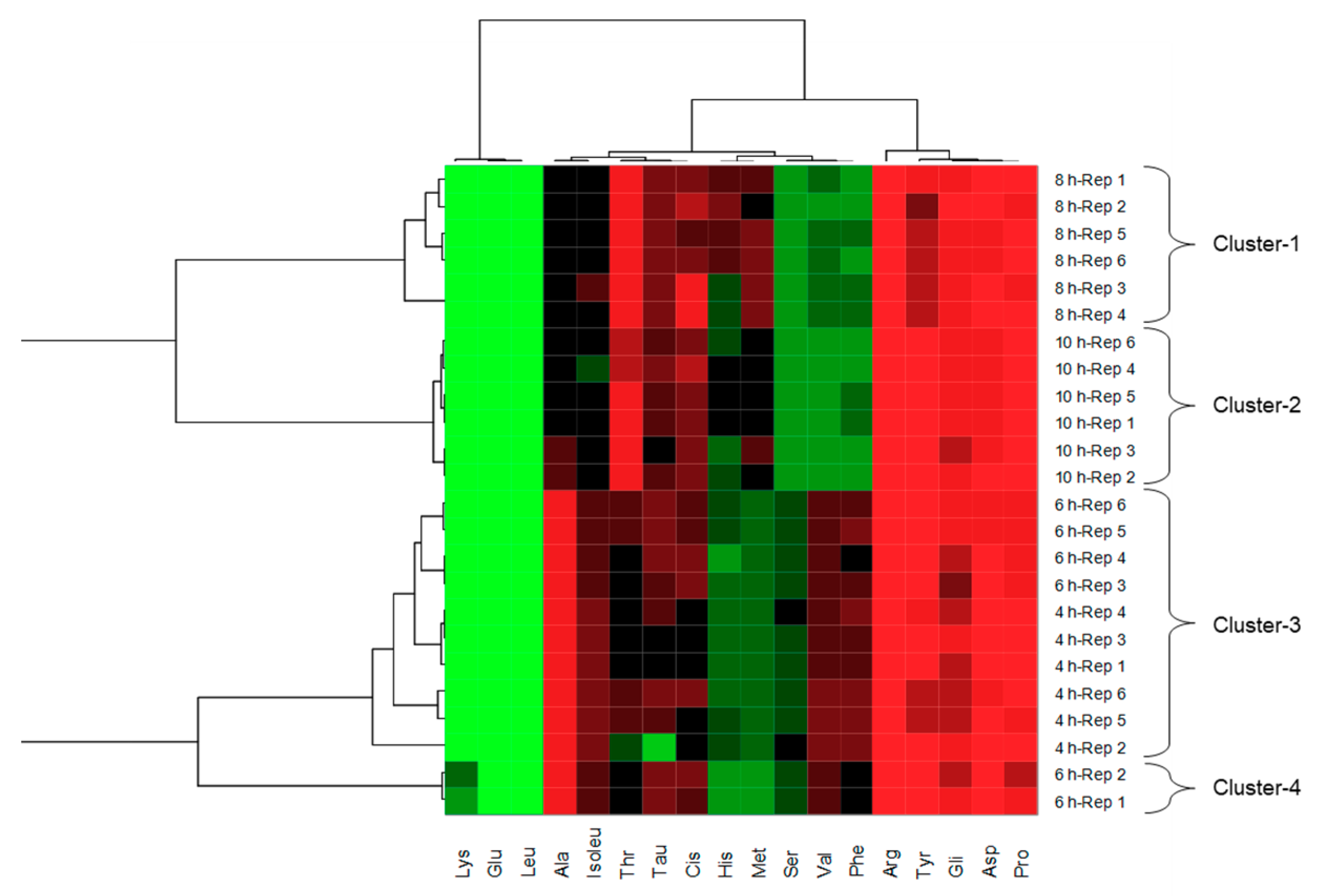
3.1. Molecular Weight Profile and Amino Acid Profile of Pork Liver Hydrolysates

The hydrolysis of the porcine liver was analyzed by sodium dodecyl sulfate polyacrylamide gel electrophoresis (SDS-PAGE). The protein profile of the control and the four Flavourzyme treatments at different times (4, 6, 8, and 10 h) are displayed in Figure 1. Porcine liver protein used as control was separated into seven bands at 100, 76, 64, 54, 49, 41, and 23 kDa. After the treatment with Flavourzyme, most of the protein bands disappeared, and only, four weak bands (68, 60, 54, 41 kDa) remained to confirm the enzymatic reaction of pork liver. However, there were no significant differences among the hydrolysis times. Several authors hypothesized that Flavourzyme contains a mixture of endo- and exo-peptidases with more capacity to hydrolyze peptide bonds than other enzymes which contain only one type of peptidase [34], although red blood fractions from different species required 24 h for total digestion with Flavourzyme [35]. In another case, proteolysis of Cantonese bacon by Flavourzyme was carried out in 28 h [36]. This suggests us that complete degradation of the pork liver need longer time of enzymatic reaction. The peptides with higher bioactivity are small peptides of approximately 4–16 amino acids (400–2000 Da) [37]. Consequently, the bioactive peptides are not showed in the SDS-PAGE gel, they are smaller degradation products. In these sense, the bioactivity depends on the amino acid sequence and length [38].
The amino acids may also have bioactivity, consequently the free amino acid content is of great importance. The free amino acid content was analyzed in each porcine liver hydrolysate at different hydrolysis time of Flavourzyme (Table 1). In general, hydrolysates revealed that they are rich in glutamic acid, lysine, and leucine. Both lysine and leucine are essential amino acids, hence they cannot be synthesized by the body so they must be supplied from the diet [39]. Total free amino acids significantly increased with the hydrolysis time as expected. The presence of free amino acids within the hydrolysates could increment its antioxidant activity [40]. This suggests that the porcine liver hydrolysates obtained from Flavourzyme at 10 h could present the highest antioxidant capacity. The effect of hydrolysis time significantly affected almost all the analyzed amino acids. The hydrolysates subjected to the longer reaction time of enzyme, generated more amount of free amino acids, excluding, arginine, threonine, tyrosine, and methionine. This fact implies that a longer reaction time of enzyme produced a greater protein degradation releasing peptides and amino acids. It has been reported that hydrophobic amino acids such as aliphatic ones (proline, valine, methionine, isoleucine, and leucine) increase the solubility in the lipid tissue, while aromatic amino acids (phenylalanine and tyrosine) facilitate the scavenging of free radicals [41]. Particularly interesting is the increase of these type of amino acids to enhance the antioxidant activity of the hydrolysates.
To evaluate the amino acid profiles, a heat map cluster analysis was performed as shown in Figure 2. To build the heat map analysis, we use the six replicates of the amino acid profile of the porcine liver hydrolysates generated at different reaction times. The samples were mainly separated into four clusters demonstrating the strong changes in the amino acid profile. The first cluster only contains samples which were hydrolyzed during 8 h, meanwhile, the second cluster only links samples hydrolyzed during 10 h. Nevertheless, the third and fourth clusters display that the amino acid pattern in reaction times of 6 and 4 h did not show differences. It is possible to think that reaction times between 4 and 6 h produce the same pattern of amino acids. Porcine liver hydrolysates generated in reaction times above 6 h were became more differentiated. Optimization of enzymatic hydrolysis is critical for the production of protein hydrolysates at industrial level, and hydrolysis time and enzyme: substrate ratio are usually the most two parameters assessed [42].

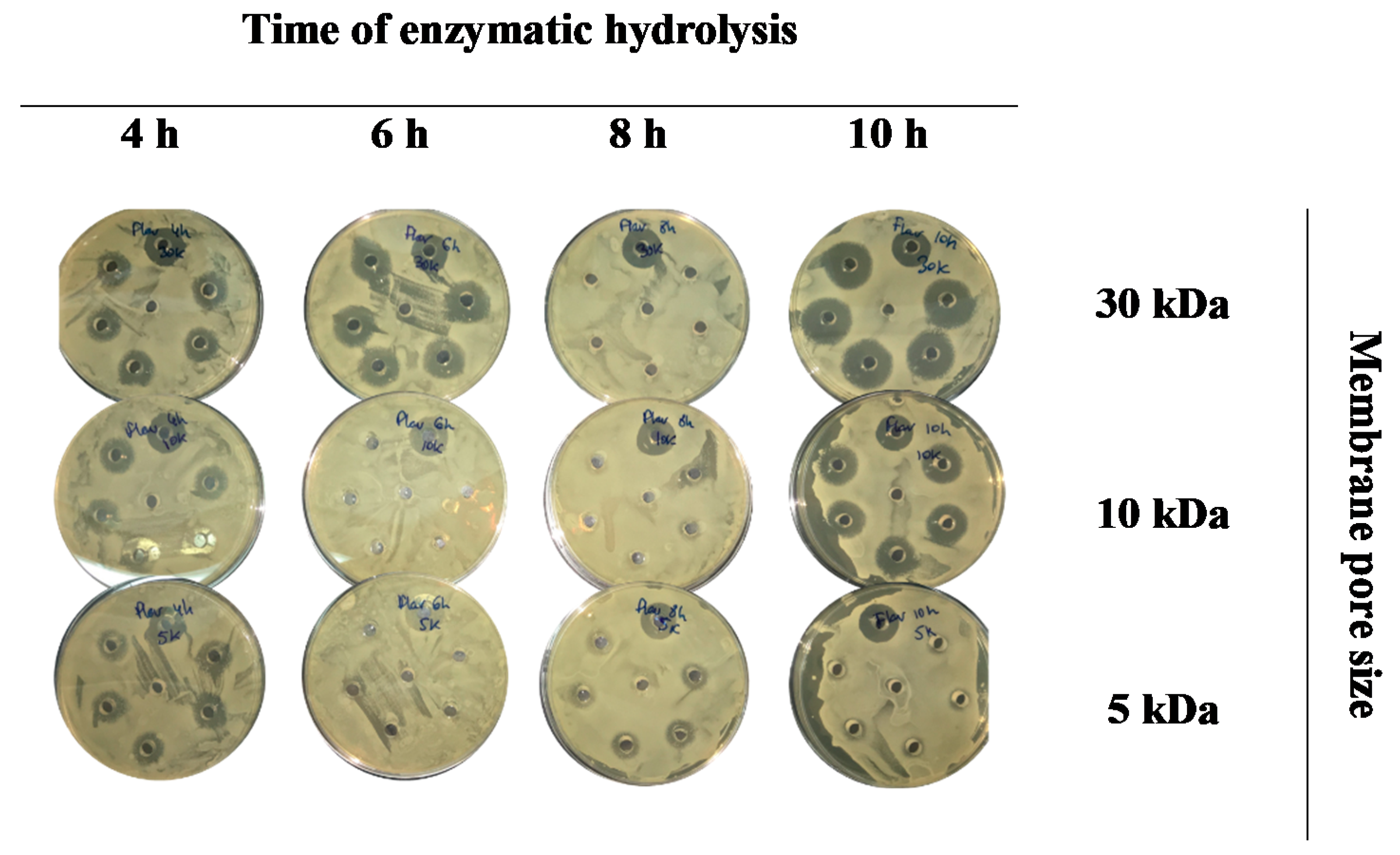
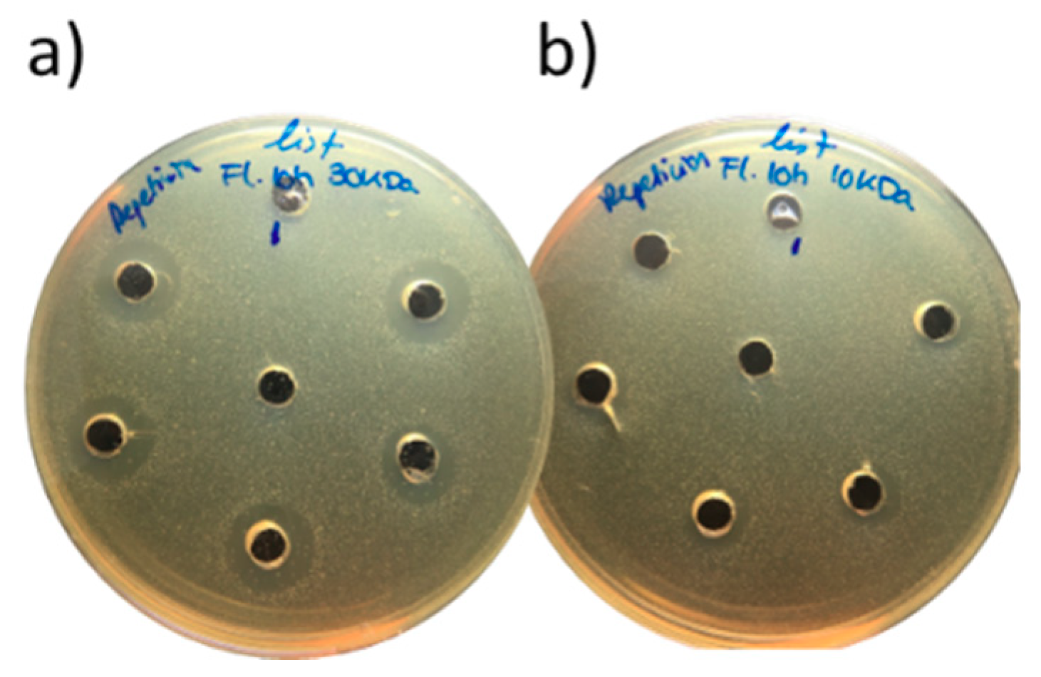
3.2. Antimicrobial Activity of Porcine Liver Hydrolysates

The antimicrobial activity of porcine liver hydrolysates was evaluated by Gram-positive (B. thermosphacta, L. monocytogenes, and S. aureus) and Gram-negative (P. aeruginosa, E. coli, and S. enterica) bacteria. The zone of inhibition (mm) was measured as percentage of inhibition for porcine liver hydrolysates generated by Flavourzyme at different hydrolysis times (4, 6, 8, and 10 h) and filtration through three pore sizes (5, 10, and 30 kDa). The outcomes revealed that most hydrolysates caused growth inhibition of Brochothrix thermosphacta but at different level (Figure 3).
The highest antimicrobial activity was reached by a hydrolysis time of 10 h using filters with a pore size of 30-kDa. The effect of hydrolysis time showed that higher hydrolysis time led to higher inhibition. For instance, in the case of 30 kDa membrane, the inhibition percentage was 83.3% at 4 h and 116.7% at 10 h. Similarly, the same trend was obtained with 10 kDa membrane, where the inhibition percentage was 66.7% at 4 h, and 100% at 10 h. These findings are in accordance with the data reported by Verma et al. [43], which showed the same behavior in the three microorganisms studied (L. monocytogenes, E. coli, S. aureus). On the other hand, the effect of the membrane seemed to indicate that the higher pore size achieved a greater level of inhibition except for hydrolysis time of 8 h. For the study of fresh meat, the Brochothrix strains are one of the most predominant spoilage organisms [44]. Hence, the incorporation of these peptides into foods could be interesting from the antimicrobial safety point of view. Regarding the inhibition of Listeria monocytogenes, none of the extracts could inhibit it except for 30 and 10 kDa hydrolysates fractions obtained by Flavourzyme for 10 h (Figure 4). Both displayed antimicrobial capacity against this strain, reaching the same activity as the nisin standard in the case of 10 kDa and the double in 30 kDa fraction (the percentage inhibition of diameter growth was 100% and 200%, respectively).
On the contrary, none of the porcine liver hydrolysates inhibited the growth of Gram-negative bacteria due to their greater resistance to antimicrobials. This higher resistance is conferred by the existence of an outer membrane in the cell wall structure of the bacteria [45]. This outer cell envelope is a highly hydrophobic asymmetric bilayer, which consists of lipopolysaccharides and phospholipids providing negative charge. This electrostatic charge may provide impermeability against antimicrobial agents, acting as a protecting selective barrier against hydrophobic molecules. Moreover, this bilayer contains porins, which are pore-forming proteins as transport channels [46], allowing the passive penetration of small hydrophilic molecules (<600 Da). Therefore, the permeability of hydrophilic compounds is compromised by size-exclusion [47]. The interactions of antimicrobial peptides with inner and outer membranes could also depend on the structural characteristics of the peptides, such as the total density of cationic charges, length of the peptide sequence, amount of hydrogen bonds donors, and 3-D conformation [48]. Overall, the antimicrobial activity of hydrolysates extracts is influenced by the cationic and hydrophobic character of the peptides, which can interact with the lipopolysaccharides of the bilayer, causing the cell disruption [43].

3.3. Antioxidant Activity of Extracts from Flavourzyme Hydrolysates

ABTS and DPPH scavenging activity was determined for liver hydrolysates generated at different hydrolysis times (4, 6, 8, and 10 h) and filtered with the three pore sizes (5, 10, and 30 kDa) (Table 2). All extracts were significantly affected (p < 0.001) by the membrane pore size employed for collecting the fractions. The ABTS activity increased with pore size, reaching the highest values in the 30 kDa fraction (433.77, 497.26, 474.40, and 426.15 mg ascorbic acid/100 g at 4, 6, 8, and 10 h, respectively). Our results were lower than those reported in poultry viscera with values of 794.6 mM TEAC/mg and 675.5 mM TEAC/mg for fractions below 10 and 3 kDa, respectively [49]. Concerning to the hydrolysis time, porcine liver hydrolysates subjected to ultrafiltration through 5 kDa and 30 kDa showed significant differences. The highest ABTS value was achieved at 6 h with a filtration of 5 kDa and 30 kDa (388.91 and 497.26 mg ascorbic acid/100 g, respectively), hence there is no need to extend the hydrolysis to maximize the ABTS activity.
Another antioxidant assay is the DPPH which is based on the ability of compounds as hydrogen donors or free radical scavengers [50,51]. As ABTS assay, the antioxidant activity was significantly higher in hydrolysates filtered through a pore size of 30 kDa (970.06, 484.00 and 441.67 µg Trolox/g for 4, 6, and 8 h, respectively) except for a hydrolysis time of 10 h. Indeed, the antioxidant activity was not significantly different (p > 0.05) at 10 h of hydrolysis (400.82, 397.85, and 403.05 µg Trolox/g for 5, 10, and 30 kDa, respectively). These outcomes followed the same trend as ABTS assay, as expected. Regarding hydrolysis time, the maximum of antioxidant activity was observed at 6 h for 5 kDa and 4 h for 10 and 30 kDa. Considering ABTS and DPPH assays, it does not seem necessary to reach 8 h to achieve the highest antioxidant capacity. Similar results were observed by Bah et al. [35] in peptide hydrolysates from pig red blood cell fractions using papain and bromelain. Although a slight increase in DPPH values was observed after two hours of hydrolysis, little effect of hydrolysis time was observed over time. In addition, small decreases were also observed at 4 h and 24 h in the case of bromelain and papain, respectively.
The effect was observed in porcine plasma and hemoglobin protein hydrolysates, which lower molecular weight fraction of <3 kDa presented higher values of antioxidant capacity than >10 kDa fraction [52,53]. In this sense, Wang et al. [54] reported that hydrolyzed extracts of duck breasts filtered through (>30 kDa) showed 63% lesser antioxidant activity than corresponding ones (<10 kDa). These authors have separately evaluated the antioxidant capacity with respect to peptide size. In our work, the smallest (<5 kDa) peptides of porcine liver hydrolysates are included within the three types of hydrolysates. Additionally, the other two hydrolysates had higher amount of intermediate peptides. For this reason, it seems logically that the fraction with more amount of peptides including small peptides display a higher antioxidant capacity. Indeed, the antioxidant capacity of pork liver hydrolysates have been evaluated in fractions (<1 kDa, 1–5 kDa, 5–10 kDa, and >10 kDa) resulting lower than the whole fraction [55]. Regarding hydrolysis time of the enzymes such as alcalase, trypsin and papain, it has been demonstrated that the antioxidant capacity of liver hydrolysates increased with the time [43]. Although, our data did not follow the same trend, maybe due to the difference in antioxidant capacity was not too high.
Moreover, the oxygen radical absorbance capacity (ORAC) assay was tested (Table 2). There were significant differences among the hydrolysis times except for hydrolysates filtered through 10 kDa. Nevertheless, the highest antioxidant capacity was not exhibit at longest hydrolysis time of Flavourzyme studied. Regarding the molecular size of peptides, the lowest values were achieved for 10 kDa hydrolysates (21.72, 9.80, and 18.06 mg Trolox/g at 6, 8, and 10 h). Pork liver hydrolysates filtered through 30 kDa displayed the highest value at 6 h (33.71 mg Trolox/g). However, most cases the 30 kDa hydrolysates did not show the highest antioxidant value. This finding was unexpected because of the higher amount of peptide types (whole fraction) should exhibit the highest antioxidant capacity. A possible explanation for these results may be the presence of prooxidant molecules in the larger fraction or compounds could interact with the ORAC method.
The ferric reducing antioxidant power (FRAP) assay was also carried out on porcine liver hydrolysates (Table 2). FRAP values increased with the pore size of membrane, 30 kDa filtrates displayed the highest values (41.81, 48.43, 42.54, and 36. 77 µmol Fe+2/100 g at 4, 6, 8, and 10 h). This finding followed a similar trend as the values of ABTS and DPPH as discussed above. For proteins of plasma, it was found that the FRAP activity of <3 kDa hydrolysate fraction reached more than twice with respect to >10 kDa fraction (1206.4 vs. 557.3 µmol) (Liu et al., 2010). The peptides of each size were evaluated separately in contrast to the results of the present study. For long hydrolysis times, the antioxidant activity was significantly lower (33.19, 36.03, and 36.77 µmol Fe+2/100 g for 5, 10, and 30 kDa, respectively). Although, the behavior was not the same in all the samples, since the highest values were not found for the same hydrolysis time. In 30 kDa filtrates, 6 h of hydrolysis were enough to obtain the best results, while only 4 h were necessary for 10 kDa filtrates. Finally, the filtrated obtained with the lower pore size were those than need a higher hydrolysis time.

4. Conclusions

Based on our results, it could be concluded that enzymatic hydrolysis of pork liver with Flavourzyme generated a large amount of free amino acids shifting the profile for each hydrolysis time studied. The aliphatic and aromatic amino acids, as well as small peptides, caused an increase in antimicrobial and antioxidant activity of pork liver hydrolysates. The highest antimicrobial and antioxidant activities were often displayed in the fractions with peptides at 30 kDa. Peptide mixtures revealed different bioactivities depending on the composition of peptides and amino acids. Consequently, the optimization of enzymatic hydrolysis must consider the specific bioactivity. Moreover, pork liver resulted to be a good source of biopeptides enhancing the swine industry. Further studies in other pork by-products should be performed and new sources of peptides should be thoroughly researched.

Author Contributions

Conceptualization, P.B., D.F., and J.M.L.; writing—original draft preparation, P.B., M.L.-P., and D.F.; writing—review and editing, P.B., M.L.-P., D.F., M.P., and J.M.L. All authors have read and agreed to the published version of the manuscript.

Funding

This research received external funding by grant RTA 2017-00024-CO4-04 from INIA (Instituto Nacional de Investigación y Tecnología Agraria y Alimentaria, Spain).

Acknowledgments

Authors would like to acknowledge to INIA for granting Paula Borrajo with a predoctoral scholarship (grant number CPD2016-0030). José M. Lorenzo is a member of the HealthyMeat network, funded by CYTED (Programa Iberoamericano de Ciencia y Tecnología para el Desarrollo) (ref. 119RT0568). Thanks to GAIN (Axencia Galega de Innovación, Xunta de Galicia, Spain) for supporting this research (grant number IN607A2019/01).

Conflicts of Interest

The authors declare no conflict of interest.

References

  1. Pizzino, G.; Irrera, N.; Cucinotta, M.; Pallio, G.; Mannino, F.; Arcoraci, V.; Squadrito, F.; Altavilla, D.; Bitto, A. Oxidative Stress: Harms and Benefits for Human Health. Oxid. Med. Cell. Longev. 2017, 2017, 8416763. [Google Scholar] [CrossRef] [PubMed]
  2. Ghosh, N.; Das, A.; Chaffee, S.; Roy, S.; Sen, C.K. Reactive Oxygen Species, Oxidative Damage and Cell Death. In Immunity and Inflammation in Health and Disease. Emerging Roles of Nutraceuticals and Functional Foods in Immune Support; Chatterjee, S., Jungraithmayr, W., Bagchi, D., Eds.; Academic Press: London, UK, 2018; pp. 45–55. ISBN 978-0-12-805417-8. [Google Scholar]
  3. Borrajo, P.; Pateiro, M.; Gagaoua, M.; Franco, D.; Zhang, W.; Lorenzo, J.M. Evaluation of the Antioxidant and Antimicrobial Activities of Porcine Liver Protein Hydrolysates Obtained Using Alcalase, Bromelain, and Papain. Appl. Sci. 2020, 10, 2290. [Google Scholar] [CrossRef] [Green Version]
  4. Wang, T.-Y.; Libardo, M.D.J.; Angeles-Boza, A.M.; Pellois, J.-P. Membrane oxidation in cell delivery and cell killing applications. ACS Chem. Biol. 2017, 12, 1170–1182. [Google Scholar] [CrossRef] [Green Version]
  5. Zhao, K.; Zhao, G.-M.; Wu, D.; Soong, Y.; Birk, A.V.; Schiller, P.W.; Szeto, H.H. Cell-permeable peptide antioxidants targeted to inner mitochondrial membrane inhibit mitochondrial swelling, oxidative cell death, and reperfusion injury. J. Biol. Chem. 2004, 279, 34682–34690. [Google Scholar] [CrossRef] [PubMed] [Green Version]
  6. Ott, M.; Gogvadze, V.; Orrenius, S.; Zhivotovsky, B. Mitochondria, oxidative stress and cell death. Apoptosis 2007, 12, 913–922. [Google Scholar] [CrossRef] [PubMed]
  7. Gaschler, M.M.; Stockwell, B.R. Lipid peroxidation in cell death. Biochem. Biophys. Res. Commun. 2017, 482, 419–425. [Google Scholar] [CrossRef] [PubMed]
  8. Patel, R.; Rinker, L.; Peng, J.; Chilian, W.M. Reactive oxygen species: The good and the bad. In Reactive Oxygen Species (ROS) in Living Cells; Filip, C., Albu, E., Eds.; IntechOpen: London, UK, 2018; pp. 7–20. ISBN 978-1-78923-134-2. [Google Scholar]
  9. Kothari, S.; Thompson, A.; Agarwal, A.; du Plessis, S.S. Free radicals: Their beneficial and detrimental effects on sperm function. Indian J. Exp. Biol. 2010, 48, 425–435. [Google Scholar]
  10. Ozougwu, J.C. The role of reactive oxygen species and antioxidants in oxidative stress. Int. J. Res. Pharm. Biosci. 2016, 3, 1–8. [Google Scholar]
  11. Fernandes, R.P.P.; Trindade, M.A.; Lorenzo, J.M.; de Melo, M.P. Assessment of the stability of sheep sausages with the addition of different concentrations of Origanum vulgare extract during storage. Meat Sci. 2018, 137, 244–257. [Google Scholar] [CrossRef]
  12. Lorenzo, J.M.; Batlle, R.; Gómez, M. Extension of the shelf-life of foal meat with two antioxidant active packaging systems. LWT Food Sci. Technol. 2014, 59, 181–188. [Google Scholar] [CrossRef]
  13. Nikmaram, N.; Budaraju, S.; Barba, F.J.; Lorenzo, J.M.; Cox, R.B.; Mallikarjunan, K.; Roohinejad, S. Application of plant extracts to improve the shelf-life, nutritional and health-related properties of ready-to-eat meat products. Meat Sci. 2018, 145, 245–255. [Google Scholar] [CrossRef] [PubMed]
  14. Domínguez, R.; Pateiro, M.; Gagaoua, M.; Barba, F.J.; Zhang, W.; Lorenzo, J.M. A comprehensive review on lipid oxidation in meat and meat products. Antioxidants 2019, 8, 429. [Google Scholar] [CrossRef] [PubMed] [Green Version]
  15. Amaral, A.B.; da Silva, M.V.; da Lannes, S.C.S. Lipid oxidation in meat: Mechanisms and protective factors—A review. Food Sci. Technol. 2018, 38, 1–15. [Google Scholar] [CrossRef] [Green Version]
  16. Ahmed, M.; Pickova, J.; Ahmad, T.; Liaquat, M.; Farid, A.; Jahangir, M. Oxidation of Lipids in Foods. Sarhad J. Agric. 2016, 32, 230–238. [Google Scholar] [CrossRef]
  17. Echegaray, N.; Gómez, B.; Barba, F.J.; Franco, D.; Estévez, M.; Carballo, J.; Marszałek, K.; Lorenzo, J.M. Chestnuts and by-products as source of natural antioxidants in meat and meat products: A review. Trends Food Sci. Technol. 2018, 82, 110–121. [Google Scholar] [CrossRef]
  18. Munekata, P.E.S.; Rocchetti, G.; Pateiro, M.; Lucini, L.; Domínguez, R.; Lorenzo, J.M. Addition of plant extracts to meat and meat products to extend shelf-life and health-promoting attributes: An overview. Curr. Opin. Food Sci. 2020, 31, 81–87. [Google Scholar] [CrossRef]
  19. Zamuz, S.; López-Pedrouso, M.; Barba, F.J.; Lorenzo, J.M.; Domínguez, H.; Franco, D. Application of hull, bur and leaf chestnut extracts on the shelf-life of beef patties stored under MAP: Evaluation of their impact on physicochemical properties, lipid oxidation, antioxidant, and antimicrobial potential. Food Res. Int. 2018, 112, 263–273. [Google Scholar] [CrossRef]
  20. Lorenzo, J.M.M.; Pateiro, M.; Domínguez, R.; Barba, F.J.F.J.; Putnik, P.; Kovačević, D.B.B.D.B.; Shpigelman, A.; Granato, D.; Franco, D. Berries extracts as natural antioxidants in meat products: A review. Food Res. Int. 2018, 106, 1095–1104. [Google Scholar] [CrossRef]
  21. Tkaczewska, J.; Bukowski, M.; Mak, P. Identification of antioxidant peptides in enzymatic hydrolysates of carp (Cyprinus Carpio) skin gelatin. Molecules 2019, 24, 97. [Google Scholar] [CrossRef] [Green Version]
  22. Chakrabarti, S.; Guha, S.; Majumder, K. Food-derived bioactive peptides in human health: Challenges and opportunities. Nutrients 2018, 10, 1738. [Google Scholar] [CrossRef] [Green Version]
  23. Mora, L.; Gallego, M.; Aristoy, M.C.; Reig, M.; Toldrá, F. Bioactive peptides. In Innovative Thermal and Non-Thermal Processing, Bioaccessibility and Bioavailability of Nutrients and Bioactive Compounds; Barba, F.J., Saraiva, J.M.A., Cravotto, G., Lorenzo, J.M., Eds.; Woodhead Publishing: Duxford, UK, 2019; pp. 333–345. ISBN 978-0-12-814174-8. [Google Scholar]
  24. Lorenzo, J.M.; Munekata, P.E.S.; Gómez, B.; Barba, F.J.; Mora, L.; Pérez-Santaescolástica, C.; Toldrá, F. Bioactive peptides as natural antioxidants in food products—A review. Trends Food Sci. Technol. 2018, 79, 136–147. [Google Scholar] [CrossRef]
  25. Pateiro, M.; Borrajo, P.; Campagnol, P.C.B.; Domínguez, R.; Tomasevic, I.; Munekata, P.E.S.; Barba, F.J.; Lorenzo, J.M. Extraction of valuable compounds from meat by-products. In Green Extraction and Valorization of By-Products from Food Processing; Barba, F.J., Roselló-Soto, E., Brncic, M., Lorenzo, J.M., Eds.; CRC Press: Boca Raton, FL, USA, 2019; pp. 55–90. ISBN 9781138544048. [Google Scholar]
  26. Yu, H.C.; Hsu, J.L.; Chang, C.I.; Tan, F.J. Antioxidant properties of porcine liver proteins hydrolyzed using Monascus purpureus. Food Sci. Biotechnol. 2017, 26, 1217–1225. [Google Scholar] [CrossRef] [PubMed]
  27. Fu, Y.; Liu, J.; Hansen, E.T.; Bredie, W.L.P.; Lametsch, R. Structural characteristics of low bitter and high umami protein hydrolysates prepared from bovine muscle and porcine plasma. Food Chem. 2018, 257, 163–171. [Google Scholar] [CrossRef] [PubMed]
  28. Ramírez Aristizabal, L.S.; Marín, D. Evaluación de la actividad antibacteriana de aceites esenciales y extractos etanólicos utilizando métodos de difusión en agar y dilución en pozo. Sci. Tech. 2012, 17, 152–157. [Google Scholar]
  29. Re, R.; Pellegrini, N.; Proteggente, A.; Pannala, A.; Yang, M.; Rice-Evans, C.; Proteggenete, A.; Pannala, A.; Yang, M.; Rice-Evans, C.; et al. Antioxidant activity applying an improved ABTS radical cation decolorization assay. Free Radic. Biol. Med. 1999, 26, 1231–1237. [Google Scholar] [CrossRef]
  30. Huan, D.; Ou, B.; Hampsch-Woodill, M.; Flanagan, J.A.; Prior, R.L. High-throughput assay of oxygen radical absorbance capacity (ORAC) using a multichannel liquid handling system coupled with a microplate fluorescence reader in 96-well format. J. Agric. Food Chem. 2002, 50, 4437–4444. [Google Scholar] [CrossRef]
  31. Brand-Williams, W.; Cuvelier, M.E.; Berset, C. Use of a free radical method to evaluate antioxidant activity. LWT Food Sci. Technol. 1995, 28, 25–30. [Google Scholar] [CrossRef]
  32. Benzie, I.F.F.; Strain, J.J. The Ferric Reducing Ability of Plasma (FRAP) as a Measure of ‘“Antioxidant Power”’: The FRAP Assay Iris. Anal. Biochem. 1996, 239, 70–76. [Google Scholar] [CrossRef] [Green Version]
  33. Lorenzo, J.M.; Cittadini, A.; Bermúdez, R.; Munekata, P.E.; Domínguez, R. Influence of partial replacement of NaCl with KCl, CaCl 2 and MgCl 2 on proteolysis, lipolysis and sensory properties during the manufacture of dry-cured lacón. Food Control 2015, 55, 90–96. [Google Scholar] [CrossRef]
  34. Chang, C.Y.; Wu, K.C.; Chiang, S.H. Antioxidant properties and protein compositions of porcine haemoglobin hydrolysates. Food Chem. 2007, 100, 1537–1543. [Google Scholar] [CrossRef]
  35. Bah, C.S.F.; Carne, A.; McConnell, M.A.; Mros, S.; Bekhit, A.E.D.A. Production of bioactive peptide hydrolysates from deer, sheep, pig and cattle red blood cell fractions using plant and fungal protease preparations. Food Chem. 2016, 202, 458–466. [Google Scholar] [CrossRef] [PubMed]
  36. Zhang, S.; Zhang, C.; Qiao, Y.; Xing, L.; Kang, D.; Khan, I.A.; Huang, M.; Zhou, G. Effect of Flavourzyme on proteolysis, antioxidant activity and sensory qualities of Cantonese bacon. Food Chem. 2017, 237, 779–785. [Google Scholar] [CrossRef] [PubMed]
  37. Liu, R.; Xing, L.; Fu, Q.; Zhou, G.H.; Zhang, W.G. A review of antioxidant peptides derived from meat muscle and by-products. Antioxidants 2016, 5, 32. [Google Scholar] [CrossRef] [PubMed] [Green Version]
  38. Verma, A.K.; Chatli, M.K.; Kumar, P.; Mehta, N. Antioxidant and Antimicrobial Efficacy of Peptidic Hydrolysate Obtained from Porcine Blood. Agric. Res. 2019, 8, 116–124. [Google Scholar] [CrossRef]
  39. Galili, G.; Amir, R.; Fernie, A.R. The Regulation of Essential Amino Acid Synthesis and Accumulation in Plants. Annu. Rev. Plant Biol. 2016, 67, 153–178. [Google Scholar] [CrossRef]
  40. Bah, C.S.F.; Bekhit, A.E.D.A.; Carne, A.; Mcconnell, M.A. Slaughterhouse blood: An emerging source of bioactive compounds. Compr. Rev. Food Sci. Food Saf. 2013, 12, 314–331. [Google Scholar] [CrossRef]
  41. Sun, Y.; Pan, D.; Guo, Y.; Li, J. Purification of chicken breast protein hydrolysate and analysis of its antioxidant activity. Food Chem. Toxicol. 2012, 50, 3397–3404. [Google Scholar] [CrossRef]
  42. de Queiroz, A.L.M.; Bezerra, T.K.A.; de Freitas Pereira, S.; da Silva, M.E.C.; de Almeida Gadelha, C.A.; Gadelha, T.S.; Pacheco, M.T.B.; Madruga, M.S. Functional protein hydrolysate from goat by-products: Optimization and characterization studies. Food Biosci. 2017, 20, 19–27. [Google Scholar] [CrossRef]
  43. Verma, A.K.; Chatli, M.K.; Kumar, P.; Mehta, N. Antioxidant and antimicrobial activity of protein hydrolysate extracted from porcine liver. Indian J. Anim. Sci. 2017, 87, 711–717. [Google Scholar]
  44. Stanborough, T.; Fegan, N.; Powell, S.M.; Tamplin, M.; Chandry, P.S. Insight into the genome of Brochothrix thermosphacta, a problematic meat spoilage bacterium. Appl. Environ. Microbiol. 2017, 83, e02786-16. [Google Scholar] [CrossRef] [Green Version]
  45. Epand, R.M.; Walker, C.; Epand, R.F.; Magarvey, N.A. Molecular mechanisms of membrane targeting antibiotics. Biochim. Biophys. Acta Biomembr. 2016, 1858, 980–987. [Google Scholar] [CrossRef] [PubMed]
  46. Delcour, A.H. Outer membrane permeability and antibiotic resistance. Biochim. Biophys. Acta Proteins Proteom. 2009, 1794, 808–816. [Google Scholar] [CrossRef] [PubMed] [Green Version]
  47. Denyer, S.P.; Maillard, J.-Y. Cellular impermeability and uptake of biocides and antibiotics in Gram-negative bacteria. J. Appl. Microbiol. 2002, 92, 35S–45S. [Google Scholar] [CrossRef] [PubMed]
  48. Li, J.; Koh, J.J.; Liu, S.; Lakshminarayanan, R.; Verma, C.S.; Beuerman, R.W. Membrane active antimicrobial peptides: Translating mechanistic insights to design. Front. Neurosci. 2017, 11, 73. [Google Scholar] [CrossRef] [Green Version]
  49. Jamdar, S.N.; Rajalakshmi, V.; Sharma, A. Antioxidant and ace inhibitory properties of poultry viscera protein hydrolysate and its peptide fractions. J. Food Biochem. 2012, 36, 494–501. [Google Scholar] [CrossRef]
  50. Guija-Poma, E.; Inocente-Camones, M.Á.; Ponce-Pardo, J.; Zarzosa-Norabuena, E. Evaluación de la técnica 2,2-Difenil-1-Picrilhidrazilo (DPPH) para determinar capacidad antioxidante. Horiz. Med. 2015, 15, 57–60. [Google Scholar] [CrossRef] [Green Version]
  51. Sowndhararajan, K.; Kang, S.C. Free radical scavenging activity from different extracts of leaves of Bauhinia vahlii Wight & Arn. Saudi J. Biol. Sci. 2013, 20, 319–325. [Google Scholar]
  52. Liu, Q.; Kong, B.; Xiong, Y.L.; Xia, X. Antioxidant activity and functional properties of porcine plasma protein hydrolysate as influenced by the degree of hydrolysis. Food Chem. 2010, 118, 403–410. [Google Scholar] [CrossRef]
  53. Sun, Q.; Shen, H.; Luo, Y. Antioxidant activity of hydrolysates and peptide fractions derived from porcine hemoglobin. J. Food Sci. Technol. 2011, 48, 53–60. [Google Scholar] [CrossRef] [Green Version]
  54. Wang, L.S.; Huang, J.C.; Chen, Y.L.; Huang, M.; Zhou, G.H. Identification and Characterization of Antioxidant Peptides from Enzymatic Hydrolysates of Duck Meat. J. Agric. Food Chem. 2015, 63, 3437–3444. [Google Scholar] [CrossRef]
  55. Verma, A.K.; Chatli, M.K.; Kumar, P.; Mehta, N. In-vitro assessment of antioxidant and antimicrobial activity of whole porcine-liver hydrolysates and its fractions. Anim. Prod. Sci. 2019, 59, 641–646. [Google Scholar] [CrossRef]
Figure 1. SDS-PAGE of proteins from control and pork liver hydrolysates obtained from Flavourzyme at different times of hydrolysis (4, 6, 8, and 10 h).
Applsci 10 03950 g001
Figure 2. Heat map analysis of amino acid profiles for each porcine liver hydrolysates. Six replicates were generated at different hydrolysis times (4, 6, 8, and 10 h) by Flavourzyme.
Applsci 10 03950 g002
Figure 3. Antimicrobial activity of porcine liver hydrolysates generated by Flavourzyme at different hydrolysis times (4, 6, 8, and 10 h) and filtration through three pore sizes (5 kDa, 10 kDa, and 30 kDa) against Brochothrix thermosphacta.
Applsci 10 03950 g003
Figure 4. Antimicrobial activity of porcine liver hydrolysates using Flavourzyme during 10 h for 30-kDa (a) and 10-kDa (b) against Listeria monocytogenes.
Applsci 10 03950 g004
Table 1. Effect of hydrolysis time on free amino acid profile (mg/100 g of pork liver) of porcine liver hydrolysates.
Hydrolysis TimeSig.
Amino Acid4 h6 h8 h10 h
Aspartic acid118.00 ± 5.83 a158.16 ± 31.35 b211.42 ± 6.42 c246.85 ± 21.22 d***
Serine282.14 ± 17.61 a384.66 ± 75.67 b505.00 ± 15.07 c579.71 ± 52.93 d***
Glutamic acid471.42 ± 19.25 a648.66 ± 131.99 c607.28 ± 15.26 b721.71 ± 70.69 d***
Glycine178.57 ± 19.86 a238.50 ± 28.31 b232.57 ± 21.46 b279.00 ± 42.38 c***
Histidine303.28 ± 24.47 a417.66 ± 48.58 b391.57 ± 34.36 b476.00 ± 63.67 c***
Arginine22.57 ± 7.25 a29.16 ± 8.75 ab36.28 ± 11.20 b25.14 ± 9.37 a*
Threonine267.28 ± 35.57 b341.83 ± 48.61 d252.00 ± 6.27 a301.57 ± 22.21 c***
Alanine165.00 ± 11.47 a219.00 ± 35.92 b398.14 ± 9.68 c435.85 ± 36.97 d***
Proline108.57 ± 19.44 a200.83 ± 18.85 b207.71 ± 12.95 b201.28 ± 20.65 b***
Cysteine261.57 ± 32.17 a301.33 ± 66.45 b303.14 ± 46.82 b355.71 ± 44.61 c***
Tyrosine136.85 ± 29.43 a157.33 ± 18.38 c298.85 ± 25.50 d115.00 ± 14.44 a***
Valine228.00 ± 20.17 a318.66 ± 49.65 b480.57 ± 25.15 b568.17 ± 54.17 c***
Methionine320.57 ± 23.48 a433.83 ± 68.65 c356.74 ± 23.00 a436.71 ± 45.28 c***
Lysine498.85 ± 34.43 a573.50 ± 202.12 b737.28 ± 44.58 c751.71 ± 72.00 c***
Isoleucine224.57 ± 17.17 a308.67 ± 51.22 b397.57 ± 19.07 c468.42 ± 46.56 d***
Leucine477.14 ± 48.02 a658.66 ± 102.68 b706.42 ± 39.21 c825.57 ± 82.36 d***
Phenylalanine232.42 ± 22.70 a326.00 ± 28.94 b491.28 ± 24.10 c550.14 ± 60.03 d***
Total FAA4562.42 ± 322.24 a6010.00 ± 1031.88 b6936.85 ± 208.28 c7751.85 ± 753.67 d***
a–d Mean ± standard deviation values in the same row (corresponding to the same hydrolysis time) not followed by a common letter differ significantly (p < 0.05); Sig.: significance: * (p < 0.05); *** (p < 0.001).
Table 2. Effect of hydrolysis time of Flavourzyme and membrane pore size on antioxidant activity of liver porcine hydrolysates.
Hydrolysis TimePore Size of MembraneSig.
5 kDa10 kDa30 kDa
ABTS (mg Ascorbic Acid/100 g)
4 h337.27 ± 6.39 399.07 ± 3.69 β433.77 ± 2.93 ***
6 h388.91 ± 5.93 407.53 ± 5.15 β497.26 ± 2.93 ***
8 h379.60 ± 8.34 bcα410.92 ± 11.45 β474.40 ± 5.29 ***
10 h366.05 ± 0.85 410.07 ± 8.34 β426.15 ± 2.54 ***
Sig.**ns***
DPPH (µg trolox/g)
4 h395.62 ± 1.29 446.87 ± 4.64 970.06 ± 57.66 ***
6 h414.19 ± 3.93 430.53 ± 1.49 484.00 ± 16.74 **
8 h411.96 ± 1.96 bcα426.07 ± 3.71 441.67 ± 4.87 **
10 h400.82 ± 5.80 ab397.85 ± 1.29 a403.05 ± 2.68 ans
Sig.*******
ORAC (mg trolox/g)
4 h20.93 ± 1.25 a18.40 ± 3.6016.26 ± 3.31 ans
6 h30.48 ± 2.05 21.72 ± 2.24 α33.71 ± 2.39 *
8 h24.74 ± 0.13 abγ9.80 ± 1.51 α15.60 ± 1.58 ***
10 h30.02 ± 2.36 bcβ18.06 ± 3.62 α26.49 ± 1.58 bαβ*
Sig.*ns**
FRAP (µmol Fe+2/100 g)
4 h34.09 ± 0.62 40.97 ± 0.36 41.81 ± 0.58 β***
6 h35.93 ± 0.42 b38.34 ± 0.34 b48.43 ± 5.33ns
8 h36.08 ± 0.55 39.29 ± 0.34 42.54 ± 0.72 γ***
10 h33.19 ± 0.37 36.03 ± 0.68 36.77 ± 0.23 β**
Sig.*****ns
α–γ Mean ± standard deviation values in the same column (corresponding to the same parameter) not followed by a common letter differ significantly (p < 0.05); a–c Mean ± standard deviation in the same row (corresponding to the same parameter) not followed by a common letter differ significantly (p < 0.05); Sig.: significance: * (p < 0.05); ** (p < 0.01), *** (p < 0.001), ns (not significant).

