Review

Rigenera® Autologous Micrografts in Oral Regeneration: Clinical, Histological, and Radiographical Evaluations

1
Department of Life, Health and Environmental Sciences, University of L’Aquila, 67100 L’Aquila, Italy
2
Via R. G. De Ayala 9, 80100 Naples, Italy
3
Human Brain Wave SRL, Corso Galileo Ferraris 6, 10128 Turin, Italy
*
Author to whom correspondence should be addressed.
Appl. Sci. 2020, 10(15), 5084; https://doi.org/10.3390/app10155084
Submission received: 5 July 2020 / Revised: 17 July 2020 / Accepted: 20 July 2020 / Published: 23 July 2020
(This article belongs to the Special Issue Applied Biomaterials in Oral Surgery and Personalized Dentistry)

Abstract

:
Tissue engineering represents a novel approach that aims to exploit the use of biomaterials composed mainly of scaffolds, cells (or grafts), and growth factors capable of restoring a specific tissue. Biomaterials represent the future of dental and oral regeneration due to their biocompatibility and affinity with the receiving site. The aim of this review was to collect results and considerations about a new type of biomaterial based on the use of micrografts in combination with different scaffolds. Micrografts are tissue particles enriched with progenitor cells (PCs), which are defined as descendants of stem cells that can differentiate into specialized cells belonging to the same tissue. PCs in the oral cavity might be extracted from various tissues such as dental pulp, periosteum, or periodontal ligament. Moreover, these cells are easy to isolate through a mechanical process that allows for the filtration of cells with a diameter of 80 μm, in contrast with enzymatic procedures where reagents are used and various culture periods are needed. The aim of this review was to collect data regarding the use of micrografts, developed by a Rigenera® chair-side machine, in oral regeneration evaluating the clinical, histological, and radiographical outcomes. There have been encouraging results for the application of micrografts in bone and periodontal regeneration, but further randomized clinical trials are needed to validate this promising outcome.

1. Introduction

The use of grafting materials in periodontology, implantology, and oral surgery has become very common over the last two decades. New products are brought to market every year with various protocols and uses [1]. Autologous biomaterials still remain the gold standard in oral regeneration due to their osteoinductive, osteoconductive, and osteogenic properties [2]. The use of these materials is limited owing to rapid resorption, collection of inadequate amounts of tissue, high biological cost, and donor site morbidity [3,4]. During recent years, different types of allografts and xenografts have also been proposed due to their biocompatibility and their potential as scaffolds for tissue regeneration [5]. These bone substitutes are limited by the fact that they are not formed from osteogenic cells and osteoinductive molecules, which are important for better tissue regeneration [6,7]. In the literature, several studies have proposed the use of mesenchymal stem cells (MSCs) or progenitor cells (PCs) isolated from various tissues and combined with various biomaterials in oral regeneration [8,9,10]. Specifically, PCs are defined as descendants of stem cells that are able to differentiate into specialized cells belonging to the same tissue. The process from isolation through culture is still at a crucial point. The most commonly used protocol for isolation of cells is the enzymatic process which utilizes the application of different chemical solutions to extract stem cells and requires an incubator and cell storage. Obviously, this process is not suitable in clinical practice where time and handling are crucial for clinical outcomes [11]. A recent protocol proposed by Trovato et al. and later by Monti et al. [12,13], using the Rigenera® micrografting technology, suggests the use of a new type of autologous graft (micrograft) that involves a chair-side mechanical disaggregation device and utilizes PCs derived from various sites such as the periosteum or dental pulp. Such cells can be obtained from a tissue fragment a few millimeters in length that is harvested directly during the intervention phase, even from the same surgical site, and can be used without any manipulation or cell culture. A micrograft might be compared to an autologous graft due to the presence of MSCs that are able to enhance the healing process and regenerate the damaged tissue [14,15]. Considering the limitations of autologous biomaterials, such as their availability and quantity, and furthermore considering the osteoconductivity of xenografts, the use of micrografts leads to a greater regenerative potential. The aim of this review was to summarize the evidence regarding the use of micrografts in oral regeneration considering human studies.

2. Materials and Methods

A search was done on various databases such as PubMed and Scopus, and a manual search was done on relevant journals with the following words: micrografts AND Rigenera AND regeneration. Randomized clinical trials, retrospective studies, case series, and case reports were included in this review. The inclusion criteria were human studies (randomized clinical trials, case series, and case reports) using micrografts during oral regenerative procedures with clinical, histological, and radiographic evaluation (Table 1). Exclusion criteria were not assessed due to the novel technique and scarce literature regarding this novel protocol.

2.1. Population

Males and females between 25 and 69 years of age were included in these studies. Healthy subjects needing implant rehabilitation of the upper jaw with a sinus lift procedure and periodontal patients with periodontitis stage III (probing depth of ≥6 mm) were involved. All patients with systemic diseases were excluded. In each study, patients were subjected to a presurgical phase of oral hygiene, and they were instructed to perform domiciliary oral hygiene in the correct modalities using chlorhexidine associated with a brushing technique until the day of surgery.

2.2. Autologous Micrograft Generation—Rigenera® Micrografting Technology

Depending on the type of intervention, tissue samples (1–2 mm up to 10 mm) were collected from the periosteum of an access flap or from the dental pulp of a third molar extracted simultaneously for mobility or malposition. The samples were all disaggregated using a particular chair-side medical device, the Rigeneracons® (Human Brain Wave LLC, Turin, Italy), which mechanically disaggregates the tissue with a particular micro-blade grid and a filter for cells with a diameter of 80 μm. This device leads to the generation of a micrograft suspension that is ready for use and rich in PCs, thanks to the particular 80 μm filter that enables selective collection of this cellular subtype, extracellular matrix fragments, and growth factors, and facilitates and enhances the regenerative potential of the isolated tissue fragments. Briefly, the tissue sample is inserted into the Rigeneracons® in addition to 1 mL of physiological solution. Following that, the sample is disaggregated at a rotation of 75 R/min and 15 N cm, activating the disruption. After 2 min, the micrografts, suspended in 1 mL of physiological solution, are collected with a syringe through a dedicated hole on the upper part of the Rigeneracons®. The micrograft suspension is stored inside the syringe and used to embed a collagen sponge or hydroxyapatite for 10 min in order to form a biocomplex used in periodontal defects, socket preservation, or sinus floor augmentation (Figure 1).

2.3. Surgery and Grafting Procedure

2.3.1. Sinus Lift

In the three studies [16,17,18] included for the augmentation of the sinus floor, a simple flap and a lateral bone window were used; after membrane elevation, in two studies, a periosteal sample was collected, whereas in a single study by Brunelli et al. [17], the pulp of a third molar extracted for malposition was used. From the tissue samples, micrografts were generated and used as adjuncts to hydroxyapatite (Alos®, Allmed srl, Lissone, Italy) or collagen sponge (Gingistat, GABA, Rome, Italy) in order to form a regenerative complex. According to Baena et al. [16], a control group was assessed, and non-sintered porous hydroxyapatite (Alos®, Allmed srl, Lissone, Italy) or bovine bone (Bio-oss®, Geistlich Biomaterials, Wolhusen, Switzerland) was used alone. Before the closure for primary intention of the flap, a collagen membrane (Bio-Gide®, Geistlich Biomaterials, Wolhusen, Switzerland) was used in the test and control; thus, the flap was repositioned with a single suture line.

2.3.2. Periodontal Regeneration

Ferrarotti et al. [19] showed the use of micrografts combined with a minimally-invasive surgical procedure (MIST), and in association with a collagen sponge (Condress®, Istituto Gentili, Milano, Italy), the elevation of the flap was kept at a minimum to ensure cloth stability and to facilitate the regenerative procedure in the test and control sites. Aimetti et al. [20] showed the same surgical procedures with the minimally invasive flap and the use of pulp progenitor cells combined with a collagen sponge (Condress®, Istituto Gentili, Milan, Italy). The molars from which the pulp samples were extracted were washed with chlorhexidine at 0.2% (CHX) for 60 s, and after crown separation, pulp was collected and used to generate micrografts using the Rigeneracons®. All the defects were treated with scaling and root planing before the insertion of the sponge, and after the placement and complete filling of the infrabony defect, primary wound closure was achieved with horizontal interrupted mattress sutures (Gore-Tex®, WL Gore & Associates Inc., Newark, DE, USA). Graziano et al. [21] in their case report used a sample of periodontal ligament from an extracted third molar after a washing period of 60 s with CHX at 0.2%. Scaling and root planing were achieved by the use of manual Gracey curettes and ultrasonic instruments in the test and control groups; thus, all the inflammatory tissue was removed. Collagen sponge (Gingistat®, GABA, Rome, Italy) was used in combination with micrografts or alone in the control site.

2.3.3. Socket Preservation

In the studies by D’Aquino et al. in 2009 [22] and 2016 [23], respectively, an impacted third molar was used to extract pulp tissue, or a periosteal sample was collected from the inner layer of an access flap. When simple extraction was not possible in one go, a crown and root separation was performed. The socket obtained by the extraction of the third molar was then filled with a collagen sponge (Gingistat®, GABA, Rome, RM, Italy) embedded with progenitor cells. Primary closure was achieved by an interrupted suture.

2.4. Qualitative Analysis

Clinical, histological, and radiographic evaluation was performed with dissimilar follow-up periods according to the procedures of each study (Table 2). The clinical parameters were assessed during follow-ups at 1 week and 3, 6, and 12 months regarding periodontal regeneration with the use of a periodontal probe (PCP 15/11.5, Hu-Friedy, Chicago, IL, USA). The parameters evaluated were the presence or absence of plaque (PI), presence or absence of bleeding on probing (BoP) [24], periodontal depth (PD), recession (REC), and clinical attachment level (CAL) [25].
Histological evaluations were made after 40, 60, 90, and 120 days for socket preservation, whereas for sinus lift, the histological analysis was performed 4 months after the surgery. For periodontal regeneration, no histological data were found. Radiographic analysis was performed in all the studies with a periapical X-ray or a cone-beam. Follow-up was at 4 months for sinus floor augmentation; 3, 6, and 12 months for periodontal procedures; and 45 or 90 days for socket preservation after dental implant insertion.

3. Results

The micrografts in all the studies were combined with a scaffold such as collagen sponge or hydroxyapatite. In the control group, a collagen sponge or hydroxyapatite alone was used (Table 3). Sinus augmentation with micrografts was described in a retrospective study [16] and in two case reports [17,18], whereas periodontal regeneration was assessed in a randomized clinical trial [19], a case series [20], and a case report [21]. Furthermore, two case series of socket preservation with test and control were published by d’Aquino et al. in 2009 [22] and 2016 [23]. Results regarding periodontal regeneration in the RCT [19] were extracted, and various types of non-containing defects (20% incisive, 13.4% canine, 26.7% premolar, and 40% molar) were treated with micrografts combined with a collagen sponge associated with a minimally invasive flap. Statistically significant results were found in the micrograft group for PD and CAL (p < 0.001), with mean reductions of 4.9 ± 1.4 mm and 4.5 ± 1.9 mm, respectively, during a 12-month period. A statistically non-significant increase in REC was found (0.4 ± 1.1 mm), whereas in the control group, PD, CAL, and REC were, respectively, 3.4 ± 1.7 mm, 2.9 ± 2.2 mm, and 0.5 ± 0.9 mm. Other interesting data came from a case report [21] where, in a split-mouth approach, micrografts showed a reduction in PD from 12 to 3 mm, in contrast with the control site in which the collagen sponge alone showed a reduction in PD from 11 to 7 mm during a follow-up period of 6 months. The radiographic assessment at 6 months showed greater mineralization of the micrograft site than the control site. Radiological data were extracted from the RCT [19] in which the use of image analysis software (ImageJ, LOCI, University of Wisconsin, NIH, Madison, WI, USA) showed statistically significant differences in intrabony defect depth between the test and control groups. Data were observed at both the 6- (p < 0.001) and 12-month follow-ups (p < 0.001). At 12 months, the mean radiographic bone fill levels were 3.9 ± 1.5 mm and 1.6 ± 1.1 mm in the test and control groups, respectively. Regarding sinus lift, in a retrospective study [18], radiological follow-up at 4 months showed greater mineralization in the group treated with a micrograft combined with hydroxyapatite than in the two control groups in which hydroxyapatite or deproteinized bovine bone was used alone. Histological and histomorphometric data showed a statistically significant result, with more vital mineralized tissue (p<0.004) in the group treated with combined micrograft/hydroxyapatite (58.5 ± 2.5% with respect to the control group) than in those treated with hydroxyapatite (20.2 ± 3.1%) or deproteinized bovine bone alone (48 ± 2.5%), and more non-mineralized tissue (p < 0.003) in the group treated with combined micrograft/hydroxyapatite (41.4 ± 5.6%) than in those treated with hydroxyapatite (5.5 ± 1.6%) or deproteinized bovine bone alone (20.5 ± 3.1%). Results for socket preservation come from a multicentric study [23] where samples were collected 45, 60, 90, and 120 days after treatment. Evaluation of the samples treated with periosteum-derived micrografts and collagen alone showed that ossification was much faster in the test group than the control group. In particular, at 45 days, the micrograft group already exhibited bone formation, while the presence of inflammatory cells was evident in the control group; moreover, an increase of mineralized matrix at 60, 90, and 120 days was found, in contrast to the control group where the organic matrix was more evident and no bone formation appeared.

4. Discussion

The entire world of regeneration has in the last few years been completely directed by the use of biomaterials composed of various scaffolds and grafts with different origins, sizes, and generation technologies [6,26,27]. MSCs derived from human dental pulp or periosteum have been used for periodontal regeneration, bone regeneration of atrophic maxilla, and alveolar socket preservation [28]. In the literature, there are several studies showing interesting results for the use of micrografts in tissue regeneration, mainly in dermatology [15,29], orthopedic reconstruction [30,31], and cardiac surgery involving the regeneration of ischemic myocardium [32]. Studies that show the potential of this new biomaterial in oral regeneration are not numerous [16,17,18,19,21,22,23,33]. However, positive results were extracted from eight articles in which micrografts, combined with various scaffolds, were utilized for sinus augmentation, periodontal regeneration, and socket preservation. Micrografts are tissue particles composed of PCs, fragments of extracellular matrix, and growth factors extracted by a mechanical method from various tissues. Given their ease of handling, they could be directly injected into a recipient site or used along with a scaffold such as a collagen sponge or bone grafts to maintain space and lead cells to move forward toward damaged tissue. The mechanical extraction method is an alternative way of isolating stem cells (Figure 2).
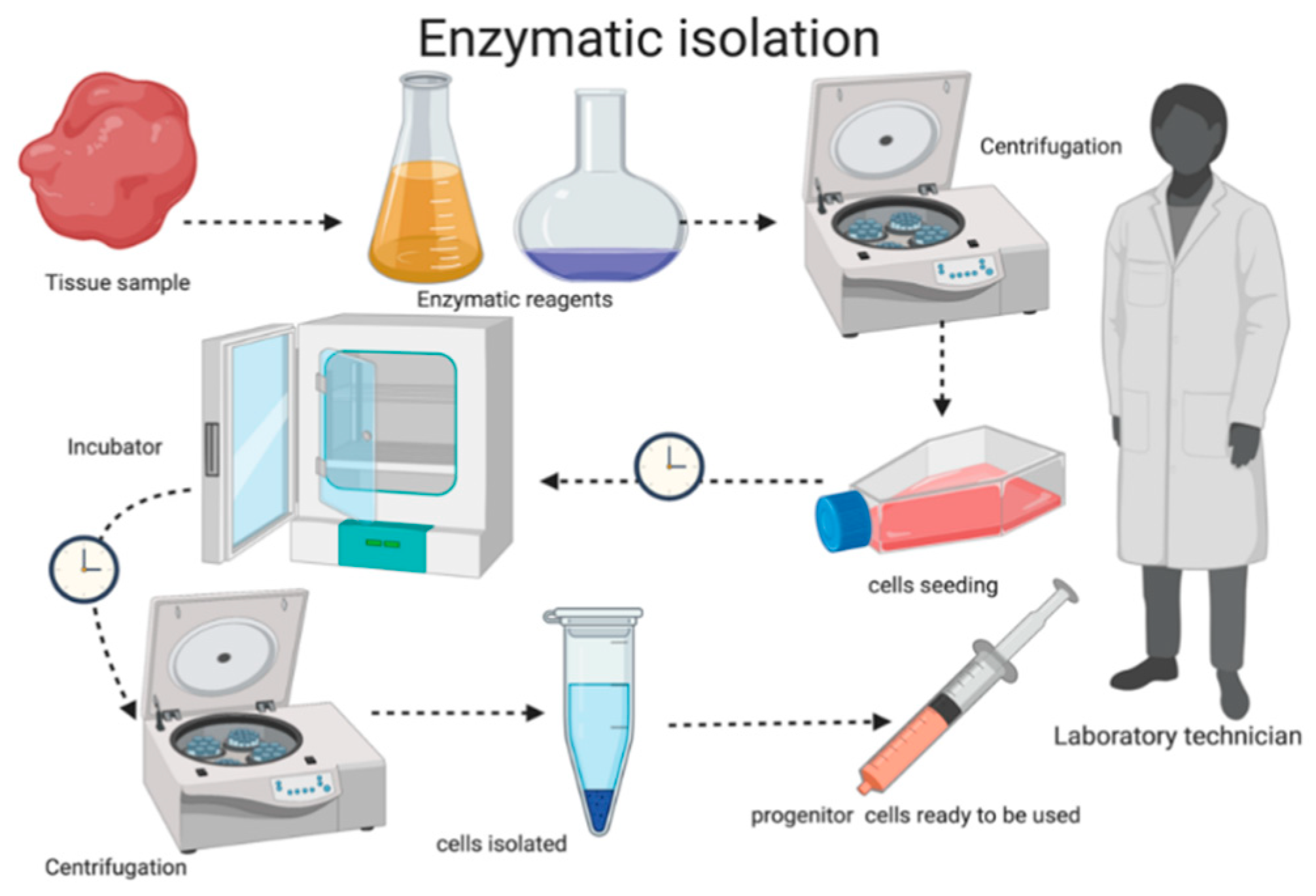
Indeed, the enzymatic technique introduced two decades ago [34] showed incredible results in vitro, but enzymatic digestion with the use of trypsin and collagenase requires a long culture time and various steps of washing and centrifugation procedures. Furthermore, all these processes require a laboratory where cells could be stored and cultured. Other studies have showed a combination of enzymatic and mechanical extraction that demonstrates excellent results due to the application of a low dose of enzymatic mixture and a 70/40 μm mechanical filter [35]; this procedure also requires the use of a medium and a culture period. The differences between the enzymatic and mechanical processes are the application of chemical reagents, the presence of an expert in cell isolation, and a dedicated laboratory (Figure 3).
By using the Rigenera® technology, all the characteristics of MSCs are preserved, ensuring viability and proliferation, which are not always guaranteed in an enzymatic process. Another aspect is that with the Rigeneracons®, the donor and acceptor sites are the same, avoiding the possible complications associated with non-autologous micrografts [13]. Moreover, the Rigeneracons® system is closed, thus avoiding contamination and unpredictability of the cell material. PCs are considered to be adult stem cells due to their origin, unlike embryonal or perinatal stem cells which are not easy to isolate or to harvest. Furthermore, to our knowledge, the behaviors of various types of these stem cells in oral regeneration, and how they might enhance the regenerative process, are not yet clear. On the other hand, it is clear that progenitor cells or MSCs have a limited life span, leading to a short period of regeneration when they are transplanted [36,37]. The future of oral regeneration might be in the use of MSCs/PCs associated with a scaffold. Furthermore, according to the literature, there are reservoirs of PCs in the oral cavity that are able to differentiate in various directions depending on the tissue to be repaired [38,39,40]. The use of micrografts enriched with PCs shows interesting and encouraging results, but further clinical studies are needed to compare this technique with traditional biomaterials.

Author Contributions

Conceptualization, L.M. and E.M.; methodology, L.M.; software, R.D.; validation, E.M., S.M. and V.Q.; formal analysis, L.M.; investigation, L.M.; resources, R.D.; data curation, V.Q.; writing—original draft preparation, L.M.; writing—review and editing, E.M.; visualization, S.M.; supervision, G.M.; project administration, E.M. and G.M. All authors have read and agreed to the published version of the manuscript.

Funding

This research received no external funding.

Conflicts of Interest

The author R.D. is a member of Human Brain Wave, the company that developed the Rigeneracons® medical device. The other authors declare that the study was conducted in the absence of any commercial or financial relationships that could be construed as a potential conflict of interest.

References

  1. Grassia, V.; Nucci, L. New Materials in Oral Surgery. Materials 2020, 13, 1034. [Google Scholar] [CrossRef] [PubMed] [Green Version]
  2. Zhang, Q.; Wu, W.; Qian, C.; Xiao, W.; Zhu, H.; Guo, J.; Meng, Z.; Zhu, J.; Ge, Z.; Cui, W. Advanced biomaterials for repairing and reconstruction of mandibular defects. Mater. Sci. Eng. C 2019, 103, 109858. [Google Scholar] [CrossRef] [PubMed]
  3. Dimitriou, R.; Mataliotakis, G.I.; Angoules, A.G.; Kanakaris, N.K.; Giannoudis, P.V. Complications following autologous bone graft harvesting from the iliac crest and using the RIA: A systematic review. Injury 2011, 42, S3–S15. [Google Scholar] [CrossRef]
  4. Nkenke, E.; Weisbach, V.; Winckler, E.; Kessler, P.; Schultze-Mosgau, S.; Wiltfang, J.; Neukam, F.W. Morbidity of harvesting of bone grafts from the iliac crest for preprosthetic augmentation procedures: A prospective study. Int, J. Oral Maxillofac. Surg. 2004, 33, 157–163. [Google Scholar] [CrossRef]
  5. Sanz, M.; Dahlin, C.; Apatzidou, D.; Artzi, Z.; Bozic, D.; Calciolari, E.; De Bruyn, H.; Dommisch, H.; Donos, N.; Eickholz, P.; et al. Biomaterials and regenerative technologies used in bone regeneration in the craniomaxillofacial region: Consensus report of group 2 of the 15th European Workshop on Periodontology on Bone Regeneration. J. Clin. Periodontol. 2019, 46, 82–91. [Google Scholar]
  6. Haugen, H.J.; Lyngstadaas, S.P.; Rossi, F.; Perale, G. Bone grafts: Which is the ideal biomaterial? J. Clin. Periodontol. 2019, 46, 92.S–102.S. [Google Scholar] [CrossRef] [PubMed]
  7. Stevens, M.M. Biomaterials for bone tissue engineering. Mater. Today 2008, 11, 18–25. [Google Scholar] [CrossRef]
  8. Chen, F.M.; Gao, L.N.; Tian, B.M.; Zhang, X.Y.; Zhang, Y.J.; Dong, G.Y.; Lu, H.; Chu, Q.; Xu, J.; Yu, Y.; et al. Treatment of periodontal intrabony defects using autologous periodontal ligament stem cells: A randomized clinical trial. Stem Cell Res. Ther 2016, 7, 33. [Google Scholar] [CrossRef] [Green Version]
  9. Bajestan, M.N.; Rajan, A.; Edwards, S.P.; Aronovich, S.; Cevidanes, L.; Polymeri, A.; Travan, S.; Kaigler, D. Stem cell therapy for reconstruction of alveolar cleft and trauma defects in adults: A randomized controlled, clinical trial. Clin. Implant. Dent. Relat. Res. 2017, 19, 793–801. [Google Scholar] [CrossRef]
  10. Barbier, L.; Ramos, E.; Mendiola, J.; Rodriguez, O.; Santamaria, G.; Santamaria, J.; Arteagoitia, I. Autologous dental pulp mesenchymal stem cells for inferior third molar post-extraction socket healing: A split-mouth randomised clinical trial. Med. Oral Patol. Oral Cir. Bucal. 2018, 23, 469–477. [Google Scholar] [CrossRef]
  11. Cheng, H.; Chen, B.P.; Soleas, I.M.; Ferko, N.C.; Cameron, C.G.; Hinoul, P. Prolonged Operative Duration Increases Risk of Surgical Site Infections: A Systematic Review. Surg. Infect. 2017, 18, 722–735. [Google Scholar] [CrossRef] [PubMed] [Green Version]
  12. Trovato, L.; Monti, M.; Del Fante, C.; Cervio, M.; Lampinen, M.; Ambrosio, L.; Redi, C.A.; Perotti, C.; Kankuri, E.; Ambrosio, G.; et al. A New Medical Device Rigeneracons Allows to Obtain Viable Micro-Grafts From Mechanical Disaggregation of Human Tissues. J. Cell Physiol. 2015, 2 30, 2299–2303. [Google Scholar] [CrossRef]
  13. Monti, M.; Graziano, A.; Rizzo, S.; Perotti, C.; Del Fante, C.; d’Aquino, R.; Redi, C.A.; Rodriguez, Y.; Baena, R. In Vitro and In Vivo Differentiation of Progenitor Stem Cells Obtained After Mechanical Digestion of Human Dental Pulp. J. Cell Physiol. 2017, 232, 548–555. [Google Scholar] [CrossRef] [PubMed]
  14. Giaccone, M.; Brunetti, M.; Camandona, M.; Trovato, L.; Graziano, A. A New Medical Device, Based on Rigenera Protocol, in the Management of Complex Wounds. J. Stem Cells Res. Rev. Rep. 2014, 1, 3–5. [Google Scholar]
  15. Ceccarelli, G.; Graziano, A.; Benedetti, L.; Imbriani, M.; Romano, F.; Ferrarotti, F.; Aimetti, M.; Cusella de Angelis, G.M. Osteogenic Potential of Human Oral-Periosteal Cells (PCs) Isolated From Different Oral Origin: An In Vitro Study. J. Cell Physiol. 2016, 231, 607–612. [Google Scholar] [CrossRef] [PubMed]
  16. Baena, R.R.; D’Aquino, R.; Graziano, A.; Trovato, L.; Aloise, A.C.; Ceccarelli, G.; Cusella, G.; Pelegrine, A.A.; Lupi, S.M. Autologous periosteum-derived micrografts and PLGA/HA enhance the bone formation in sinus lift augmentation. Front. Cell Dev. Biol. 2017, 1–7. [Google Scholar]
  17. Brunelli, G.; Motroni, A.; Graziano, A.; D’Aquino, R.; Zollino, I.; Carinci, F. Sinus lift tissue engineering using autologous pulp micro-grafts: A case report of bone density evaluation. J. Indian Soc. Periodontol. 2013, 17, 644–647. [Google Scholar]
  18. Lupi, S.M.; Rodriguez y Baena, A.; Todaro, C.; Ceccarelli, G.; Rodriguez y Baena, R. Maxillary sinus lift using autologous periosteal micrografts: A new regenerative approach and a case report of a 3-year follow-up. Case Rep. Dent. 2018, 2018, 3023096. [Google Scholar]
  19. Ferrarotti, F.; Romano, F.; Gamba, M.N.; Quirico, A.; Giraudi, M.; Audagna, M.; Aimetti, M. Human intrabony defect regeneration with micrografts containing dental pulp stem cells: A randomized controlled clinical trial. J. Clin. Periodontol. 2018, 45, 841–850. [Google Scholar] [CrossRef]
  20. Aimetti, M.; Ferrarotti, F.; Gamba, M.N.; Giraudi, M.; Romano, F. Regenerative Treatment of Periodontal Intrabony Defects Using Autologous Dental Pulp Stem Cells: A 1-Year Follow-Up Case Series. Int. J. Periodontics Restorative Dent. 2018, 38, 5–58. [Google Scholar] [CrossRef]
  21. Graziano, A.; Carinci, F.; Scolaro, S.; D’Aquino, R. Periodontal tissue generation using autologous dental ligament micro-grafts: Case report with 6 months follow-up. Ann. Oral Maxillofac. Surg. 2013, 1, 20. [Google Scholar] [CrossRef]
  22. d’Aquino, R.; De Rosa, A.; Lanza, V.; Tirino, V.; Laino, L.; Graziano, A.; Desiderio, V.; Laino, G.; Papaccio, G. Human mandible bone defect repair by the grafting of dental pulp stem/progenitor cells and collagen sponge biocomplexes. Eur. Cell Mater. 2009, 18, 75–83. [Google Scholar] [CrossRef] [PubMed]
  23. d’Aquino, R.; Trovato, L.; Graziano, A.; Ceccarelli, G.; Cusella de Angelis, G.; Marangini, A.; Nisio, A.; Galli, M.; Pasi, M.; Finotti, M.; et al. Periosteum-derived micro-grafts for tissue regeneration of human maxillary bone. J. Transl. Sci. 2016, 2, 125–129. [Google Scholar] [CrossRef] [Green Version]
  24. Lang, N.P.; Joss, A.; Orsanic, T.; Gusberti, F.A.; Siegrist, B.E. Bleeding on probing. A predictor for the progression of periodontal disease? J. Clin. Periodontol. 1986, 13, 590–596. [Google Scholar] [CrossRef] [PubMed]
  25. Lang, N.P.; Bartold, P.M. Periodontal health. J. Clin. Periodontol. 2018, 45, S9–S16. [Google Scholar] [CrossRef] [Green Version]
  26. Isola, G.; Polizzi, A.; Iorio-Siciliano, V.; Alibrandi, A.; Ramaglia, L.; Leonardi, R. Effectiveness of a nutraceutical agent in the non-surgical periodontal therapy: A randomized, controlled clinical trial. Clin. Oral Investig. 2020. [Google Scholar] [CrossRef]
  27. Lo Giudice, A.; Quinzi, V.; Ronsivalle, V.; Farronato, M.; Nicotra, C.; Indelicato, F.; Isola, G. Evaluation of Imaging Software Accuracy for 3-Dimensional Analysis of the Mandibular Condyle. A Comparative Study Using a Surface-to-Surface Matching Technique. Int. J. Environ. Res. Public Health 2020, 17, E4789. [Google Scholar] [CrossRef]
  28. Shanbhag, S.; Suliman, S.; Pandis, N.; Stavropoulos, A.; Sanz, M.; Mustafa, K. Cell therapy for orofacial bone regeneration: A systematic review and meta-analysis. J. Clin. Periodontol. 2019, 46, 162–182. [Google Scholar] [CrossRef] [Green Version]
  29. Svolacchia, F.; De Francesco, F.; Trovato, L.; Graziano, A.; Ferraro, G.A. An innovative regenerative treatment of scars with dermal micrografts. J. Cosmet. Derm.. 2016, 15, 245–253. [Google Scholar] [CrossRef]
  30. Ferretti, C. Periosteum derived stem cells for regenerative medicine proposals: Boosting current knowledge. World J. Stem Cells 2014, 6, 266. [Google Scholar] [CrossRef]
  31. Viganò, M.; Tessaro, I.; Trovato, L.; Colombini, A.; Scala, M.; Magi, A.; Toto, A.; Peretti, G.; de Girolamo, L. Rationale and pre-clinical evidences for the use of autologous cartilage micrografts in cartilage repair. J. Orthop. Surg. Res. 2018, 13. [Google Scholar] [CrossRef] [PubMed] [Green Version]
  32. Nummi, A.; Nieminen, T.; Pätilä, T.; Lampinen, M.; Lehtinen, M.L.; Kivistö, S.; Holmström, M.; Wilkman, E.; Teittinen, K.; Laine, M.; et al. Epicardial delivery of autologous atrial appendage micrografts during coronary artery bypass surgery-safety and feasibility study. Pilot Feasibility Stud. 2017, 3, 1–9. [Google Scholar] [CrossRef] [PubMed] [Green Version]
  33. Trubiani, O.; Marconi, G.D.; Pierdomenico, S.D.; Piattelli, A.; Diomede, F.; Pizzicannella, J. Human Oral Stem Cells, Biomaterials and Extracellular Vesicles: A Promising Tool in Bone Tissue Repair. Int. J. Mol. Sci. 2019, 20, 4987. [Google Scholar] [CrossRef] [PubMed] [Green Version]
  34. Nakahara, H.; Bruder, S.P.; Haynesworth, S.E.; Holecek, J.J.; Baber, M.A.; Goldberg, V.M.; Caplan, A.I. Bone and cartilage formation in diffusion chambers by subcultured cells derived from the periosteum. Bone 1990, 11, 181–188. [Google Scholar] [CrossRef]
  35. Tong, C.K.; Vellasamy, S.; Chong Tan, B.; Abdullah, M.; Vidyadaran, S.; Fong Seow, H.; Ramasamy, R. Generation of mesenchymal stem cell from human umbilical cord tissue using a combination enzymatic and mechanical disassociation method. Cell Biol. Int. 2011, 35, 221–226. [Google Scholar] [CrossRef]
  36. Abdel Meguid, E.; Ke, Y.; Ji, J.; El-Hashash, A.H.K. Stem cells applications in bone and tooth repair and regeneration: New insights, tools, and hopes. J. Cell Physiol. 2018, 233, 1825–1835. [Google Scholar] [CrossRef]
  37. Isola, G.; Giudice, A.L.; Polizzi, A.; Alibrandi, A.; Patini, R.; Ferlito, S. Periodontitis and Tooth Loss Have Negative Systemic Impact on Circulating Progenitor Cell Levels: A Clinical Study. Genes 2019, 10, 1022. [Google Scholar] [CrossRef] [Green Version]
  38. Mao, J.J.; Robey, P.G.; Prockop, D.J. Stem cells in the face: Tooth regeneration and beyond. Cell Stem Cell. 2012, 11, 291–301. [Google Scholar] [CrossRef] [Green Version]
  39. Sanz, A.R.; Carrión, F.S.; Chaparro, A.P. Mesenchymal stem cells from the oral cavity and their potential value in tissue engineering. Periodontol. 2000 2015, 67, 251–267. [Google Scholar] [CrossRef]
  40. Isola, G.; Polizzi, A.; Santonocito, S.; Alibrandi, A.; Ferlito, S. Expression of Salivary and Serum Malondialdehyde and Lipid Profile of Patients with Periodontitis and Coronary Heart Disease. Int. J. Mol. Sci. 2019, 20, 6061. [Google Scholar] [CrossRef] [Green Version]
Figure 1. Schematic representation of Rigenera® technology use. (A) Collection of an autologous sample. (B) Average size of the tissue sample. (C) Mechanical disaggregation with the Rigeneracons® leads to the generation of micrograft suspensions collected from the device with a syringe. (D) The biomaterial is generated by soaking a scaffold with the micrografts.
Applsci 10 05084 g001
Figure 2. Mechanical isolation of mesenchymal stem cells (MSCs) with the use of a micro-blade grid filter.
Applsci 10 05084 g002
Figure 3. Workflow of the enzymatic process with several passages requiring a technician.
Applsci 10 05084 g003
Table 1. Types of studies included in the review.
Authors (Year)Study DesignEffective Population (n. of Patients)n. of Chirurgical SitesStudy Group(s)Control GroupMean Age (Years)n. Malen. FemaleType of InterventIon
Baena et al. 2017 [16]Retrospective study2424Micrografts+hydroxyapatiteDeprotenized bovine bone;hydroxyapatite alone45–641212Sinus lift
Brunelli et al. 2013 [17]Case report11Micrografts+collagen sponge-451-Sinus lift
Lupi et al. 2018 [18]Case report11Micrografts+hydroxyapatite-52-1Sinus lift
Ferrarotti et al. 2018 [19]Randomized clinical trials2929Micrografts+collagen spongeCollagen sponge39–691314Periodontal regeneration
Aimetti et al. 2018 [20]Case series1111Micrografts+collagen sponge-43–5965Periodontal regeneration
Graziano et al. 2013 [21]Case report12Micrografts+collagen spongeCollagen sponge32-1Periodontal regeneration
d’Aquino et al. 2016 [22]Multicentric study3535Micrografts+collagen spongeCollagen sponge25–641421Socket preservation
d’Aquino et al. 2009 [23]Case series714Micrografts+collagen spongeCollagen sponge24–401 Socket preservation
Table 2. Characteristics of each study for type of intervention.
Sinus Lift
Authors (Year)Type of EvaluationFollow-upHistological ParametersRadiographic ParametersClinical ParametersType of TissueSite
Baena et al. 2017 [16]Periapical RX and Histological.4 MonthsVital mineralized tissue(VTM); nonmineralized tissue (NMT); nonvital mineralized tissue (NVMT).Grey scale (Mineralization).-Periosteal sampleInner layer of the flap
Brunelli et al. 2013 [17]CT scans and Periapical RX.4 Months-Hounsfield unit scale and grey scale (Mineralization).-Pulp tissueThird molar
Lupi et al. 2018 [18]CT scans and Histological.4 Months/3 YearsCalcified matrix and organic matrixHounsfield unit scale (Mineralization).-Persiosteal sampleInner layer of the flap
Periodontal Regeneration
Authors (Year)Type of EvaluationFollow-upHistological ParametersRadiographic ParametersClinical ParametersType of TissueSite
Ferrarotti et al. 2018 [19] Clinical and peripical RX.6 months/12 months-Intrabony defct depth(IBD).PI;BoP;PD;CAL; REC.Pulp tissueThird molar
Aimetti et al. 2018 [20]Clinical and peripical RX.1 year-Intrabony defct depth(IBD).PI;BoP;PD;CAL; REC.Pulp tissueThird molar
Graziano et al. 2013 [21]Clinical and CT scans.6 months-Hounsfield unit scale (Mineralization).PI;BoP;PD;CAL; REC.Periodontal ligamentThird molar
Socket Preservation
Authors (Year)Type of EvaluationFollow-upHistological ParametersRadiographic ParametersClinical ParametersType of TissueSite
d’Aquino et al. 2009 [22]Clinical, peripical RX, Histological and Immunofluorescence.3 monthsCalcified matrix, organic matrix and expression of osteonectin and osteocalcin.Grey scale (Mineralization).Horizontal and vertical dimension of the sokcet; PD;CAL.Pulp tissueThird molar
d’Aquino et al. 2016 [23]Clinical, Histological and periapical RX.1 to 4 monthsCalcified matrix and organic matrixGrey scale (Mineralization).Horizontal and vertical dimension of the sokcetPersiosteal sampleInner layer of the flap
BoP, bleeding on probing; PI, plaque index; PD, periodontal depth; CAL, clinical attachment level; CT, computed tomography; REC, gengival recession; RX, periapical radiography; VTM, vital mineralized tissue; NMT, nonmineralized tissue;NVMT, nonvital mineralized tissue; IBD, Intrabony defect depth.
Table 3. Results extracted from test and control sites with follow-up periods of 4 months for sinus lift procedures, 6 and 12 months for periodontal regeneration, and 3 and 4 months for socket preservation.
Sinus Lift
Baena et al. 2017 [16]Micro-grafts groupBone substitutes
HistologicalVMT = 58.5 ± 2.5%; NMT = 41.4 ± 5.6%; NVMT = N/AHidroxiapatite alone VMT = 20.2 ± 3.1%; NMT = 5.5 ± 1.6%; NVMT = N/A; Bovine bone alone VMT = 48 ± 2.5%; NMT = 20.5 ± 3.1%; NVMT = 31.5 ± 2.3%
RadiographicalHigh mineralizationHigh mineralization
Periodontal regeneration
Ferrarotti et al. 2018 [19]Micro-grafts groupCollagen sponge group
ClinicalPD = 8.3 ± 1.2 mm; CAL = 10.0 ± 1.6 mm;PD = 7.9 ± 1.3 mm; CAL = 9.4 ± 1.5 mm;
RadiographicalIBD = 6.4 ± 1.4 mmIBD = 5.6 ± 1.0 mm
Graziano et al. 2013 [21]Micro-grafts groupCollagen sponge group
ClinicalPD reduction from 12 to 3 mmPD reduction from 11 to 7 mm
RadiographicalHigh mineralizationLow mineralization
Socket preservation
d’Aquino et al. 2009 [22]Micro-grafts groupCollagen sponge group
ClinicalCAL = 6.2±2.3 mmCAL = 4.4±1.2 mm
Histologicallamellar architectureimmature Bone
RadiographicalHigh mineralizationLow mineralization
d’Aquino et al. 2016 [23]Micro-grafts groupCollagen sponge group
ClinicalHorizontal reduction = 25% Vertical reduction = 0.60%Horizontal reduction = 30% Vertical reduction = 1.5%
Histologicalcalcified matrixorganic matrix
RadiographicalHigh mineralizationLow mineralization
VMT, vital mineralized tissue; NMT, non-mineralized tissue; NVMT, nonvital mineralized tissue; PD, probing depth; CAL, clinical attachment level; IBD, intrabony defect depth.