Review

Overview of the Cerebellar Function in Anticipatory Postural Adjustments and of the Compensatory Mechanisms Developing in Neural Dysfunctions

Human Physiology Section of the Department of Pathophysiology and Transplantation, Università degli Studi di Milano, 20133 Milan, Italy
*
Author to whom correspondence should be addressed.
Appl. Sci. 2020, 10(15), 5088; https://doi.org/10.3390/app10155088
Submission received: 26 June 2020 / Revised: 21 July 2020 / Accepted: 22 July 2020 / Published: 24 July 2020
(This article belongs to the Special Issue Cerebellar Genetic Diseases: Diagnostic and Monitoring)

Abstract

:
This review aims to highlight the important contribution of the cerebellum in the Anticipatory Postural Adjustments (APAs). These are unconscious muscular activities, accompanying every voluntary movement, which are crucial for optimizing motor performance by contrasting any destabilization of the whole body and of each single segment. Moreover, APAs are deeply involved in initiating the displacement of the center of mass in whole-body reaching movements or when starting gait. Here we present literature that illustrates how the peculiar abilities of the cerebellum i) to predict, and contrast in advance, the upcoming mechanical events; ii) to adapt motor outputs to the mechanical context, and iii) to control the temporal relationship between task-relevant events, are all exploited in the APA control. Moreover, recent papers are discussed which underline the key role of cerebellum ontogenesis in the correct maturation of APAs. Finally, on the basis of a survey of animal and human studies about cortical and subcortical compensatory processes that follow brain lesions, we propose a candidate neural network that could compensate for cerebellar deficits and suggest how to verify such a hypothesis.

1. Introduction

The aim of this review is to highlight the important contribution of the cerebellum in the genesis and control of the Anticipatory Postural Adjustments (APAs). After all, prediction and anticipation of incoming information are two of the most important cerebellar functions, which guarantee that the anticipated actions are properly set up, also taking into account the changes in the environment [1,2].
APAs are unconscious muscular activities aimed to contrast the reaction forces caused by the primary movement, in order to grant whole-body balance, as well as to set up the mechanical context for initiating the displacement of the center of mass in whole-body reaching movements or when starting gait. APAs are associated with movements that involve tiny to large masses and build up one or more fixation chains throughout postural muscles. This biological mechanism has an extreme value not only in the maintenance of upright posture, in balance, and in locomotion, but also in optimizing the performance of each voluntary movement by avoiding any destabilization, both of the whole body and of each single segment. As detailed in Section 3, such chains may spread over different limbs (inter-limb APAs) or also develop within the same limb in which one of the distal segments is voluntarily moved (intra-limb APAs).
For a critical and rich analysis of the cerebellar role in this anticipatory mechanism, it has been necessary to reach a wide overview of this argument, encompassing studies on both inter- and intra-limb APAs, taking also into account literature regarding the contribution of other neural structures.

2. Overview of Cerebellar Functions

From classical literature, the cerebellum is well known to govern movement coordination and motor learning [3,4,5] (for more recent reviews, see [6,7]), but it has been also recognized as involved in cognitive and emotional processing [8,9]. Taking into account the peculiar anatomy of the cerebellum with a parallel repetition of microcircuits [8], its dense connectivity with cerebral cortex, basal ganglia, brainstem, and spinal cord, and that it contains about 50% of the brain neurons [10], it is not inappropriate to think that the cerebellum is involved in so many different processes.
One of the most important cerebellar functions is correlated to ensure that the anticipated actions are correctly in tune with changes in the environment [1,2]. The cerebellum is thought to contain neural representations reproducing the dynamic properties of the body and to exploit them to create sensorimotor predictions; this allows performing accurate motor forecasts linked to environmental stimuli and to body kinematics [11].
This subcortical structure is essential not only in the prediction of incoming information, but also in controlling the temporal relationship between task-relevant events [12]. To grant a correct output in motor coordination, it is necessary to define spatiotemporal sequences of body segment movements. In this regard, the cerebellum is able to operate as an internal “timing machine”, providing a precise temporal representation for motor and nonmotor tasks [13]. Another characteristic function of the cerebellum is sequence learning; in fact, in a work of Shin and Ivry (2003) [14], patients with cerebellar damage did not show learning the spatial and temporal sequences simultaneously presented.
The wide neural network in which the cerebellum communicates with the cerebral cortex, basal ganglia, and limbic system allows not only the control of timing, as well as the predictive and learning function, but also some high-level cognitive and emotional processing, like attention, language, memory, and reasoning [15,16,17]. Consequently, patients with cerebellar lesions can also show cognitive-affective alterations [18].
The most common motor outcome of cerebellar lesions is ataxia (from the Greek word αταξισ, i.e., lack of order). Ataxia is a neuropathological state consisting of lack of movement coordination. This condition is characterized by hypotonia, dysmetria, asynergy, dyschronometria, and dysdiadochokinesia [11,19]. In addition, the cognitive-affective alterations linked to this pathology lead to a lack in the organization of thought, called “dysmetria of thought”, comprising impairment of executive functions (abstract reasoning, working memory, planning) and difficulties with spatial cognition [20]. Nevertheless, the “lack of order” mainly affects the control of balance and gait.
Babinski [5] reported that a cerebellar lesion disrupted the coordination between voluntary movement and equilibrium stabilization, demonstrating the important involvement of the cerebellum in postural organization [21]. Cerebellar patients typically show enhanced postural sway [22], abnormal response to perturbations, reduced control of equilibrium during movements of other body parts, and irregular oscillations of the trunk. Moreover, these patients often suffer from dysfunctional co-contractions, that is, co-activation of muscle pairs with opposing actions of major limb joints, during postural control [23]. Gait ataxia is often described as a “drunken gait” characterized by walking incoordination, variable foot placement, abnormal foot trajectories, a widened base of support, a deviating path of movement, and irregular interjoint coordination patterns [24,25,26]. Indeed, it has been reported that the cerebellum contributes in generating appropriate patterns of limb movements and in modulating their activation duration during locomotion [27]; moreover, since it processes sensory-motor information integrating feedforward and feedback mechanisms [28], it is also able to provide adaptability to the locomotor output through error-feedback learning [29].

3. Overview of Anticipatory Postural Adjustments

Considering the human body as an “articulated chain” in which a combination of anatomical rigid structures work together, we should consider that every anatomical segment is closely linked to others. Therefore, when a segment is voluntarily moved (primary movement), a reaction force occurs which perturbates all adjacent segments. For this reason, each motor action should be associated with a specific anticipatory program which is able to counterbalance the reaction force caused by the prime mover [30,31]. This anticipatory program consists of unconscious muscular activities, called Anticipatory Postural Adjustments (APAs), which create one or more fixation chains spreading over several muscles of the same limb or of different limbs, in order to counteract the perturbating reaction force [32]. When voluntary actions involve large masses, APAs spread over several muscles of different limbs and they are called inter-limb APAs. They aim to stabilize the whole-body postural equilibrium. When voluntary actions concern tiny masses, APAs develop also in the same limb in which one of the distal segments is moved. These latter activities are called intra-limb APAs and they have been shown to optimize movement performance by stabilizing the limb’s proximal segments [33,34]. Therefore, basically, the aim of APAs can be summarized as the stabilization and fine-tuning of the whole-body equilibrium as well as of the local equilibrium of body segments.
The majority of APAs literature has been focused on the inter-limb postural chains which precede voluntary actions such as shoulder flexion and extension or elbow flexion [35,36,37] and also movements involving the lower limbs, hips, and trunk [38,39]. For example, when flexing both arms at the shoulder level, a dorsal muscle postural chain develops, comprising Erector Spinae (ES), Biceps Femoris (BF), and Soleus (SOL), in order to counterbalance the reaction force due to arms movement [40]. In fact, the perturbation induced by this primary movement may dislocate the projection on the ground of the body Center of Mass (CoM) and cause a whole-body equilibrium disturbance [30,31,36]. The maintenance of the body’s dynamic stability needs inter-limb APAs; this is particularly evident during gait initiation since they create the propulsive forces to move the CoM forwards. Gait initiation is the transient period between quiet standing posture and steady-state walking. This is the most used functional task to investigate how the Central Nervous System (CNS) controls equilibrium during a whole-body movement involving modifications in the base of support and CoM progression [41]. During gait initiation in healthy subjects, the Centre of Pressure (CoP, the barycenter of the ground reaction forces) first moves backwards and towards the future swing foot. The onset of such CoP shift is usually defined as APA onset. The horizontal gap created between CoP and CoM produces an “imbalance” torque that drives the CoM forwards and towards the future stance foot (imbalance phase). Subsequently, the CoP moves laterally towards the stance foot (unloading phase), so that the body weight is supported on that side and the swing foot can execute the first step [42]. The CoP shift during APA is associated with a typical electromyographic sequence involving inhibition of the activity in the Soleus (SOL) muscles, tonically active during quiet stance, shortly followed by activation of Tibialis Anterior (TA) muscles, normally silent during quiet stance. In particular, in the stance leg, the SOL inhibition precedes TA excitation by about 100 ms [39].
In parallel, the intra-limb APAs associated with movements that involve little masses optimize the movement performance by preserving the local equilibrium of the limb [43]. Examples of intra-limb APAs are reported for elbow movements [44,45,46], as well as wrist flexions as documented by Aoki et al. in 1991 [47], who reported a pattern of muscular activity in various arm muscles about 50 to 60 ms before the movement. Moreover, Caronni and Cavallari [33] described an anticipatory postural chain developing in several upper-limb muscles, that stabilizes the segmental equilibrium of the arm during index-finger tapping. With the prone hand, a brisk finger flexion was preceded by an excitatory burst in Extensor Carpi Radialis (ECR), Triceps Brachii (TB), and Superior Trapezius (ST), while Flexor Carpi Radialis (FCR), Biceps Brachii (BB), and Anterior Deltoid (AD) showed a concomitant inhibition of their tonic activity. The coupled activities of TB–BB and ST–AD counterbalanced the elbow and shoulder flexion torques produced by the reaction force that the index-finger flexion discharged on the metacarpophalangeal (MP) joint. Interestingly, when the hand posture was changed from prone to supine, the APA pattern reverted in sign in the elbow and shoulder postural muscles but not in ECR and FCR. Therefore, BB and AD showed an excitation pattern, while TB resulted inhibited. These observations demonstrate the ability of APAs to adapt to the mechanical requirements of the postural context.
Despite the different classification, inter- and intra-limb APAs share similar control mechanisms. Indeed, APAs create fixation chains in several muscles to avoid the destabilization produced by the reaction forces. As mentioned above, APAs revert in sign when movement direction is reverted [33,40] and adapt to changes in the postural context and also in movement speed [48,49,50]; moreover, they have an important link with the movement precision [34,51]. Besides the kinematic aspects, inter- and intra-limb APAs also share the same neural structures. The involvement of Primary Motor Cortex (PMC) in both inter- [52] and intra-limb APAs has been studied [33]. For example, the inhibition of PMC through transcranial magnetic stimulation on the left M1 induced a delay of the APA onset in the contralateral Latissimus Dorsi muscle while the subject abducted his left arm [53]. Even the Supplementary Motor Area (SMA) has been correlated to the modulation of APAs. In fact, during a bimanual task, patients suffering unilateral SMA lesion showed impaired inter-limb APAs in the forearm contralateral to the lesion [54]. Furthermore, a study carried out with the transcranial direct current stimulation highlighted the role of SMA in modifying intra-limb APA amplitude associated to the index-finger tapping motor task [55]. Schmitz et al. (2005) [56] reported the involvement of Sensorimotor Areas, using functional Magnetic Resonance Imaging, while Schepens and Drew (2004) [57] reported that the Pontomedullary Reticular Formation, a site of integration of signals from both cortical and subcortical structures, is able to mediate APAs in time and magnitude, to optimize motor control of posture and movement in the cat. Moreover, basal ganglia have been reported to contribute to APA control (Figure 1); in particular, a disruption in intra-limb APAs associated with the index finger flexion [58] and an impairment in inter-limb APAs during a bimanual load-lifting task [59] have been found in patients with Parkinson’s disease.

4. Cerebellum and Anticipatory Postural Control

The contribution of the cerebellum to APA regulation (Figure 1) has been deeply documented. In 2005, Diedrichsen et al. [60] studied APAs associated with the “barman task” in patients with unilateral or bilateral cerebellar damage. In this bimanual movement, subjects supported an object with one hand, while they had to lift the object voluntarily with the other hand (active lifting) or the object was lifted by an experimenter (passive lifting). In healthy subjects, when the object was lifted by the other hand, an inhibitory APA chain was observed, synchronous to the recruitment of the prime mover and preceding the lifting. However, when the object was unexpectedly lifted by an external force (by the experimenter), an impaired balance of the arm occurred, which could only be compensated by a sensory feedback, a postural reflex [61]. Therefore, the APA feedforward command, generated during the active lifting, allowed greater stabilization of balance. On the contrary, patients with cerebellar damage showed poorly timed adjustments, with the APAs beginning earlier than in healthy participants, confirming the essential role played by the cerebellum in timing motor sequences [12,13] and extending it also to the context of the feedforward APA control. Moreover, in this work, it has been reported that cerebellar damage abolished APA “plasticity” since these patients were unable to learn new APA schemes. This evidence suggests the engagement of the cerebellum not only in the predictive but also in the adaptive control of motor timing (Figure 1).
As stated before, inter-limb APAs are evident in gait initiation. Literature showed altered gait initiation in several neurological dysfunctions characterized by poor motor control [62,63] and in particular, in pathologies involving the cerebellum. For example, Timman and Horak (2001) [64] described adults with cerebellar deficits showing reduced force production, length, and velocity of the first step, accompanied by impairments in the use of predictive information to adapt APAs to the mechanical needs of gait initiation. More recently, Richard et al. (2017) [65] tested the involvement of the SMA–pontine–cerebello–thalamo–cortical pathway in gait initiation throughout a technique of repetitive transcranial magnetic stimulation, called continuous theta burst stimulation (cTBS), which is able to consistently reduce motor cortical excitability with long-term depression effects [66]. The cTBS functional inhibition over the cerebellum showed a considerable role of this structure in the muscle coordination, as well as in coupling between the anticipatory postural control and the execution phase during gait initiation.
Literature regarding cerebellar control in intra-limb APA is generally poor. In this regard, it is worth recalling that cerebellar damage altered timing and amplitude scale of muscular activity during arm movements [67,68] and that patients with acute cerebellar lesions did not show a normal intra-limb anticipatory adjustment in unimanual motor tasks, like in grip force when lifting or moving an object [69,70]. In this regard, Bruttini et al. (2015) [71] focused on the involvement of the cerebellum in intra-limb APAs, testing the postural chain that stabilizes the arm during a brisk index-finger flexion [33] in ataxic adult patients. Electromyographyc recordings of postural muscles were analyzed in a group of adult subjects with a slowly progressive adult-onset cerebellar syndrome and then compared with those from a group of age-matched healthy subjects. Results showed that the intra-limb APA pattern associated with the index-finger flexion remained unchanged between the two groups, while a timing disruption of intra-limb APAs occurred in ataxic patients. The delayed APAs described in Bruttini’s work are in line with those found by Yamaura at al. (2013) [72] in transgenic Spinocerebellar ataxic mice. These animals activated hindlimb postural muscles markedly later than neck prime movers, in order to reach and drink from a flask while standing; that is, they showed delayed APAs with respect to the wild-type mice. Actually, delayed APAs during the index-finger flexion task would diverge from the anticipated APAs observed in the bimanual barman task after cerebellar lesion [60]. However, the early APAs in cerebellar subjects might be interpreted as a safety strategy to avoid a violent elbow flexion during the unloading of the hand, but such strategy could be thought of as unnecessary during the index-finger flexion task; consequently, anticipated APAs would have not occurred. From a speculative perspective, it could be argued that when the cerebellum is impaired, so that its “automation” facility is unavailable, APA control should be devolved to other brain areas, maybe at higher levels in the motor control pathway. Should this be true, it is no wonder that APA deficits differ from one motor task to another, because the higher the hierarchical level that takes care of APAs, the more complex and task-specific would be the underlying decision process.

5. Role of Cerebellum in the Ontogenesis of APA Control

Taking into account the rich connections between the cerebellum and several regions of the cerebral cortex involved in motor control, posture, locomotion, and cognitive/emotional processes, aberrant cerebellar growth might have significant consequences on the functional organization of the cerebral cortex [73,74]. In fact, the cerebellum supports the optimization of behavior, especially in procedural learning and skill acquisition. Therefore, an early damage of the cerebellum may induce significant changes in the structure and function of cerebro-cerebellar systems, with long-term effects on motor behavior.
Ontogenesis of posture starts from infants (0–6 months) [75,76,77] and toddlers (6–18 months) [78,79,80]. In particular, the postural activities in sitting and reaching movements have been observed as early as three months, but only from six months do infants develop the ability to adapt postural activity to the specific mechanical situation [81]. Interestingly, three months and six months are the ages in which an increasing functional activity is detected in specific cerebral areas such as the cerebellum, basal ganglia, and frontal cortices [82,83]. The development of APAs starts later (13–14 months) [81], and is related to the development of independent walking [78,80]. During growth, other functional tasks were studied to detect APAs, such as load lifting and release [84,85], upright stance without support [86], rising arms during standing [87], and gait initiation [88,89]. From these works, it emerges that the feedforward control of posture is present but still immature at the age of 4–5 and it approaches that of adults only at 8 years of age [90]. However, the neural network for APA control seems to complete its maturation only after the age of 11 [91].
Despite the importance of the cerebellum during the developmental stages, there is poor literature regarding APAs in children with cerebellar deficit. Lesions of the cerebellum lead to posture and muscle tone disorders. Particularly in early development, cerebellar deficits produce long-term alterations which underlay the potential contribution of this structure also to atypical development [74]. In this context, we analyzed quiet stance and gait initiation parameters in children affected by Pediatric Cerebellar Ataxia (PCA) and compared them to those measured in healthy children [92]. PCAs are a heterogeneous group of cerebellar developmental disorders characterized by dysfunctional motor coordination and very early cerebellar symptoms. Static posturography highlighted alterations in cerebellar children, while the spatial and temporal parameters of APAs during gait initiation were not notably disturbed by the pathology. First step length and velocity were instead different between the groups. From a descriptive analysis of electromyographic recordings, cerebellar patients showed more alterations in the timing distribution of the muscular actions with respect to healthy controls, while the muscular pattern preceding the first step was preserved. The alteration in time delay between SOL inhibition and TA activation, in both stance and swing legs, agreed with literature that assigns to the cerebellum the involvement in feedforward muscle synergies [23,64] and in the timing relationship among motor events [12,93,94,95]. Therefore, this work supports the hypothesis that the cerebellum plays a key role also during human development, in particular, in building up internal models of gait initiation timings. Moreover, considering that the APA pattern was not affected by cerebellar dysfunction, pattern selection is a functional skill which may be attributed to other brain structures, like the basal ganglia [58].

6. Compensatory Strategies in Cerebellar Dysfunctions

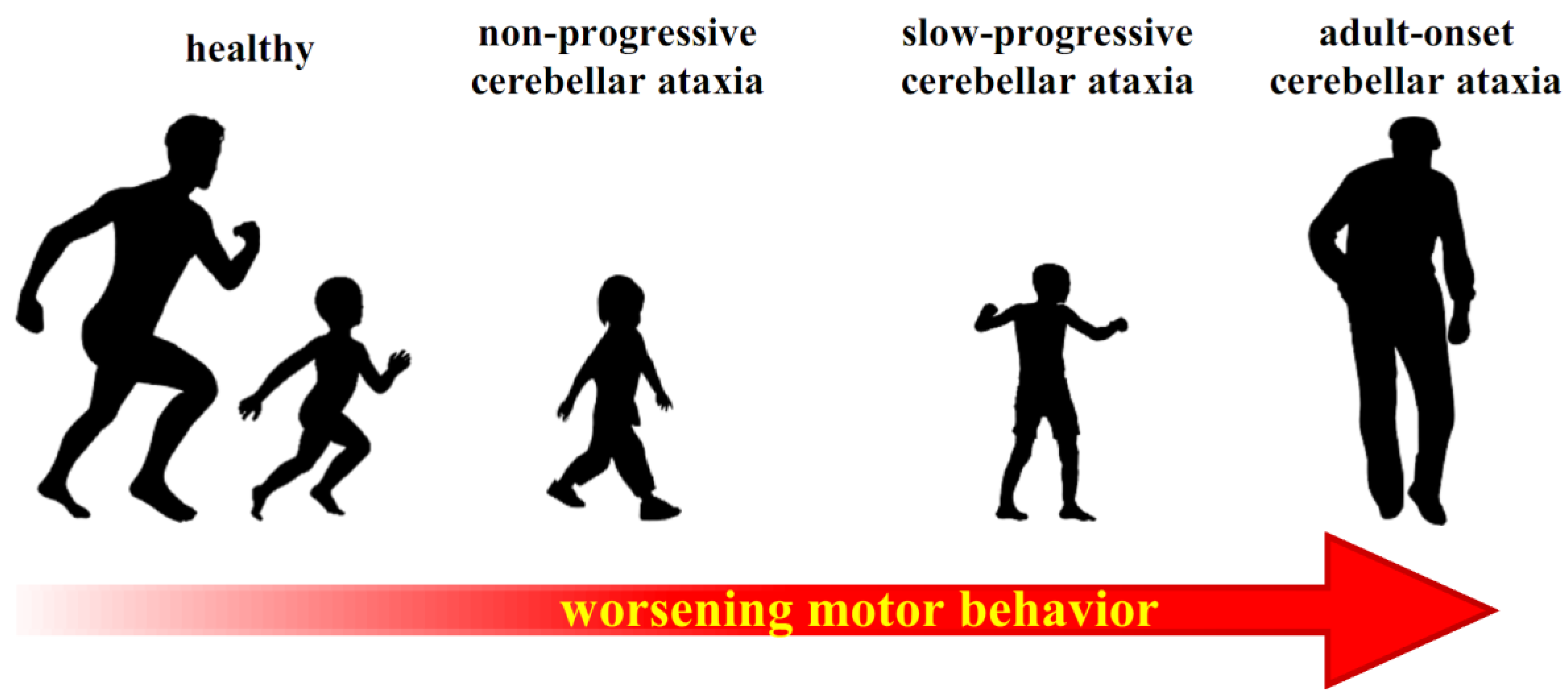
Another considerable issue emerged from our work on PCA. Children who had a slow-progressive course of disease showed a worse postural behavior with respect to both children with non-progressive disease and healthy children, a result which might be due to the different nature of the pathology. In fact, the still intact cerebral areas in children with non-progressive PCA could cope with a stable lesion since embryogenesis by creating new neural pathways. On the contrary, the continuous degeneration in children with slow-progressive PCA might conflict with the consolidation of compensatory functional strategies. In this context, the possibility to exploit neural plasticity in order to overcome the deficits produced by a stable lesion is also supported by the case of a 17-year-old subject suffering cerebellar complete agenesis, who showed only a mild ataxia without apparent difficulty in performing complex motor tasks [96]. It is also interesting to note that patients with adult-onset cerebellar lesions described in Bruttini’s work (2015) [71] showed more pronounced deficits with respect to children with slow-progressive PCA. The gradual worsening of motor control from children with non-progressive ataxia to children with slow-progressive ataxia and to adult ataxic patients (Figure 2) agrees with the observation that the compensation abilities in intact brain areas gradually but consistently decrease over the lifespan [97]. This idea dates back to the early 1940s, as proposed in the seminal papers of Margaret Kennard [98,99], who firstly showed a negative correlation between age and extent of compensation ensuing from motor cortex lesions in the monkey. Plasticity is an argument of increasing interest and considering that the cerebellum is fully involved in motor learning and plasticity, it should be looked into.

6.1. Neural Plasticity and Compensatory Mechanisms

The idea that intact brain areas may reorganize their activity to compensate for other functionally impaired areas (e.g., because of cerebral lesions or degenerations) is not new. For example, back at the end of the last century, several papers by Spear, Tong, et al. [100,101,102] reported that the posteromedial lateral suprasylvian (PMLS) visual cortical area in the cat showed physiological compensation after damage to hierarchically lower visual areas (Brodmann’s 17, 18, and 19), provided that the damage occurred early in the animal’s life. Even if such compensation could not completely overcome the deficits [101], PMLS neurons could develop the properties they would have had in the absence of brain damage. Moreover, tracing methods revealed an increased projection from the retina through the thalamus to the PMLS after lesion [103], showing that neural pathway reorganization plays a role in the compensatory process. Later, Bridge et al. [104] reported such neural pathway reorganization in a blindsight man, who suffered the loss of the left primary visual cortex. In addition to the normal pathways found in healthy subjects, he showed two major features: a pathway from right lateral geniculate nucleus to left MT+ / V5 area and a strong cortico-cortical bilateral connection between MT+ / V5.
Further evidence supporting a compensatory activation of intact brain areas comes from studies of language production and comprehension deficits, following prefrontal stroke [105] and left temporal lobe damage [106]. In both cases, a specific compensatory pathway involving extra-lesion areas was highlighted. Another study recorded event-related brain potentials in patients with a lesion in the perisylvian area of the language-dominant hemisphere that impaired their ability to apply syntax and grammar rules [107]. Results suggested that these patients overcame their syntactic deficit by relying on another, more semantic processing route. A recent study on patients with cognitive dysfunctions following pontine ischemia [108], probably due to damages in the fronto-cerebellar circuits, reported a hyperactivity of frontal areas and suggested that it could compensate for the cognitive impairments.
Another masterpiece of brain compensation regards the recovery of hand motor functions after stroke in the primary motor area (M1). In this condition, multiple structures are called into play, such as the contralateral undamaged M1, the bilateral premotor, and the supplementary and somatosensory areas, as well as the cerebellum and basal ganglia (for a review, see [109]). More recently, a PET study in the monkey [110] showed not only an enhanced activity in the ventral premotor cortex during the early post-recovery period after M1 lesion, but also an increased functional connectivity within the perilesional M1 in the late post-recovery. Another study on macaque [111] provided evidence that fronto-cerebellar circuits may reorganize to sustain functional recovery after M1 lesions. Furthermore, the neural pathway reorganization quoted above has been observed in rats after lesions of the sensorimotor cortices [112]. After neonatal hemidecortication, corticospinal fibers from the intact contralateral sensorimotor cortex send collateral sprouts to the ipsilateral spinal cord, mediating cortical excitation to ipsilateral forelimb, with a different contribution of rostral and caudal forelimb motor areas.
Evidence of compensation in several brain functions has also been reported after complete hemispherectomy, both in children [113] and in adults [114], highlighting the considerable brain ability to reorganize and rewire the cortex in order to cope with such an extensive lesion, especially early in life.
On these premises, as well as on the bases of many other observations, Herbet and Duffau recently published a review [115] forwarding the idea that the traditional “localizationist view”, in which any given function is sustained by a discrete cortical area, isolated from the others, should be abandoned. They indeed propose an alternative “meta-networking” theory, in which brain functions stem from the spatiotemporal integration of many relatively specialized networks, a view that fits not only with the brain ability to learn complex tasks but also with the frequent observation that postlesional reshaping is associated with functional compensation after brain damage.

6.2. A Possible Pathway for Compensating Cerebellar Dysfunction

By applying the above considerations to the cerebellum, it would be interesting to figure out which brain areas might be involved in compensating for cerebellar dysfunction. Even though the cerebellum is engaged in a wide and rich neural network, so that many cortical areas may contribute to the compensation, recent discoveries suggest a new candidate: the basal ganglia network. Classically, the cerebellum and basal ganglia were considered independent systems, which play distinct roles in motor, cognitive, and behavioral control. Actually, there is increasing evidence showing subcortical bidirectional connections between basal ganglia and cerebellum [16] (Figure 3a). In particular, transneuronal transport of the rabies virus in monkeys demonstrated that the subthalamic and the dentate nuclei have disynaptic projections addressed to, respectively, the cerebellar cortex [116] and the striatum [117]. Moreover, it has been recently reported that the pedunculopontine tegmental nucleus (PPTg), which is known to communicate with the basal ganglia, also activates the deep cerebellar nuclei [118]. Therefore, the PPTg could act as an interface between the basal ganglia and cerebellum, involving the latter in motor and cognitive functions [119]. The densely interconnected network among the cerebellum, basal ganglia, and cerebral cortex allows focusing on a different way in which these areas could influence cerebral functions. An abnormal activity at one node could thus spread throughout the entire network and cause dysfunctions at other nodes in the network [120]. In this regard, it has been observed that patients with Parkinson’s disease (PD) show abnormal functioning also in the cerebellum [121,122] and when they perform simple motor tasks, functional MRI highlighted an increase of cerebellar–putamen activity correlated with better motor performance [123] (Figure 3b). This evidence suggests a compensative role played by the cerebellum in the basal ganglia dysfunctions. It has also been observed that this compensatory ability contributes to avoiding the full manifestation of the motor symptoms during the initial stage of PD, but this mechanism saturates with time, leading these patients to develop cerebellar symptoms too [124]. Altogether, these observations allow hypothesizing that intact basal ganglia might compensate for cerebellar deficits, as well as intact cerebellum compensates in patients with basal ganglia impairments (Figure 3c).

7. Conclusions and Future Research

This review aimed to collect information regarding the role of the cerebellum in the control of Anticipatory Postural Adjustments. It also supports the view that the function of the cerebellum is to coordinate, like an “orchestra director”, the timing of muscular events, which follow each other both in the execution of the voluntary action and in the postural activities that accompany it.
Considering the interesting observations carried out on inter-limb APAs in children with PCA (Section 5), it would be interesting to insert the missing piece of this puzzle: to investigate the intra-limb APA control in cerebellar children, in order to gain more information regarding the cerebellar ontogenesis in stabilizing and optimizing motor performance. In this context, it might be possible to test activities of postural muscles that stabilize the arm when the index finger is briskly flexed in children with pediatric cerebellar ataxia. If a timing disruption occurs, we might further confirm the essential function of the cerebellum in controlling the temporal relationships between muscular activities, but also verify its important role during human development. In this way, we could deepen the knowledge on the cerebellar contribution to postural control since human embryogenesis. Moreover, to deepen the knowledge regarding the neural areas, like basal ganglia, possibly involved in the compensatory role in children with cerebellar impairments, it would be interesting to study the connectivity of the brain networks through MRI. Indeed, recent methods of functional and diffusion MRI are able to highlight the involvement of specific areas and to reconstruct the streams among these neural structures.
The study of APAs in patients with cerebellar damage is not only crucial for understanding the essential functions of the cerebellum, but also for clinical monitoring. Indeed, the analysis of APAs during a motor task (from gait initiation to index-finger flexion) may be an additional tool to the established diagnostic methods for a complete clinical evaluation of cerebellar patients.
Finally, a better knowledge of which neural structures actually compensate for the effects of cerebellar dysfunction could be of great help in therapy and rehabilitation, as it could suggest new targets for pharmacological and brain stimulation treatments, as well as for rehabilitation procedures. As a purely speculative example, if basal ganglia actually compensate for cerebellar dysfunction, it would then be important to test whether such compensation may overload and/or be detrimental for basal ganglia over time. If so, a pharmacological or brain stimulation approach similar to that used for PD patients might be envisaged.

Author Contributions

Conceptualization, P.C. and S.M.M.; writing—original draft preparation, S.M.M.; writing—review and editing, S.M.M., R.E., V.F., F.B., P.C.; funding acquisition, P.C. All authors have read and agreed to the published version of the manuscript.

Funding

This study was supported by Fondazione Mariani for Child Neurology.

Conflicts of Interest

The authors declare no conflict of interest.

References

  1. Moberget, T.; Karns, C.M.; Deouell, L.Y.; Lindgren, M.; Robert, T.; Ivry, R.B. Detecting Violations of Sensory Expectancies Following Cerebellar Degeneration: A Mismatch Negativity Study. Neuropsychologia 2009, 46, 2569–2579. [Google Scholar] [CrossRef] [Green Version]
  2. Mauk, M.D.; Medina, J.F.; Nores, W.L.; Ohyama, T. Cerebellar Function: Coordination, Learning or Timing? Curr. Biol. 2000, 10, 522–525. [Google Scholar] [CrossRef] [Green Version]
  3. Luciani, L. Il Cervelletto. Nuovi Studi Di Fisiologia Normale e Patologica. Philos. Rev. 1893, 2, 475–477. [Google Scholar]
  4. Albus, J.S. A Theory of Cerebellar Function. Math. Biosci. 1971, 10, 25–61. [Google Scholar] [CrossRef]
  5. Babinski, J. De l’asynergie Cérebelleuse. Rev. Neurol. 1899, 7, 806–816. [Google Scholar]
  6. Manto, M.; Bower, J.M.; Conforto, A.B.; Molinari, M.; Naito, E.; Nowak, D.A.; Oulad, N.; Taib, B. Consensus Paper: Roles of the Cerebellum in Motor Control—The Diversity of Ideas on Cerebellar Involvement in Movement. The Cerebellum 2012, 11, 457–487. [Google Scholar] [CrossRef]
  7. De Zeeuw, C.I.; Ten Brinke, M.M. Motor Learning and the Cerebellum. Cold Spring Harb. Perspect. Biol. 2015, 7. [Google Scholar] [CrossRef] [Green Version]
  8. D’Angelo, E.; Casali, S. Seeking a Unified Framework for Cerebellar Function and Dysfunction: From Circuit Operations to Cognition. Front. Neural Circuits 2012, 6, 116. [Google Scholar] [CrossRef] [Green Version]
  9. Ito, M. Control of Mental Activities by Internal Models in the Cerebellum. Nat. Rev. Neurosci. 2008, 9, 304–313. [Google Scholar] [CrossRef]
  10. Zagon, I.S.; McLaughlin, P.J.; Smith, S. Neural Populations in the Human Cerebellum: Estimations from Isolated Cell Nuclei. Brain Res. 1977, 127, 279–282. [Google Scholar] [CrossRef]
  11. Manto, M. Chapter 9—Cerebellar Motor Syndrome from Children to the Elderly. In The Cerebellum: From Embryology to Diagnostic Investigations; Elsevier: New York, NY, USA, 2018; Volume 154, pp. 151–166. [Google Scholar] [CrossRef]
  12. Ivry, R.B.; Spencer, R.M.; Zelaznik, H.N.; Diedrichsen, J. The Cerebellum and Event Timing. Ann. N. Y. Acad. Sci. 2002, 978, 302–317. [Google Scholar] [CrossRef] [PubMed]
  13. Bareš, M.; Apps, R.; Avanzino, L.; Breska, A.; D’Angelo, E.; Filip, P.; Gerwig, M.; Ivry, R.B.; Lawrenson, C.L.; Louis, E.D.; et al. Consensus Paper: Decoding the Contributions of the Cerebellum as a Time Machine. From Neurons to Clinical Applications. Cerebellum 2019, 18, 266–286. [Google Scholar] [CrossRef] [PubMed] [Green Version]
  14. Shin, J.C.; Ivry, R.B. Spatial and Temporal Sequence Learning in Patients with Parkinson’s Disease or Spatial and Temporal Sequence Learning in Patients with Parkinson’s Disease or Cerebellar Lesions. J. Cogn. Neurosci. 2003, 15, 1232–1243. [Google Scholar] [CrossRef]
  15. Palesi, F.; De Rinaldis, A.; Castellazzi, G.; Calamante, F.; Muhlert, N.; Chard, D.; Tournier, J.D.; Magenes, G.; D’Angelo, E.; Wheeler-Kingshott, C.A.M.G. Contralateral Cortico-Ponto-Cerebellar Pathways Reconstruction in Humans in Vivo: Implications for Reciprocal Cerebro-Cerebellar Structural Connectivity in Motor and Non-Motor Areas. Sci. Rep. 2017, 7, 1–13. [Google Scholar] [CrossRef]
  16. Bostan, A.C.; Dum, R.P.; Strick, P.L. Cerebellar Networks with the Cerebral Cortex and Basal Ganglia. Trends Cogn. Sci. 2013, 241–254. [Google Scholar] [CrossRef] [Green Version]
  17. Blatt, G.J.; Oblak, A. Cerebellar Connections with Limbic Circuits: Anatomy and Functional Implications. Handb. Cerebellum Cerebellar Disord. 2013, 479–496. [Google Scholar] [CrossRef]
  18. Schmahmann, J.D.; Sherman, J.C. The Cerebellar Cognitive Affective Syndrome. Brain 1998, 121, 561–579. [Google Scholar] [CrossRef]
  19. Mochizuki, H.; Ugawa, Y. Cerebellar Ataxic Gait. Brain Nerve 2010, 62, 1203–1210. [Google Scholar]
  20. Schmahmann, J.D. Disorders of the Cerebellum: Ataxia, Dysmetria of Thought, and the Cerebellar Cognitive Affective Syndrome. J. Neuropsychiatry Clin. Neurosci. 2004, 16, 367–378. [Google Scholar] [CrossRef]
  21. Diener, H.-C.; Dichgans, J.; Guschlbauer, B.; Bacher, M.; Rapp, H.; Klockgether, T. The Coordination of Posture and Voluntary Movement in Patients with Cerebellar Dysfunction. Mov. Disord. 1992, 7, 14–22. [Google Scholar] [CrossRef]
  22. Diener, H.C.; Dichgans, J.; Bacher, M.; Gompf, B. Quantification of Postural Sway in Normals and Patients with Cerebellar Diseases. Electroencephalogr. Clin. Neurophysiol. 1984, 57, 134–142. [Google Scholar] [CrossRef] [Green Version]
  23. Asaka, T.; Wang, Y. Feedforward Postural Muscle Modes and Multi-Mode Coordination in Mild Cerebellar Ataxia. Exp. Brain Res. 2011, 210, 153–163. [Google Scholar] [CrossRef] [PubMed]
  24. Earhart, G.M.; Bastian, A.J. Selection and Coordination of Human Locomotor Forms Following Cerebellar Damage. J. Neurophysiol. 2001, 85, 759–769. [Google Scholar] [CrossRef] [PubMed]
  25. Morton, S.M.; Bastian, A.J. Cerebellar Control of Balance and Locomotion. Neuroscientist 2004, 10, 247–259. [Google Scholar] [CrossRef]
  26. Manto, M. Mechanisms of Human Cerebellar Dysmetria: Experimental Evidence and Current Conceptual Bases. J. Neuroeng. Rehabil. 2009, 18, 1–18. [Google Scholar] [CrossRef]
  27. Martino, G.; Ivanenko, Y.P.; Serrao, M.; Ranavolo, A.; D’Avella, A.; Draicchio, F.; Conte, C.; Casali, C.; Lacquaniti, F. Locomotor Patterns in Cerebellar Ataxia. J. Neurophysiol. 2014, 112, 2810–2821. [Google Scholar] [CrossRef]
  28. Bastian, A.J. Learning to Predict the Future: The Cerebellum Adapts Feedforward Movement Control. Curr. Opin. Neurobiol. 2006, 645–649. [Google Scholar] [CrossRef]
  29. Morton, S.M.; Bastian, A.M.Y.J. Mechanisms of Cerebellar Gait Ataxia. Cerebellum 2007, 18, 79–86. [Google Scholar] [CrossRef]
  30. Bouisset, S.; Do, M. Posture, Dynamic Stability and Voluntary Movement. Posture, Stabilité, Dynamique et Mouvement Volontaire. Neurophysiol Clin. 2008, 38, 345–362. [Google Scholar] [CrossRef]
  31. Bouisset, S.; Zattara, M. A Sequence of Postural Movements Precedes Voluntary Movement. Neurosci. Lett. 1981, 22, 263–270. [Google Scholar] [CrossRef]
  32. Massion, J. Movement, Posture and Equilibrium: Interaction and Coordination. Prog. Neurobiol. 1992, 38, 35–56. [Google Scholar] [CrossRef]
  33. Caronni, A.; Cavallari, P. Anticipatory Postural Adjustments Stabilise the Whole Upper-Limb Prior to a Gentle Index Finger Tap. Exp. Brain Res. 2009, 194, 59–66. [Google Scholar] [CrossRef] [PubMed]
  34. Bruttini, C.; Esposti, R.; Bolzoni, F.; Cavallari, P. Higher Precision in Pointing Movements of the Preferred vs. Non-Preferred Hand Is Associated with an Earlier Occurrence of Anticipatory Postural Adjustments. Front. Hum. Neurosci. 2016, 10, 365. [Google Scholar] [CrossRef] [PubMed] [Green Version]
  35. Belen’kii, V.Y.; Gurfinkel’, V.S.; Pal’tsev, Y.I. Elements of Control of Voluntary Movements. Biophys. Oxf. 1967, 12, 154–161. [Google Scholar]
  36. Bouisset, S.; Zattara, M. Biomechanical Study of the Programming of Anticipatory Postural Adjustments Associated with Voluntary Movement. J. Biomech. 1987, 20, 735–742. [Google Scholar] [CrossRef]
  37. Friedli, W.G.; Hallett, M.; Simon, S.R. Postural Adjustments Associated with Rapid Voluntary Arm Movements. I. Electromyographic Data. J. Neurol. Neurosurg. Psychiatry 1984, 47, 611–622. [Google Scholar] [CrossRef] [Green Version]
  38. Honeine, J.; Schieppati, M.; Crisafulli, O.; Do, M. The Neuro-Mechanical Processes That Underlie Goal-Directed Medio-Lateral APA during Gait Initiation. Front. Hum. Neurosci. 2016, 10, 1–17. [Google Scholar] [CrossRef] [Green Version]
  39. Crenna, P.; Frigo, C. A Motor Programme for the Initiation of Forward-oriented Movements in Humans. J. Physiol. 1991, 437, 635–653. [Google Scholar] [CrossRef]
  40. Aruin, A.S.; Latash, M.L. The Role of Motor Action in Anticipatory Postural Adjustments Studied with Self-Induced and Externally Triggered Perturbations. Exp. Brain Res. 1995, 106, 291–300. [Google Scholar] [CrossRef]
  41. Yiou, E.; Caderby, T. Balance Control during Gait Initiation: State-of-the-Art and Research Perspectives. World J. Orthop. 2017, 8, 815–828. [Google Scholar] [CrossRef]
  42. Crenna, P.; Carpinella, I.; Rabuffetti, M.; Rizzone, M.; Lopiano, L.; Lanotte, M.; Ferrarin, M. Impact of Subthalamic Nucleus Stimulation on the Initiation of Gait in Parkinson’s Disease. Exp. Brain Res. 2006, 172, 519–532. [Google Scholar] [CrossRef] [PubMed]
  43. Cavallari, P.; Bolzoni, F.; Bruttini, C.; Esposti, R. The Organization and Control of Intra-Limb Anticipatory Postural Adjustments and Their Role in Movement Performance. Front. Hum. Neurosci. 2016, 10, 525. [Google Scholar] [CrossRef] [PubMed] [Green Version]
  44. Almeida, G.L.; Hong, D.A.; Corcos, D.; Gottlieb, G.L. Organizing Principles for Voluntary Movement: Extending Single-Joint Rules. J. Neurophysiol. 1995, 74, 1374–1381. [Google Scholar] [CrossRef] [PubMed]
  45. Gribble, P.L.; Ostry, D.J. Compensation for Interaction Torques during Single- and Multijoint Limb Movement. J. Neurophysiol. 1999, 82, 2310–2326. [Google Scholar] [CrossRef] [Green Version]
  46. Hopf, H.C.; Hufschmidt, H.J. Coordination of Adjoining Muscles in Simple Voluntary Movements. Dtsch. Z. Nervenheilkd. 1963, 185, 191–202. [Google Scholar]
  47. Aoki, F. Activity Patterns of Upper Arm Muscles in Relation to Direction of Rapid Wrist Movement in Man. Exp. Brain Res. 1991, 83, 679–682. [Google Scholar] [CrossRef]
  48. Bruttini, C.; Esposti, R.; Bolzoni, F.; Cavallari, P. Ischemic Block of the Forearm Abolishes Finger Movements but Not Their Associated Anticipatory Postural Adjustments. Exp. Brain Res. 2014, 232, 1739–1750. [Google Scholar] [CrossRef]
  49. Esposti, R.; Bruttini, C.; Bolzoni, F.; Cavallari, P. Intended Rather than Actual Movement Velocity Determines the Latency of Anticipatory Postural Adjustments. Exp. Brain Res. 2015, 233, 397–403. [Google Scholar] [CrossRef]
  50. Hall, L.M.; Brauer, S.; Horak, F.; Hodges, P.W. Adaptive Changes in Anticipatory Postural Adjustments with Novel and Familiar Postural Supports. J. Neurophysiol. 2010, 103, 968–976. [Google Scholar] [CrossRef]
  51. Caronni, A.; Bolzoni, F.; Esposti, R.; Bruttini, C.; Cavallari, P. Accuracy of Pointing Movements Relies upon a Specific Tuning between Anticipatory Postural Adjustments and Prime Mover Activation. Acta Physiol. 2013. [Google Scholar] [CrossRef]
  52. Petersen, T.H.; Rosenberg, K.; Petersen, N.C.; Nielsen, J.B. Cortical Involvement in Anticipatory Postural Reactions in Man. Exp. Brain Res. 2009, 193, 161–171. [Google Scholar] [CrossRef] [PubMed]
  53. Palmer, E.; Cafarelli, E.; Ashby, P. The Processing of Human Ballistic Movements Explored by Stimulation over the Cortex. J. Physiol. 1994, 481, 509–520. [Google Scholar] [CrossRef] [PubMed] [Green Version]
  54. Massion, J.; Ioffe, M.; Fran, C.S.; Gantcheva, R. Acquisition of Anticipatory Postural Adjustments in a Bimanual Load-Lifting Task: Normal and Pathological Aspects. Exp. Brain Res. 1999, 128, 229–235. [Google Scholar] [CrossRef] [PubMed]
  55. Bolzoni, F.; Bruttini, C.; Esposti, R.; Castellani, C.; Cavallari, P. Transcranial Direct Current Stimulation of SMA Modulates Anticipatory Postural Adjustments without Affecting the Primary Movement. Behav. Brain Res. 2015, 291, 407–413. [Google Scholar] [CrossRef]
  56. Schmitz, C.; Jenmalm, P.; Westling, G.; Ehrsson, H.H.; Forssberg, H. 8.5 Anticipatory Postural Adjustments in a Bimanualload-Lifting Task: Central Aspects. Gait Posture 2005, 21. [Google Scholar] [CrossRef]
  57. Schepens, B.; Drew, T. Independent and Convergent Signals From the Pontomedullary Reticular Formation Contribute to the Control of Posture and Movement During Reaching in the Cat. J. Neurophysiol. 2004, 92, 2217–2238. [Google Scholar] [CrossRef]
  58. Bolzoni, F.; Esposti, R.; Marchese, S.M.; Pozzi, N.G.; Ramirez-pasos, U.E.; Isaias, I.U.; Cavallari, P. Disrupt of Intra-Limb APA Pattern in Parkinsonian Patients Performing Index-Finger Flexion. Front. Physiol. 2018, 9, 1–8. [Google Scholar] [CrossRef]
  59. Viallet, F.; Massion, J.; Massarino, R.; Khalil, R. Performance of a Bimanual Load-Lifting Task by Parkinsonian Patients. J. Neurol. Neurosurg. Psychiatry 1987, 50, 1274–1283. [Google Scholar] [CrossRef] [Green Version]
  60. Diedrichsen, J.; Verstynen, T.; Lehman, S.L.; Ivry, R.B. Cerebellar Involvement in Anticipating the Consequences of Self-Produced Actions During Bimanual Movements. J Neurophysiol 2005, 93, 801–812. [Google Scholar] [CrossRef] [Green Version]
  61. Hugon, M.; Massion, J.; Wiesendanger, M. Anticipatory Postural Changes Induced by Active Unloading and Comparison with Passive Unloading in Man. Pfliigers Arch. 1982, 393, 292–296. [Google Scholar] [CrossRef]
  62. Isaias, I.U.; Dipaola, M.; Michi, M.; Marzegan, A.; Volkmann, J.; Roidi, M.L.R.; Frigo, C.A.; Cavallari, P. Gait Initiation in Children with Rett Syndrome. PLoS ONE 2014, 9, e92736. [Google Scholar] [CrossRef] [PubMed]
  63. Dipaola, M.; Frigo, C.A.; Cavallari, P.; Iasias, I.U. Alterations of Load Transfer Mechanism during Gait Initiation in Parkinson’s Disease. In Proceedings of the E-Health and Bioengineering Conference, EHB 2017, Sinaia, Romania, 22–24 June 2017; pp. 579–582. [Google Scholar] [CrossRef]
  64. Timmann, D.; Horak, F.B. Perturbed Step Initiation in Cerebellar Subjects: 2. Modification of Anticipatory Postural Adjustments. Exp. Brain Res. 2001, 141, 110–120. [Google Scholar] [CrossRef]
  65. Richard, A.; Van Hamme, A.; Drevelle, X.; Golmard, J.; Meunier, S.; Welter, M. Contribution of the Supplementary Motor Area and the Cerebellum to the Anticipatory Postural Adjustments and Execution Phases of Human Gait Initiation. Neuroscience 2017. [Google Scholar] [CrossRef] [PubMed] [Green Version]
  66. Di Lazzaro, V.; Pilato, F.; Saturno, E.; Oliviero, A.; Dileone, M.; Mazzone, P.; Insola, A.; Tonali, P.A.; Ranieri, F.; Huang, Y.Z.; et al. Theta-Burst Repetitive Transcranial Magnetic Stimulation Suppresses Specific Excitatory Circuits in the Human Motor Cortex. J. Physiol. 2005, 3, 945–950. [Google Scholar] [CrossRef] [PubMed]
  67. Brown, S.H.; Hefter, H.; Mertens, M.; Freund, H. Disturbances in Human Arm Movement Trajectory Due to Mild Cerebellar Dysfunction. J. Neurol. Neurosurg. Psychiatry 1990, 53, 306–313. [Google Scholar] [CrossRef] [Green Version]
  68. Manto, M.; Godaux, E.; Jacquy, J. Cerebellar Hypermetria Is Larger When the Inertial Load Is Artificially Increased. Ann. Neurol. 1994, 35, 45–52. [Google Scholar] [CrossRef]
  69. Müller, F.; Dichgans, J. Impairments of Precision Grip in Two Patients with Acute Unilateral Cerebellar Lesions: A Simple Parametric Test for Clinical Use. Neuropsychologia 1994. [Google Scholar] [CrossRef]
  70. Babin-Ratté, S.; Sirigu, A.; Gilles, M.; Wing, A. Impaired Anticipatory Finger Grip-Force Adjustments in a Case of Cerebellar Degeneration. Exp. Brain Res. 1999. [Google Scholar] [CrossRef]
  71. Bruttini, C.; Esposti, R.; Bolzoni, F.; Vanotti, A.; Mariotti, C.; Cavallari, P. Temporal Disruption of Upper-Limb Anticipatory Postural Adjustments in Cerebellar Ataxic Patients. Exp. Brain Res. 2015, 233, 197–203. [Google Scholar] [CrossRef]
  72. Yamaura, H.; Hirai, H.; Yanagihara, D. Postural Dysfunction in a Transgenic Mouse Model of Spinocerebellar Ataxia Type 3. Neuroscience 2013. [Google Scholar] [CrossRef]
  73. Wang, S.S.; Kloth, A.D.; Badura, A. The Cerebellum, Sensitive Periods, and Autism. Neuron 2014, 83, 518–532. [Google Scholar] [CrossRef] [PubMed] [Green Version]
  74. Stoodley, C.J. The Cerebellum and Neurodevelopmental Disorders. Cerebellum 2017, 15, 34–37. [Google Scholar] [CrossRef] [PubMed]
  75. De Graaf-Peters, V.B.; Bakker, H.; Van Eykern, L.A.; Otten, B.; Hadders-Algra, M. Postural Adjustments and Reaching in 4- and 6-Month-Old Infants: An EMG and Kinematical Study. Exp. Brain Res. 2007, 181, 647–656. [Google Scholar] [CrossRef] [PubMed] [Green Version]
  76. Hedberg, A.; Carlberg, E.B.; Forssberg, H.; Hadders-Algra, M. Development of Postural Adjustments in Sitting Position during the First Half Year of Life. Dev. Med. Child Neurol. 2005, 47, 312–320. [Google Scholar] [CrossRef]
  77. Van der Fits, I.B.; Klip, A.W.; van Eykern, L.A.; Hadders-Algra, M. Postural Adjustments during Spontaneous and Goal-Directed Arm Movements in the First Half Year of Life. Behav. Brain Res. 1999, 106, 75–90. [Google Scholar] [CrossRef]
  78. Barela, J.A.; Jeka, J.J.; Clark, J.E. The Use of Somatosensory Information during the Aquisition of Independent Upright Stance. Infant. Behav. Dev. 1999, 22, 87–102. [Google Scholar] [CrossRef]
  79. Van der Fits, I.B.; Otten, E.; Klip, A.W.; Van Eykern, L.A.; Hadders-Algra, M. The Development of Postural Adjustments during Reaching in 6- to 18-Month-Old Infants. Evidence for Two Transitions. Exp. Brain Res. 1999, 126, 517–528. [Google Scholar] [CrossRef]
  80. Witherington, D.C.; Von Hofsten, C.; Rosander, K.; Robinette, A.; Woollacott, M.H.; Bertenthal, B.I. The Development of Anticipatory Postural Adjustments in Infancy. Infancy 2002, 3, 495–517. [Google Scholar] [CrossRef]
  81. Hadders-Algra, M. Development of Postural Control during the First 18 Months of Life. Neural Plast. 2005, 12, 99–108. [Google Scholar] [CrossRef] [Green Version]
  82. Rubinstein, M.; Denays, R.; Ham, H.R.; Piepsz, A.; VanPachterbeke, T.; Haumont, D.; Noël, P. Functional Imaging of Brain Maturation in Humans Using Iodine-123 Iodoamphetamine and SPECT. J. Nucl. Med. 1989, 30, 1982–1985. [Google Scholar]
  83. Chugani, H.T. A Critical Period of Brain Development: Studies of Cerebral Glucose Utilization with PET. Prev. Med. Baltim. 1998, 27, 184–188. [Google Scholar] [CrossRef] [PubMed] [Green Version]
  84. Jover, M.; Schmitz, C.; Bosdure, E.; Chabrol, B.; Assaiante, C. Anticipatory Postural Adjustments in a Bimanual Load-Lifting Task in Children with Duchenne Muscular Dystrophy. Neurosci. Lett. 2006, 403, 271–275. [Google Scholar] [CrossRef] [PubMed]
  85. Hay, L.; Redon, C. Feedforward versus Feedback Control in Children and Adults Subjected to a Postural Disturbance. Exp. Brain Res. 1999, 125, 153–162. [Google Scholar] [CrossRef] [PubMed]
  86. Haas, G.; Diener, H.C.; Rapp, H.; Dichgans, J. Development of Feedback and Feedforward Control of Upright Stance. Dev. Med. Child Neurol. 1989, 31, 481–488. [Google Scholar] [CrossRef] [PubMed]
  87. Riach, C.L.; Hayes, K.C. Anticipatory Postural Control in Children. J. Mot. Behav. 1990, 22, 250–266. [Google Scholar] [CrossRef]
  88. Ledebt, A.; Bril, B.; Brenière, Y. The Build-up of Anticipatory Behaviour: An Analysis of the Development of Gait Initiation in Children. Exp. Brain Res. 1998, 120, 9–17. [Google Scholar] [CrossRef]
  89. Malouin, F.; Richards, C.L. Preparatory Adjustments during Gait Initiation in 4–6-Year-Old Children. Gait Posture 2000, 11, 239–253. [Google Scholar] [CrossRef]
  90. Schmitz, C.; Martin, N.; Assaiante, C. Building Anticipatory Postural Adjustment during Childhood: A Kinematic and Electromyographic Analysis of Unloading in Children from 4 to 8 Years of Age. Exp. Brain Res. 2002, 142, 354–364. [Google Scholar] [CrossRef]
  91. Cignetti, F.; Vaugoyeau, M.; Decker, L.M.; Grosbras, M.-H.; Girard, N.; Chaix, Y.; Péran, P.; Assaiante, C. Brain Network Connectivity Associated with Anticipatory Postural Control in Children and Adults. Cortex 2018, 108, 210–221. [Google Scholar] [CrossRef]
  92. Farinelli, V.; Palmisano, C.; Marchese, S.M.; Strano, C.M.M.; Arrigo, S.D.; Pantaleoni, C.; Ardissone, A.; Nardocci, N.; Esposti, R.; Cavallari, P. Postural Control in Children with Cerebellar Ataxia. Appl. Sci. 2020, 10, 1606. [Google Scholar] [CrossRef] [Green Version]
  93. Ivry, R.B.; Keele, S.W. Timing Functions of the Cerebellum. J. Cogn. Neurosci. 1989, 1, 136–152. [Google Scholar] [CrossRef] [PubMed]
  94. Cerri, G.; Esposti, R.; Locatelli, M.; Cavallari, P. Coupling of Hand and Foot Voluntary Oscillations in Patients Suffering Cerebellar Ataxia: Different Effect of Lateral or Medial Lesions on Coordination. Progress Brain Res. 2005, 227–241. [Google Scholar] [CrossRef]
  95. D’Angelo, E. Rebuilding Cerebellar Network Computations from Cellular Neurophysiology. Front. Cell. Neurosci. 2010. [Google Scholar] [CrossRef] [PubMed] [Green Version]
  96. Titomanlio, L.; Romaniello, R.; Borgatti, R. Cerebellar Agenesis. Handb. Cerebellum Cerebellar Disord. 2005, 1855–1872. [Google Scholar] [CrossRef] [Green Version]
  97. Lu, C.; Huffmaster, S.L.A.; Harvey, J.C.; MacKinnon, C.D. Anticipatory Postural Adjustment Patterns during Gait Initiation across the Adult Lifespan. Gait Posture 2017, 57, 182–187. [Google Scholar] [CrossRef]
  98. Kennard, M.A. Age and Other Factors in Motor Recovery from Precentral Lesions in Monkeys. Am. J. Physiol. Content 1936, 115, 138–146. [Google Scholar] [CrossRef]
  99. Kennard, M.A. Cortical Reorganization of Motor Function: Studies on Series of Monkeys of Various Ages from Infancy to Maturity. Arch. Neurol. Psychiatry 1942, 48, 227–240. [Google Scholar] [CrossRef]
  100. Tong, L.; Spear, P.D.; Kalil, R.E. Effects of Corpus Callosum Section on Functional Compensation in the Posteromedial Lateral Suprasylvian Visual Area after Early Visual Cortex Damage in Cats. J. Comp. Neurol. 1987, 256, 128–136. [Google Scholar] [CrossRef]
  101. Guido, W.; Spear, P.D.; Tong, L. How Complete Is Physiological Compensation in Extrastriate Cortex after Visual Cortex Damage in Kittens? Exp. Brain Res. 1992, 91, 455–466. [Google Scholar] [CrossRef]
  102. Guido, W.; Spear, P.D.; Tong, L. Functional Compensation in the Lateral Suprasylvian Visual Area Following Bilateral Visual Cortex Damage in Kittens. Exp. Brain Res. 1990, 83, 219–224. [Google Scholar] [CrossRef]
  103. Spear, P.D. Plasticity Following Neonatal Visual Cortex Damage in Cats. Can. J. Physiol. Pharmacol. 1995, 73, 1389–1397. [Google Scholar] [CrossRef] [PubMed]
  104. Bridge, H.; Thomas, O.; Jbabdi, S.; Cowey, A. Changes in Connectivity after Visual Cortical Brain Damage Underlie Altered Visual Function. Brain 2008, 131, 1433–1444. [Google Scholar] [CrossRef] [PubMed] [Green Version]
  105. Buckner, R.; Corbetta, M.; Schatz, J.; Raichle, M.; Petersen, S. Preserved Speech Abilities and Compensation Following Prefrontal Damage. Proc. Natl. Acad. Sci. USA 1996, 93, 1249–1253. [Google Scholar] [CrossRef] [Green Version]
  106. Léonard, B.; de Partz, M.-P.; Grandin, C.; Pillon, A. Domain-Specific Reorganization of Semantic Processing after Extensive Damage to the Left Temporal Lobe. Neuroimage 2009, 45, 572–586. [Google Scholar] [CrossRef] [PubMed]
  107. Hagoort, P.; Wassenaar, M.; Brown, C. Real-Time Semantic Compensation in Patients with Agrammatic Comprehension: Electrophysiological Evidence for Multiple-Route Plasticity. Proc. Natl. Acad. Sci. USA 2003, 100, 4340–4345. [Google Scholar] [CrossRef] [Green Version]
  108. Obayashi, S. Frontal Dynamic Activity as a Predictor of Cognitive Dysfunction after Pontine Ischemia. NeuroRehabilitation 2019, 44, 251–261. [Google Scholar] [CrossRef]
  109. Fregni, F.; Pascual-Leone, A. Hand Motor Recovery After Stroke: Tuning the Orchestra to Improve Hand Motor Function. Cogn. Behav. Neurol. 2006, 19, 21–33. [Google Scholar] [CrossRef]
  110. Murata, Y.; Higo, N.; Hayashi, T.; Nishimura, Y.; Sugiyama, Y.; Oishi, T.; Tsukada, H.; Isa, T.; Onoe, H. Temporal Plasticity Involved in Recovery from Manual Dexterity Deficit after Motor Cortex Lesion in Macaque Monkeys. J. Neurosci. 2015, 35, 84–95. [Google Scholar] [CrossRef] [Green Version]
  111. Yamamoto, X.T.; Hayashi, X.T.; Murata, X.Y.; Ose, T.; Higo, X.N. Premotor Cortical-Cerebellar Reorganization in a Macaque Model of Primary Motor Cortical Lesion and Recovery. J. Neurosci. 2019, 39, 8484–8496. [Google Scholar] [CrossRef]
  112. Umeda, T.; Isa, T. Differential Contributions of Rostral and Caudal Frontal Forelimb Areas to Compensatory Process after Neonatal Hemidecortication in Rats. Eur. J. Neurosci. 2011, 34, 1453–1460. [Google Scholar] [CrossRef]
  113. Graveline, C.J.; Mikulis, D.J.; Crawley, A.P.; Hwang, P.A. Regionalized Sensorimotor Plasticity after Hemispherectomy FMRI Evaluation. Pediatr. Neurol. 1998, 19, 337–342. [Google Scholar] [CrossRef]
  114. Sebastianelli, L.; Versace, V.; Taylor, A.; Brigo, F.; Nothdurfter, W.; Saltuari, L.; Trinka, E.; Nardone, R. Functional Reorganization after Hemispherectomy in Humans and Animal Models: What Can We Learn about the Brain’s Resilience to Extensive Unilateral Lesions? Brain Res. Bull. 2017, 131, 156–167. [Google Scholar] [CrossRef]
  115. Herbet, G.; Duffau, H. Revisiting the Functional Anatomy of the Human Brain: Toward a Meta-Networking Theory of Cerebral Functions. Physiol. Rev. 2020, 100, 1181–1228. [Google Scholar] [CrossRef]
  116. Bostan, A.C.; Dum, R.P.; Strick, P.L. The Basal Ganglia Communicate with the Cerebellum. Proc. Natl. Acad. Sci. USA 2010, 107, 8452–8456. [Google Scholar] [CrossRef] [PubMed] [Green Version]
  117. Hoshi, E.; Tremblay, L.; Féger, J.; Carras, P.L.; Strick, P.L. The Cerebellum Communicates with the Basal Ganglia. Nat. Neurosci. 2005, 8, 1491–1493. [Google Scholar] [CrossRef] [PubMed]
  118. Vitale, F.; Mattei, C.; Capozzo, A.; Pietrantoni, I.; Mazzone, P.; Scarnati, E. Cholinergic Excitation from the Pedunculopontine Tegmental Nucleus to the Dentate Nucleus in the Rat. Neuroscience 2016, 317, 12–22. [Google Scholar] [CrossRef]
  119. Mori, F.; Okada, K.; Nomura, T.; Kobayashi, Y. The Pedunculopontine Tegmental Nucleus as a Motor and Cognitive Interface between the Cerebellum and Basal Ganglia. Front. Neurosci. 2016, 10, 1–8. [Google Scholar] [CrossRef] [Green Version]
  120. Bostan, A.C.; Strick, P.L. The Basal Ganglia and the Cerebellum: Nodes in an Integrated Network. Nat. Rev. Neurosci. 2018, 19, 338–350. [Google Scholar] [CrossRef]
  121. Wu, T.; Hallett, M. The Cerebellum in Parkinson’s Disease. Brain 2013, 136, 696–709. [Google Scholar] [CrossRef] [Green Version]
  122. Yu, H.; Sternad, D.; Corcos, D.M.; Vaillancourt, D.E. Role of Hyperactive Cerebellum and Motor Cortex in Parkinson’s Disease. Neuroimage 2007, 35, 222–233. [Google Scholar] [CrossRef] [Green Version]
  123. Simioni, A.C.; Dagher, A.; Fellows, L.K. Compensatory Striatal-Cerebellar Connectivity in Mild-Moderate Parkinson’s Disease. NeuroImage Clin. 2016, 10, 54–62. [Google Scholar] [CrossRef] [PubMed] [Green Version]
  124. Martinu, K.; Monchi, O. Cortico-Basal Ganglia and Cortico-Cerebellar Circuits in Parkinson’s Disease: Pathophysiology or Compensation? Behav. Neurosci. 2013, 127, 222–236. [Google Scholar] [CrossRef] [PubMed]
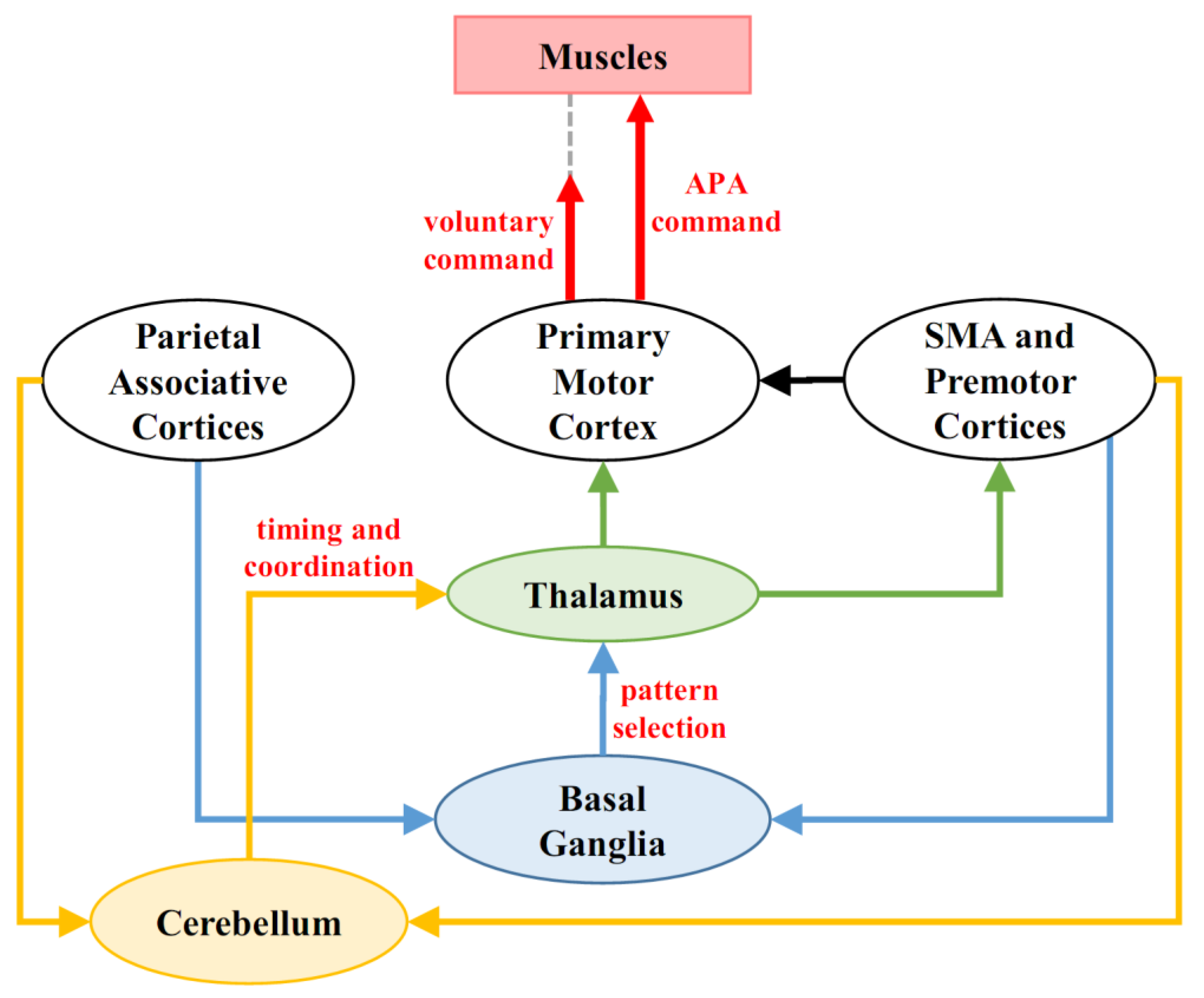
Figure 1. Role of Cerebellum and Basal Ganglia in APA programming. Cerebellum and Basal Ganglia receive information about the environmental context and body schema from the Parietal Associative Cortices, as well as about the motor goal from SMA and Premotor Cortices. In turn, Basal Ganglia define the pattern of muscular recruitment (i.e., the muscles which should be excited or inhibited), while the timing and the coordination of such actions is defined by the cerebellum. Through their Thalamic projections, the final motor program reaches the Primary Motor Cortex, which routes it to both the focal and postural muscles. Note that the APA command reaches the postural muscles before the voluntary command activates the focal muscles.
Applsci 10 05088 g001
Figure 2. Role of the cerebellum in the ontogenesis of the APA control. Cerebellar deficits interfere with the correct development of postural control. The later the appearance of the cerebellar deficit, the weaker seems to be the ability of the CNS to develop a compensatory strategy. This is particularly apparent when the disease has a progressive course, since it contrasts with the strategy consolidation.
Applsci 10 05088 g002
Figure 3. Subcortical compensatory networks. (a) Reciprocal interconnections between cerebellum and basal ganglia have been described in healthy conditions [116,117]. Panel (b) shows how the pathway from cerebellum to basal ganglia enhances its activity (thick black arrow) in Parkinson’s disease, to compensate the basal ganglia deficit [123]. Symmetrically, in (c), it is proposed that the pathway from basal ganglia to cerebellum (thick red arrow) may allow the former to compensate for cerebellar deficits in ataxia.
Applsci 10 05088 g003