Article

Changes in Electroencephalography by Modulation of Interferential Current Stimulation

1
Department of Physical Therapy, Nambu University, 23 Cheomdanjungang-ro, Gwangsan-gu, Gwangju 62271, Korea
2
Department of Biomedical Engineering, School of Medicine, Keimyung University, 1095 Dalgubeol-daero, Daegu 42601, Korea
*
Author to whom correspondence should be addressed.
Appl. Sci. 2020, 10(17), 6028; https://doi.org/10.3390/app10176028
Submission received: 16 July 2020 / Revised: 17 August 2020 / Accepted: 27 August 2020 / Published: 31 August 2020

Abstract

:
Interferential current (IFC) stimulation can alter pain perception. This study aimed to investigate the effects of IFC stimulation on motor cortex signals and observe how electroencephalography changes depend on IFC stimulation parameters. Forty-five healthy adults were divided into high frequency (HF)–low intensity (LI), HF–high intensity (HI), and low frequency (LF)–HI groups to compare their electroencephalography before, immediately after, and 30 min after current stimulation. The changes in relative beta power according to the intervention time showed significant differences between the HF–LI and HF–HI, as well as the LF–HI and HF–HI, groups in the C3 and P3 regions immediately after IFC stimulation. Similarly, the gamma band showed significant differences according to the intervention time between the LF–HI and HF–HI groups in the P3 region immediately following IFC intervention. For relative theta power, the interaction between group and time was significantly different in the Fp2, F3, F4, C3, C4, and P4 regions. Based on these results, we were able to map the activation in cerebral cortex regions according to the stimulation level, confirming changes in electroencephalogram activation through peripheral nerve stimulation. This study provides a foundation for future applications for selectively controlling feedback at a proper stimulation level in young adults.

1. Introduction

Electrical stimulation can be used as a therapeutic tool to relieve bone disorder or neurological pain conditions. This approach can expand the size of the cortical receptor area and improve the sensitivity of the somatosensory system [1]. The transcutaneous application of electrical current activates neurons, which in turn can lead to physiological changes such as enhanced blood circulation and muscle activity through increased muscle metabolism [2]. Therefore, electrical stimulation has been reported to be effective in controlling pain, retraining and strengthening muscles, and healing wounds [3]. However, the stimulation of peripheral sensory receptors that affect the excitatory state of the motor cortex can differ depending on the type of electrical stimulation applied and the time of intervention [4]. In addition, in the absence of established indicators, there is a tendency to evaluate the various therapeutic effects of electrical stimulation according to the patient’s pain intensity, so applying an appropriate level of electrical stimulation is a difficult task [5]. Thus, many studies from different fields have emphasized the necessity for an effective clinical approach based on current stimulation tailored to each patient’s specific needs [6].
The use of computers enables visual judgments and quantitative mathematical analyses of the state of brain function and makes it possible to analyze brain function objectively [7]. Electroencephalography has been used to objectively evaluate the variability of the current thresholds and the adaptation of peripheral nerves induced by electrical stimulation [8]. By measuring cortical electrical activity, these tests can objectively control the variable current threshold, thus modifying the peripheral nerve induction by electrical stimulation. The increased excitability of nerve cells due to physical stimulation may also increase muscular motor-evoked potentials [9].
Thus, electroencephalogram patterns representing changes in electrical signals between brain neuronal cells are widely used as diagnostic indicators in various fields of medicine [10]. The electrical waveforms of electroencephalography are a means for the cerebral cortex to maintain contact with the rest of the body and allow the processing of external information for comparison, analysis, judgment, and decision making [11]. During tasks requiring concentration, the frontal lobe is involved in memory encoding and planning resulting from current actions. The parietal lobe is involved in the reception and processing of sensory information from the body [6]. By revealing brain activity and activation states, which are regulated by a balance between central and peripheral organs, the existence of distinct patterns affected by the brain’s conscious condition, mental activity, pathology, and electrical stimulation may be confirmed [12].
Interferential current (IFC) stimulation is an intervention method combining the advantages of high permeability from middle-frequency currents and efficient tissue stimulation from low-frequency currents [13]. IFC stimulation has a much shorter pulse duration than transcutaneous nerve stimulation and, therefore, can deliver maximum current to tissues with high tissue permeability. Thus, this intervention can be applied in various clinical settings due to its physiological mechanism of deep current permeation into a specific area of the body during current conduction [3]. In patients with symptoms of hypertonicity in the spinal erector muscle or imbalance in the autonomic nervous system, electrical stimulation methods are used, modifying the amplitude modulation frequency and waveform through electrode placement to the paravertebral area [14].
Human tissue comprises cells suspended in an aqueous medium, making its electrical conductivity inhomogeneous on a microscopic scale [15]. Therefore, the acquired electroencephalography response patterns to maximum electrical stimulation are reportedly various and irregular [10]. Diagnosis and evaluation of intervention efficacy using electrical stimulation are important in clinical practice. Hence, when selecting stimulation conditions, it is important to consider intervention variables that are affected by electrical stimulation parameters and consider how they may act differently on biorhythm activation mechanisms [12].
The effects of electrical stimulation on electroencephalography (EEG) have previously been reported [16,17]. In healthy volunteers, high-frequency transcutaneous electrical nerve stimulation (TENS) reduces enhanced gamma-band activity following tonic pain induction, which can serve as a functional brain biomarker for pain treatment, specifically for EEG-based neurofeedback approaches [16]. TENS increases alpha activity in young subjects, who had higher alpha activity than elderly subjects, in occipital and somatosensory areas. Thus, TENS may be used as an objective method to examine sensory impairments and evaluate pain treatment [17].
IFC is widely used in clinical settings; its therapeutic effects have been well documented using various methods under different conditions. The present study investigates how electrical stimulation can affect electroencephalogram changes in healthy individuals by measuring brain signals from the motor cortex and observing how changes in electroencephalogram activation are dependent on the IFC stimulation parameters.

2. Methods

2.1. Participants

This study enrolled 45 healthy male and female students from Gwangju Metropolitan City. All participants were fully informed about the purpose of the study and the study design, and all participants provided written informed consent. This study was carried out in accordance with the ethical standards laid out in the Declaration of Helsinki and with the approval of the Institutional Review Board (IRB: 1040621-201410-HR-010-09), South Gwangju University, Korea. Participants were excluded if they had metal implants, a history of neurological or psychiatric disorders, diseases affecting vital signs (blood pressure, pulse, body temperature, and respiratory rate) or blood test results, or contraindications to electrical stimulation interventions. Participants were instructed not to engage in strenuous exercise, and tobacco, drink, or food intake that may have affected the autonomic nervous system was restricted to 1 h before the experiment.
To determine the sample size, the G*Power version 3.19 statistical power analysis software program was used. Assuming a power of 90%, a type 1 error (α) of 5%, a type 2 error (β) of 10%, and a confidence interval of 95%, we calculated a sample size of 15 for each of the three experimental groups [18,19]. After recording age, sex, and weight as the main parameters, participants were randomly assigned to one of the following three groups: a high frequency–low intensity group (HF–LI; n = 15), a low frequency–high intensity group (LF–HI; n = 15), and a high frequency–high intensity group (HF–HI; n = 15). The three groups were found to be homogeneous after evaluating the demographic characteristics of each group (Table 1).

2.2. EEG Recordings

EEG is used to analyze brain activity recorded from multiple electrodes placed on the scalp [20]. The EEG signal reflects the functional state of the brain allied to the person’s mental condition, from which we can extract vital information, monitor the patient’s health [21], and identify different brain conditions [22,23]. Quantitative EEG (QEEG) allows the objective diagnosis of neurological changes in the brain [24]. In the current study, a QEEG-8 system (Poly G-I, LAXTHA Inc., Daejeon, Korea) was used to obtain electroencephalography measurements. The electrodes were placed according to the international 10/20 system on the following scalp locations: prefrontal 1 (Fp1), prefrontal 2 (Fp2), frontal 3 (F3), frontal 4 (F4), central 3 (C3), central 4 (C4), parietal 3 (P3), and parietal 4 (P4; Figure 1). In addition, a reference electrode and a ground electrode were placed behind the right and left earlobes, respectively. Gold-plated disc-shaped electrodes were used, and a paste was used to attach and apply gauze. The skin resistance of all electrodes was set below 5 kΩ, with a sampling rate of 256 Hz/channel.
After noise removal, the electroencephalography obtained from the participants comprised diverse frequencies. Therefore, the corrected frequency was extracted by filtering the electroencephalography through a 0–50 Hz band-pass filter. Other settings included a 12-bit analog–digital converter, a 0.6-Hz low-frequency pass filter, a 46-Hz high-frequency pass filter, and a 60-Hz notch filter [25]. The waveforms of electroencephalography were monitored using the computer’s record mode, which collected filtered data. Possible interferences from the participant’s condition and noise were minimized [26,27]. During the experiment, the laboratory temperature and humidity were controlled, and the light and noise levels were kept as low as possible.
Quantitative analysis was performed on the collected electroencephalography data, using the electroencephalography analysis system Complexity 2.0 (Laxtha Inc., Daejeon, Korea). Data from 10 epochs, each lasting 30 s, were corrected using a Blackman–Harris window. Fast Fourier Transform was performed to digitize the data and show quantitative relationships between the frequency and amplitude of electroencephalography, and power spectrum analysis values were obtained at various frequencies. The absolute power produced the periodic amplitude, whereas relative power produced the ratios between magnitudes of various frequencies and that of the entire frequency band, which provided equalized values among different individuals [11].
Electroencephalography is categorized according to the shape of the changes in the electrical currents measured due to the combination of action potentials of brain neurons. Alpha (α) waves of 8.0–12.9 Hz appear during the relaxation of consciousness and resting states. The amplitude of alpha waves increases in proportion to stability. Alpha waves are regular pulse waves and are greatest in the parietal and occipital regions. Beta waves (β) of 13.0–29.9 Hz appear broadly across the whole brain during attentive conscious mental activity. Thus, when beta waves increase, the person is considered to be in a state of concentration. Gamma waves (γ) of 30–50 Hz appear during tension and in the performance of active, highly complex mental functions, as well as in strongly excited states [28]. Theta waves (θ) of 4.0–7.9 Hz appear mainly during sleeping or the stable state and are generally found in the frontal cortical areas that are involved in emotion. They also appear during strong internal conscious attention [29].
The spectral analysis-based relative power value was calculated by recording the EEG signal for a short period of time, whereas the calculation of the relative power value was performed for 3 min at 20–30 s per frequency band using a clean EEG with artifacts removed [30]. This led to a precise evaluation of the brain’s electrical activity. Raw electroencephalography data were obtained for 220-s periods at three time points: before the electrical stimulation, immediately after IFC stimulation, and 30 min after IFC stimulation. From the 220-s of EEG raw data obtained in each period, 180 s were analyzed, excluding the first and last 20 s [31].

2.3. IFC Application and EEG Measurements

Participants were instructed to close their eyes to prevent eye-induced artifacts during EEG recordings. Participants rested comfortably in bed for 10 min before the experiment to enhance psychological stability, minimize EEG artifacts, and reduce errors due to postural or behavioral changes [12]. IFC is produced by the interaction between two medium-frequency currents from two electrodes. The currents pass through the tissue simultaneously and interfere with each other, generating an interference current that stimulates the tissue [32]. A pad electrode (4 × 4 cm; Protens Electrodes; Bio-Protech Inc., Gangwon-do, Korea) was attached to the transverse process 2 cm from the left spinous processes, corresponding to the T1–T4 spinal cord segment levels. IFC stimulation was applied for 20 min using the ENDOMED 582 IFC device (Enraf Nonius, Rotterdam, The Netherlands). After a preliminary test, the experiment was conducted in three groups: HF–LI group (burst frequency: 100 bps, intensity: 10–20 mA), LF–HI group (burst frequency: 5 bps, intensity: 45–50 mA), and HF–HI group (burst frequency: 100 bps, intensity: 80–90 mA) [33,34,35].

2.4. Statistical Analysis

All data were analyzed using SPSS version 20.0 for Windows (IBM Corp., Armonk, NY, USA) and are presented as means ± standard deviations. The Shapiro–Wilk test was conducted to determine the normal distribution of the variables in each category. The results of the Shapiro–Wilk test confirmed the normal distribution of the data, and one-way analysis of variance was conducted to determine the differences between groups based on the participants’ basic characteristics. Repeated measures analysis of covariance (ANCOVA) was performed to analyze the EEG changes in this study before, immediately after, and 30 min after treatment between groups [36].
The covariate was used as the baseline measurement value for each EEG measurement. The repeated measures ANCOVA results confirmed whether the sphericity and equal variance assumptions were satisfied. If not, Wilks’ lambda value was determined by multivariate analysis. In addition, to explain the effect size between the independent variable and the dependent variable, the interaction between group and time was analyzed and expressed as partial eta square (η2). If the partial η2 is 0.02, the effect size is small, if it is 0.13, the effect size is medium, and if it is 0.26, the effect size is large. Therefore, as the value of the partial η2 approaches 1, the average difference between groups increases, and the error is small [37,38,39].
If a group showed a main effect in an effective treatment period, one-way analysis of variance was conducted to analyze the differences in average measurements between groups and between time periods. Tukey’s test was used to evaluate significant differences between groups. The significance level α was set to 0.05.

3. Results

To check the changes in relative beta power, relative gamma power, and relative theta power for each brain region, the pre-test measurement values of electroencephalography were covariantly processed with the differences in initial result values. After confirming sphericity with Mauchly’s test, the two-way repeated measures ANCOVA showed that group differences in IFC-induced beta wave changes were statistically significant (p < 0.05). However, the interactions between group and IFC stimulation were not significant (p > 0.05). The post-hoc analysis of the main effects of relative beta power in each group revealed significant differences in the C3 and P3 regions between the HF–LI and HF–HI groups, as well as between the LF–HI and HF–HI groups (p < 0.05; Table 2, Figure 2).
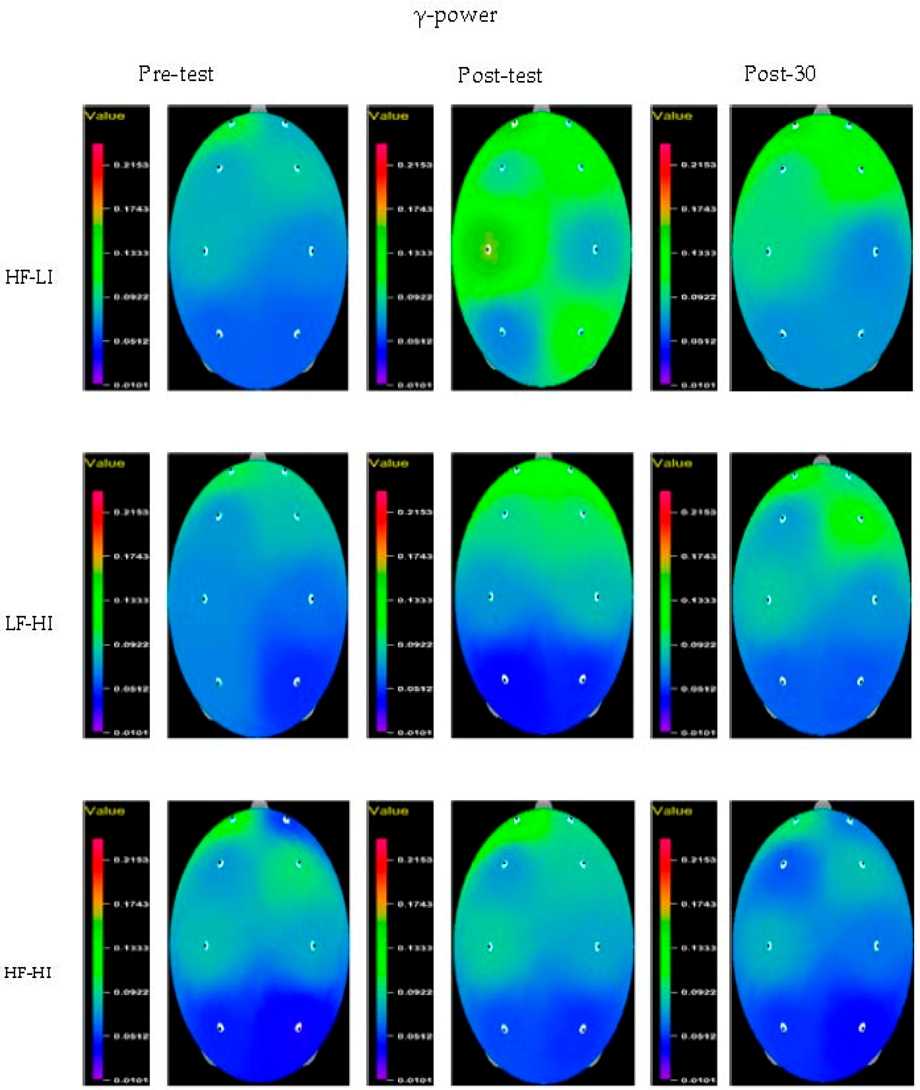
Mauchly’s sphericity test validated that the repeated measures ANCOVA regarding changes in relative gamma waves in each group caused by IFC was statistically significant (p < 0.05). No interaction was observed between groups and time. The post-hoc analysis of the main effects of relative gamma power in each group showed significant differences in the P3 region between the HF–LI and HF–HI groups, as well as between the LF–HI and HF–HI groups (p < 0.05; Table 3, Figure 3).
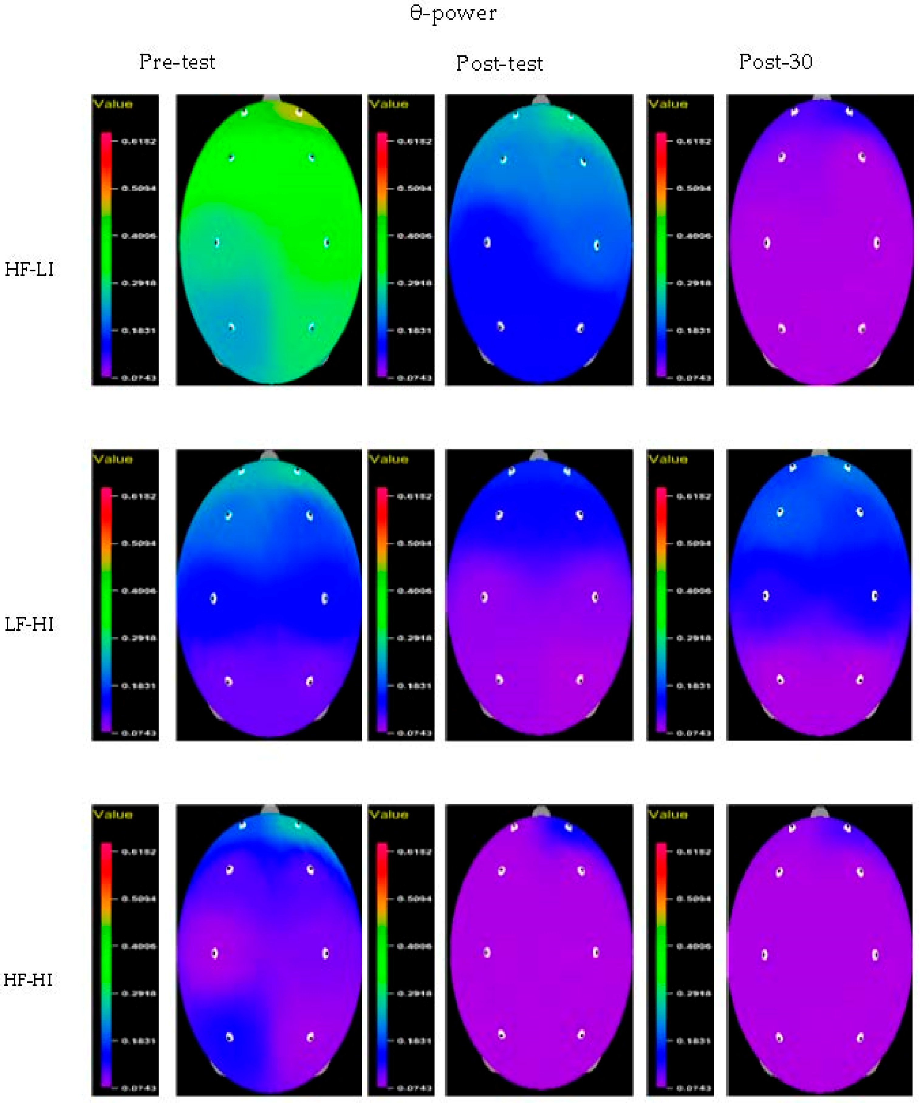
For relative theta power, there was a significant difference in the interaction between group and time in the Fp2, F3, F4, C3, C4, and P4 regions according to the two-way repeated measures ANCOVA with covariate processing of the pre-measured values (p < 0.05). The partial η2 values, representing the effects of intervention according to group and time, were 0.188 in the Fp2 region, 0.192 in the F3 region, 0.208 in the F4 region, 0.211 in the C3 region, 0.251 in the C4 region, and 0.229 in the P4 region (Table 4, Figure 4). No significant differences in the main effects were observed after IFC stimulation for relative theta power.

4. Discussion

IFC is a treatment method primarily used to control pain symptoms. It presents advantages in the physiological measurement of pain and has been shown to decrease the frequency of pain medication use [4]. IFC is widely used in clinical settings and is favored over other forms of low-frequency stimulation since a greater maximum current can be delivered to specific tissues, with greater penetration, due to the lower skin or tissue resistance [40]. IFC uses amplitude-modulated alternating currents at a medium frequency of 1–100 kHz, which are produced when two independent alternating currents of slightly different frequencies intersect and deliver a biologically acceptable burst frequency under the flow of a sustained electric charge [14].
IFC stimulation near the spinal cord might result in hypertonic sympathetic nervous system and spontaneous activity, causing antagonistic activity in various organs. Thus, an understanding of the spinal segments and controlled areas is required [41]. However, both the sympathetic and parasympathetic systems act in numerous organs to maintain an optimal internal environment. Therefore, segmental reflexes in the spinal cord act to combine the descending effects of higher centers and the inhibitory or excitatory effects of the spinal cord. These reflex effects are based on the anatomical basis of the segmental relationship between the skin and organs [42]. Based on these principles, IFC stimulation is performed during spinal segment examination, which can confirm the responses of the dermatome and myotome using a medium-frequency current. To wit, two electrodes are applied to the T1–T4 locations near the thoracic vertebrae [43]. In the current study, we used the frequency and intensity of IFC stimulation as the main parameters to investigate the direct effects of the three stimulation types on EEG activation. The electrical activity of the brain, which is reflected in the EEG signals, arises from biochemical interactions of glial cells and neurons due to the flow of ions [28].
Electroencephalography can be used as a diagnostic tool to provide spatiotemporal information about the electrical activity originating from neural cell populations of the cerebral cortex. The power spectrum analysis method can be used to extract waveforms resulting from the highly complex electrical activity of the neurons of the cerebral cortex [44]. Sensory receptors are located near the skin surface rather than on motor nerve fibers or myofibers to reach sensory thresholds at low levels of stimulation to enhance their effect and modulatory regulation [45].
During the induction of arousal waves, changes in skeletal muscles caused by exercise and sensory receptors revealed significant changes in the activation of the motor cortex area that controls the stimulated muscles. Furthermore, under aroused, nervous, or tense states, brain activity changes in the beta wave range may be caused by external stimulation [26]. Our findings are supported by previous reports that physical exercise increases the secretion of brain-derived neurotrophic factors, preventing the destruction of existing brain cells and aiding the creation of new cells.
Gamma waves appear during high levels of tension or during the active performance of complex mental functions, especially when the participant’s performance peaks, but also appear under pathological conditions in cases of brain injury [46]. Moreover, a previous report mentioned that the participant’s response is related to the activity of gamma waves between the cerebral cortex and spinal cord [28]. In the current study, there was no statistically significant change in the gamma waves according to the electrical stimulation variables. However, gamma waves were reduced in each group after IFC intervention, and more stable results were confirmed in the occipital lobe.
Theta waves appear during deep sleep and are characterized by slow, large amplitude vibrations in different sleep states; the pattern turns into predictable characteristic waveforms in various awake states [47]. Theta waves are not related to emotional anxiety or deep sleep but are related to internalized and quiet physical, emotional, and thinking activities [48,49]. However, the appearance of theta waves during wakefulness can be a sign of the brain being in an abnormal state, and asymmetrical waves in the stable state can be a sign of brain dysfunction [50]. Our ANCOVA results revealed significant differences in the interaction between group and time in the Fp2, F3, F4, C3, C4, and P4 regions.
Disorders of the endogenous pain control system represent a potential mechanism underlying increased pain [7]. When pain-controlled electrical stimulation is applied to alleviate severe pain, high-level pain catastrophizing is inversely related to the brain’s electrical activity in specific regions of the cortex, including an inhibitory network associated with top-down pain control [51]. Previous studies suggest that HF–HI electrical stimulation leads to descending pain control via the secretion of endogenous opioids within the central nervous system [52]. Besides, receptor-based skin pressure stimulation and skin stretching showed the highest sensitivity at frequencies of 0.3–3 Hz and 25–400 Hz, respectively, and produced a variety of stimuli such as sensation, movement, and pain [53].
The current study provided supporting data needed in the use of therapeutic methods to improve cognitive ability, muscular stability, and bodily self-awareness when simple attention is required. Some limitations should be noted. First, First, baseline differences between the groups require the results to be treated with some caution. It was difficult to detect differences over time within each group due to differences in baseline values for each EEG measurement. Second, this study did not fully reveal the cortical potential changes for each frequency of each cortical region involved in motor activity. Third, since the state of brain development and age may affect EEG signals, future research should also examine electroencephalography changes according to age groups. Lastly, it will be necessary to standardize the electrical stimulation types with relevance to neurologic changes or pain control in future studies.

5. Conclusions

We found diverse changes in electroencephalography in relation to the IFC stimulation level. Furthermore, EEG-based brain mapping by analysis of electroencephalography frequency bands is routinely used in experimental and clinical pain studies. Based on the frequency band results, we were able to map activation in regions of the cerebral cortex according to the stimulation level, by confirming changes in electroencephalography activation through peripheral nerve stimulation. The results of this study could be utilized in healthy adults by controlling feedback at a proper stimulation level.

Author Contributions

S.-H.C.; project administration, S.-H.C. and S.-C.K.; investigation, S.-H.C. and S.-C.K.; writing—original draft preparation, S.-H.C. and S.-C.K.; writing—review and editing. All authors have read and agreed to the published version of the manuscript.

Funding

This work was funded by the National Research Foundation of Korea (NRF) grant funded by the Korean government (MSIT) (no. 2017R1C1B5076499).

Conflicts of Interest

The authors declare no conflict of interest.

References

  1. Dhruv, N.T.; Niemi, J.B.; Harry, J.D.; Lipsitz, L.A.; Collins, J.J. Enhancing tactile sensation in older adults with electrical noise stimulation. Neuroreport 2002, 13, 597–600. [Google Scholar] [CrossRef] [Green Version]
  2. Valeriani, M.; Tonali, P.; Le Pera, D.; Restuccia, D.; De Armas, L.; Del Vesco, C.; Miliucci, R.; Fiaschi, A.; Vigevano, F.; Arendt-Nielsen, L.; et al. Modulation of laser-evoked potentials by experimental cutaneous tonic pain. Neuroscience 2006, 140, 1301–1310. [Google Scholar] [CrossRef] [PubMed]
  3. Araújo, F.M.; Menezes, M.A.; De Araújo, A.M.; Sousa, T.A.D.S.; Lima, L.V.; Carvalho, E.Á.N.; DeSantana, J.M. Validation of a New Placebo Interferential Current Method: A New Placebo Method of Electrostimulation. Pain Med. 2016, 18, 86–94. [Google Scholar] [CrossRef] [PubMed] [Green Version]
  4. Corrêa, J.B.; Costa, L.O.; Oliveira, N.T.; Lima, W.P.; Sluka, K.A.; Liebano, R.E. Effects of the carrier frequency of interferential current on pain modulation and central hypersensitivity in people with chronic nonspecific low back pain: A randomized placebo-controlled trial. Eur. J. Pain 2016, 20, 1653–1666. [Google Scholar] [CrossRef] [PubMed]
  5. Kottink, A.I.; Oostendorp, L.J.; Buurke, J.H.; Nene, A.V.; Hermens, H.J.; IJzerman, M.J. The orthotic effect of functional electrical stimulation on the improvement of walking in stroke patients with a dropped foot: A systematic review. Artif. Organs 2004, 28, 577–586. [Google Scholar] [CrossRef] [PubMed]
  6. Sullivan, M.J.; Lynch, M.E.; Clark, A.J. Dimensions of catastrophic thinking associated with pain experience and disability in patients with neuropathic pain conditions. Pain 2005, 113, 310–315. [Google Scholar] [CrossRef] [PubMed]
  7. Sokhadze, T.M.; Cannon, R.L.; Trudeau, D.L. EEG biofeedback as a treatment for substance use disorders: Review, rating of efficacy, and recommendations for further research. Appl. Psychophysiol. Biofeedback 2008, 33, 1–28. [Google Scholar] [CrossRef] [PubMed] [Green Version]
  8. Nangini, C.; Ross, B.; Tam, F.; Graham, S.J. Magnetoencephalographic study of vibrotactile evoked transient and steady-state responses in human somatosensory cortex. Neuroimage 2006, 33, 252–262. [Google Scholar] [CrossRef]
  9. Kaelin-Lang, A.; Luft, A.R.; Sawaki, L.; Burstein, A.H.; Sohn, Y.H.; Cohen, L.G. Modulation of human corticomotor excitability by somatosensory input. J. Physiol. 2002, 540, 623–633. [Google Scholar] [CrossRef]
  10. Wang, L. Brain plasticity: Dynamic changes of the cerebral activity as effect of pain modulation. Ph.D. Thesis, Aalborg University, Aalborg, Denmark, 2007. [Google Scholar]
  11. Niemiec, A.; Lithgow, B. Alpha-band characteristics in EEG spectrum indicate reliability of frontal brain asymmetry measures in diagnosis of depression. In Proceedings of the IEEE Engineering in Medicine and Biology 27th Annual Conference, Shanghai, China, 17–18 January 2006; pp. 7517–7520. [Google Scholar] [CrossRef]
  12. Dornhege, G.; Millán, J.R.; Hinterberger, T.; McFarland, D.; Müller, K.R. Toward Brain-Computer Interfacing; MIT Press: Cambridge, MA, USA, 2007; pp. 31–42. [Google Scholar]
  13. Eslamian, F.; Farhoudi, M.; Jahanjoo, F.; Sadeghi-Hokmabadi, E.; Darabi, P. Electrical interferential current stimulation versus electrical acupuncture in management of hemiplegic shoulder pain and disability following ischemic stroke-a randomized clinical trial. Arch. Physiother. 2020, 10, 2. [Google Scholar] [CrossRef]
  14. Furuta, T.; Takemura, M.; Tsujita, J.; Oku, Y. Interferential electric stimulation applied to the neck increases swallowing frequency. Dysphagia 2012, 27, 94–100. [Google Scholar] [CrossRef] [PubMed]
  15. Peters, M.J.; Stinstra, G.; Hendriks, M. Estimation of the electrical conductivity of human tissue. Electromagnetics 2001, 21, 545–557. [Google Scholar] [CrossRef]
  16. Ebrahimian, M.; Razeghi, M.; Zamani, A.; Bagheri, Z.; Rastegar, K.; Motealleh, A. Does high frequency transcutaneous electrical nerve stimulation (TENS) affect EEG gamma band activity? J. Biomed. Phys. Eng. 2018, 8, 271–280. [Google Scholar] [CrossRef] [Green Version]
  17. Yıldırım, E.; Güntekin, B.; Hanoğlu, L.; Algun, C. EEG alpha activity increased in response to transcutaneous electrical nervous stimulation in young healthy subjects but not in the healthy elderly. PeerJ 2020, 8, e8330. [Google Scholar] [CrossRef] [PubMed] [Green Version]
  18. Faul, F.; Erdfelder, E.; Lang, A.G.; Buchner, A. G*Power 3: A flexible statistical power analysis program for the social, behavioral, and biomedical sciences. Behav. Res. Methods 2007, 39, 175–191. [Google Scholar] [CrossRef]
  19. Faul, F.; Erdfelder, E.; Buchner, A.; Lang, A.G. Statistical power analyses using G*Power 3.1: Tests for correlation and regression analyses. Behav. Res. Methods 2009, 41, 1149–1160. [Google Scholar] [CrossRef] [Green Version]
  20. Niedermeyer, E.; da Silva, F.H.L. Electroencephalography: Basic Principles, Clinical Applications, and Related Fields, 5th ed.; Lippincott Williams & Wilkins: Philadelphia, PA, USA, 2005. [Google Scholar]
  21. Hirsch, L.J.; Brenner, R.P. Atlas of EEG in Critical Care; John Wiley and Sons: Hoboken, NJ, USA, 2010; Volume 30, pp. 187–216. [Google Scholar]
  22. Nunez, P.L.; Srinivasan, R. Electric Fields of the Brain: The Meurophysics of EEG, 2nd ed.; Oxford University Press: New York, NY, USA, 2005; pp. 154–169. [Google Scholar]
  23. Wang, H.; Lei, X.; Zhan, Z.; Yao, L.; Wu, X. A new fMRI informed mixed-norm constrained algorithm for EEG source localization. IEEE Access 2018, 6, 8258–8269. [Google Scholar] [CrossRef]
  24. Babiloni, C.; Ferri, R.; Moretti, D.V.; Strambi, A.; Binetti, G.; Dal Forno, G.; Ferreri, F.; Lanuzza, B.; Bonato, C.; Nobili, F.; et al. Abnormal fronto-parietal coupling of brain rhythms in mild Alzheimer’s disease: A multicentric EEG study. Eur. J. Neurosci. 2004, 19, 2583–2590. [Google Scholar] [CrossRef]
  25. Murugappan, M.; Ramachandran, N.; Sazali, Y. Combining spatial filtering and wavelet transform for classifying human emotions using EEG signals. J. Med. Biol. Eng. 2010, 31, 45–51. [Google Scholar] [CrossRef]
  26. Fisch, B. Fisch and Spehlmann’s EEG Primer: Basic Principles of Digital and Analog EEG, 3rd ed.; Elsevier: Amsterdam, The Netherlands, 2016. [Google Scholar]
  27. Selkowitz, D.M. Improvement in isometric strength of the quadriceps femoris muscle after training with electrical stimulation. Phys. Ther. 1985, 65, 186–196. [Google Scholar] [CrossRef]
  28. Schoffelen, J.M.; Oostenveld, R.; Fries, P. Neuronal coherence as a mechanism of effective corticospinal interaction. Science 2005, 308, 111–113. [Google Scholar] [CrossRef] [PubMed] [Green Version]
  29. Sanei, S.; Chambers, J.A. EEG Signal Processing, 1st ed.; John Wiley & Sons: Hoboken, NJ, USA, 2007; pp. 10–13. [Google Scholar]
  30. Jin, H.K.; Cho, S.H. Comparison of relative α-power spectral electroencephalogram activity analysis according to electrical stimulation levels in normal adults. J. Psychiatry 2015, 18, 5. [Google Scholar] [CrossRef]
  31. Jiang, X.; Bian, G.B.; Tian, Z. Removal of artifacts from EEG signals: A review. Sensors 2019, 19, 987. [Google Scholar] [CrossRef] [PubMed] [Green Version]
  32. Grabiańska, E.; Leśniewicz, J.; Pieszyński, I.; Kostka, J. Comparison of the analgesic effect of interferential current (IFC) and TENS in patients with low back pain. Wiad. Lek. 2015, 68, 13–19. (In Polish) [Google Scholar] [PubMed]
  33. Cincotti, F.; Kauhanen, L.; Aloise, F.; Palomäki, T.; Caporusso, N.; Jylänki, P.; Mattia, D.; Babiloni, F.; Vanacker, G.; Nuttin, M.; et al. Vibrotactile feedback for brain-computer interface operation. Comput. Intell. Neurosci. 2007, 2007, 48937. [Google Scholar] [CrossRef] [Green Version]
  34. Cho, S.H. Frequency and intensity of electrical stimulation of human sympathetic ganglia affect heart rate variability and pain threshold. Appl. Sci. 2019, 9, 4490. [Google Scholar] [CrossRef] [Green Version]
  35. Ozcan, J.; Ward, A.R.; Robertson, V.J. A comparison of true and premodulated interferential currents. Arch. Phys. Med. Rehabil. 2004, 85, 409–415. [Google Scholar] [CrossRef]
  36. Norman, G.R.; Streiner, D.L. Biostatistics: The Bare Essentials, 2nd ed.; B.C. Decker Inc.: London, UK, 2000. [Google Scholar]
  37. Bakeman, R. Recommended effect size statistics for repeated measures designs. Behav. Res. Methods 2005, 37, 379–384. [Google Scholar] [CrossRef]
  38. Cohen, J. Statistical Power Analysis for the Behavioral Sciences, 2nd ed.; Erlbaum: Hillsdale, NJ, USA, 1988; pp. 413–414. [Google Scholar]
  39. Field, A. Discovering Statistics Using IBM SPSS Statistics, 4th ed.; Sage Publications Ltd.: London, UK, 2013; p. 267. [Google Scholar]
  40. Robertson, V.; Ward, A.; Low, J.; Reed, A. Electrotherapy Explained: Principles and Practice, 4th ed.; Elsevier Butterworth-Heinemann: Philadelphia, PA, USA, 2006. [Google Scholar]
  41. Ge, H.Y.; Fernández-de-las-Peñas, C.; Arendt-Nielsen, L. Sympathetic facilitation of hyperalgesia evoked from myofascial tender and trigger points in patients with unilateral shoulder pain. Clin. Neurophysiol. 2006, 117, 1545–1550. [Google Scholar] [CrossRef]
  42. Watson, N.F.; Buchwald, D.; Goldberg, J.; Noonan, C.; Ellenbogen, R.G. Neurologic signs and symptoms in fibromyalgia. Arthritis Rheum. 2009, 60, 2839–2844. [Google Scholar] [CrossRef] [Green Version]
  43. Sadock, B.J.; Sadock, V.A. Kaplan and Sadock’s Synopsis of Psychiatry: Behavioral Sciences/Clinical Psychiatry, 10th ed.; Lippincott Williams & Wilkins: Philadelphia, PA, USA, 2007. [Google Scholar]
  44. Walker, J.E. Recent advances in quantitative EEG as an aid to diagnosis and as a guide to neurofeedback training for cortical hypofunctions, hyperfunctions, disconnections, and hyperconnections: Improving efficacy in complicated neurological and psychological disorders. Appl. Psychophysiol. Biofeedback 2010, 35, 25–27. [Google Scholar] [CrossRef] [PubMed]
  45. Ridding, M.C.; McKay, D.R.; Thompson, P.D.; Miles, T.S. Changes in corticomotor representations induced by prolonged peripheral nerve stimulation in humans. Clin. Neurophysiol. 2001, 112, 1461–1469. [Google Scholar] [CrossRef]
  46. Thornton, K.E.; Carmody, D.P. Electroencephalogram biofeedback for reading disability and traumatic brain injury. Child Adolesc. Psychiatr. Clin. N. Am. 2005, 14, 137–162. [Google Scholar] [CrossRef] [PubMed]
  47. Gazzaniga, M.S.; Ivry, R.B.; Mangun, G.R. Cognitive Neuroscience—The Biology of the Mind, 2nd ed.; Norton & Company. Ltd.: New York, NY, USA, 2002; pp. 301–350. [Google Scholar]
  48. Klimesch, W. Memory processes described as brain oscillations in the EEG-alpha and theta bands. Psycoloquy 1995, 11, 134–143. [Google Scholar]
  49. Klimesch, W. EEG alpha and theta oscillations reflect cognitive and memory performance: A review and analysis. Brain Res. Brain Res. Rev. 1999, 29, 169–195. [Google Scholar] [CrossRef]
  50. Vernon, D.; Egner, T.; Cooper, N.; Compton, T.; Neilands, C.; Sheri, A.; Gruzelier, J. The effect of training distinct neurofeedback protocols on aspects of cognitive performance. Int. J. Psychophysiol. 2003, 47, 75–85. [Google Scholar] [CrossRef]
  51. Seminowicz, D.A.; Davis, K.D. Cortical responses to pain in healthy individuals depends on pain catastrophizing. Pain 2006, 120, 297–306. [Google Scholar] [CrossRef]
  52. Cheing, G.L.; Chan, W.W. Influence of choice of electrical stimulation site on peripheral neurophysiological and hypoalgesic effects. J. Rehabil. Med. 2009, 41, 412–417. [Google Scholar] [CrossRef] [Green Version]
  53. Bolanowski, S.J.; Verrillo, R.T.; McGlone, F. Passive, active and intra-active (self) touch. Somatosens. Mot. Res. 1999, 16, 304–311. [Google Scholar] [CrossRef]
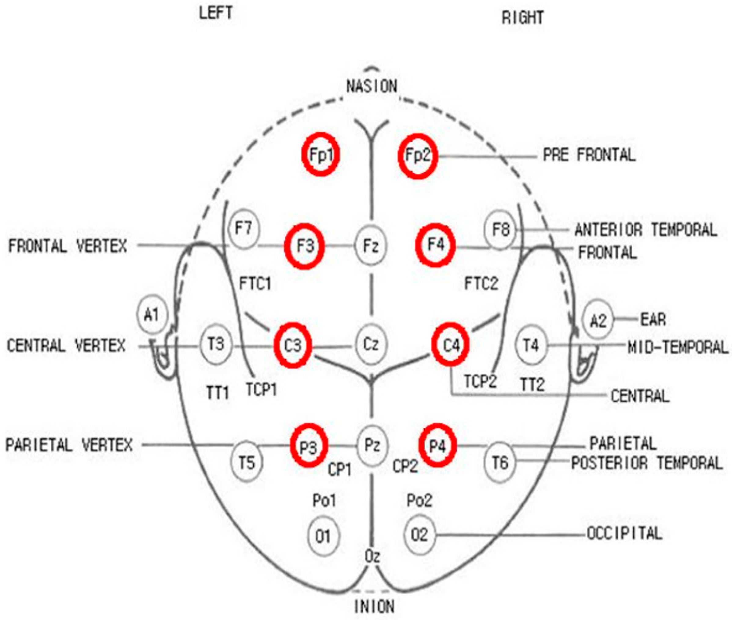
Figure 1. International 10/20 electrode system. Fp1: left prefrontal lobe; Fp2: right prefrontal lobe; F3: left frontal lobe; F4: right frontal lobe; C3: left central lobe; C4: right central lobe; P3: left parietal lobe; P4: right parietal lobe.
Applsci 10 06028 g001
Figure 2. Brain mapping of the electroencephalographic (EEG) activity in the β-power band. Left: Scales range from 0.07 (minimum) to 0.59 (maximum) in steps of 0.08. Right: EEG electrodes are depicted in their location on the head, and colors represent the values of relative β-power. Pre-test: before electrical stimulation; Post-test: directly after electrical stimulation; Post-30: 30 min after electrical stimulation; high frequency–low intensity group (HF–LI); low frequency–high intensity group (LF–HI); high frequency–high intensity group (HF–HI).
Applsci 10 06028 g002
Figure 3. Brain mapping of the electroencephalographic (EEG) activity in the γ-power band. Left: Scales range from 0.07 (minimum) to 0.59 (maximum) in steps of 0.08. Right: EEG electrodes are depicted in their location on the head, and colors represent the values of relative γ-power. Pre-test: before electrical stimulation; Post-test: directly after electrical stimulation; Post-30: 30 min after electrical stimulation; high frequency–low intensity group (HF–LI); low frequency–high intensity group (LF–HI); high frequency–high intensity group (HF–HI).
Applsci 10 06028 g003
Figure 4. Brain mapping of the electroencephalographic (EEG) activity in the θ-power band. Left: Scales range from 0.07 (minimum) to 0.59 (maximum) in steps of 0.08. Right: EEG electrodes are depicted in their location on the head, and colors represent the values of relative θ-power. Pre-test: before electrical stimulation; Post-test: directly after electrical stimulation; Post-30: 30 min after electrical stimulation; high frequency–low intensity group (HF–LI); low frequency–high intensity group (LF–HI); high frequency–high intensity group (HF–HI).
Applsci 10 06028 g004
Table 1. General characteristics of the study population.
CharacteristicsHF–LI
(N = 15)
LF–HI
(N = 15)
HF–HI
(N = 15)
FP
Sex (male/female)7/88/78/7
Age (years)22.64 ± 1.4122.45 ± 1.4422.20 ± 1.630.5270.566
Height (cm)170.27 ± 7.46167.09 ± 10.50166.64 ± 11.080.6150.753
Weight (kg)63.55 ± 4.5561.82 ± 4.0864.09 ± 6.930.2120.210
Values are presented as the mean ± standard deviation. High frequency–low intensity group (HF–LI); low frequency–high intensity group (LF–HI); high frequency–high intensity group (HF–HI).
Table 2. Changes in the relative β-power spectrum.
VariableGroupPre-TestPost-TestPost-30F (P)Group × Time η2
GroupTimeGroup × Time
Fp1HF–LI0.17 ± 0.070.19 ± 0.050.18 ± 0.091.834 (0.178)0.521 (0.476)0.114 (0.892)0.008
LF–HI0.18 ± 0.080.18 ± 0.070.19 ± 0.08
HF–HI0.23 ± 0.100.25 ± 0.080.25 ± 0.08
Fp2HF–LI0.16 ± 0.060.18 ± 0.040.17 ± 0.072.098 (0.141)0.894 (0.352)0.010 (0.990)0.001
LF–HI0.18 ± 0.070.18 ± 0.070.18 ± 0.08
HF–HI0.21 ± 0.110.24 ± 0.080.24 ± 0.10
F3HF–LI0.18 ± 0.050.19 ± 0.060.17 ± 0.052.279 (0.120)0.054 (0.818)0.307 (0.738)0.021
LF–HI0.18 ± 0.070.18 ± 0.070.18 ± 0.07
HF–HI0.24 ± 0.100.26 ± 0.090.25 ± 0.10
F4HF–LI0.18 ± 0.050.18 ± 0.050.17 ± 0.061.882 (0.170)0.342 (0.563)0.317 (0.731)0.021
LF–HI0.19 ± 0.080.18 ± 0.070.18 ± 0.08
HF–HI0.25 ± 0.100.26 ± 0.100.25 ± 0.11
C3HF–LI0.17 ± 0.040.17 ± 0.050.17 ± 0.053.507 (0.043 *)0.537 (0.470)0.004 (0.996)0.000
LF–HI0.17 ± 0.070.17 ± 0.060.17 ± 0.07
HF–HI0.23 ± 0.090.26 ± 0.080.25 ± 0.10
C4HF–LI0.17 ± 0.050.16 ± 0.060.15 ± 0.053.275 (0.052)0.206 (0.653)0.334 (0.718)0.023
LF–HI0.17 ± 0.080.17 ± 0.060.17 ± 0.07
HF–HI0.24 ± 0.090.26 ± 0.090.25 ± 0.10
P3HF–LI0.16 ± 0.050.16 ± 0.050.16 ± 0.064.379 (0.022)0.712 (0.406)0.578 (0.567)0.038
LF–HI0.17 ± 0.080.15 ± 0.050.16 ± 0.08
HF–HI0.20 ± 0.100.25 ± 0.080.23 ± 0.10
P4HF–LI0.17 ± 0.050.17 ± 0.070.16 ± 0.063.453 (0.045 *)0.173 (0.680)0.079 (0.924)0.005
LF–HI0.16 ± 0.080.16 ± 0.060.16 ± 0.08
HF–HI0.22 ± 0.090.26 ± 0.090.25 ± 0.11
* p < 0.05. Values are presented as the mean ± standard deviation. Pre-test: before electrical stimulation; Post-test: directly after electrical stimulation; Post-30: 30 min after electrical stimulation; high frequency–low intensity group (HF–LI); low frequency–high intensity group (LF–HI); high frequency–high intensity group (HF–HI).
Table 3. Changes in the relative γ-power spectrum.
VariableGroupPre-TestPost-TestPost-30F (P)Group × Time η2
GroupTimeGroup × Time
Fp1HF–LI0.11 ± 0.120.09 ± 0.060.08 ± 0.071.916 (0.165)0.292 (0.593)0.453 (0.640)0.030
LF–HI0.09 ± 0.100.07 ± 0.020.08 ± 0.04
HF–HI0.12 ± 0.080.12 ± 0.050.10 ± 0.05
Fp2HF–LI0.09 ± 0.100.07 ± 0.040.06 ± 0.041.528 (0.234)0.456 (0.505)0.145 (0.866)0.010
LF–HI0.08 ± 0.090.07 ± 0.030.06 ± 0.04
HF–HI0.08 ± 0.050.09 ± 0.040.08 ± 0.03
F3HF–LI0.08 ± 0.080.07 ± 0.050.05 ± 0.040.708 (0.501)0.000 (0.989)0.139 (0.871)0.009
LF–HI0.07 ± 0.070.05 ± 0.040.05 ± 0.03
HF–HI0.10 ± 0.090.08 ± 0.040.07 ± 0.05
F4HF–LI0.09 ± 0.090.08 ± 0.050.07 ± 0.060.485 (0.621)0.046 (0.832)0.766 (0.474)0.050
LF–HI0.07 ± 0.070.06 ± 0.030.07 ± 0.06
HF–HI0.11 ± 0.090.09 ± 0.050.09 ± 0.05
C3HF–LI0.09 ± 0.080.10 ± 0.110.05 ± 0.031.298 (0.288)0.025 (0.876)1.698 (0.201)0.105
LF–HI0.06 ± 0.070.05 ± 0.020.06 ± 0.03
HF–HI0.10 ± 0.100.09 ± 0.040.09 ± 0.05
C4HF–LI0.08 ± 0.070.06 ± 0.050.04 ± 0.022.686 (0.085)0.164 (0.689)0.633 (0.538)0.042
LF–HI0.06 ± 0.050.05 ± 0.020.05 ± 0.04
HF–HI0.10 ± 0.080.08 ± 0.050.08 ± 0.04
P3HF–LI0.07 ± 0.060.06 ± 0.050.04 ± 0.033.670 (0.038 *)0.027 (0.870)1.172 (0.324)0.075
LF–HI0.06 ± 0.060.03 ± 0.020.05 ± 0.04
HF–HI0.08 ± 0.080.07 ± 0.030.07 ± 0.03
P4HF–LI0.07 ± 0.060.08 ± 0.100.04 ± 0.031.130 (0.337)0.301 (0.588)1.065 (0.358)0.068
LF–HI0.05 ± 0.050.03 ± 0.020.05 ± 0.04
HF–HI0.07 ± 0.060.07 ± 0.030.06 ± 0.03
* p < 0.05. Values are presented as the mean ± standard deviation. Pre-test: before electrical stimulation; Post-test: directly after electrical stimulation; Post-30: 30 min after electrical stimulation; high frequency–low intensity group (HF–LI); low frequency–high intensity group (LF–HI); high frequency–high intensity group (HF–HI).
Table 4. Changes in the relative θ-power spectrum.
VariableGroupPre-TestPost-TestPost-30F (P)Group × Time η2
GroupTimeGroup × Time
Fp1HF–LI0.40 ± 0.240.39 ± 0.150.33 ± 0.110.835 (0.444)0.118 (0.734)2.212 (0.128)0.132
LF–HI0.40 ± 0.210.32 ± 0.070.36 ± 0.06
HF–HI0.33 ± 0.150.31 ± 0.080.31 ± 0.08
Fp2HF–LI0.41 ± 0.230.41 ± 0.160.34 ± 0.120.628 (0.541)0.508 (0.482)3.351 (0.049 *)0.188
LF–HI0.41 ± 0.210.32 ± 0.070.37 ± 0.08
HF–HI0.39 ± 0.190.34 ± 0.110.33 ± 0.09
F3HF–LI0.35 ± 0.190.38 ± 0.170.31 ± 0.100.232 (0.794)0.594 (0.447)3.435 (0.046 *)0.192
LF–HI0.37 ± 0.210.30 ± 0.090.37 ± 0.09
HF–HI0.28 ± 0.150.30 ± 0.090.30 ± 0.10
F4HF–LI0.35 ± 0.190.38 ± 0.160.30 ± 0.120.374 (0.691)0.023 (0.880)3.803 (0.034 *)0.208
LF–HI0.36 ± 0.210.30 ± 0.100.36 ± 0.11
HF–HI0.28 ± 0.140.28 ± 0.100.29 ± 0.09
C3HF–LI0.32 ± 0.200.34 ± 0.190.27 ± 0.090.035 (0.965)1.111 (0.301)3.872 (0.032 *)0.211
LF–HI0.34 ± 0.220.27 ± 0.100.33 ± 0.11
HF–HI0.26 ± 0.150.28 ± 0.080.28 ± 0.09
C4HF–LI0.33 ± 0.200.37 ± 0.180.30 ± 0.110.371 (0.693)0.939 (0.341)4.851 (0.015 *)0.251
LF–HI0.35 ± 0.230.27 ± 0.110.34 ± 0.12
HF–HI0.27 ± 0.150.27 ± 0.090.29 ± 0.09
P3HF–LI0.30 ± 0.210.34 ± 0.180.25 ± 0.100.150 (0.861)0.000 (0.994)2.863 (0.073)0.165
LF–HI0.32 ± 0.240.27 ± 0.150.30 ± 0.11
HF–HI0.30 ± 0.170.26 ± 0.080.28 ± 0.08
P4HF–LI0.32 ± 0.200.34 ± 0.190.25 ± 0.100.199 (0.820)0.590 (0.449)4.301 (0.023 *)0.229
LF–HI0.32 ± 0.240.24 ± 0.110.30 ± 0.12
HF–HI0.26 ± 0.150.26 ± 0.080.28 ± 0.08
* p < 0.05. Values are presented as the mean ± standard deviation. Pre-test: before electrical stimulation; Post-test: directly after electrical stimulation; Post-30: 30 min after electrical stimulation; high frequency–low intensity group (HF–LI); low frequency–high intensity group (LF–HI); high frequency–high intensity group (HF–HI).