Article

Beta-Catenin in Pseudoexfoliation Syndrome

by
Joanna Stafiej
1,*,
Marta Hałas-Wiśniewska
2,
Magdalena Izdebska
2,
Maciej Gagat
2,
Alina Grzanka
2 and
Grażyna Malukiewicz
1
1
Department of Ophthalmology, Faculty of Medicine, Nicolaus Copernicus University in Toruń, Collegium Medicum in Bydgoszcz, Sklodowskiej-Curie 9, 85-094 Bydgoszcz, Poland
2
Department of Histology and Embryology, Faculty of Medicine, Nicolaus Copernicus University in Toruń, Collegium Medicum in Bydgoszcz, Karlowicza 24, 85-092 Bydgoszcz, Poland
*
Author to whom correspondence should be addressed.
Appl. Sci. 2020, 10(18), 6199; https://doi.org/10.3390/app10186199
Submission received: 31 July 2020 / Revised: 22 August 2020 / Accepted: 2 September 2020 / Published: 7 September 2020
(This article belongs to the Special Issue New Trends in Biosciences)

Abstract

:
To find whether it is possible that beta-catenin, associated with the development of serious systemic diseases, as well as the neoplastic process, plays a role in the development of pseudoexfoliation syndrome (PEX). If so, identifying PEX, an age-related, vision-threatening disorder of elastic fibers, which is manifested in eyes by the accumulation of an abnormal fibrillar material on the tissues of the anterior segment, with its poorly understood pathogenesis, may be an early indicator of other systemic diseases. The specimens of anterior lens capsules were obtained during routine cataract surgeries from patients with PEX (study group) and those without it (control group). Patients with previously diagnosed renal, cardiac or neoplasm diseases were excluded. In order to determine the localization of β-catenin at the ultrastructural level, the post-embedding colloidal gold (AU) method was used. For the analysis of the presence of proteins involved in cell–cell junctions, including β-catenin, fluorescence staining was performed. An enhanced accumulation of AU in the area of cell junctions in the PEX group was observed in comparison to control patients. A statistically significant increase in the level of β-catenin expression in lens epithelial cells (LECs) for the PEX group (MFI = 808.98) in comparison to the control patients (MFI = 731.6) was also noted. Our study presented the increase in the β-catenin in LECs of PEX group in comparison to control patients. It might be possible for PEX, due to it being easily recognizable, to be the first indicator of serious kidney or cardiac diseases, as well as cancer metastases. Further studies are needed in order to confirm this hypothesis.

1. Introduction

Pseudoexfoliation syndrome (PEX) is a well-known age-related systemic disorder of elastic fibers. It should not, however, be considered an integral part of normal aging. It is manifested in eyes by the accumulation of an abnormal fibrillar material in the extracellular matrix of tissues of the anterior segment and may be identified in a standard ophthalmological examination. The pathogenesis of PEX is still not fully understood, and the biomarkers for its clinical detection are limited. While research is currently being conducted to identify the etiology and the risk factors associated with PEX, it is equally important to determine the interconnections between PEX and various systemic diseases.
One of the proteins involved in the process of regulating the gene expression is β-catenin, which participates in the Wnt/β-catenin pathway. At the same time, it plays a key role in regulating the cytoskeleton which is closely linked with adherens junctions. The cytoplasmic domain of cadherins forms a complex that regulates the cytoskeleton by directing the formation of actin filaments beneath the plasma membrane which has an impact on the whole cell structure [1]. The Wnt/β-catenin signaling pathway is a multifunctional pathway that is not only involved in embryonic development but also in the control of homeostatic self-renewal in various adult tissues [2]. Wnt proteins are a family of secreted glycoproteins responsible for the stabilization and accumulation of β-catenin. Subsequently, β-catenin translocates into the nucleus and activates transcription of multiple target genes including some inflammatory and angiogenic factors [3,4]. Over the past few decades, it has been identified by some authors that Wnt/β-catenin is a developmental signaling pathway that plays an essential role in the regulation of the nephron formation during embryogenesis as well as in the injury repair, and the pathogenesis of kidney diseases [5,6,7]. Some results also suggest that β-catenin is not only an important player in the aortic calcification in chronic kidney disease (CKD) [8] but also in the case of hypertension, renal and cardiac fibrosis induced by prorenin-overexpression [9]. Previously, the role of beta-catenin was mostly associated with the development of the neoplastic process [10,11,12,13,14]. Recently, Shepherd’s team discovered the protein’s impact on the insulin level in blood [15,16]. Our goal was to find whether it is possible that beta-catenin also plays a role in the development of PEX. In such case, identifying PEX could be the first indicator of other diseases linked to β-catenin expression disturbances.

2. Materials and Methods

2.1. Tissue Extraction

In order to continue our previously conducted study on identifying the etiology of PEX, the anterior lens capsules and lens epithelial cells (LECs) were obtained and classified from clinical patients who underwent routine cataract surgery in the Department of Ophthalmology, Nicolaus Copernicus University Collegium Medicum in Bydgoszcz (Poland) [17]. All subjects gave their informed consent for inclusion before they participated in the study. The study was conducted in accordance with the Declaration of Helsinki, and the protocol was approved by the Bioethics Committee of the Nicolaus Copernicus University Collegium Medicum in Bydgoszcz (KB 61/2015). For the study group, 29 patients with diagnosed PEX were selected, while the control group consisted of 23 patients of a similar demographic profile. Patients with previously diagnosed renal, cardiac or neoplasm diseases were excluded in order to maintain the objectivity of the study. In the study group, the average age was 74 years with a 20:9 women to men ratio, while for the control group the average age was 71 years with a 16:7 women to men ratio.

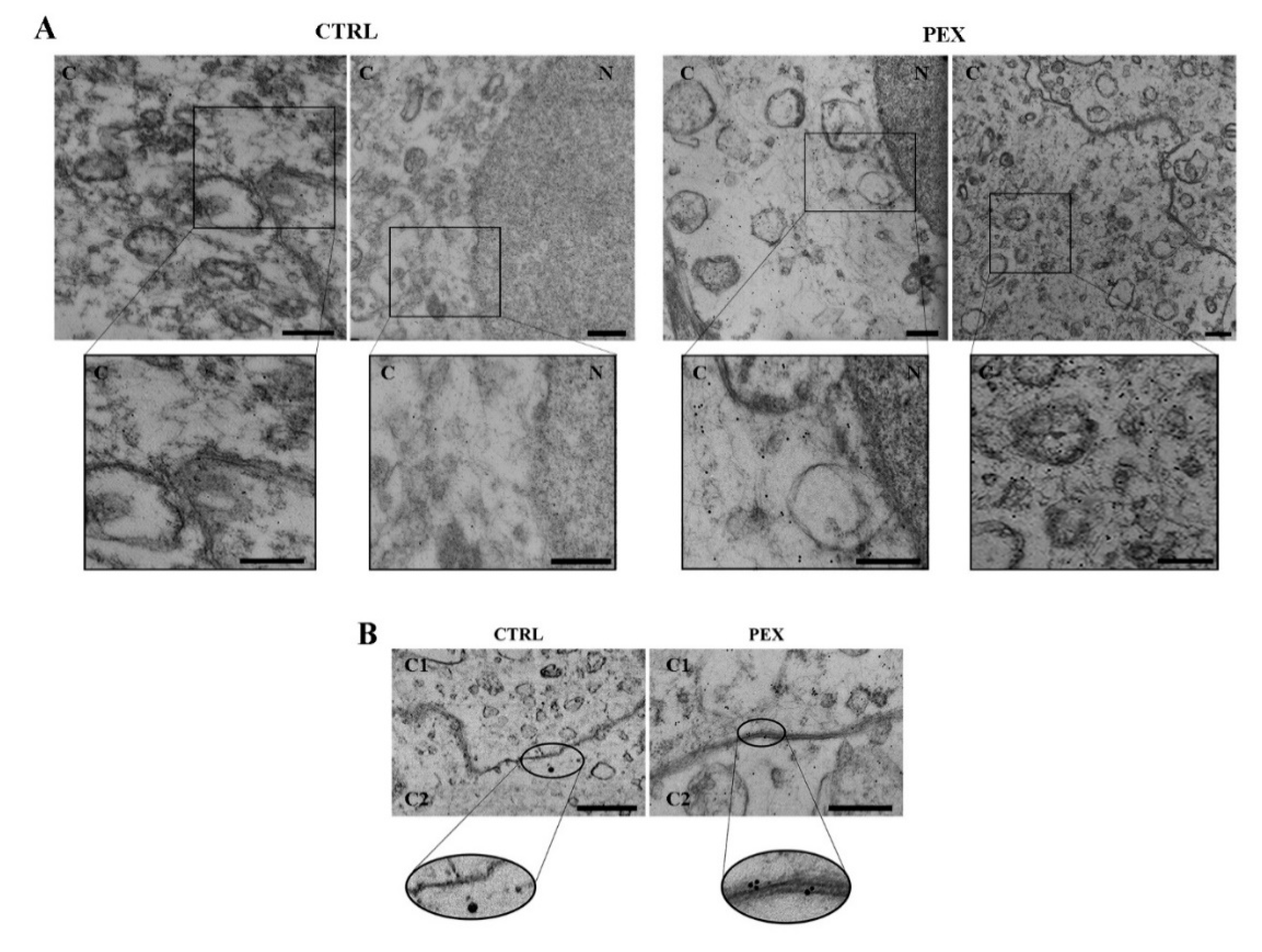
2.2. The Ultrastructural Localization of β-Catenin Using AU

In order to determine the localization of β-catenin at the ultrastructural level, the post-embedding colloidal gold (AU) method was used after introduction of certain modifications [18]. The anterior lens capsule and lens epithelial cells were fixed in 2% (w/v) paraformaldehyde (Serva, Heidelberg, Germany) in PBS for 20 min at room temperature (RT). Next, the surgical specimens were washed in PBS and post-fixed in 2% (w/v) OsO4 (Serva) for 1 h (RT). After the dehydration in a graded series of ethanol (POCH S.A, Gliwice, Poland), the tissue was embedded in LR White (Sigma-Aldrich, Merck KGaA, Darmastadt, Germany), cut by ultramicrotome (Reichert, Wien, Austria) and collected on nickel grids (Sigma-Aldrich). The ultra-thin sections were permeabilized by adding 0.25% Triton X-100 (Serva) in PBS (15 min, RT) and blocked with 5% BSA (Sigma-Aldrich) (1:200, 1 h, RT). The immune detection of β-catenin was performed by incubating primary antibodies dedicated to β-catenin (1 h, RT; Sigma-Aldrich). Later, the material was washed with PBS and incubated with anti-rabbit secondary antibodies conjugated with 10 nm gold particles (1:100, 1 h, RT; Aurion, Toowong, Australia). Finally, the ultra-thin sections were rinsed in redistilled water (5 ×5 min, RT) and stained with uranyl acetate (Ted Pella, Inc, Redding, CA, USA) and lead citrate (Sigma-Aldrich). The ultrastructural localization of β-catenin was examined by transmission electron microscope (JEM 100 CX, Jeol, Ltd., Tokyo, Japan).

2.3. The Fluorescent Labelling of Cell–Cell Junctions

For the analysis of the presence of the proteins involved in cell–cell junctions, including β-catenin, in surgical specimens of human anterior lens capsules with adherent LECs, fluorescence staining was performed. The surgical specimens were embedded in paraffin and then cut into thin sections according to the method described previously [17]. The next step included the dipping of material in xylene (POCH S.A), xylene/ethanol and series of ethanol. Next, the antigen retrieval was performed (Target Retrieval Solution, water bath 92 °C, 20 min; Dako, Agilent Technologies, Santa Clara, CA, USA). The tissue samples were placed on the slides and incubated with detergent (0.25% Triton X-100, 10 min, RT), BSA (1%, 20 min, RT) and rabbit primary antibodies anti-β-catenin (1:100, 1 h, RT). Each step was preceded by PBS washing (3 × 5 min, RT). The presence of cell–cell junctions was visualized using secondary antibodies AlexaFluor 555 (1:500, 1 h, RT; Invitrogen; Thermo Fisher Scientific, Inc., Waltham, MA, USA). After a final washing step in PBS, the cell nuclei were stained with DAPI (1:20,000, 10 min, RT; Sigma-Aldrich). The preparations were mounted in Aqua-Poly/Mount (Polysciences, Inc., Warrington, PA, USA) and examined using a C1 laser-scanning confocal microscope (Nikon, Tokyo, Japan). Additionally, the fluorescent intensity of cell–cell junctions including background emitted by the paraffin-embedded section was analyzed using ImageJ (Ver1.45s, NIH, Bethesda, MD, USA).

2.4. Statistical Analysis

The statistical analysis was performed using GraphPad Prism 7.0 (GraphPad Software, La Jolla, CA, USA). The differences in fluorescent intensity of β-catenin between the control group and the PEX group were analyzed using the Mann-Whitney U statistical test (p < 0.05) and statistically significant results were marked by an asterisk (*).

3. Results

3.1. The Ultrastructural Localization of β-Catenin

The post-embedding AU method was used to localize β-catenin at the ultrastructural level. The results are presented in Figure 1. The analysis of the electronograms from patients without the PEX syndrome (control group) revealed a single molecule of colloid gold at the cell surface (Figure 1A). In turn, the electronograms from the study group (PEX patients) showed an increased amount of AU at the cytoplasm of lens epithelial cells (Figure 1A). Additionally, the method chosen allowed us to determine the localization of β-catenin at the cell–cell contacts site. We observed the enhanced accumulation of AU in the area of cell junction in PEX group in comparison to control patients. In the control group, the long tight junctions (TJs), were observed, while in the PEX group, long adherens junctions (AJs) were noticed (Figure 1B).

3.2. The Fluorescence Staining of β-Catenin

To determine the presence of β-catenin in the context of cell–cell contacts, fluorescence staining was used. The results, presented in Figure 2, indicated an increased concentration of β-catenin in the area between lens epithelial cells in the material obtained from the PEX patients in comparison to the control group (Figure 2A). In turn, the intensity measurement of fluorescence staining showed a statistically significant (p = 0.035) increase in the level of β-catenin expression in lens epithelial cells for the PEX group (MFI = 808.98) in comparison to the control patients (MFI = 731.6) (Figure 2B). The presented results confirmed the earlier observation from the transmission electron microscope.

4. Discussion

To our knowledge, this is the first study assessing the β-catenin concentration in the lens epithelial cells in patients suffering from the cataract and the pseudoexfoliation syndrome, and in patients with the cataract alone. The etiology and pathophysiology of PEX still remain unexplained. For over 17 years, PEX has been considered a systemic disease. For example, PEX was thought to be connected with disorders impairing blood vessels [19]. It is commonly known that vascular calcification is a risk factor for causing cardiovascular events. However, the molecular mechanism underlying this pathogenic process is still obscure. Yao et al. noticed that β-catenin was involved in rat abdominal aortic calcification induced by high Pi. When the knockdown expression of β-catenin in the rat model was investigated, they found that aortic calcification was reduced [8]. Chronic and progressive upregulation of β-catenin is a final pathway common to a wide variety of fibrotic CKDs [20,21], including diabetic nephropathy, obstructive nephropathy, adriamycin nephropathy, chronic allograft nephropathy, polycystic kidney disease and remnant 5/6-nephrectomy [22]. Lin et al. revealed that protein expression levels of β-catenin were significantly increased by 2.8-fold (p = 0.002) in CKD group kidney tissues compared with the control group [22]. Similarly, our study showed a statistically significant increase in the level of β-catenin in PEX lens epithelial cells (MFI = 808.98) compared to control patients (MFI = 731.6). Furthermore, the analysis of the electronograms from PEX patients showed a larger amount of β-catenin in the cytoplasm of lens epithelial cells. Moreover, in our study, we observed an enhanced accumulation of β-catenin in the area of cell–cell junctions in the PEX group in comparison to the control patients. The increase was confirmed by fluorescence microscope and transmission electron microscope. We suggested that the mechanism of beta-catenin overexpression in PEX cases is associated with the changes in the shape of lens epithelial cells into a flat shape and in the organization of intercellular connections. We observed a change from tight epithelium (TJs) in CTRL to a more point-like pattern of AJs in the PEX patients. As a result, an increase in a level of fluorescence intensity of beta-catenin was noticed. This is a consequence of fact that mentioned types of intercellular junctions are important in contact with neighbouring cells, but they have different structures and functions [23]. Tight junctions are created by occludins and claudins, and are involved in selective barriers and maintaining the polarity of cells [23,24]. In turn, adherens junctions are formed by the cadherin/catenin complex and play key roles in cell–cell interactions, intracellular signalling and transcriptional regulation [23,25]. One of the components of AJs, β-catenin, is important in cell adhesion [26]. Kam and Quaranta also presented the key role of β-catenin in cadherin-based adherens junctions. [27]. Similarly, Valenta et al. indicated the involvement of protein in the formation and stabilization of cell–cell junctions [28]. Cited authors described that alterations in beta-catenin caused the destabilization in cell–cell connection [26,27]. Thus, cell flattening and changes in type of cell–cell junction may induce an increase in the level expression of β-catenin. The presence of point intercellular junctions and a change in epithelial permeability may be associated with an accumulation of pseudoexfoliative material on the anterior lens capsule. In addition, destabilization of cell–cell junctions is common in the cancer metastasis, which is characterized by the loss of contact inhibition, epithelial-to-mesenchymal transition (EMT), and abnormal cell invasiveness [29,30]. On the other hand, β-catenin plays a key function in the Wnt signaling pathway involved in many cellular processes including cell proliferation, adhesion, differentiation, and migration [27,31]. Antosova et al. presented that alterations in Wnt/β-catenin by ectopic activation in lens fiber cells of the transgenic mouse model resulted in delayed differentiation and promotes cataracts [32]. In turn, the study presented by Chong et al. indicated that in cataracts, an increase in Wnt expression is correlated with the EMT of lens epithelial cells. Moreover, the author draws attention to the relationship between Wnt signaling and the formation of fibrosis in the lens [33]. Similar results were reported by Bao et al. who observed that the upregulation expression of Wnt signaling is correlated with an overexpression of the β-catenin level and the nuclear translocation of protein in human lens epithelial (HLE). Thus, the EMT induced in this way promotes the fibrosis process in posterior capsular opacity (PCO) [34]. Furthermore, literature has reported that disturbances in β-catenin levels may contribute to disease promotion [28]. In our study, the higher level of β-catenin was caused by a change in the shape of the lens epithelial cells and disturbed cell–cell connections. These alterations in comparison to the control group may also be caused by a disturbance of Wnt/β-catenin through alterations in the nuclear transport of protein. The increase in the level of protein expression in the cytoplasm may be the result of a decrease in the level or an absence of β-catenin in the nucleus. The above observation may indicate that the Wnt/β-catenin pathway is impaired in LECs of PEX patients. Moreover, Zhang et al. described that a low level of the Wnt signaling pathway enhances aging in the eyes [35]. Our study presented an increase in β-catenin level at the cytoplasm of LECs among PEX patients. These results may correlate with the observations regarding disturbances in Wnt signaling homeostasis in the context of accelerating the aging processes, which is considered one of the factors in this disease entity [35].
The connection described above may indicate that PEX is one of multiple diseases simultaneously developing in the presence of impaired β-catenin expression. While many of them are challenging to identify in early stages, in the case of PEX, a slit lamp examination performed routinely during ophthalmological examination, makes it possible to recognize the disease. Confirming the β-catenin connection between other diseases could provide an opportunity to identify patients at risk of developing other systemic diseases or allow for an early diagnosis in cases where their development has already commenced.

5. Conclusions

Our study presented the increase in the β-catenin in LECs of PEX group in comparison to control patients. We suggest that this phenomenon is caused by a change in the cell shape and the organization of cell–cell junctions, which was confirmed by fluorescence microscope and TEM. In addition, it might be possible for PEX, due to it being easily recognizable, to be the first indicator of serious kidney or cardiac diseases, as well as cancer metastases. In such case, identifying it would present an opportunity for an earlier introduction of treatment. Further studies are needed in order to confirm this hypothesis.

Author Contributions

Conceptualization, J.S.; methodology, J.S., M.I., M.G.; validation, M.I., M.G.; formal analysis, M.H.-W., M.I.; investigation, J.S., M.H.-W., M.I.; resources, J.S.; data curation, M.I., M.H.-W.; writing—original draft preparation, J.S., M.H.-W.; writing—review and editing, J.S., M.I.; visualization, J.S., M.H.-W.; supervision, G.M., A.G.; project administration, J.S. All authors have read and agreed to the published version of the manuscript.

Funding

This research did not receive any specific grants from funding agencies in the public, commercial, or not-for-profit sectors.

Conflicts of Interest

The authors declare no conflict of interest.

References

  1. Shapiro, L.; Weis, W.I. Structure and biochemistry of cadherins and catenins. Cold Spring Harb. Perspect. Biol. 2009, 1, a003053. [Google Scholar] [CrossRef] [PubMed]
  2. Clevers, H. Wnt/beta-catenin signaling in development and disease. Cell 2006, 127, 469–480. [Google Scholar] [CrossRef] [PubMed] [Green Version]
  3. Tamai, K.; Semenov, M.; Kato, Y.; Spokony, R.; Liu, C.; Katsuyama, Y.; Hess, F.; Saint-Jeannet, J.P.; He, X. LDL-receptor-related proteins in Wnt signal transduction. Nature 2000, 407, 530–535. [Google Scholar] [CrossRef] [PubMed]
  4. Zhou, T.; Hu, Y.; Chen, Y.; Zhou, K.K.; Zhang, B.; Gao, G.; Ma, J.X. The pathogenic role of the canonical Wnt pathway in age-related macular degeneration. Investig. Ophthalmol. Vis. Sci. 2010, 51, 4371–4379. [Google Scholar] [CrossRef] [PubMed]
  5. Huang, H.; He, X. Wnt/beta-catenin signaling: New (and old) players and new insights. Curr. Opin. Cell Biol. 2008, 20, 119–125. [Google Scholar] [CrossRef] [Green Version]
  6. Fodde, R.; Brabletz, T. Wnt/beta-catenin signaling in cancer stemness and malignant behavior. Curr. Opin. Cell Biol. 2007, 19, 150–158. [Google Scholar] [CrossRef]
  7. Xiao, L.; Zhou, D.; Tan, R.J.; Fu, H.; Zhou, L.; Hou, F.F.; Liu, Y. Sustained Activation of Wnt/β-Catenin Signaling Drives AKI to CKD Progression. J. Am. Soc. Nephrol. 2016, 27, 1727–1740. [Google Scholar] [CrossRef] [Green Version]
  8. Yao, L.; Sun, Y.T.; Sun, W.; Xu, T.H.; Ren, C.; Fan, X.; Sun, L.; Liu, L.L.; Feng, J.M.; Ma, J.F.; et al. High phosphorus level leads to aortic calcification via β-catenin in chronic kidney disease. Am. J. Nephrol. 2015, 41, 28–36. [Google Scholar] [CrossRef]
  9. Zhou, G.; Wu, J.; Gu, C.; Wang, B.; Abel, E.D.; Cheung, A.K.; Huang, Y. Prorenin independently causes hypertension and renal and cardiac fibrosis in cyp1a1-prorenin transgenic rats. Clin. Sci. 2018, 132, 1345–1363. [Google Scholar] [CrossRef] [Green Version]
  10. Heiser, P.W.; Cano, D.A.; Landsman, L.; Kim, G.E.; Kench, J.G.; Klimstra, D.S.; Taketo, M.M.; Biankin, A.V.; Hebrok, M. Stabilization of beta-catenin induces pancreas tumor formation. Gastroenterology 2008, 135, 1288–1300. [Google Scholar] [CrossRef] [Green Version]
  11. Zeng, G.; Germinaro, M.; Micsenyi, A.; Monga, N.K.; Bell, A.; Sood, A.; Malhotra, V.; Sood, N.; Midda, V.; Monga, D.K.; et al. Aberrant Wnt/beta-catenin signaling in pancreatic adenocarcinoma. Neoplasia 2006, 8, 279–289. [Google Scholar] [CrossRef] [Green Version]
  12. Wang, L.; Heidt, D.G.; Lee, C.J.; Yang, H.; Logsdon, C.D.; Zhang, L.; Fearon, E.R.; Ljungman, M.; Simeone, D.M. Oncogenic function of ATDC in pancreatic cancer through Wnt pathway activation and beta-catenin stabilization. Cancer Cell 2009, 15, 207–219. [Google Scholar] [CrossRef] [PubMed] [Green Version]
  13. Pramanik, K.C.; Fofaria, N.M.; Gupta, P.; Ranjan, A.; Kim, S.H.; Srivastava, S.K. Inhibition of β-Catenin signaling suppresses pancreatic tumor growth by disrupting nuclear β-Catenin/TCF-1 complex: Critical role of STAT-3. Oncotarget 2015, 10, 11561–11574. [Google Scholar] [CrossRef] [PubMed] [Green Version]
  14. Reddy, H.S.; Keene, C.D.; Chang, S.H.; Jian-Amadi, A.; Cimino, P.J. Immunohistochemical profiling including beta-catenin in conjunctival melanocytic lesions. Exp. Mol. Pathol. 2017, 102, 198–202. [Google Scholar] [CrossRef] [PubMed]
  15. Dissanayake, W.C.; Sorrenson, B.; Shepherd, P.R. The role of adherens junction proteins in the regulation of insulin secretion. Biosci. Rep. 2018, 38, 2. [Google Scholar] [CrossRef] [PubMed] [Green Version]
  16. Sorrenson, B.; Cognard, E.; Lee, K.L.; Dissanayake, W.C.; Fu, Y.; Han, W.; Hughes, W.E.; Shepherd, P.R. A critical role for β-catenin in modulating levels of insulin secretion from β-cells by regulating actin cytoskeleton and insulin vesicle localization. J. Biol. Chem. 2016, 291, 25888–25900. [Google Scholar] [CrossRef] [Green Version]
  17. Stafiej, J.; Hałas-Wiśniewska, M.; Izdebska, M.; Gagat, M.; Grzanka, D.; Grzanka, A.; Malukiewicz, G. Immunohistochemical analysis of microsomal glutathione S-transferase 1 and clusterin expression in lens epithelial cells of patients with pseudoexfoliation syndrome. Exp. Ther. Med. 2017, 13, 1057–1063. [Google Scholar] [CrossRef]
  18. Izdebska, M.; Gagat, M.; Grzanka, D.; Grzanka, A. Ultrastructural localization of F-actin using phalloidin and quantum dots in HL-60 promyelocytic leukemia cell line after cell death induction by arsenic trioxide. Acta Histochem. 2013, 115, 487–495. [Google Scholar] [CrossRef]
  19. Schumacher, S.; Schlötzer-Schrehardt, U.; Martus, P.; Lang, W.; Naumann, G.O. Pseudoexfoliation syndrome and aneurysms of the abdominal aorta. Lancet 2001, 357, 359–360. [Google Scholar] [CrossRef]
  20. Dai, C.; Stolz, D.B.; Kiss, L.P.; Monga, S.P.; Holzman, L.B.; Liu, Y. Wnt/beta-catenin signaling promotes podocyte dysfunction and albuminuria. J. Am. Soc. Nephrol. 2009, 20, 1997–2008. [Google Scholar] [CrossRef] [Green Version]
  21. von Toerne, C.; Schmidt, C.; Adams, J.; Kiss, E.; Bedke, J.; Porubsky, S.; Gretz, N.; Lindenmeyer, M.T.; Cohen, C.D.; Gröne, H.J.; et al. Wnt pathway regulation in chronic renal allograft damage. Am. J. Transplant. 2009, 9, 2223–2239. [Google Scholar] [CrossRef]
  22. Lin, X.; Zha, Y.; Zeng, X.Z.; Dong, R.; Wang, Q.H.; Wang, D.T. Role of the Wnt/β-catenin signaling pathway in inducing apoptosis and renal fibrosis in 5/6-nephrectomized rats. Mol. Med. Rep. 2017, 15, 3575–3582. [Google Scholar] [CrossRef] [PubMed]
  23. Hartsock, A.; Nelson, W.J. Adherens and tight junctions: Structure, function and connections to the actin cytoskeleton. Biochim. Biophys. Acta 2008, 1778, 660–669. [Google Scholar] [CrossRef] [PubMed] [Green Version]
  24. Vermette, D.; Hu, P.; Canarie, M.F.; Funaro, M.; Glover, J.; Pierce, R.W. Tight junction structure, function, and assessment in the critically ill: A systematic review. Intensive Care Med. Exp. 2018, 6, 37. [Google Scholar] [CrossRef] [PubMed] [Green Version]
  25. McEwen, A.E.; Escobar, D.E.; Gottardi, C.J. Signaling from the adherens junction. Sub-Cell. Biochem. 2012, 60, 171–196. [Google Scholar]
  26. Cadigan, K.M.; Peifer, M. Wnt signaling from development to disease: Insights from model systems. Cold Spring Harb. Perspect. Biol. 2009, 1, a002881. [Google Scholar] [CrossRef] [Green Version]
  27. Kam, Y.; Quaranta, V. Cadherin-Bound β-Catenin Feeds into the Wnt Pathway upon Adherens Junctions Dissociation: Evidence for an Intersection between β-Catenin Pools. PLoS ONE 2009, 4, e4580. [Google Scholar] [CrossRef] [PubMed]
  28. Valenta, T.; Hausmann, G.; Basler, K. The many faces and functions of β-catenin. EMBO J. 2012, 31, 2714–2736. [Google Scholar] [CrossRef] [PubMed] [Green Version]
  29. Vasioukhin, V. Adherens junctions and cancer. Sub-Cell. Biochem. 2012, 60, 379–414. [Google Scholar]
  30. Yang, S.; Liu, Y.; Li, M.Y.; Ng, C.S.H.; Yang, S.L.; Wang, S.; Zou, C.; Dong, Y.; Du, J.; Long, X.; et al. FOXP3 promotes tumor growth and metastasis by activating Wnt/β-catenin signaling pathway and EMT in non-small cell lung cancer. Mol. Cancer 2017, 16, 124. [Google Scholar] [CrossRef]
  31. Sedgwick, A.E.; D’Souza-Schorey, C. Wnt Signaling in Cell Motility and Invasion: Drawing Parallels between Development and Cancer. Cancers 2016, 8, 80. [Google Scholar] [CrossRef] [PubMed] [Green Version]
  32. Antosova, B.; Smolikova, J.; Borkovcova, R.; Strnad, H.; Lachova, J.; Machon, O.; Kozmik, Z. Ectopic activation of Wnt/β-catenin signaling in lens fiber cells results in cataract formation and aberrant fiber cell differentiation. PLoS ONE 2013, 8, e78279. [Google Scholar] [CrossRef] [Green Version]
  33. Chong, C.C.; Stump, R.J.; Lovicu, F.J.; McAvoy, J.W. TGFbeta promotes Wnt expression during cataract development. Exp. Eye Res. 2009, 88, 307–313. [Google Scholar] [CrossRef] [PubMed] [Green Version]
  34. Bao, X.L.; Song, H.; Chen, Z.; Tang, X. Wnt3a promotes epithelial-mesenchymal transition, migration, and proliferation of lens epithelial cells. Mol. Vis. 2012, 18, 1890–1983. [Google Scholar] [PubMed]
  35. Zhang, Y.; Jeffrey, J.; Dong, F.; Zhang, J.; Kao, W.W.; Liu, C.Y.; Yuan, Y. Repressed Wnt Signaling Accelerates the Aging Process in Mouse Eyes. J. Ophthalmol. 2019, 2019, 7604396. [Google Scholar] [CrossRef] [Green Version]
Figure 1. The analysis of the ultrastructural localization of β-catenin in control group vs.pseudoexfoliation syndrome (PEX) group. A single molecule of colloid gold at the cell surface in control group and increased amount of AU in the cytoplasm in the study group. N—nucleus; C—cytoplasm. (A). The enhanced accumulation of AU in the area of cell junction in the PEX group in comparison to the control group. C1—cell 1; C2—cell 2. (B). Bar—100 nm (inserts—200 nm).
Applsci 10 06199 g001
Figure 2. The fluorescence staining of β-catenin. The increased concentration of β-catenin in the area between lens epithelial cells in the PEX group vs. the control group (A). A significant increase in the level of expression of β-catenin in lens epithelial cells of the PEX group, compared to the control group. Data shown represent the mean ± SD obtained from PEX n = 29 and CTRL n = 23. The statistically significant results were marked by an asterisk (* p < 0.05; Mann-Whitney U test) (B).
Applsci 10 06199 g002

