Development of a Musculoskeletal Model of Hyolaryngeal Elements for Understanding Pharyngeal Swallowing Mechanics
Abstract
:Featured Application
Abstract
1. Introduction
2. Materials and Methods
2.1. Motion Analysis of the Hyoid Bone and Thyroid Cartilage during Swallowing
2.1.1. Method of the Analysis
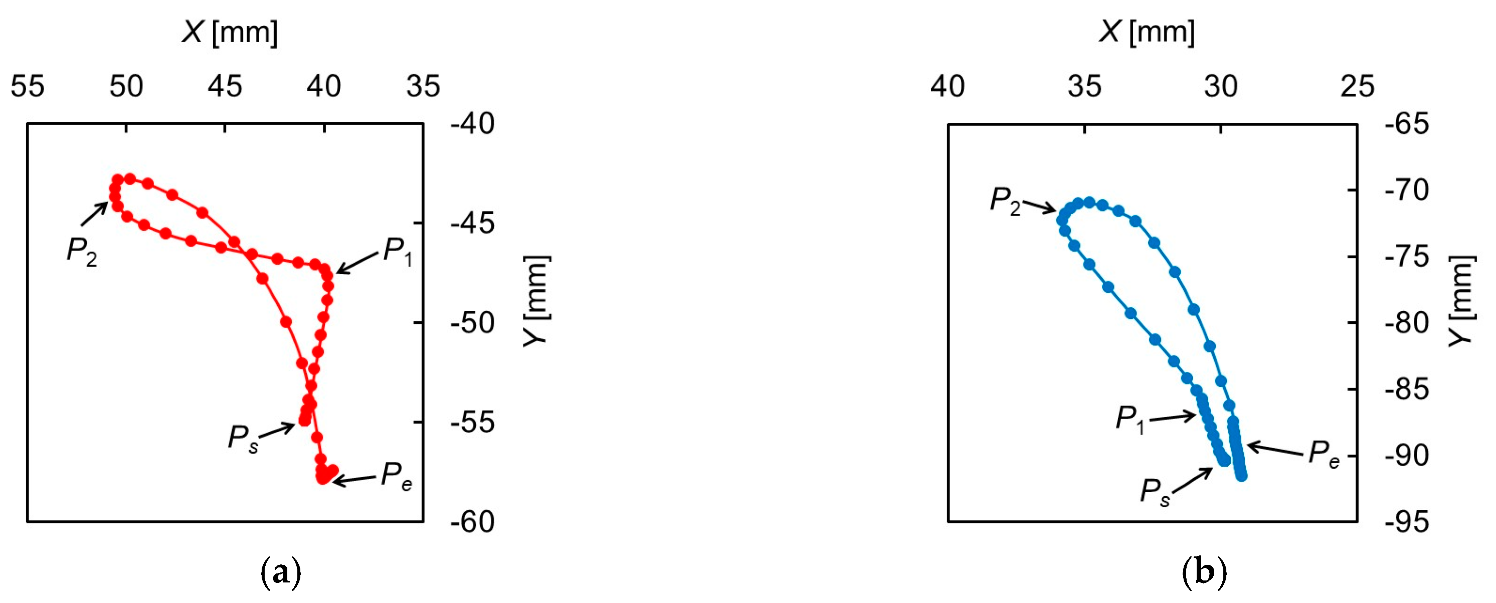
2.1.2. Results of the Analysis
2.2. Development of the Musculoskeletal Model of Swallowing
2.2.1. Modeling of the Swallowing-Related Muscles
2.2.2. Modeling of Damping and Stiffness of Biological Tissue
2.2.3. Modeling of the Mechanical Properties of Muscles
2.2.4. Physiological Cross-Sectional Area (PCSA)
2.2.5. Muscle Force Estimation Using an Optimization Program
3. Results
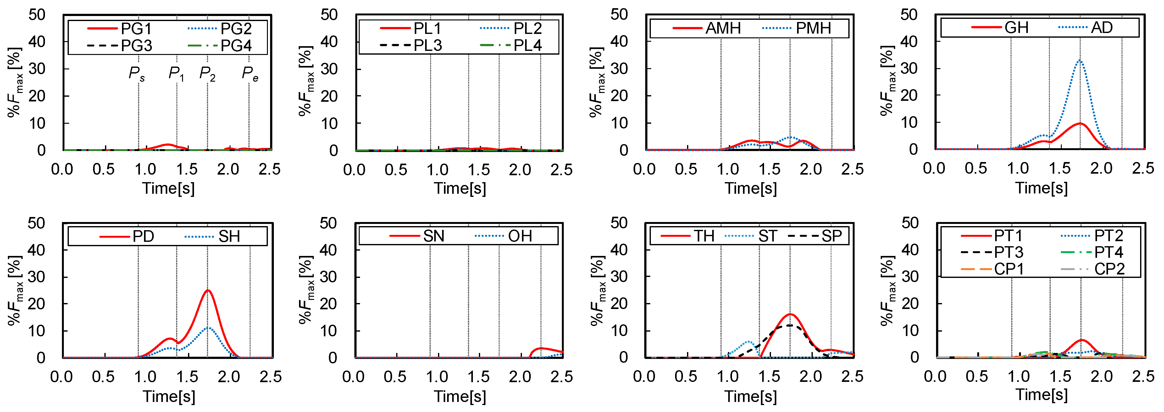
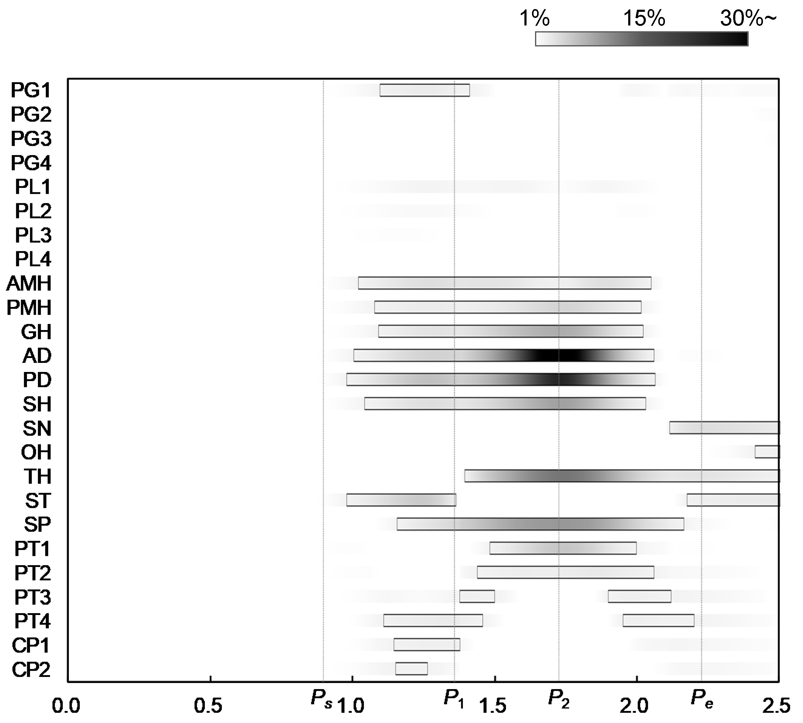
3.1. Results of Muscle Force Estimation
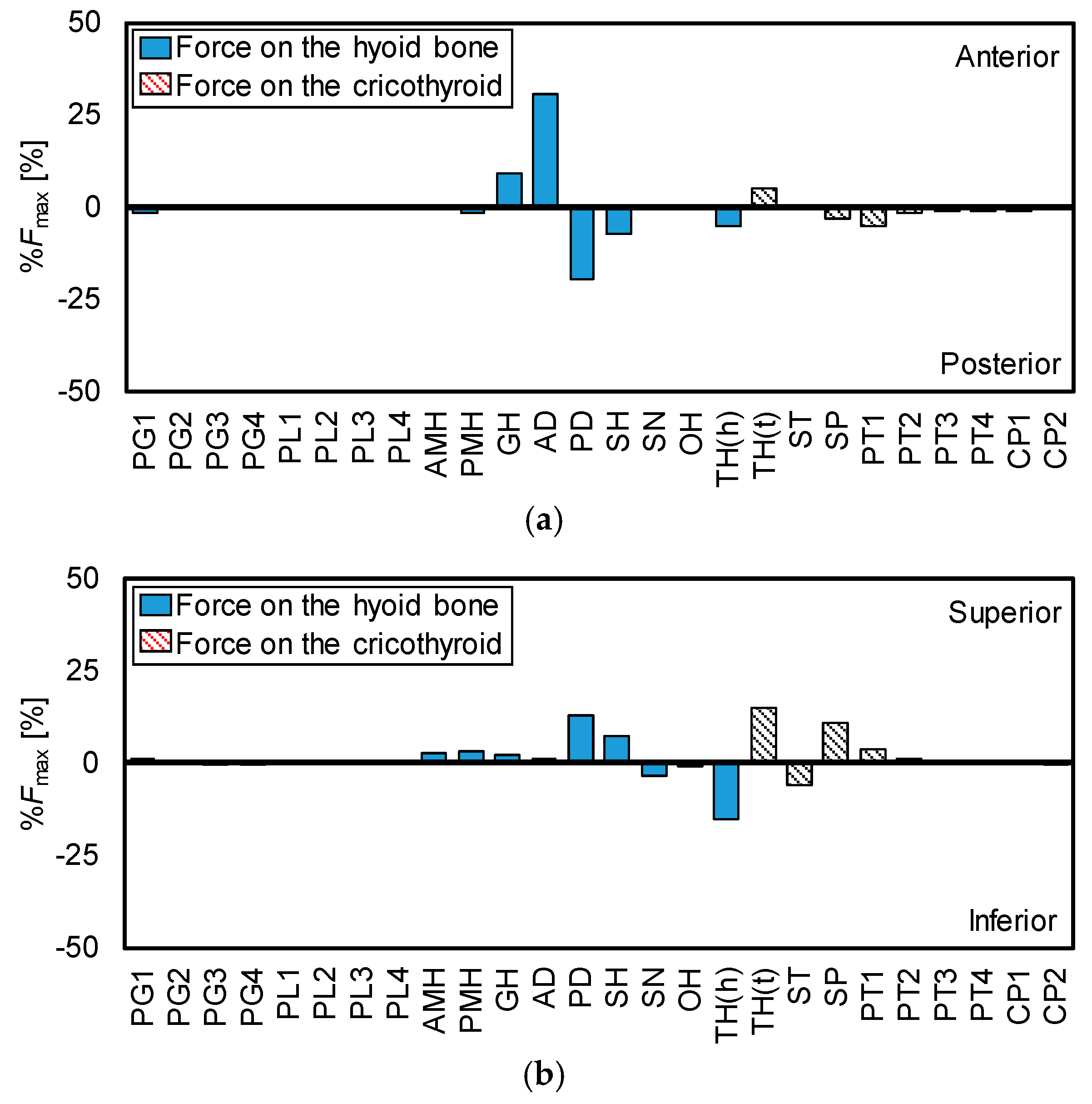
3.2. Force Direction of the Muscles
4. Discussion
4.1. Activity Pattern of the Muscles
- −
- Some studies have indicated that activation of AD, GH, MH, and SN varied with food characteristics and that there were no obvious consistent activation patterns [43,44,45]. However, a more recent study suggested that there was a typical sequence of muscle activation [46]. That is, the masseter (MA) is first, the next is GH and AD, and the last is SN.
- −
- −
- The onset of muscle activation of MH before that of TH was significant [47].
- −
- Normal activity of CP exhibits an inhibition phase in swallowing: Preswallowing and rebound bursts across the pause [48].
4.2. Role of the Muscles
4.3. Limitations
5. Conclusions
Author Contributions
Funding
Conflicts of Interest
References
- Statistics Bureau of Japan. Current Population Estimates as of 1 October 1 2016. Available online: http://www.stat.go.jp/english/data/jinsui/2016np/index.htm (accessed on 1 March 2017).
- Statistics and Information Department Minister’s Secretariat of Ministry of Health, Labour and Welfare. Vital Statistics of Japan 2015—Trends in Leading Causes of Death: Japan. Available online: http://www.e-stat.go.jp/SG1/estat/ListE.do?lid=000001158057 (accessed on 1 March 2017).
- Teramoto, S.; Fukuchi, Y.; Sasaki, H.; Sato, K.; Sekizawa, K.; Matsuse, T. High incidence of aspiration pneumonia in community- and hospital-acquired pneumonia in hospitalized patients: A multicenter, prospective study in Japan. J. Am. Geriatr. Soc. 2008, 56, 577–579. [Google Scholar] [CrossRef] [PubMed]
- Kaplan, V.; Angus, D.C.; Griffin, M.F.; Clermont, G.; Watson, R.S.; Linde-Zwirble, W.T. Hospitalized Community-acquired Pneumonia in the Elderly Age- and Sex-related Patterns of Care and Outcome in the United States. Am. J. Respir. Crit. Care Med. 2002, 165, 766–772. [Google Scholar] [CrossRef]
- Cunningham, E.T., Jr.; Jones, B. Anatomical and Physiological Overview. In Normal and Abnormal Swallowing: Imaging in Diagnosis and Therapy, 2nd ed.; Jones, B., Ed.; Springer: New York, NY, USA, 2003; pp. 11–34. [Google Scholar]
- Palmer, J.B.; Kuhlemeier, K.V.; Tippett, D.C.; Lynch, C. A protocol for the videofluorographic swallowing study. Dysphagia 1993, 8, 209–214. [Google Scholar] [CrossRef] [PubMed]
- Scharitzer, M.; Pokieser, P. Videofluoroscopy: Current Clinical Impact in Deglutology. J. Gastroenterol. Hepatol. Res. 2014, 3, 1061–1065. [Google Scholar]
- Leder, S.B.; Sasaki, C.T.; Burrell, M.I. Fiberoptic endoscopic evaluation of dysphagia to identify silent aspiration. Dysphagia 1998, 13, 19–21. [Google Scholar] [CrossRef] [PubMed]
- Aviv, J.E.; Kaplan, S.T.; Thomson, J.E.; Spitzer, J.; Diamond, B.; Close, L.G. The Safety of Flexible Endoscopic Evaluation of Swallowing with Sensory Testing (FEESST): An Analysis of 500 Consecutive Evaluations. Dysphagia 2000, 15, 39–44. [Google Scholar] [CrossRef]
- Neis, L.R.; Logemann, J.; Larson, C. Viscosity effects on EMG activity in normal swallow. Dysphagia 1994, 9, 101–106. [Google Scholar]
- Crary, M.A.; Baldwin, B.O. Surface Electromyographic Characteristics of Swallowing in Dysphagia Secondary to Brainstem Stroke. Dysphagia 1997, 12, 180–187. [Google Scholar] [CrossRef]
- Delp, S.T.; Loan, J.P. A computational framework for simulating and analyzing human and animal movement. IEEE Comput. Sci. Eng. 2000, 2, 46–55. [Google Scholar] [CrossRef] [Green Version]
- Damsgaard, M.; Rasmussen, J.; Christensen, S.T.; Surma, E.; de Zee, M. Analysis of musculoskeletal systems in the AnyBody Modeling System. Simul. Model. Pract. Theory 2006, 14, 1100–1111. [Google Scholar] [CrossRef]
- Delp, S.L.; Anderson, F.C.; Arnold, A.S.; Loan, P.; Habib, A.; John, C.T.; Guendelman, E.; Thelen, D.G. OpenSim: Open-source software to create and analyze dynamic simulations of movement. IEEE Trans. Biomed. Eng. 2007, 54, 1940–1950. [Google Scholar] [CrossRef] [PubMed] [Green Version]
- Nakamura, Y.; Yamane, K.; Fujita, Y.; Suzuki, I. Somatosensory Computation for Man-Machine Interface from Motion Capture Data and Musculoskeletal Human Model. IEEE Trans. Robot. 2005, 21, 58–66. [Google Scholar] [CrossRef]
- Crowninshield, R.D.; Brand, R.A. A physiologically based criterion of muscle force prediction in locomotion. J. Biomech. 1981, 14, 793–801. [Google Scholar] [CrossRef]
- Ravera, E.P.; Crespo, M.J.; Braidot, A.A. Estimation of muscle forces in gait using a simulation of the electromyographic activity and numerical optimization. Comput. Methods Biomech. Biomed. Eng. 2014, 19, 1–12. [Google Scholar] [CrossRef] [PubMed]
- Sasaki, K.; Neptune, R.R. Muscle mechanical work and elastic energy utilization during walking and running near the preferred gait transition speed. Gait Posture 2006, 23, 383–390. [Google Scholar] [CrossRef] [PubMed]
- Anderson, F.C.; Pandy, M.G. A Dynamic Optimization Solution for Vertical Jumping in Three Dimensions. Comput. Methods Biomech. Biomed. Eng. 1999, 2, 201–231. [Google Scholar] [CrossRef]
- Holzbaur, K.R.S.; Murray, W.M.; Delp, S.L. A Model of the Upper Extremity for Simulating Musculoskeletal Surgery and Analyzing Neuromuscular Control. Ann. Biomed. Eng. 2005, 33, 829–840. [Google Scholar] [CrossRef]
- Nikooyan, A.A.; Veerger, H.E.J.; Chadwick, E.K.J.; Praagman, M.; Helm, F.C. Development of a comprehensive musculoskeletal model of the shoulder and elbow. Med. Biol. Eng. Comput. 2011, 49, 1425–1435. [Google Scholar] [CrossRef] [Green Version]
- Lin, H.T.; Nakamura, Y.; Su, F.C.; Elias, J.J.; Hashimoto, J.; Chao, E.Y.S. Use of Virtual, Interactive, Musculoskeletal System (VIMS) in Modeling and Analysis of Shoulder Throwing Activity. J. Biomech. Eng. 2005, 127, 525–530. [Google Scholar] [CrossRef] [Green Version]
- Sasaki, M.; Stefanov, D.; Ota, Y.; Miura, H.; Nakayama, A. Shoulder joint contact force during lever-propelled wheelchair propulsion. Robomech J. 2015, 2, 1–10. [Google Scholar] [CrossRef] [Green Version]
- Stavness, I.; Hannam, A.G.; Lloyd, J.E.; Fels, S. An Integrated Dynamic Jaw and Laryngeal Model Constructed from CT Data. In Proceedings of the Third International Symposium for Biomedical Simulation, Zurich, Switzerland, 10–11 July 2006; pp. 169–177. [Google Scholar]
- Hannam, A.G.; Stavness, I.; Lloyd, J.E.; Fels, S. A dynamic model of jaw and hyoid biomechanics during chewing. J. Biomech. 2008, 41, 1069–1076. [Google Scholar] [CrossRef] [PubMed]
- Hashimoto, T.; Murakoshi, A.; Kikuchi, T.; Michiwaki, Y.; Koike, T. Development of musculoskeletal model for the hyoid bone during swallowing. In Proceedings of the 2016 IEEE-EMBS International Conference on Biomedical and Health Informatics, Las Vegas, NV, USA, 24–27 February 2016; pp. 457–460. [Google Scholar]
- Hashimoto, T.; Murakoshi, A.; Kikuchi, T.; Michiwaki, Y.; Koike, T. Development of Musculoskeletal Model to Estimate Muscle Activities during Swallowing. In Digital Human Modeling: Applications in Health, Safety, Ergonomics and Risk Management (Lecture Note in Computer Science); Duffy, V.G., Ed.; Springer: Cham, Switzerland, 2016; Volume 9745, pp. 82–91. [Google Scholar]
- Sonomura, M.; Mizunuma, H.; Numamori, T.; Michiwaki, H.; Nishinari, K. Numerical simulation of the swallowing of liquid bolus. J. Texture Stud. 2011, 42, 203–211. [Google Scholar] [CrossRef]
- Ho, A.K.; Tsou, L.; Green, S.; Fels, S. A 3D swallowing simulation using smoothed particle hydrodynamics. Comput. Methods Biomech. Biomed. Eng. Imaging Vis. 2014, 2, 237–244. [Google Scholar] [CrossRef]
- Kikuchi, T.; Michiwaki, Y.; Kamiya, T.; Toyama, Y.; Tamai, T.; Koshizuka, S. Human swallowing simulation based on videofluorography images using Hamiltonian MPS method. Comput. Part. Mech. 2015, 2, 247–260. [Google Scholar] [CrossRef] [Green Version]
- Pearson, W.G.; Taylor, B.K.; Blair, J.; Martin-Harris, B. Computational analysis of swallowing mechanics underlying impaired epiglottic inversion. Laryngoscope 2016, 126, 1854–1858. [Google Scholar] [CrossRef]
- Ishida, R.; Palmer, J.B.; Hiiemae, K.M. Hyoid Motion during Swallowing: Factors Affecting Forward and Upward Displacement. Dysphagia 2002, 17, 262–272. [Google Scholar] [CrossRef]
- McFarland, D.H. Netter’s Atlas of Anatomy for Speech, Swallowing, and Hearing, 2nd ed.; Elsevier—Health Sciences Division: Amsterdam, The Netherlands, 2008; pp. 109–170. [Google Scholar]
- BodyParts3D/Anatomography. Available online: http://lifesciencedb.jp/bp3d/ (accessed on 1 March 2019).
- Mitsuhashi, N.; Fujieda, K.; Tamura, T.; Kawamoto, S.; Takagi, T.; Okubo, K. BodyParts3D: 3D structure database for anatomical concepts. Nucleic Acids Res. 2009, 37 (Suppl. 1). [Google Scholar] [CrossRef] [Green Version]
- Pailler-Mattei, C.; Bee, S.; Zahouani, H. In vivo measurements of the elastic mechanical properties of human skin by indentation test. Med. Eng. Phys. 2008, 30, 599–606. [Google Scholar] [CrossRef]
- Hill, A.V. The heat of shortening and the dynamic constants of muscle. Proc. R. Soc. Lond. Ser. B Biol. Sci. 1938, 126, 136–195. [Google Scholar]
- Strove, S. Learning combined feedback and feedforward control of a musculoskeletal system. Biol. Cybern. 1996, 75, 73–83. [Google Scholar] [CrossRef]
- Strove, S. Impedance Characteristics of a Neuromusculoskeletal model of the human arm I. Posture Control. Biol. Cybern. 1999, 81, 475–494. [Google Scholar] [CrossRef]
- Borst, J.; Forbes, P.A.; Happee, R.; Veeger, D.H. Muscle parameters for musculoskeletal modelling of the human neck. Clin. Biomech. 2011, 26, 343–351. [Google Scholar] [CrossRef] [Green Version]
- Pearson, W.G., Jr.; Langmore, S.E.; Zumwalt, A.C. Evaluating the Structural Properties of Suprahyoid Muscles and their Potential for Moving the Hyoid. Dysphagia 2011, 26, 345–351. [Google Scholar] [CrossRef] [PubMed] [Green Version]
- Pearson, W.G., Jr.; Langmore, S.E.; Yu, L.B.; Zumwalt, A.C. Structural Analysis of Muscles Elevating the Hyolaryngeal Complex. Dysphagia 2012, 27, 445–451. [Google Scholar] [CrossRef] [PubMed] [Green Version]
- Palmer, J.B.; Rudin, N.J.; Lara, G.; Crompton, A.W. Coordination of Mastication and Swallowing. Dysphagia 1992, 7, 187–200. [Google Scholar] [CrossRef] [PubMed]
- Spiro, J.; Rendell, J.; Gay, T. Activation and coordination patterns of the suprahyoid muscles during swallowing. Laryngoscope 1994, 104, 1376–1382. [Google Scholar] [CrossRef]
- Palmer, P.M.; McCulloch, T.M.; Jaffe, D.; Neel, A.T. Effects of a Sour Bolus on the Intramuscular Electromyographic (EMG) Activity of Muscles in the Submental Region. Dysphagia 2005, 20, 210–217. [Google Scholar] [CrossRef]
- Inokuchi, H.; González-Fernández, M.; Matsuo, K.; Brodsky, M.B.; Yoda, M.; Taniguchi, H.; Okazaki, H.; Hiraoka, T.; Palmer, J.B. Electromyography of Swallowing with Fine Wire Intramuscular Electrodes in Healthy Human: Activation Sequence of Selected Hyoid Muscles. Dysphagia 2014, 29, 713–721. [Google Scholar] [CrossRef]
- Burnett, T.A.; Mann, E.A.; Stoklosa, J.B.; Ludlow, C.L. Self-Triggered Functional Electrical Stimulation during Swallowing. J. Neurophysiol. 2005, 94, 4011–4018. [Google Scholar] [CrossRef]
- Perlman, A.L.; Palmer, P.M.; McCulloch, T.M.; Vandaele, D.J. Electromyographic activity from human laryngeal, pharyngeal, and submental muscles during swallowing. J. Appl. Physiol. 1999, 86, 1663–1669. [Google Scholar] [CrossRef]
- Rohrlea, O.; Pullana, A.J. Three-dimensional finite element modelling of muscle forces during mastication. J. Biomech. 2007, 40, 3363–3372. [Google Scholar] [CrossRef] [PubMed]


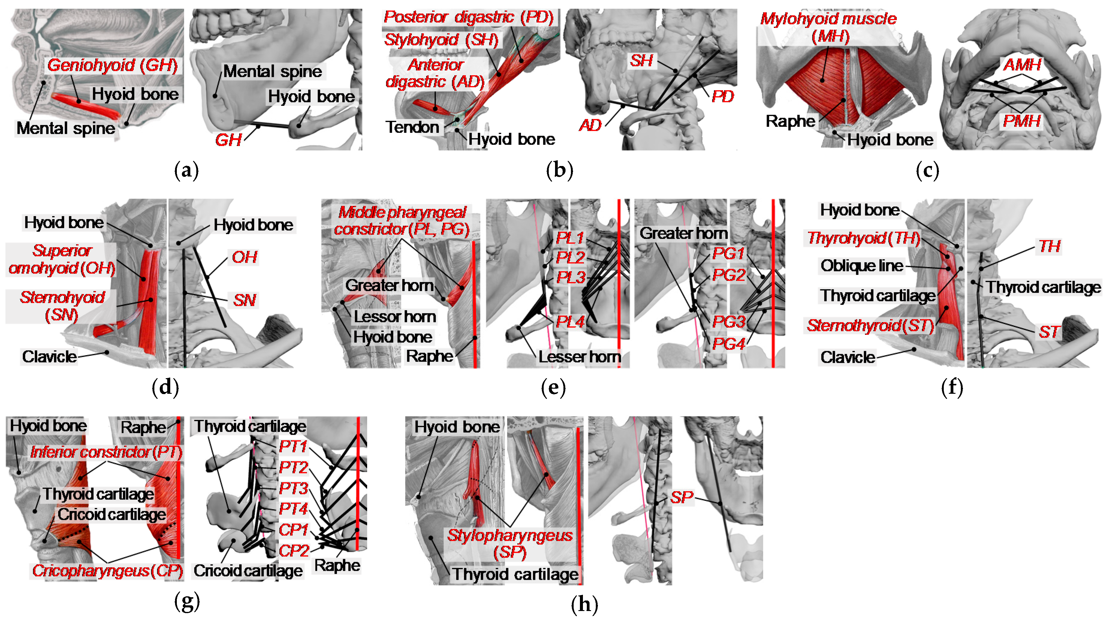

| Muscle Name | Symbol | Properties | |||
|---|---|---|---|---|---|
| Origin | Insertion | PCSA | |||
| Geniohyoid | GH | mental spine | anterior surface of the hyoid bone | 4.60 | |
| Stylohyoid | SH | posterior surface of the styloid process | lessor horn of the hyoid bone | 2.70 | |
| Digastric | anterior belly | AD | digastric fossa of the mandible | lessor horn of the hyoid bone | 5.50 |
| posterior belly | PD | mastoid process | lessor horn of the hyoid bone | 6.40 | |
| Mylohyoid | anterior part | AMH | anterior part of mylohyoid line | anterior surface of the hyoid bone | 8.20 |
| posterior part | PMH | posterior part of mylohyoid line | anterior surface of the hyoid bone | 4.30 | |
| Sternohyoid | SN | posterior part of the manubrium of sternum | lower border of the hyoid bone | 3.41 | |
| Omohyoid | superior belly | OH | intermediate tendon | lower border of the hyoid bone | 2.75 |
| inferior belly | - | - | - | - | |
| Middle pharyngeal constrictor | greater horn | PG1–4 | greater horn of the hyoid bone | pharyngeal raphe | 1.65 (*) |
| lesser horn | PL1–4 | lessor horn of the hyoid bone | pharyngeal raphe | 1.13 (*) | |
| Thyrohyoid | TH | oblique line on the thyroid cartilage | lower border of the hyoid bone | 6.04 | |
| Sternothyroid | ST | posterior part of the manubrium of sternum | oblique line on the thyroid cartilage | 5.14 | |
| Inferior pharyngeal constrictor | PT1–4 | oblique line of thyroid lamina | pharyngeal raphe | 2.25 (*) | |
| Cricopharyngeus | CP1, 2 | rear part of cricoid | pharyngeal raphe | 1.15 (*) | |
| Stylopharyngeus | SP | medial side of the styloid process | posterior border of the thyroid cartilage | 1.90 | |
| Parameter | Value | Unit |
|---|---|---|
| [N/m2] | ||
| Derived from the model | [m] | |
| [m] | ||
| 0.5 | [m] | |
| 6 | [m/s] | |
| 0.3 | [-] | |
| 0.23 | [-] | |
| 1.3 | [-] | |
| 0.2 | [-] | |
| 4.0 | [-] | |
| 0.4 | [m] |
© 2020 by the authors. Licensee MDPI, Basel, Switzerland. This article is an open access article distributed under the terms and conditions of the Creative Commons Attribution (CC BY) license (http://creativecommons.org/licenses/by/4.0/).
Share and Cite
Hashimoto, T.; Urabe, M.; Chee-Sheng, F.; Murakoshi, A.; Kikuchi, T.; Michiwaki, Y.; Koike, T. Development of a Musculoskeletal Model of Hyolaryngeal Elements for Understanding Pharyngeal Swallowing Mechanics. Appl. Sci. 2020, 10, 6276. https://doi.org/10.3390/app10186276
Hashimoto T, Urabe M, Chee-Sheng F, Murakoshi A, Kikuchi T, Michiwaki Y, Koike T. Development of a Musculoskeletal Model of Hyolaryngeal Elements for Understanding Pharyngeal Swallowing Mechanics. Applied Sciences. 2020; 10(18):6276. https://doi.org/10.3390/app10186276
Chicago/Turabian StyleHashimoto, Takuya, Mariko Urabe, Foo Chee-Sheng, Atsuko Murakoshi, Takahiro Kikuchi, Yukihiro Michiwaki, and Takuji Koike. 2020. "Development of a Musculoskeletal Model of Hyolaryngeal Elements for Understanding Pharyngeal Swallowing Mechanics" Applied Sciences 10, no. 18: 6276. https://doi.org/10.3390/app10186276
APA StyleHashimoto, T., Urabe, M., Chee-Sheng, F., Murakoshi, A., Kikuchi, T., Michiwaki, Y., & Koike, T. (2020). Development of a Musculoskeletal Model of Hyolaryngeal Elements for Understanding Pharyngeal Swallowing Mechanics. Applied Sciences, 10(18), 6276. https://doi.org/10.3390/app10186276


