Article

Antimicrobial Air Filter Coating with Plant Extracts Against Airborne Microbes

1
Korea Institute of Ceramic Engineering and Technology (KICET), Osong 28160, Korea
2
Department of Chemical Engineering, Hanyang University, Seoul 04763, Korea
3
Department of Food Biotechnology, Dong-A University, Busan 49315, Korea
4
Graduate School of Semiconductor and Chemical Engineering, Jeonbuk National University, Jeonju 54896, Korea
*
Author to whom correspondence should be addressed.
Appl. Sci. 2020, 10(24), 9120; https://doi.org/10.3390/app10249120
Submission received: 25 November 2020 / Revised: 17 December 2020 / Accepted: 17 December 2020 / Published: 20 December 2020
(This article belongs to the Special Issue Techniques for Sustainable Processing of Natural Resources II)

Abstract

:
Antimicrobial air filters are required to protect humans from the risk of secondary bioaerosol pollution as well as airborne particles. Three plant extracts (tea-tree oil, rosemary, and garlic) were selected to replace antimicrobial chemicals in air filters. The antimicrobial activity of plant extracts was investigated using Micrococcus luteus and Escherichia coli. Phytochemicals present in the three plant extracts were identified using a gas chromatograph coupled with a mass spectrometer. The extracts were spray-coated on polyethylene terephthalate filter surfaces using silicate polymeric coating and evaluated via X-ray photoelectron spectroscopy and a scanning electron microscope with energy dispersive spectroscopy. After coating, an increase of 9.1% in the pressure drop was observed. The strain Micrococcus luteus was used to evaluate the antimicrobial activity of the air filter. After bioaerosol exposure, the tea-tree oil-coated filters immediately induced M. luteus cell inactivation (40–55%), whereas the rosemary and garlic coated filters did not. However, 48 h after exposure, a significant M. luteus inactivation of 99.99%, 99.0%, and 99.9% was recorded for concentrations of 2.89, 6.73, and 11.51 mg/cm2 for the tea-tree, rosemary, and garlic extracts, respectively. The coated filters exhibited high antimicrobial activity, thereby indicating significant potential for application as self-cleaning air filters.

1. Introduction

As people engage in indoor activities for increasing amounts of time, keeping indoor air clean is becoming increasingly important [1]. Fine dust pollution and air quality deterioration induce a negative effect on humans, including contagious diseases, allergies, acute toxicity, and cancer [2]. To reduce these adverse effects, air filtering is used to improve indoor air quality. An air filter allows clean air to pass and rejects fine particles, including bioaerosols. Bioaerosols are airborne particles consisting of bacteria, viruses, fungi, their fragments, and various antigens [3]. Bioaerosols can accumulate in the filters of air purifiers, air conditioners, heaters, etc., causing a variety of diseases, including hypersensitivity pneumonia and respiratory problems, such as rhinitis and asthma [4,5,6,7]. Methods for removing biological contaminants from indoor environments include UV irradiation [8], photocatalytic oxidation [9], negative air ionization [10], thermal inactivation [11], ozone [12], and electrostatic precipitation [13]. Among these methods, the most noticeable is an antimicrobial filtration system that involves filter surface treatments with antibacterial material to physically capture the bioaerosols entering the air filter and then inactivate them on the surface [14]. The antimicrobial materials used include silver (Ag) nanoparticles [15], carbon nanotubes (CNT) [16], iodine powders [17], titanium-based nanoparticles [18], copper [19], and other inorganic materials.
However, it is well-known that exposure to nanoparticles causes various problems [20]. In some cases, copper oxide nanoparticles result in DNA damage and oxidative stress on cells [21]. Silver nanoparticles have been found to induce toxicity in mammalian cells and organs, because these penetrate the skin [22]. Moreover, CNTs have also been reported to initiate unwanted mechanisms, such as disturbance of electron transfer through the cell membranes, penetration through the cell envelope, and oxidation of cell components [23]. In addition, the prolonged exposure or inhalation of these nanoparticles can lead to respiratory illness or abnormalities [24].
To overcome the disadvantages of these chemicals and inorganic materials, various materials have been proposed [25]. Natural plant extracts are regarded as antimicrobial agents that can improve indoor air quality because they typically exhibit less toxicity than other chemicals or nanomaterials [26]. Their biological activities, such as anti-inflammatory, antiviral, and antibacterial effects, depend on the type or characteristics of natural plant extracts and are well documented [27]. Sophora flavescens, used in oriental medicine and cosmetics for anti-inflammatory, detoxification, and neuralgia relief, was applied to air filters with successful antimicrobial activity against gram-positive bacteria such as Staphylococcus epidermidis and Bacillus subtilis [25]. An air filter using Mukdenia rossii showed an inactivation rate of approximately 70% against S. epidermidis when treated with natural plant extracts, the inactivation increasing by 20% when combined with unipolar ion emission [28]. Several plant extracts are good candidates for applying to antimicrobial air filters even if they have not been used to air filter yet. Grapefruit seed extract has a strong antimicrobial activity due to its flavonoids and polyphenol compounds [29,30,31]. Shiitake mushrooms are well-known for antimicrobial potential as well as hypocholesterolemic, hypoglycemic, antitumor, antiviral properties [32].
In this study, tea-tree oil, rosemary, and garlic extracts were selected to prepare antimicrobial air filters. These plant extracts are readily available and widely used as topical antiseptics and complementary medicine for bacterial and fungal infections [33]. Tea-tree oil, as reported in many studies, possesses antifungal, antiviral, antiprotozoal, and anti-inflammatory properties. It is well-known to have antimicrobial activity due to its components such as terpinen-4-ol, γ-terpinene, and α-terpinene, as reported in various studies published by medical and pharmaceutical associations [26]. The rosemary extract is also known for its antioxidant and antibacterial activities and is used as a spice and as a cosmetic ingredient [34]. Alliin, a garlic component, is converted by enzymes in allicin when garlic is cut or chopped and been reported to have broad antibacterial activity against gram-negative and gram-positive bacteria as well as antifungal and antiprotozoal activity [35]. The above three extracts are already commercially available for other industrial applications, making them easy to procure and apply to air filters.
In this study, we investigated the antimicrobial activity of these three plant extracts against gram-negative and gram-positive bacteria. Then, the surface of the filter material (PET; polyethylene terephthalate) of the air filters was coated by a silicate polymer containing plant extracts. The pressure drops in the air filters thus manufactured was measured and compared before and after coating. The antimicrobial activity of the air filter was investigated by measuring the inactivation of bacteria on the surface of the filter. To evaluate the antimicrobial activity of the air filter, Micrococcus luteus was selected because it is the most prevalent airborne bacteria found in indoor air [36]. For anti-pathogen activity, Staphylococcus aureus and Klebsiella pneumonia were selected because those were used as representative airborne pathogens as recommended by the US Environmental Protection Agency (EPA) [37].

2. Materials and Methods
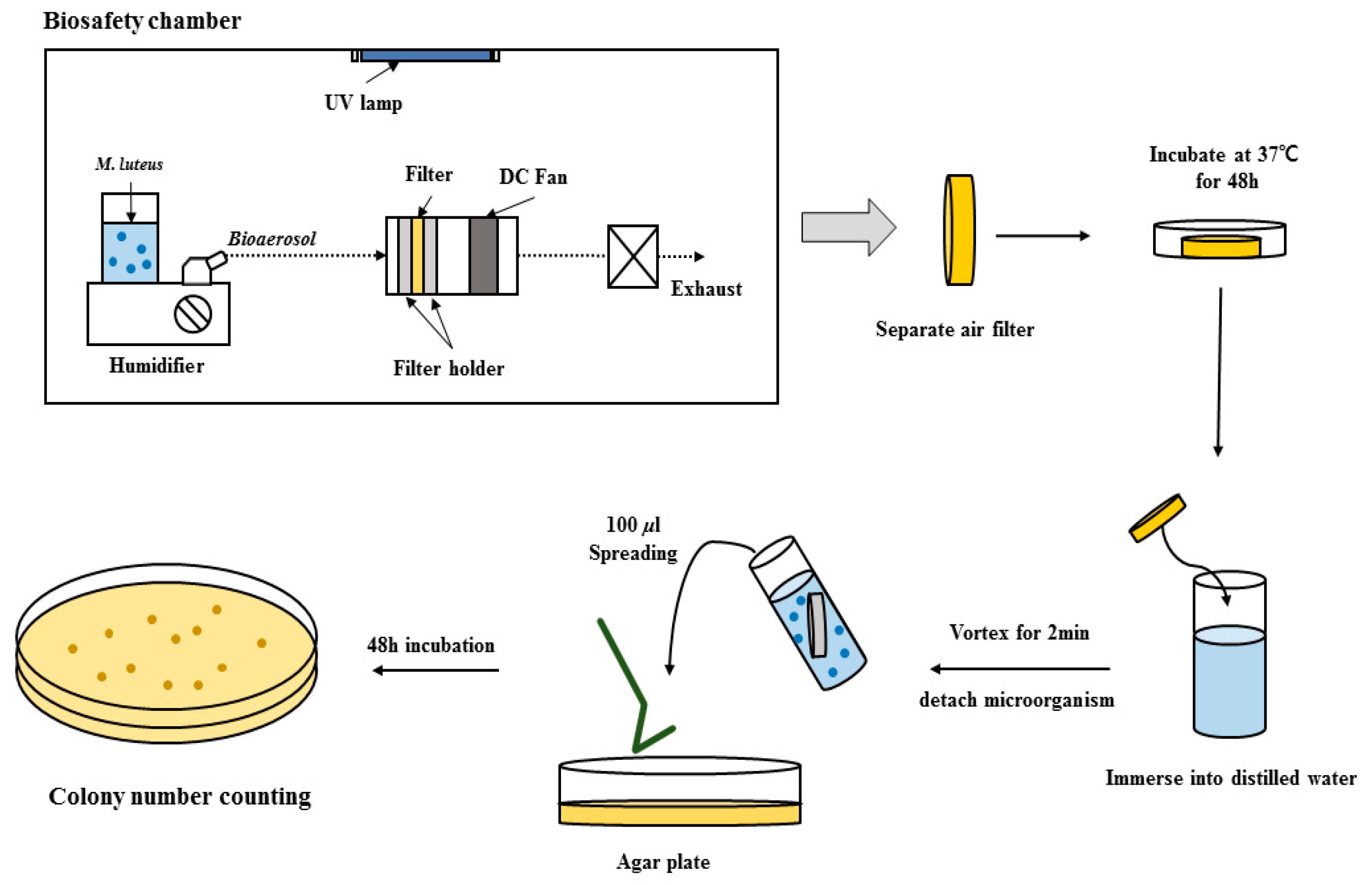
2.1. Materials

Natural plant extracts of tea-tree oil, rosemary, and garlic were purchased from Koreasimilac Co., Pocheon, Rep. Korea. Micrococcus luteus (KCTC 1056) and Escherichia coli (KCTC 22003), gram-positive and gram-negative, respectively, were selected as representative strains and sourced from the Korean collection for type cultures, Daejeon, Rep. Korea. Staphylococcus aureus (ATCC6538) and Klebsiella pneumonia (ATCC4352) were selected as representative airborne gram-positive and gram-negative pathogens, respectively, and were sourced from the Korean Collection for Type Cultures, Daejeon, Rep. Korea. Heart infusion broth, nutrient broth, yeast extract, beef extract, and peptone were purchased from Becton Dickinson, USA. Luria–Bertani (LB) broth and agar were purchased from MBcell Co., Seoul, Rep. Korea. Tetramethyl orthosilicate (TMOS) and hydrogen chloride (HCl) were purchased from Sigma-Aldrich Co., St. Louis, MO, USA. The PET filter material was provided by Saemyonghite Co., Ltd., Paju, Rep. Korea.

2.2. Antimicrobial Activity of Plant Extracts

All strains of M. luteus and E. coli were grown from lyophilized stock in 2 mL enriched nutrient broth and LB broth, respectively. They were incubated at 37 °C and shaken at 180 rpm for 24 h. The enriched nutrient broth was prepared by dissolving 12.5 g of heart infusion broth, 5.4 g of nutrient broth, and 2.5 g of yeast extract in 1 L of distilled water. The LB broth was prepared by dissolving 25 g of LB powder in 1 L of distilled water. Two bacteria cultured in the liquid phase were streaked on agar plates and incubated for 48 h at 37 °C. One colony was inoculated into 5 mL of liquid medium and cultured overnight. From this culture, 500 μL of the seeded culture was inoculated into 50 mL of liquid medium and incubated until it reached the exponential growth phase. When the bacterium reached this phase, the plant extracts were added into the cell cultures after dilution with distilled water to the desired concentrations. The cell growth was analyzed by measuring the 600 nm absorbance after 1 h using a UV/Vis spectrophotometer (Human Co., Seoul, Rep. Korea). All experiments were repeated three times.

2.3. Preparation of Plant Extract-Coated Filters and the Antimicrobial Test

Before usage, the PET was sterilized for 15 min at 121 °C. The coating solution was prepared by mixing 6 mL of TMOS and plant extract solutions with 0.8 mL of 0.2 M HCl. Before mixing to TMOS, each plant extract solution was prepared by diluting in distilled water. After adding the plant extract solution, the mixture was sonicated for 10 min and aged in a refrigerator for 12 h. One milliliter of plant extract coating solution was dispersed onto the surface of the air filter material using an atomizer (TAMIYA, Inc., Shizuoka, Japan). The coating process was carried out on a clean bench, and after coating, the filter material was dried at room temperature for 12 h. Negative control samples of non-coated PET were prepared by using PET materials as received without surface modification.
The antimicrobial activities of the air filters were measured following the SPS-KACA009-139 protocol developed by the Korea Air Cleaning Association [38]. One colony of M. luteus was removed from the agar plate and cultured in 5 mL enriched nutrient broth in a shaking incubator (180 rpm) at 37 °C for 24 h. The cell culture so obtained was injected into three flasks each containing 50 mL enriched nutrient broth and cultured until the exponential growth phase was reached. Then, 40 mL of cell solution was centrifuged at 1610 g for 10 min. After centrifugation, the supernatant was discarded, and the cell pellet was re-suspended in 10 mL of fresh enriched nutrient broth. Three re-suspended cell solutions were collected and injected into a bottle of humidifier containing 270 mL of distilled water. The air filter antimicrobial test was performed in a chamber (Figure 1) sterilized by 30 min of ultraviolet (UV) irradiation at 365 nm. The humidifier containing the cell solution was operated for 2 min to spray the bacterial aerosols. The fan located at the back of the air filter pushed the bacterial aerosols through the air filter. After bacterial aerosols exposure, the air filter was separated from the test apparatus and then incubated at 37° for 48 h to investigate long-term survival on the air filter surface. To detach the microorganisms, the air filter was immersed in 30 mL of distilled water and vortexed for 2 min. In this work, we considered that bacteria were detached from the air filters with the same efficiency in each experiment. The cell solution was streaked on nutrient agar plates and incubated at 37 °C for 48 h. The resulting colonies were counted and used to calculate the inactivation rate, using Equation (1):
Inactivation rate (%) = ((CFU control − CFUexperiment)/(CFUcontrol)) × 100,
where CFUexperiment and CFUcontrol are the number of bacteria retrieved from the antimicrobial and pristine (control) filters, respectively. CFU means colony-forming unit.
The anti-pathogen activities of the plant extract-coated air filters were investigated following the Korean standard testing method, KS K 0693:2016 [39], using Staphylococcus aureus and Klebsiella pneumonia. In this test, the cell exposing manner was different. Cell solution was poured on air filter placed in glass bottle. After incubating for 48 h, distilled water was added to the bottle and used it to count cell number. All experiments were conducted in triplicate.

2.4. Analytical Methods

Identification of phytochemicals present in the three plant extracts was performed using a gas chromatography coupled to a mass spectrometer (GC/MS; QP-2010 Plus, Shimazu Ltd., Kyoto, Japan) with a capillary column (Rxi-5MS, 30 m × 0.25 mm × 0.25 µm). The column oven was heated from 70 °C (held for 3 min) to 320 °C (held for 5 min) at 10 °C per minute. Samples were analyzed by GC/MS in scan mode (m/z = 45–550), and major peaks in the chromatograms were identified via the comparison of mass spectra with the spectrum library. Microstructure images and energy dispersive spectroscopy (EDS) mapping of the plant extract-coated air filters were obtained using a scanning electron microscope (SEM) (FE-SEM; JSM-7610F, JEOL Ltd., Tokyo, Japan). The coating layers were characterized by X-ray photoelectron spectroscopy (XPS, NEXSA, ThermoFisher ScientificTM, Waltham, MA, USA) with monochromatic Al Kα (hv = 1486.6 eV) radiation under ultrahigh vacuum (0.1 eV step, 50 eV pass energy and 400 µm spot size). The x-binding energy of the XPS data was calibrated with reference to the C–C bond in C1s (284.8 eV). The spectra were decomposed with the Avantage program, using a Gaussian/Lorenzian product function after subtraction of a Shirley x-baseline. The filter pressure drop was evaluated with a filter pressure measurement booth (Saemyonghite Co., Ltd., Pocheon, Rep. Korea). In the booth, the 280 mm × 364 mm air filter was placed in a filter holder and an air flow of 1 m/s was maintained throughout the measurement. The pressure drop was measured with a micromanometer from FC-2000, Furness Control, Ltd., Bexhill, UK. R(version 3.6.3) embedded in RStudio(version 1.2.1335) was used for statistical analysis and a p-value less than 0.05 was considered statistically significant.

3. Results and Discussion

3.1. Comparison of the Antimicrobial Activities of the Three Plant Extracts

The antimicrobial activity of the tea-tree oil, rosemary, and garlic against two microbial strains was investigated and is presented in Figure 2. These plant extracts showed antimicrobial activities against both gram-positive and negative strains. All plant extracts showed a dose-dependent pattern of the antimicrobial function. Cell inactivation increased with the concentrations of plant extracts.
Typically, the tea-tree oil showed a higher cell inactivation than the other two plant extracts. To quantitatively compare the extract’s antimicrobial activities, the MIC50 (minimum inhibition concentration) values, where 50% of cell inactivation is found, were calculated. The MIC50 values against the E. coli were 1.62, 224, and 407 mg/mL for tea-tree oil, rosemary, and garlic, respectively (Table 1). The MIC50 values against the M. luteus were 2.06, 181, and 419 mg/mL for tea-tree oil, rosemary, and garlic, respectively. Tea-tree oil showed potent cell inactivation effects on both E. coli and M. luteus and caused cell inactivation even at low concentration. In contrast, adding rosemary and garlic extracts caused cell inactivation initially, but the cell growth recovered at longer cultivation times (Figures S1 and S2). This suggests that the microorganisms adapted to the antimicrobial mechanism of both rosemary and garlic extracts. However, the tea-tree oil maintained its antimicrobial activity for a long time. For example, a concentration of 0.9 mg/mL of tea-tree oil-induced an approximately 22% inactivation at 1 h after being added and maintained this level of antimicrobial activity for 6 h (Figures S1 and S2). A concentration of 9 mg/mL tea-tree oil inhibited the cell growth by approximately 53% after 1 h, and by 88 and 82% for M. luteus and E. coli, respectively, at 6 h (Figures S1a and S2a). These results indicate that the antimicrobial activity of the tea-tree oil increased as the culturing time increased, implying that the microorganisms did not adapt to the tea-tree oil during cultivation time. Rosemary and garlic extracts showed cell inactivation at higher concentrations and a clear decrease of antimicrobial activity during cell culturing. This could be due to their weaker antimicrobial properties or their instability in aqueous solutions.
The phytochemicals present in the plant extracts chosen are listed in Table 2. The plant extracts were composed of 30, 6, and 6 phytochemicals for tea-tree oil, rosemary, and garlic, respectively. Typically, tea-tree oil contains various phytochemicals. We found through a literature survey that twenty-two phytochemicals in tea-tree oil are related to antimicrobial activities. Among them, thirteen chemicals—α-pinene, β-pinene, α-phellandrene, α-terpinene, cymene, dipentene (D-limonene), eucalyptol (1,8-cineole), γ-terpinene, terpinolene, linalool, α-terpineol, (–)-terpinen-4-ol, and (–)-globulol—exhibited an antimicrobial activity in previous studies. Nine compounds—sabinene, trans-β-ocimene, (–)-trans-pinocarveol, β-terpineol, isoborneol, (–)-α-gurjunene, (+)-aromadendrene, β-selinene, viridiflorol—were proven to feature antimicrobial activities in other studies (references are listed in Table 2). A further nine phytochemicals—sabinene, trans-β-ocimene, (–)-trans-pinocarveol, β-terpineol, isobeorneol, (–)-α-gurjunene, (+)-aromadendrene, β-selinene, and viridiflorol—were found as components in antimicrobial plant extracts. Meanwhile, only trimethylsilyl-glycerol in rosemary and garlic extracts was found in antimicrobial active plant extracts. The higher antimicrobial function of tea-tree oil, presented in Table 1 and Figure 2, could result from a higher number of antimicrobial components as compared to rosemary and garlic extracts. The combinational effects of 22 antimicrobial phytochemicals in tea-tree oil results in its strong antimicrobial activity.

3.2. Characteristics of Plant Extract-Coated Filters

To investigate the composition and chemical states of the coating layer surface, we performed an XPS analysis. A silicate polymer was used as a coating binder. The atomic composition of the coating layer was determined to be 68.6% of O1s, 28.3% of Si 2p, and 3.1% of C1s, and the formation of Si–O–Si bonds was confirmed (Figure S3). The SEM results show that the filter without plant extract had a smooth nonwoven fiber surface (Figure S4a). After coating with the plant extract, the coating layer was homogeneously formed with no aggregates on the filter surface (Figure S4b). An EDS analysis showed that the predominant element in the coating layer was Si (Figure S4c). These results confirmed that the silicate polymer coating layer was well-formed on the surface of the filter material by spray-coating.
Table 3 presents the pressure drops across the filters for various coating materials. The density of the filter will increase after the surface of air filter is coated by plant extracts. It will cause the reduced airflow rate across the filter, which can be measured using pressure drop. A higher pressure drop means that the device equipped with the air filter consumes more energy in order to maintain proper airflow. The PET filter without coating had a pressure drop of 1.97 mmAq. After coating with silicate polymer containing plant extracts, the pressure drop increased to an average of 2.15 mmAq; approximately 9.1% more than for the control filter, probably because the fiber thickness increased, although the fiber morphology did not change. However, this was not significantly different when compared to the control (p > 0.05). The plant extract type induced no significant difference in the pressure drop changes measured. Hwang et al. [1] used the nanoparticle deposition method to fabricate antimicrobial air filters; in their study, the pressure drop across the filters increased from 1.1 to 13.4 mmAq, depending on the number of nanoparticles deposited; i.e., nanoparticle deposition could clog the pores of the air filter. However, the silicate polymer coating used in this study did not block the pores in the filter fibers and did not cause a significant pressure drop increase. This result indicates that the coating method used in this study is superior and can be successfully applied to prepare antimicrobial air filters.

3.3. Inactivation of Microorganisms on Plant Extract-Coated Air Filters

The antimicrobial function of the plant extract-coated air filters was investigated by measuring the inactivation rate of M. luteus. M. luteus was recommended as a testing microorganism in air filter antimicrobial tests by the Korea Air Cleaning Association. Figure S5 presents the colony-forming units of M. luteus detached from the air filter immediately after exposure to bioaerosols. The tea-tree oil-coated air filter inactivation of M. luteus is presented in Figure S5a. When coated with 0.57 mg/cm2 of tea-tree oil, the colony-forming unit decreased from 5.23 × 10⁵ to 2.47 × 10⁵ CFU/mL, with a 52.87% inactivation rate. Other tea-tree oil-coated air filters exhibited a cell inactivation between 40 and 55%. However, neither rosemary nor garlic extract-coated air filters exhibited inactivation of M. luteus at any extract concentration (Figure S5b,c). As shown in Table 1, tea-tree oil showed the highest cell inactivation among the three plant extracts tested, and similar results were found in plant extract-coated air filters. Although the tea-tree oil-coated air filter initiated the inactivation of M. luteus, its antimicrobial functions were not severe after the initial exposure to bioaerosols (Figure S5a). The reduced initial antimicrobial activity of the plant extracts in air filters could be attributed to their mass transfer limitation in a silicate polymer matrix. However, significant cell inactivations were found after 48 h of cultivation (Figure 3a). The bioaerosol formed 4.73 × 10⁶ CFU/mL in non-coated air filters when it was incubated for 48 h, but the tea-tree oil-coated air filters formed only 1.63 × 10⁴ and 1.33 × 10³ CFU/mL for 0.57 and 2.89 mg/cm2 coated air filters, respectively, which induced a 99.65 and 99.97% cell inactivation (Figure 3a). No bacteria colonies formed for tea-tree oil concentrations in excess of 2.83 mg/cm2. Rosemary coated air filters exhibited a 93.3%, 99.0%, and 98.7% inactivation of M. luteus for 4.47, 6.73, and 13.40 mg/cm2 solution concentrations, respectively (Figure 3b). Garlic extract-coated air filters also showed a 99.9%, 99.7%, and 99.8% cell inactivation for 11.51, 16.98, and 33.96 mg/cm2 treated filters, respectively (Figure 3c). Typically, cell inactivation rates exhibited a dose-dependent increase pattern, indicating that the inactivation was due solely to the antimicrobial activity of the plant extracts. An air filter allows clean air to pass and rejects fine particles including bioaerosols. Even though air filters capture bioaerosols, they are active on the surface of the filters and can cause a variety of diseases. In contrast to common air filters, which can serve as microorganism reservoirs and pose the risk of secondary infections, our results clearly show that the plant extract-coated air filters fabricated in this study reduce the risk of secondary infections.
To investigate the anti-pathogen function of the plant extract-coated air filters, we evaluated the inactivation of Klebsiella pneumonia and Staphylococcus aureus. In this experiment, three air filter samples were prepared by coating the filters with 11.32 mg/cm2 tea-tree oil, 13.40 mg/cm2 rosemary, and 33.96 mg/cm2 garlic extract, respectively. Strains of K. pneumonia and S. aureus are usually found in air filters and cause microbial contamination. The plant extract-coated air filters showed a 99.99% inactivation of both K. pneumonia and S. aureus (Figure 4).
For larger-scale applications, follow-up studies should be conducted. Some of the plant extracts are unstable at room temperature; the stability of such extracts coated on air filters needs to be evaluated in future work. In addition, the effects of the coated plant extracts on human health should be evaluated for field applications. In this study, the word “cell inactivation” was used to describe the antimicrobial activity of plant extracts. Inactivation include both means of “inhibiting growth” and “killing microbial cells”. This study did not investigate the mechanism of the plant extracts used in this study, but it can be studied in future work. This study used only plant extracts instead of antimicrobial chemicals, but essential oils are also good candidates to replace antimicrobial chemicals; they could be utilized to develop antimicrobial air filters in future studies. The results of this study are useful in improving the safety of indoor environments in an environmental-friendly manner.

4. Conclusions

Three plant extracts (i.e., tea-tree oil, rosemary, and garlic) were investigated their antimicrobial activities and phytochemicals. All plant extracts tested showed an inactivation of M. luteus and E. coli. The tea-tree oil proved to be the most efficient in inhibiting both strains, even at low concentrations. The plant extracts were coated onto PET air filters with a silicate polymer by spray-coating. The characteristics of the plant extract-coated air filters were evaluated using XPS, SEM, and EDS. No significant pressure drop was found for plant extract coating when compared to the control air filters. Tea-tree oil coating on air filters resulted in some inactivation of M. luteus right after exposure to the bioaerosol, while rosemary and garlic extract-coated air filters did not show any inactivating effect. However, all three plant extract-coated air filters achieved a significant inactivation of M. luteus 48 h after the initial bioaerosol exposure.

Supplementary Materials

The following are available online at https://www.mdpi.com/2076-3417/10/24/9120/s1, Figure S1 Inactivation rate of M. luteus vs. time for (a) tea-tree oil, (b) rosemary, and (c) garlic extracts for various extract solution concentrations, Figure S2 Inactivation rate of E. coli vs. time for (a) tea-tree oil, (b) rosemary, and (c) garlic extracts for various extract solution concentrations, Figure S3 XPS spectra of the silicate polymer coating layer: (a) survey spectra, (b) O1s, and (c) Si2p, Figure S4 Scanning electron micrographs of the (a) pristine (control) and (b) tea tree oil-coated filters at a concentration of 9 mg/ml, and (c) results of Si mapping. Bars indicate (a) 33.3µm and (b,c) 100µm, Figure S5 Colony-forming units of M. luteus, immediately after bioaerosol exposure, vs. weight of plant extract per unit area: (a) tea-tree oil, (b) rosemary, and (c) garlic.

Author Contributions

Conceptualization and design, J.H.L.; Writing, H.R.B.; experiments, H.R.B., S.Y.P., E.T.H.; data acquisition, B.I.S.; data interpretation, E.T.H., J.M., and B.I.S.; data analysis, D.S., W.I.C., S.K. and J.H.L. All authors have read and agreed to the published version of the manuscript.

Funding

This research was funded by the National Research Foundation of Korea, South Korea (NRF), grant number 2020R1F1A1069515.

Acknowledgments

We acknowledge and appreciate the technical support provided by Seung-Huck Hong for the pressure drop experiment and Seo Jin Ki for the ANOVA analysis

Conflicts of Interest

The authors declare no conflict of interest.

References

  1. Hwang, G.B.; Heo, K.J.; Yun, J.H.; Lee, J.E.; Lee, H.J.; Nho, C.W.; Bae, G.-N.; Jung, J.H. Antimicrobial air filters using natural Euscaphis japonica nanoparticles. PLoS ONE 2015, 10, 0126481. [Google Scholar] [CrossRef] [PubMed]
  2. Douwes, J.; Thorne, P.; Pearce, N.; Heederik, D. Bioaerosol health effects and exposure assessment: Progress and prospects. Ann. Occup. Hyg. 2003, 47, 187–200. [Google Scholar] [PubMed] [Green Version]
  3. Kalogerakis, N.; Paschali, D.; Lekaditis, V.; Pantidou, A.; Eleftheriadis, K.; Lazaridis, M. Indoor air quality—Bioaerosol measurements in domestic and office premises. J. Aerosol Sci. 2005, 36, 751–761. [Google Scholar] [CrossRef]
  4. Sim, K.M.; Kim, K.H.; Hwang, G.B.; Seo, S.; Bae, G.N.; Jung, J.H. Development and evaluation of antimicrobial activated carbon fiber filters using Sophora flavescens nanoparticles. Sci. Total Environ. 2014, 493, 291–297. [Google Scholar] [CrossRef] [PubMed]
  5. Dales, R.E.; Zwanenburg, H.; Burnett, R.; Franklin, C.A. Respiratory health effects of home dampness and molds among Canadian children. Am. J. Epidemiol. 1991, 134, 196–203. [Google Scholar] [CrossRef]
  6. Siersted, H.C.; Gravesen, S. Extrinsic allergic alveolitis after exposure to the yeast Rhodotorula rubra. Allergy 1993, 48, 298–299. [Google Scholar] [CrossRef]
  7. Beaumont, F. Clinical manifestations of pulmonary Aspergillus infections. Mycoses 1988, 31, 15. [Google Scholar]
  8. Lin, C.Y.; Li, C.S. Control effectiveness of ultraviolet germicidal irradiation on bioaerosols. Aerosol Sci. Technol. 2002, 36, 474–478. [Google Scholar] [CrossRef]
  9. Chen, F.; Yang, X.; Mak, H.K.; Chan, D.W. Photocatalytic oxidation for antimicrobial control in built environment: A brief literature overview. Build. Environ. 2010, 45, 1747–1754. [Google Scholar] [CrossRef]
  10. Escombe, A.R.; Moore, D.A.; Gilman, R.H.; Navincopa, M.; Ticona, E.; Mitchell, B.; Noakes, C.; Martínez, C.; Sheen, P.; Ramirez, R.; et al. Upper-room ultraviolet light and negative air ionization to prevent tuberculosis transmission. PLoS Med. 2009, 6. [Google Scholar] [CrossRef] [Green Version]
  11. Jung, J.H.; Lee, J.E.; Lee, C.H.; Kim, S.S.; Lee, B.U. Treatment of fungal bioaerosols by a high-temperature, short-time process in a continuous-flow system. Appl. Environ. Microbiol. 2009, 75, 2742–2749. [Google Scholar] [CrossRef] [PubMed] [Green Version]
  12. Huang, H.L.; Lee, M.G.; Tai, J.H. Controlling indoor bioaerosols using a hybrid system of ozone and catalysts. Aerosol Air Qual. Res. 2012, 12, 73–82. [Google Scholar] [CrossRef] [Green Version]
  13. Li, C.S.; Wen, Y.M. Control effectiveness of electrostatic precipitation on airborne microorganisms. Aerosol Sci. Technol. 2003, 37, 933–938. [Google Scholar] [CrossRef]
  14. Han, B.; Kang, J.S.; Kim, H.J.; Woo, C.G.; Kim, Y.J. Investigation of antimicrobial activity of grapefruit seed extract and its application to air filters with comparison to propolis and shiitake. Aerosol Air Qual. Res. 2015, 15, 1035–1044. [Google Scholar] [CrossRef] [Green Version]
  15. Jin, W.J.; Jeon, H.J.; Kim, J.H.; Youk, J.H. A study on the preparation of poly (vinyl alcohol) nanofibers containing silver nanoparticles. Synth. Met. 2007, 157, 454–459. [Google Scholar] [CrossRef]
  16. Kang, S.; Pinault, M.; Pfefferle, L.D.; Elimelech, M. Single-walled carbon nanotubes exhibit strong antimicrobial activity. Langmuir 2007, 23, 8670–8673. [Google Scholar] [CrossRef]
  17. Lee, J.H.; Wu, C.Y.; Lee, C.N.; Anwar, D.; Wysocki, K.M.; Lundgren, D.A.; Farrah, S.; Wander, J.; Heimbuch, B.K. Assessment of iodine-treated filter media for removal and inactivation of MS2 bacteriophage aerosols. J. Appl. Microbiol. 2009, 107, 1912–1923. [Google Scholar] [CrossRef]
  18. Li, Y.; Leung, P.; Yao, L.; Song, Q.W.; Newton, E. Antimicrobial effect of surgical masks coated with nanoparticles. J. Hosp. Infect. 2006, 62, 58–63. [Google Scholar] [CrossRef]
  19. Ramyadevi, J.; Jeyasubramanian, K.; Marikani, A.; Rajakumar, G.; Rahuman, A.A. Synthesis and antimicrobial activity of copper nanoparticles. Mat. Lett. 2012, 71, 114–116. [Google Scholar] [CrossRef]
  20. Moore, M.N. Do nanoparticles present ecotoxicological risks for the health of the aquatic environment? Environ. Int. 2006, 32, 967–976. [Google Scholar] [CrossRef]
  21. Karlsson, H.L.; Cronholm, P.; Gustafsson, J.; Moller, L. Copper oxide nanoparticles are highly toxic: A comparison between metal oxide nanoparticles and carbon nanotubes. Chem. Res. Toxicol. 2008, 21, 1726–1732. [Google Scholar] [CrossRef] [PubMed]
  22. Chen, X.; Schluesener, H.J. Nanosilver: A nanoproduct in medical application. Toxicol. Lett. 2008, 176, 1–12. [Google Scholar] [CrossRef] [PubMed]
  23. Pulskamp, K.; Diabaté, S.; Krug, H.F. Carbon nanotubes show no sign of acute toxicity but induce intracellular reactive oxygen species in dependence on contaminants. Toxicol. Lett. 2007, 168, 58–74. [Google Scholar] [CrossRef] [PubMed]
  24. Ji, J.H.; Jung, J.H.; Kim, S.S.; Yoon, J.U.; Park, J.D.; Choi, B.S.; Chung, Y.H.; Kwon, I.H.; Jeong, J.; Han, B.S. Twenty-eight-day inhalation toxicity study of silver nanoparticles in Sprague-Dawley rats. Inhal. Toxicol. 2007, 19, 857–871. [Google Scholar] [CrossRef] [PubMed]
  25. Jung, J.H.; Hwang, G.B.; Park, S.Y.; Lee, J.E.; Nho, C.W.; Lee, B.U.; Bae, G.N. Antimicrobial air filtration using airborne Sophora flavescens natural-product nanoparticles. Aerosol Sci. Technol. 2011, 45, 1510–1518. [Google Scholar] [CrossRef] [Green Version]
  26. Carson, C.F.; Hammer, K.A.; Riley, T.V. Melaleuca alternifolia (tea tree) oil: A review of antimicrobial and other medicinal properties. Clin. Microbiol. Rev. 2006, 19, 50–62. [Google Scholar] [CrossRef] [PubMed] [Green Version]
  27. Rios, J.L.; Recio, M.C. Medicinal plants and antimicrobial activity. J. Ethnopharmacol. 2005, 100, 80–84. [Google Scholar] [CrossRef]
  28. Lee, C.J.; Chen, L.W.; Chen, L.G.; Chang, T.L.; Huang, C.W.; Huang, M.C.; Wang, C.C. Correlations of the components of tea tree oil with its antibacterial effects and skin irritation. J. Food Drug Anal. 2013, 21, 169–176. [Google Scholar] [CrossRef] [Green Version]
  29. Aloui, H.; Khwaldia, K.; Sánchez-González, L.; Muneret, L.; Jeandel, C.; Hamdi, M.; Desobry, S. Alginate coatings containing grapefruit essential oil or grapefruit seed extract for grapes preservation. Int. J. Food Sci. Technol. 2014, 49, 952–959. [Google Scholar] [CrossRef]
  30. Saito, M.; Hosoyama, H.; Ariga, T.; Kataoka, S.; Yamaji, N. Antiulcer activity of grape seed extract and procyanidins. J. Agric. Food Chem. 1998, 46, 1460–1464. [Google Scholar] [CrossRef]
  31. Schlüter, B.; Pflegel, P.; Lindequist, U.; Jülich, W.D. Aspects of the antimicrobial efficacy of grapefruit seed extract and its relation to preservative substances contained. Die Pharm. 1999, 54, 452–456. [Google Scholar]
  32. Hearst, R.; Nelson, D.; McCollum, G.; Millar, B.C.; Maeda, Y.; Goldsmith, C.E.; Rooney, P.J.; Loughrey, A.; Rao, J.R.; Moore, J.E. An examination of antibacterial and antifungal properties of constituents of Shiitake (Lentinula edodes) and Oyster (Pleurotus ostreatus) mushrooms. Complementary Ther. Clin. Pract. 2009, 15, 5–7. [Google Scholar] [CrossRef] [PubMed]
  33. May, J.; Chan, C.H.; King, A.; Williams, L.; French, G.L. Time–kill studies of tea tree oils on clinical isolates. J. Antimicrob. Chemother. 2000, 45, 639–643. [Google Scholar] [CrossRef]
  34. Moreno, S.; Scheyer, T.; Romano, C.S.; Vojnov, A.A. Antioxidant and antimicrobial activities of rosemary extracts linked to their polyphenol composition. Free Radic. Res. 2006, 40, 223–231. [Google Scholar] [CrossRef] [PubMed]
  35. Ankri, S.; Mirelman, D. Antimicrobial properties of allicin from garlic. Microb. Infect. 1999, 1, 125–129. [Google Scholar] [CrossRef]
  36. Li, T.C.; Ambu, S.; Mohandas, K.; Wah, M.J.; Sulaiman, L.H.; Murgaivah, M. Bacterial constituents of indoor air in a high throughput building in the tropics. Trop. Biomed. 2014, 31, 540–556. [Google Scholar]
  37. US Environmental Protection Agency. Air Sanitizers—Efficacy Data Recommendations; Test Guideline No. OCSPP 810.2500-Air Sanitizers-2013-03-12 [EPA 730-C-11-003]; US Environmental Protection Agency: Washington, DC, USA, 2012.
  38. KACA (Korea Air Cleaning Association). Antibiotic Filter; SPS-KACA009-139; Korea Air Cleaning Association: Seoul, Korea, 2002. [Google Scholar]
  39. Korean Standards Association. Test Method for Antibacterial Activity of Textile Materials; KS K, 0693:2016; Korean Standards Association: Seoul, Korea, 2016. [Google Scholar]
  40. Silva, A.C.R.D.; Lopes, P.M.; Azevedo, M.M.B.D.; Costa, D.C.M.; Alviano, C.S.; Alviano, D.S. Biological activities of a-pinene and β-pinene enantiomers. Molecules 2012, 17, 6305–6316. [Google Scholar] [CrossRef] [Green Version]
  41. Andrade, B.F.M.T.; Barbosa, L.N.; Probst, I.S.; Fernandes Júnior, A. Antimicrobial activity of essential oils. J. Essent. Oil Res. 2014, 26, 34–40. [Google Scholar] [CrossRef]
  42. Leite, A.M.; Lima, E.D.O.; Souza, E.L.D.; Diniz, M.D.F.F.M.; Trajano, V.N.; Medeiros, I.A.D. Inhibitory effect of beta-pinene, alpha-pinene and eugenol on the growth of potential infectious endocarditis causing Gram-positive bacteria. Rev. Bras. Cienc. Farm. 2007, 43, 121–126. [Google Scholar] [CrossRef] [Green Version]
  43. İşcan, G.; Kırımer, N.; Demirci, F.; Demirci, B.; Noma, Y.; Başer, K.H.C. Biotransformation of (­)-(R)-α-Phellandrene: Antimicrobial Activity of Its Major Metabolite. Chem. Biodivers. 2012, 9, 1525–1532. [Google Scholar] [CrossRef]
  44. Carson, C.F.; Riley, T.V. Antimicrobial activity of the major components of the essential oil of Melaleuca alternifolia. J. Appl. Bacterol. 1995, 78, 264–269. [Google Scholar] [CrossRef] [PubMed]
  45. Kiskó, G.; Roller, S. Carvacrol and p-cymene inactivate Escherichia coli O157: H7 in apple juice. BMC Microbiol. 2005, 5, 36. [Google Scholar] [CrossRef] [PubMed] [Green Version]
  46. Zahi, M.R.; Liang, H.; Yuan, Q. Improving the antimicrobial activity of D-limonene using a novel organogel-based nanoemulsion. Food Control. 2015, 50, 554–559. [Google Scholar] [CrossRef]
  47. Safaei-Ghomi, J.; Ahd, A.A. Antimicrobial and antifungal properties of the essential oil and methanol extracts of Eucalyptus largiflorens and Eucalyptus intertexta. Pharmacogn. Mag. 2010, 6, 172. [Google Scholar] [CrossRef] [PubMed] [Green Version]
  48. Shafaghat, A. Comparison of the antimicrobial activity and chemical constituents of the essential oil and hexanic extract from Chaerophyllum macropodum. J. Essent. Oil Bear. Plants 2017, 20, 835–843. [Google Scholar] [CrossRef]
  49. Giweli, A.; Džamić, A.M.; Soković, M.; Ristić, M.S.; Marin, P.D. Antimicrobial and antioxidant activities of essential oils of Satureja thymbra growing wild in Libya. Molecules 2012, 17, 4836–4850. [Google Scholar] [CrossRef] [Green Version]
  50. Lee, D.H.; Jung, J.H.; Lee, B.U. Effect of treatment with a natural extract of Mukdenia Rossii (Oliv) Koidz and unipolar ion emission on the antibacterial performance of air filters. Aerosol Air Qual. Res. 2013, 13, 771–776. [Google Scholar] [CrossRef] [Green Version]
  51. Park, S.N.; Lim, Y.K.; Freire, M.O.; Cho, E.; Jin, D.; Kook, J.K. Antimicrobial effect of linalool and α-terpineol against periodontopathic and cariogenic bacteria. Anaerobe 2012, 18, 369–372. [Google Scholar] [CrossRef]
  52. Alma, M.H.; Nitz, S.; Kollmannsberger, H.; Digrak, M.; Efe, F.T.; Yilmaz, N. Chemical composition and antimicrobial activity of the essential oils from the gum of Turkish pistachio (Pistacia vera L.). J. Agric. Food Chem. 2004, 52, 3911–3914. [Google Scholar] [CrossRef]
  53. Oyedemi, S.O.; Okoh, A.I.; Mabinya, L.V.; Pirochenva, G.; Afolayan, A.J. The proposed mechanism of bactericidal action of eugenol, α-terpineol and g-terpinene against Listeria monocytogenes, Streptococcus pyogenes, Proteus vulgaris and Escherichia coli. Afr. J. Biotechnol. 2009, 8, 1280–1286. [Google Scholar]
  54. George, V.; Mathew, J.; Sabulal, B.; Dan, M.; Shiburaj, S. Chemical composition and antimicrobial activity of essential oil from the rhizomes of Amomum cannicarpum. Fitoterapia 2006, 77, 392–394. [Google Scholar] [CrossRef] [PubMed]
  55. Stojanović, G.; Palić, I.; Irsić-Janković, J. Composition and antimicrobial activity of the essential oil of Micromeria cristata and Micromeria juliana. Flavour Fragr. J. 2006, 21, 77–79. [Google Scholar] [CrossRef]
  56. Carson, C.F.; Mee, B.J.; Riley, T.V. Mechanisms of action of Melaleuca alternifolia (tea tree) oil on Staphylococcus aureus determined by time-kill, lysis, leakage, and salt tolerance assays and electron microscopy. Antimicrob. Agents Chemother. 2002, 46, 1914–1920. [Google Scholar] [CrossRef] [PubMed] [Green Version]
  57. Kumar Tyagi, A.; Bukvicki, D.; Gottardi, D.; Veljic, M.; Guerzoni, M.E.; Malik, A.; Marin, P.D. Antimicrobial potential and chemical characterization of Serbian liverwort (Porella arboris-vitae): SEM and TEM observations. J. Evid. Based Complementary Altern. Med. 2013, 2013, 382927. [Google Scholar] [CrossRef] [PubMed] [Green Version]
  58. Mulyaningsih, S.; Sporer, F.; Reichling, J.; Wink, M. Antimicrobial activity of essential oils from Eucalyptus and of selected components against multidrug-resistant bacterial pathogens. Pharm. Biol. 2011, 49, 893–899. [Google Scholar] [CrossRef] [PubMed]
  59. Ho, C.L.; Wang, E.I.C.; Tseng, Y.H.; Liao, P.C.; Lin, C.N.; Chou, J.C.; Su, Y.C. Composition and antimicrobial activity of the leaf and twig oils of Litsea mushaensis and L. linii from Taiwan. Nat. Prod. Commun. 2010, 5, 1823–1828. [Google Scholar] [CrossRef] [Green Version]
  60. Tan, M.; Zhou, L.; Huang, Y.; Wang, Y.; Hao, X.; Wang, J. Antimicrobial activity of globulol isolated from the fruits of Eucalyptus globulus Labill. Nat. Prod. Res. 2008, 22, 569–575. [Google Scholar] [CrossRef]
  61. Cunico, M.M.; Lopes, A.R.; Côcco, L.C.; Yamamoto, C.I.; Plocharski, R.C.; Miguel, M.D.; Miguel, O.G. Phytochemical and antibacterial evaluation of essential oils from Ottonia mariana Miq. (Piperaceae). J. Braz. Chem. Soc. 2007, 18, 184–188. [Google Scholar] [CrossRef] [Green Version]
  62. Ertürk, Ö.; Çíl, E.; Yoloğlu, N.; Yavuz, C. An in vitro study on antimicrobial and antioxidant activity of propolis from rize province of Turkey. Mellifera 2016, 16, 4–18. [Google Scholar]
Figure 1. Schematic illustration of the experimental process for air filter antimicrobial testing.
Applsci 10 09120 g001
Figure 2. Cell inactivations of (a) Micrococcus luteus and (b) Escherichia coli depending on the concentrations of plant extracts of tea-tree oil (●), rosemary (■), and garlic (▲). The graph in the inset presents the enlarged graph of tea-tree oil.
Applsci 10 09120 g002
Figure 3. M. luteus colony-forming units 48 h after bioaerosol exposure vs. weight of plant extract per unit area. (a) tea-tree oil, (b) rosemary, and (c) garlic.
Applsci 10 09120 g003
Figure 4. Images of the anti-pathogen activity of air filter samples for S. aureus and K. pneumonia.
Applsci 10 09120 g004
Table 1. Minimum inhibitory concentration 50 (MIC50) of the plant extracts against E. coli and M. luteus (unit: mg/mL).
E. coliM. luteus
Tea-tree oil1.622.06
Rosemary224181
Garlic407419
Table 2. Phytochemicals present in plant extracts used in this study.
Plant ExtractPhytochemicalFormulaMW 1Ref 2Amount (wt%)
Tea-tree oilα-PineneC10H16136[40]1.93
SabineneC10H16136[41]0.21
β-PineneC10H16136[42]0.32
α-PhellandreneC10H16136[43]0.17
α-TerpineneC10H16136[44]4.99
CymeneC10H14134[45]9.79
DipenteneC10H16136[46]15.00
EucalyptolC10H18O154[47]3.11
Trans-β-OcimeneC10H16136[48]1.18
γ-TerpineneC10H16136[49]24.30
Bicyclo[3.1.0]hexan-2-ol, 2-methyl-5-(1-methylethyl)-, (1α, 2α, 5α)-C10H18O154-0.14
2-Furanmethanol, 5-ethenyltertrahydro- α, α, 5-trimethyl-, cis-C10H18O2170-0.49
TerpinoleneC10H16136[50]3.32
LinaloolC10H18O154[51]0.09
(-)-trans-PinocarveolC10H16O152[52]0.43
α-TerpineolC10H18O154[53]0.16
β-TerpineolC10H18O154[54]0.08
IsoborneolC10H18O154[55]0.12
(-)-Terpinen-4-olC10H18O154[56]24.86
L-α-TerpineolC10H18O154-4.68
Cyclohexene, 4-ethenyl-4-methyl-3-(1-methylethenyl)-1-(1-methylethyl)-, (3R-trans)-C15H24204-0.35
7-Ethylnonan-4-oneC11H22O170-0.34
2,2-Dimethyl-4,5-di(1-propenyl)-1,3-dioxolaneC11H18O2182-0.06
(-)-α-gurjuneneC15H24204[57]0.22
(+)-AromadendreneC15H24204[58]1.84
β-selineneC15H24204[59]0.17
α-GuaieneC15H24204-0.15
(+)-LedeneC15H24204-1.02
(−)-GlobulolC15H26O222[60]0.38
ViridiflorolC15H26O222[61]0.09
Rosemary3,6-Dioxa-2,7-disilaoctane, 2,2,4,7,7-pentamethyl-C9H24O2Si2220-15.04
IsobutoxytrimethylsilaneC7H18OSi146-19.35
[(1-Methyl-1,3-propanediyl)bis(oxy)]bis(trimethylsilane)C10H26O2Si2234-17.18
Trimethyl(1-methylpropoxy)silaneC7H18OSi146-0.42
Trimethylsilyl-GlycerolC12H32O3Si3308[62]31.89
Butane, 1,2,3-tris(trimethylsiloxy)-C13H34O3Si3322-12.18
Garlic3,6-Dioxa-2,7-disilaoctane, 2,2,4,7,7-pentamethyl-C9H24O2Si2220-22.99
[(1-Methyl-1,3-propanediyl)bis(oxy)]bis(trimethylsilane)C10H26O2Si2234-11.21
Trimethylsilyl-GlycerolC12H32O3Si3308[62]0.62
Butane, 1,2,3-tris(trimethylsiloxy)-C13H34O3Si3322-0.79
2,2,4,4,6,6,-hexamethyl-2,4,6-trisila-heptaneC10H28Si3232-64.14
2-O,3-O,5-O,6-O,7-O-Pentakis(trimethylsilyl)-D-glycero-L-manno-heptonic acid 1,4-lactoneC22H52O7Si5568-0.07
1 MW: molecular weight 2 Ref: antimicrobial activity reference.
Table 3. Pressure drop of the control and plant extract-coated air filters.
ControlTea-tree oilRosemaryGarlic
Pressure drop (mmAq)1.97 ± 0.062.17 ± 0.172.13 ± 0.222.16 ± 0.15
Publisher’s Note: MDPI stays neutral with regard to jurisdictional claims in published maps and institutional affiliations.

