Article

Analysis and Identification of Active Compounds from Salviae miltiorrhizae Radix Toxic to HCT-116 Human Colon Cancer Cells

1
Department of Korean International Medicine, College of Korean Medicine, Gachon University, Seongnam 13120, Korea
2
Department of Life Science, College of Bio-Nano Technology, Gachon University, Seongnam 13120, Korea
3
Herbal Medicine Research Division, Korea Institute of Oriental Medicine, Daejeon 34054, Korea
4
College of Korean Medicine, Gachon University, Seongnam 13120, Korea
*
Authors to whom correspondence should be addressed.
Appl. Sci. 2020, 10(4), 1304; https://doi.org/10.3390/app10041304
Submission received: 23 January 2020 / Revised: 5 February 2020 / Accepted: 11 February 2020 / Published: 14 February 2020
(This article belongs to the Special Issue Biological Activity and Applications of Natural Compounds)

Abstract

:
Colorectal cancer is one of the most frequently diagnosed cancers worldwide. The aim of the present study was to simultaneously analyze compounds of Salviae miltiorrhizae Radix (SMR) and determine their cytotoxic effects on HCT-116 human colorectal cancer cells. We established a simultaneous analysis method of five compounds (salvianic acid A, salvianolic acid B, caffeic acid, tanshinone IIA, and rosmarinic acid) contained in SMR, and found that among the various compounds in SMR, tanshinone IIA significantly decreased cell viability in a concentration-dependent manner. Hoechst staining also showed that both SMR and tanshinone IIA increased nuclear condensation, suggesting induction of apoptosis. By Western blotting, we found that tanshinone IIA induced apoptotic cell death, significantly increased Bax, but decreased Bcl-2 in the course of apoptosis. Tanshinone IIA increased the expression of cleaved caspases-7 and -8. Tanshinone IIA was shown to be an active ingredient of SMR that may be a useful chemotherapeutic strategy for patients with colorectal cancer.

Graphical Abstract

1. Introduction

Colorectal cancer is the third-most commonly diagnosed cancer worldwide [1]. Although surgery plays a key role in the diagnosis and treatment of colorectal cancer, there are still increasing attempts to stop the progression of this cancer via the application of new synthetic and naturally-occuring compounds [2,3]. Bioactive compounds from plants have been screened for anticancer activities [4,5]. Approximately 50–60% of cancer patients in the United States utilize complementary and alternative medicines with traditional therapeutic regimens, such as radiation therapy and chemotherapy [6].
Apoptosis pathways are important targets in cancer-related therapies, and insufficient apoptosis results in uncontrolled cancer cell proliferation [7]. The use of natural phytochemicals for inhibiting cancer cell proliferation and inducing apoptosis contributes to promoting cancer cell death [8,9]. Natural phytochemicals are multiple-target molecules found in plants and microorganisms, and they exert strong anticancer activity [10,11]. Phytochemicals isolated from natural sources also exhibit various beneficial effects against inflammation, cancer, and neurodegenerative disorders [10]. This broad spectrum of biological and pharmacological activities has made natural compounds suitable candidates for treating multifactorial diseases, such as colorectal cancer.
Salviae miltiorrhizae Radix (SMR) is one of the well-known traditional herbal medicines and has been used in Asian countries [8]. Recently, there has been increasing scientific attention towards SMR for its remarkable bioactivity against cardiovascular disease, renal damage, tumor angiogenesis, and tumor cell invasion [12,13,14]. In the last decade, accumulating evidence has shown that SMR exerts a significant anticancer effect against promyelocytic leukemia, breast cancer, ovarian carcinomas, and hepatocellular carcinoma (HCC) [15,16,17]. In a recent network pharmacology-based study on the anti- HCC effect of SMR, 62 chemical compounds form SMR yielded 101 putative targets that played a critical role in HCC via multiple targets and pathways, especially the EGFR and phosphatidyl-inositol 3-kinase (PI3K)/Akt signaling pathways [18]. However, the effect of SMR and its compounds on human colon cancer cells has not been fully elucidated. The aim of the present study was to simultaneously analyze the compounds of SMR and determine their cytotoxic effects on HCT-116 human colorectal cancer cells.

2. Materials and Methods

2.1. Plant Materials

Salviae Miltiorrhizae Radix (SMR) was obtained from Kwangmyungdag Medicinal Herbs (Ulsan, Korea) and identified by Dr. Goya Choi, Herbal Medicine Resources Research Center, Korea Institute of Oriental Medicine (HMRRC, KIOM; Naju, Korea). A voucher specimen (SMR-2-14-0073) was stored at the herbarium of the HMRRC, KIOM.

2.2. Chemicals and Reagents

Five reference standard compounds, salvianic acid A (98.0%), caffeic acid (99.0%), rosmarinic acid (97.0%), salvianolic acid B (98.0%), and tanshinone IIA (98.8%) were purchased from standard manufacturers: Acros Organics (Pittsburgh, PA, USA), Merck KGaA (Darmstadt, Germany), and ChemFaces Biochemical Co., Ltd. (Wuhan, China).
The solvents including methanol, acetonitrile, and water (HPLC-grade) and formic acid (≥98.0%, ACS reagent-grade) for quantitative analysis were obtained from Merck KGaA (Darmstadt, Germany) and J. T. Baker (Phillipsburg, NJ, USA), respectively.

2.3. Preparation of 70% Ethanol SMR Extract

Dried SMR (0.3 kg) was extracted with 70% ethanol (3.0 L, 3 times) for 1 h at room temperature by a Branson 8510 ultrasonicator (Denbury, CT, USA). The extract solution was filtered with 150 mm Ø filter paper (Whatman, Maidstone, Kent, UK) under vacuum, concentrated to remove the organic extract solvent (ethanol) using a Büchi rotary evaporator R-210 (Flawil, Switzerland), and then lyophilized using a Ilshin BioBase FD-5525L freeze-drier (Dongducheon, Korea) to obtain powdered extract. The yield of lyophilized 70% ethanol extract of SMR was 69.8 g (23.3%).

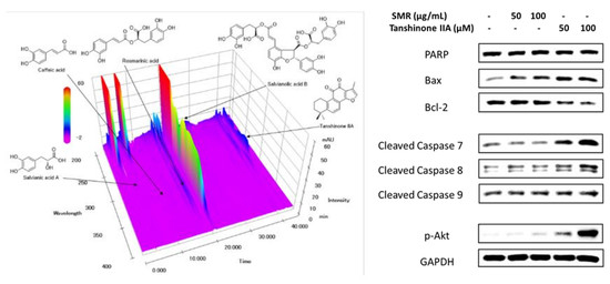
2.4. HPLC Analysis of Five Components in SMR

HPLC analysis was conducted using the Prominence LC-20A Series instruments (Shimadzu, Kyoto, Japan) consisting of a DGU-20A3 degasser, LC-20AT solvent delivery unit, SIL-20A auto sample injector, CTO-20A column oven, and SPD-M20A photodiode array detector. All chromatographic data were obtained and analyzed with the LabSolution software (Version 5.53; SP3, Kyoto, Japan). Five components were separated using a reverse-phase SunFireTM C18 analytical column (4.6 × 250 mm, 5 μm; Waters, Torrance, CA, USA) at 40 °C with gradient solvent condition. The mobile phases consisted of 0.1% (v/v) aqueous formic acid (A) and 0.1% (v/v) formic acid in acetonitrile (B) and were adjusted following the gradient condition: 0–30 min, 10–60% B; 30–40 min, 60–100% B; 40–45 min, 100% B; 45–50 min, and 100–10% B. The re-equilibrium time was adjusted for 10 min. The flow rate of the mobile phase was 1.0 mL/min, and the injection volume of the standard and test solution was 10 μL each. For quantitative determination of five marker components (salvianic acid A, caffeic acid, rosmarinic acid, salvianolic acid B, and tanshinone IIA) in SMR, 200.0 mg of lyophilized SMR extract was liquefied with 20 mL of 70% methanol and sonicated for 30 min. It was also diluted 20-fold for quantification of salvianolic acid B. All samples were filtered using a membrane filter (0.2-μm, Pall Life Sciences, Ann Arbor, MI, USA) before analysis.

2.5. Cell Culture

The human colon cancer cell (HCT-116) was purchased from the ATCC (American Type Culture Collection, Manassas, VA, USA). The cell was maintained and grown in RPMI 1640 medium (Roswell Park Memorial Institute 1640; Corning, Manassas, VA, USA) contained with 10% FBS (fetal bovine serum; Gibco BRL, Carlsbad, MD, USA) and penicillin/streptomycin (Life Technologies, Waltham, MA, USA). The condition of the incubator was 37 °C and humidified atmosphere containing 5% CO2.

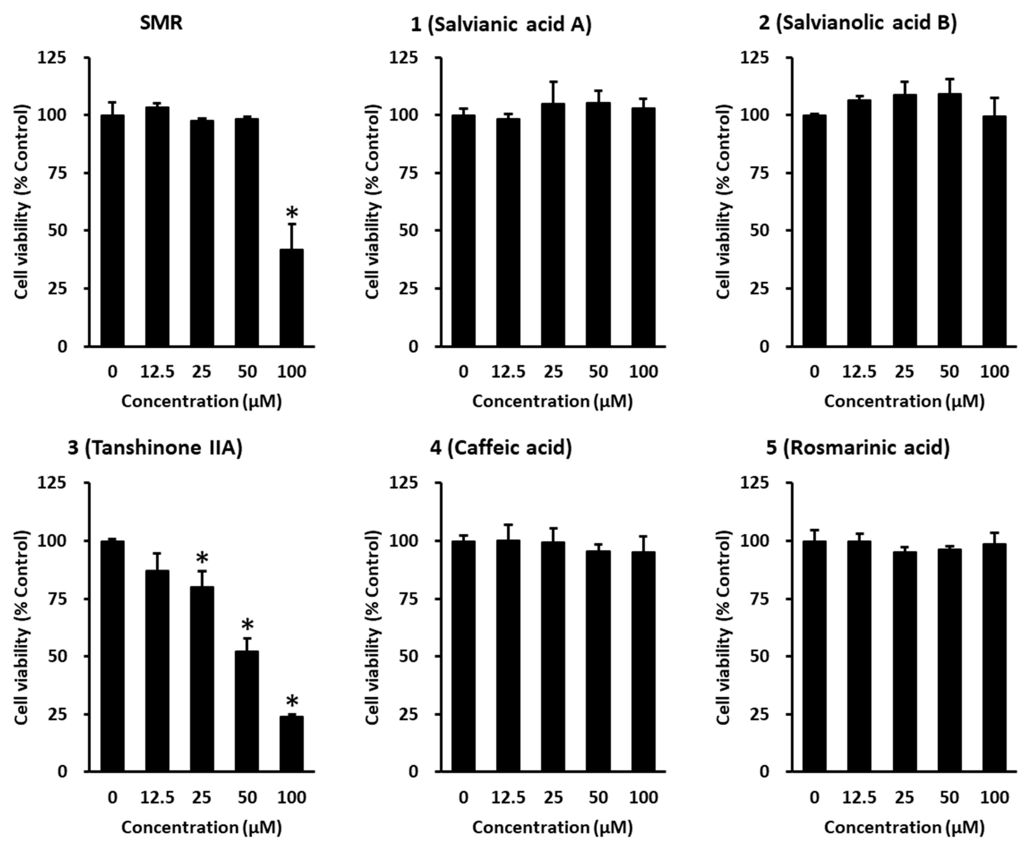
2.6. Cell Viability Assay

The cell viability assay was assessed using an Ez-Cytox Kit (Dail Lab Service Co., Seoul, Korea) based on the manufacturer’s instructions [19]. Briefly, the cells were seeded in a 96-well plate at 1 × 10⁴ cells/well and then incubated. After 24 h, the cells were treated with the indicated concentrations of each sample, and the cells were then incubated for 24 h. Following incubation, Ez-Cytox solution was mixed with medium in each well and incubated for 1 h. The absorbance at 450/600 nm was determined using a SPARK 10M (Tecan Group Ltd., Männedorf, Switzerland). The cell viability of 100% was calculated from control cells.

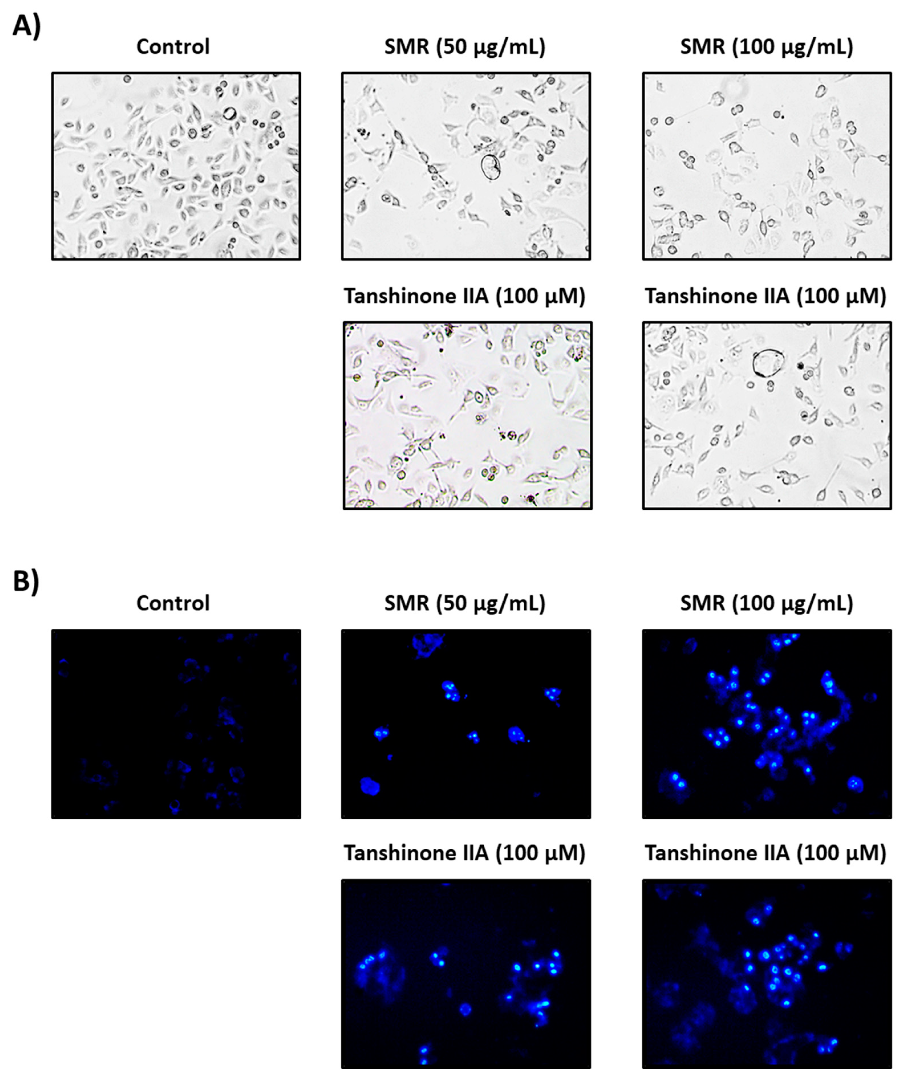
2.7. Hoechst 33342 Cell Staining

Sample-induced nuclear condensation of HCT-116 cells was observed using Hoechst 33342 staining (Sigma Aldrich, St. Louis, MO, USA) [20]. Briefly, the cells were seeded in a 6-well plate at 4 × 10⁵ cells per well. Following incubation for 24 h, the cells were treated with various concentrations of each sample, and the cells were then incubated for 24 h. Following incubation, Hoechst 33342 solution was added to the cells and incubated for 10 min. The stained cells were observed using a CCD camera conjugated IX50 fluorescent microscope (Olympus, Tokyo, Japan).

2.8. Western Blotting

The apoptosis signaling pathways of HCT-116 cells induced by samples were performed using Western blot analysis [21,22]. Briefly, the cells were seeded in a 6-well plate at 4 × 10⁵ cells/well and then incubated. After 24 h, the cells were treated with the indicated concentrations of each sample, and the cells were then incubated for 24 h. Following incubation, the cells were harvested with a scraper and lysed with radio-immunoprecipitation assay buffer (Elpis Biotech, Daejeon, Korea). The protein concentrations were calculated with the Pierce BCA Protein Assay Kit (Thermo Scientific, Carlsbad, CA, USA). The protein samples were separated by electrophoresis in a SDS-PAGE. Then, the proteins were transferred to PVDF membranes (Merck Millipore, Darmstadt, Germany). The membranes were conducted blockading by 5% skim milk. Then, the membranes were probed with primary antibodies for Bax, B-cell lymphoma 2 (Bcl-2), cleaved caspase-7, cleaved caspase-8, cleaved caspase-9, glyceraldehyde 3-phosphate dehydrogenase (GAPDH) and poly(ADP-ribose) polymerase (PARP) followed by incubating with secondary antibodies for anti-rabbit IgG (Cell Signaling Technology, Inc., Danvers, MA, USA).

2.9. Statistical Analysis

All experiments were performed in triplicate, and the quantitative data were shown as mean ± SD. Statistical analysis using Student’s t-test was conducted and considered statistically significant based on p-values less than 0.05.

3. Results and Discussion

In the present study, we analyzed five bioactive marker components found in SMR, consisting of four phenolic acids (salvianic acid A, caffeic acid, rosmarinic acid, and salvianolic acid B) and one terpenoid (tanshinone IIA). These compounds were separated with resolution >5.0 within 45 min and retention times of 5.87, 11.11, 17.72, 20.42, and 42.51 min, respectively (Figure 1). The content of rosmarinic acid, caffeic acid, salvianic acid A, salvianolic acid B, and tanshinone IIA in the samples was 3.72, 0.123, 1.27, 64.36, and 4.96 mg/g, respectively.
We initially performed a cytotoxic evaluation using HCT-116 human colorectal carcinoma cells. As shown in Figure 2, only tanshinone IIA significantly decreased cell viability in a concentration-dependent manner, whereas 61.6 μM SMR showed approximately 50% suppression.
Many clinical anticancer drugs are known to exert their effects by inducing apoptosis [23]. Apoptosis is a gene-regulated response and, from the morphological point of view, is distinguished by the specific structural changes in cells, such as plasma membrane bleb formation, cell and nuclear shrinkage, oligonucleosomal DNA fragmentation, and chromatin condensation [24]. Morphological analyses showed that both SMR and tanshinone IIA decreased the number of cells and induced signs of cellular apoptosis, such as cellular shrinkage (Figure 3). Moreover, as shown in Figure 4, Hoechst staining also showed that both SMR and tanshinone IIA increased nuclear condensation, suggesting that SMR and tanshinone IIA successfully induced apoptosis, not necrosis, in human colorectal cancer cells. However, tanshinone IIA was not cytotoxic to LLC-PK1 pig kidney epithelial cell, which is normal cell lines, up to 100 μM (Supplementary Figure S1).
Two major molecular pathways that trigger programmed cell death are the caspase-mediated intrinsic pathway, which is induced by cellular stresses, and the extrinsic pathway, which is related to the death receptor [25]. Both pathways activate the apoptotic caspases, resulting in morphological and biochemical cellular alterations related to apoptosis [26]. In addition, the extrinsic pathway controls cell turnover by decreasing mutant cells. In the extrinsic pathway, cancer cell death is triggered by the interaction with death ligands (such as tumor necrosis factor) and its death receptors. The cancer cell death-initiating complex stimulates the activation of caspase-3 and -8, which are effector and starter caspases, respectively [27,28]. The intrinsic pathway, which is typically activated in response to DNA or cellular damage, stimulates the expression of proteins in mitochondria, such as cytochrome c, which then activates caspase-3 and -9. [27,29]. It was also reported that after cleavage by caspase-9, caspase-3 inhibits reactive oxygen species production and is thus required for efficient induction of apoptosis, whereas caspase-7 is required for apoptotic cell elimination [30]. In our present study, the expressions of cleaved caspase-7 and -8 were significantly increased by tanshinone IIA, but there was no change in that of cleaved caspase-9 (Figure 4).
Furthermore, anti- and pro-apoptotic Bcl-2 members play critical roles in the mitochondria-mediated pathway. That is, the ratio of anti- and pro-apoptotic proteins (e.g., Bax/Bcl-2) is considered as a determinant of survival or apoptosis of cancer cells [31]. Earlier studies have reported that the anti-apoptotic Bcl-2 members, which consist of Bcl-xl, Bcl-2, Bcl-w, and Mcl-1, exert an important role in the resistance of cancer cells to chemotherapy. Therefore, a reduction in Bcl-2 and an increase in Bax stimulate the apoptosis process and eliminate cancer cells [32]. Our western blotting analysis results showed increased Bax expression and decreased Bcl-2 expression in cells co-treated with tanshinone IIA, which was stronger than SMR (Figure 4); however, no difference was observed in poly (ADP-ribose) polymerase (PARP) expression, which is a parameter for stress and DNA damage in cells.
In summary, we simultaneously analyzed five compounds (salvianic acid A, rosmarinic acid, salvianolic acid B, caffeic acid, and tanshinone IIA) from SMR, and determined their cytotoxic effects on HCT-116 human colon cancer cells. Among the five compounds in SMR, only tanshinone IIA significantly decreased cell viability in a concentration-dependent manner. Both SMR and tanshinone IIA increased nuclear condensation, suggesting that SMR and tanshinone IIA successfully induced apoptosis. We also found that tanshinone IIA induced apoptotic cell death and significantly increased cleaved caspases-7, -8, and Bax expression, as well as decreased Bcl-2 expression in the course of apoptosis. Taken together, our data show that tanshinone IIA is an active ingredient of SMR and may be a useful chemotherapeutic strategy for patients with colorectal cancer.

Supplementary Materials

The following are available online at https://www.mdpi.com/2076-3417/10/4/1304/s1.

Author Contributions

Conceptualization, Y.-K.C. and K.S.K.; performing experiments and analyzing data, B.K., S.L., and C.-S.S.; C.-S.S.; validation, B.K. and K.S.K.; writing—original draft preparation, Y.-K.C. and K.S.K.; writing—review and editing, K.S.K.; funding acquisition. All authors have read and agreed to the published version of the manuscript.

Funding

The present study was also supported by the Basic Science Research Program through the National Research Foundation of Korea (NRF) (2019R1F1A1059173).

Conflicts of Interest

The authors declare no conflict of interest.

References

  1. Bray, F.; Ferlay, J.; Soerjomataram, I.; Siegel, R.L.; Torre, L.A.; Jemal, A. Global cancer statistics 2018: GLOBOCAN estimates of incidence and mortality worldwide for 36 cancers in 185 countries. CA Cancer J. Clin. 2018, 68, 394–424. [Google Scholar] [CrossRef] [PubMed] [Green Version]
  2. Cheraghi, O.; Dehghan, G.; Mahdavi, M.; Rahbarghazi, R.; Rezabakhsh, A.; Charoudeh, H.N.; Iranshahi, M.; Montazersaheb, S. Potent anti-angiogenic and cytotoxic effect of conferone on human colorectal adenocarcinoma HT-29 cells. Phytomedicine 2016, 23, 398–405. [Google Scholar] [CrossRef] [PubMed]
  3. Mignani, S.; Rodrigues, J.; Tomas, H.; Zablocka, M.; Shi, X.; Caminade, A.-M.; Majoral, J.-P. Dendrimers in combination with natural products and analogues as anti-cancer agents. Chem. Soc. Rev. 2018, 47, 514–532. [Google Scholar] [CrossRef]
  4. Yang, E.-J.; An, J.-H.; Son, Y.K.; Yeo, J.-H.; Song, K.-S. The cytotoxic constituents of Betula platyphylla and their effects on human lung A549 cancer cells. Nat. Prod. Sci. 2018, 24, 219–224. [Google Scholar] [CrossRef] [Green Version]
  5. Ahuja, A.; Kim, J.H.; Kim, J.H.; Yi, Y.S.; Cho, J.Y. Functional role of ginseng-derived compounds in cancer. J. Ginseng Res. 2018, 42, 248–254. [Google Scholar] [CrossRef]
  6. Meeran, S.M.; Ahmed, A.; Tollefsbol, T.O. Epigenetic targets of bioactive dietary components for cancer prevention and therapy. Clin. Epigenetics 2010, 1, 101–116. [Google Scholar] [CrossRef] [Green Version]
  7. Gezici, S.; Şekeroğlu, N. Current perspectives in the application of medicinal plants against cancer: Novel therapeutic agents. Anticancer Agents Med. Chem. 2019, 19, 101–111. [Google Scholar] [CrossRef]
  8. Wang, Z.J.; Cui, L.J.; Chen, C.X.; Liu, J.; Yan, Y.P.; Wang, Z.Z. Down regulation of cinnamoyl CoA reductase affects lignin and phenolic acids biosynthesis in Salvia miltiorrhiza Bunge plant. Plant Mol. Biol. Rep. 2012, 30, 1229–1236. [Google Scholar] [CrossRef]
  9. Guon, T.-E.; Chung, H.S. Induction of apoptosis with Moringa oleifera fruits in HCT116 human colon cancer cells via intrinsic pathway. Nat. Prod. Sci. 2017, 23, 227–234. [Google Scholar] [CrossRef] [Green Version]
  10. Rahman, I.; Chung, S. Dietary polyphenols, deacetylases and chromatin remodeling in inflammation. J. Nutrigenet Nutrigenomics 2010, 3, 220–230. [Google Scholar] [CrossRef] [Green Version]
  11. Choi, J.H.; Jang, M.; Nah, S.Y.; Oh, S.; Cho, I.H. Multitarget effects of Korean Red Ginseng in animal model of Parkinson’s disease: Antiapoptosis, antioxidant, antiinflammation, and maintenance of blood-brain barrier integrity. J. Ginseng Res. 2018, 42, 379–388. [Google Scholar] [CrossRef]
  12. Zhao, W.; Yuan, Y.; Zhao, H.; Han, Y.; Chen, X. Aqueous extract of Salvia miltiorrhiza Bunge-Radix Puerariae herb pair ameliorates diabetic vascular injury by inhibiting oxidative stress in streptozotocin-induced diabetic rats. Food Chem. Toxicol. 2019, 129, 97–107. [Google Scholar] [CrossRef]
  13. Li, W.; Jiang, Y.H.; Wang, Y.; Zhao, M.; Hou, G.J.; Hu, H.Z.; Zhou, L. Protective Effects of Combination of Radix Astragali and Radix Salviae Miltiorrhizae on Kidney of Spontaneously Hypertensive Rats and Renal Intrinsic Cells. Chin. J. Integr. Med. 2019. (Epub ahead of print). [Google Scholar] [CrossRef]
  14. Zhang, L.J.; Chen, L.; Lu, Y.; Wu, J.M.; Xu, B.; Sun, Z.G.; Zheng, S.Z.; Wang, A.Y. Danshensu has anti-tumor activity in B16F10 melanoma by inhibiting angiogenesis and tumor cell invasion. Eur. J. Pharmacol. 2010, 643, 195–201. [Google Scholar] [CrossRef]
  15. Gu, M.; Zhang, G.; Su, Z.; Ouyang, F. Identification of major active constituents in the fingerprint of Salvia miltiorrhiza Bunge developed by high-speed counter-current chromatography. J. Chromatogr. A 2004, 1041, 239–243. [Google Scholar] [CrossRef]
  16. Li, Y.G.; Song, L.; Liu, M.; Hu, Z.B.; Wang, Z.T. Advancement in analysis of Salviae miltiorrhizae Radix et Rhizoma (Danshen). J. Chromatogr. A 2009, 1216, 1941–1953. [Google Scholar] [CrossRef]
  17. Bae, W.J.; Choi, J.B.; Kim, K.S.; Syn, H.U.; Hong, S.H.; Lee, J.Y.; Hwang, T.K.; Hwang, S.Y.; Wang, Z.P.; Kim, S.W. Inhibition of proliferation of prostate cancer cell line DU-145 in vitro and in vivo using Salvia miltiorrhiza Bunge. Chin. J. Integr. Med. 2017, 1, 1. [Google Scholar] [CrossRef]
  18. Luo, Y.; Feng, Y.; Song, L.; He, G.Q.; Li, S.; Bai, S.S.; Huang, Y.J.; Li, S.Y.; Almutairi, M.M.; Shi, H.L.; et al. A network pharmacology-based study on the anti-hepatoma effect of Radix Salviae Miltiorrhizae. Chin. Med. 2019, 14, 27. [Google Scholar] [CrossRef] [Green Version]
  19. Trinh, T.A.; Park, E.-J.; Lee, D.; Song, J.H.; Lee, H.L.; Kim, K.H.; Kim, Y.; Jung, K.; Kang, K.S.; Yoo, J.-E. Estrogenic activity of Sanguiin H-6 through activation of estrogen receptor α Coactivator-binding Site. Nat. Prod. Sci. 2019, 25, 28–33. [Google Scholar] [CrossRef]
  20. Kim, D.H.; Kim, D.W.; Jung, B.H.; Lee, J.H.; Lee, H.; Hwang, G.S.; Kang, K.S.; Lee, J.W. Ginsenoside Rb2 suppresses the glutamate-mediated oxidative stress and neuronal cell death in HT22 cells. J Ginseng Res 2019, 43, 326–334. [Google Scholar] [CrossRef]
  21. Lee, D.; Lee, D.S.; Jung, K.; Hwang, G.S.; Lee, H.L.; Yamabe, N.; Lee, H.J.; Eom, D.W.; Kim, K.H.; Kang, K.S. Protective effect of ginsenoside Rb1 against tacrolimus-induced apoptosis in renal proximal tubular LLC-PK1 cells. J. Ginseng Res. 2018, 42, 75–80. [Google Scholar] [CrossRef] [PubMed]
  22. Roy, A.; Park, H.-J.; Jung, H.A.; Choi, J.S. Estragole exhibits anti-inflammatory activity with the regulation of NF-κB and Nrf-2 signaling pathways in LPS-induced RAW 264.7 cells. Nat. Prod. Sci. 2018, 24, 13–20. [Google Scholar] [CrossRef] [Green Version]
  23. Debatin, K.-M. Activation of apoptosis pathways by anticancer treatment. Toxicol. Lett. 2000, 112, 41–48. [Google Scholar] [CrossRef]
  24. Bai, R.; Li, W.; Li, Y.; Ma, M.; Wang, Y.; Zhang, J.; Hu, F. Cytotoxicity of two water-soluble polysaccharides from Codonopsis pilosula Nannf. var. modesta (Nannf.) LT Shen against human hepatocellular carcinoma HepG2 cells and its mechanism. Int. J. Biol. Macromol. 2018, 120, 1544–1550. [Google Scholar] [CrossRef]
  25. Ghasemian, M.; Mahdavi, M.; Zare, P.; Feizi, M.A.H. Spiroquinazolinone-induced cytotoxicity and apoptosis in K562 human leukemia cells: Alteration in expression levels of Bcl-2 and Bax. J. Toxicol. Sci. 2015, 40, 115–126. [Google Scholar] [CrossRef] [Green Version]
  26. Wong, R.S.Y. Apoptosis in cancer: From pathogenesis to treatment. JECCR 2011, 30, 87. [Google Scholar] [CrossRef] [Green Version]
  27. Adams, J.M.; Cory, S. The BCL-2 arbiters of apoptosis and their growing role as cancer targets. Cell Death Differ 2018, 25, 27. [Google Scholar] [CrossRef] [Green Version]
  28. Waziri, P.M.; Abdullah, R.; Rosli, R.; Omar, A.R.; Abdul, A.B.; Kassim, N.K.; Malami, I.; Etti, I.C.; Sani, J.A.M.; Lila, M.A.M. Clausenidin induces caspase 8-dependent apoptosis and suppresses production of VEGF in liver cancer cells. Asian Pac. J. Cancer Prev. 2018, 19, 917. [Google Scholar]
  29. Shamas-Din, A.; Kale, J.; Leber, B.; Andrews, D.W. Mechanisms of action of Bcl-2 family proteins. Cold Spring Harb. Perspect. Biol. 2013, 5, a008714. [Google Scholar] [CrossRef] [Green Version]
  30. Brentnall, M.; Rodriguez-Menocal, L.; De Guevara, R.L.; Cepero, E.; Boise, L.H. Caspase-9, caspase-3 and caspase-7 have distinct roles during intrinsic apoptosis. BMC Cell Biol. 2013, 14, 32. [Google Scholar] [CrossRef] [Green Version]
  31. Keskin-Aktan, A.; Akbulut, K.G.; Yazici-Mutlu, Ç.; Sonugur, G.; Ocal, M.; Akbulut, H. The effects of melatonin and curcumin on the expression of SIRT2, Bcl-2 and Bax in the hippocampus of adult rats. Brain Res. Bull 2018, 137, 306–310. [Google Scholar] [CrossRef] [PubMed]
  32. Papadatos-Pastos, D.; Rabbie, R.; Ross, P.; Sarker, D. The role of the PI3K pathway in colorectal cancer. Crit. Rev. Oncol/Hematol. 2015, 94, 18–30. [Google Scholar] [CrossRef] [PubMed]
Figure 1. Three-dimensional high-performance liquid chromatogram of 70% ethanol extract of Salviae Miltiorrhizae Radix.
Applsci 10 01304 g001
Figure 2. Cytotoxic effect of 70% ethanol extract of Salviae Miltiorrhizae Radix (SMR), salvianic acid A (1), caffeic acid (2), rosmarinic acid (3), salvianolic acid B (4), and tanshinone IIA (5) on HCT-116 cells.
Applsci 10 01304 g002
Figure 3. Effects of 70% ethanol extract of Salviae Miltiorrhizae Radix (SMR) and tanshinone IIA on apoptosis in HCT-116 cells. (A) Morphology changes in HCT-116 cells. (B) Fluorescence microscopic images of apoptotic HCT-116 cells stained with Hoechst 33342.
Applsci 10 01304 g003
Figure 4. Effects of 70% ethanol extract of Salviae Miltiorrhizae Radix (SMR) and tanshinone IIA on apoptosis in HCT-116 cells. (A) Protein expression of PARP, Bax, Bcl-2, cleaved caspase-7, cleaved caspase-8, cleaved caspase-9, and GAPDH. (B) Graph of relative protein expression. Data are the means of experiments performed in triplicate. Data are presented as the mean ± SD. and were analyzed using the Student’s t-test. * p < 0.05 versus non-treated cells.
Applsci 10 01304 g004

